Embed Size (px)
Citation preview
Surgical Approach to Early
Esophageal Cancer:
What to Do?
Raphael Bueno, MD
Associate Chief, Division of Thoracic Surgery
AATS/STS General Thoracic Surgery Symposium April 29, 2012 San Francisco
Associate Chief, Division of Thoracic Surgery
Brigham and Women’s Hospital
Professor of Surgery
Harvard Medical School
Esophageal Cancer Epidemiology
• Esophageal Cancer is the 8th most common
cancer world-wide
• Cases in 2012: 17,460, Deaths: 15,070
• Lifetime chance of developing 1/126• Lifetime chance of developing 1/126
• Risk factors for SCC: smoking and ETOH
• Risk factors for ADC: GERD, Barrett’s
Esophagitis, high BMI, age, male gender,
Caucasian race
Types of Esophageal Cancer
• By histology
– Squamous cell, Adenocarcinoma, Small cell
– In presence or absence of Barrett’s
– Signet cell– Signet cell
• By location
– Upper third
– Middle third
– Lower third/GE junction
What to Do?
• Depends on the precise stage and histology– T and N
• Depends on your colleagues– Quality of EUS and interventional
– Quality of patient follow up– Quality of patient follow up
– Quality of pathology and specimen processing
• Depends on your institution– Experience and outcome
• Depends on the patient– Medically fit?
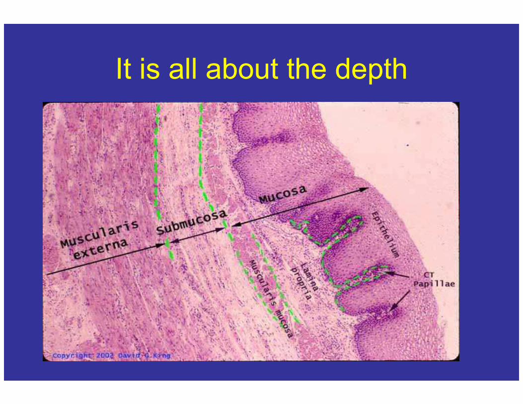
It is all about the depth
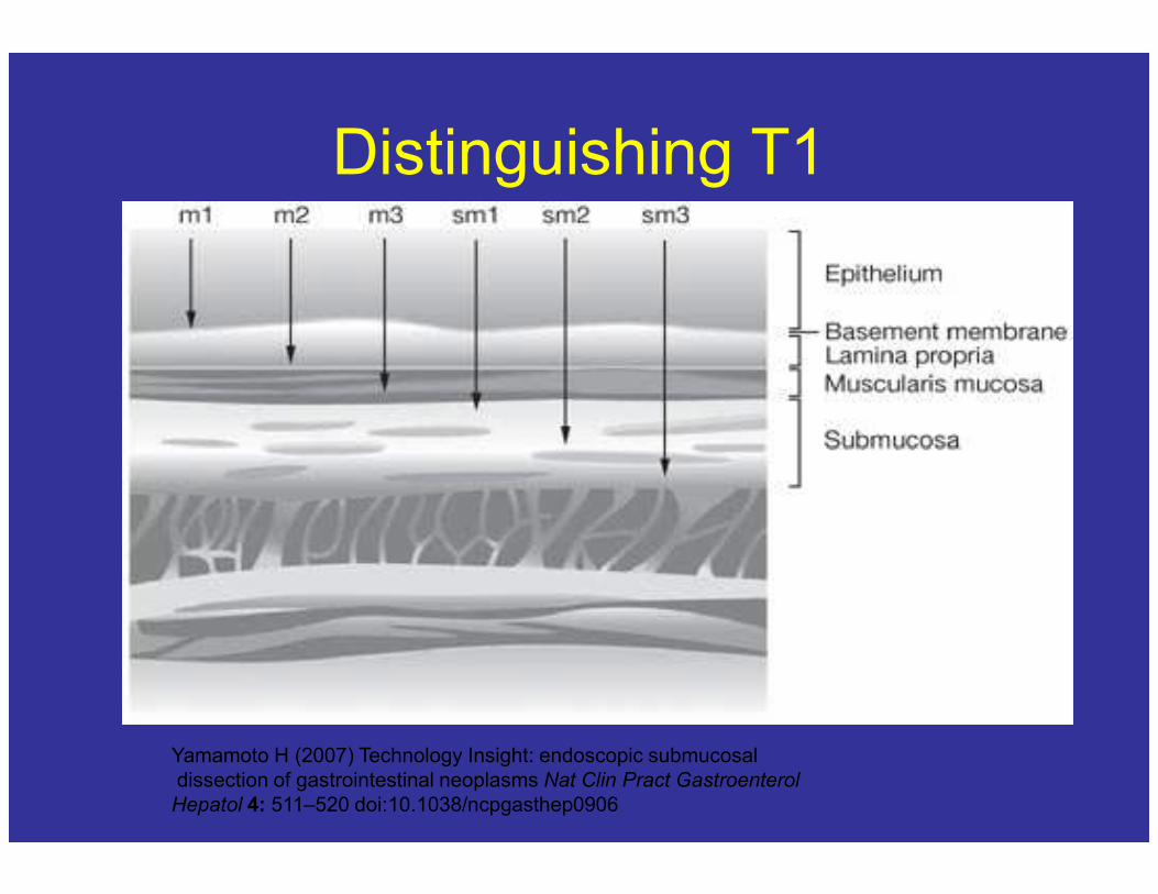
Distinguishing T1
Yamamoto H (2007) Technology Insight: endoscopic submucosal
dissection of gastrointestinal neoplasms Nat Clin Pract Gastroenterol
Hepatol 4: 511–520 doi:10.1038/ncpgasthep0906
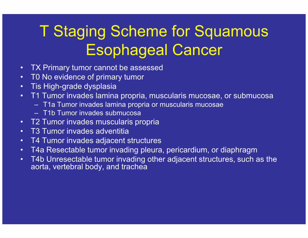
T Staging Scheme for Squamous
Esophageal Cancer• TX Primary tumor cannot be assessed
• T0 No evidence of primary tumor
• Tis High-grade dysplasia
• T1 Tumor invades lamina propria, muscularis mucosae, or submucosa – T1a Tumor invades lamina propria or muscularis mucosae
– T1b Tumor invades submucosa
• T2 Tumor invades muscularis propria • T2 Tumor invades muscularis propria
• T3 Tumor invades adventitia
• T4 Tumor invades adjacent structures
• T4a Resectable tumor invading pleura, pericardium, or diaphragm
• T4b Unresectable tumor invading other adjacent structures, such as the aorta, vertebral body, and trachea
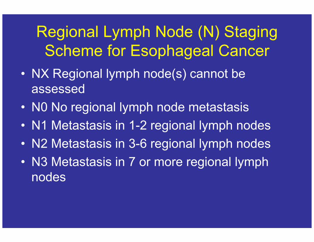
Regional Lymph Node (N) Staging
Scheme for Esophageal Cancer
• NX Regional lymph node(s) cannot be
assessed
• N0 No regional lymph node metastasis
• N1 Metastasis in 1-2 regional lymph nodes • N1 Metastasis in 1-2 regional lymph nodes
• N2 Metastasis in 3-6 regional lymph nodes
• N3 Metastasis in 7 or more regional lymph
nodes
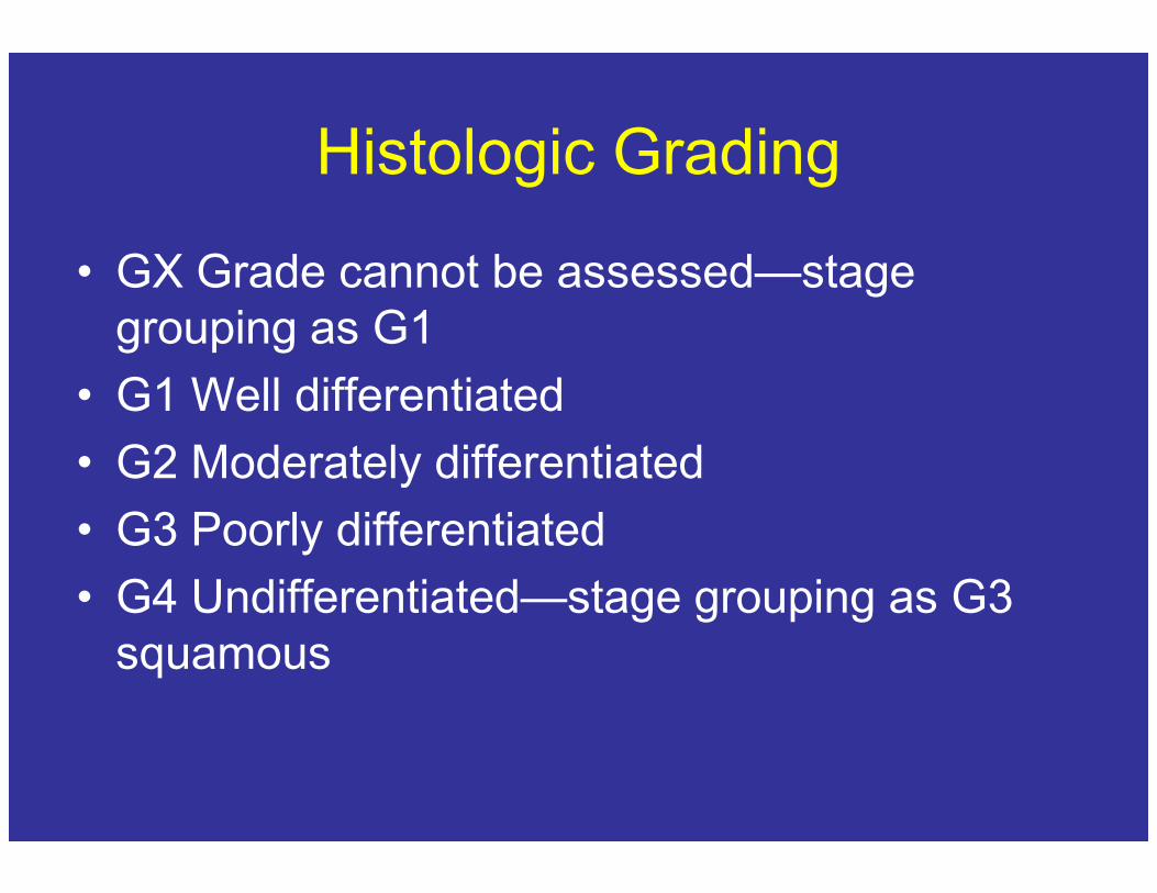
Histologic Grading
• GX Grade cannot be assessed—stage
grouping as G1
• G1 Well differentiated
• G2 Moderately differentiated • G2 Moderately differentiated
• G3 Poorly differentiated
• G4 Undifferentiated—stage grouping as G3
squamous
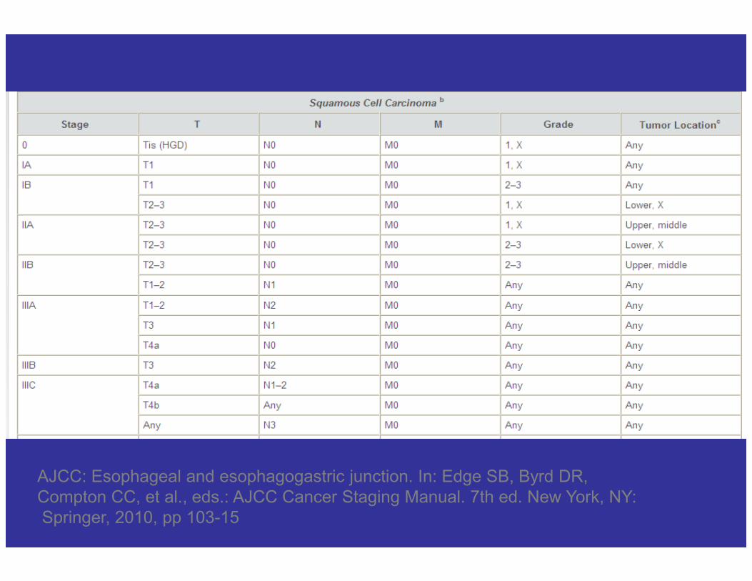
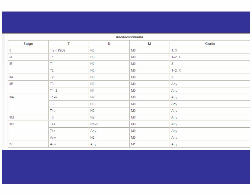
AJCC: Esophageal and esophagogastric junction. In: Edge SB, Byrd DR,
Compton CC, et al., eds.: AJCC Cancer Staging Manual. 7th ed. New York, NY:
Springer, 2010, pp 103-15
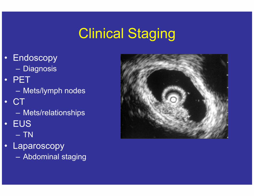
Clinical Staging
• Endoscopy– Diagnosis
• PET– Mets/lymph nodes
• CT• CT– Mets/relationships
• EUS– TN
• Laparoscopy– Abdominal staging
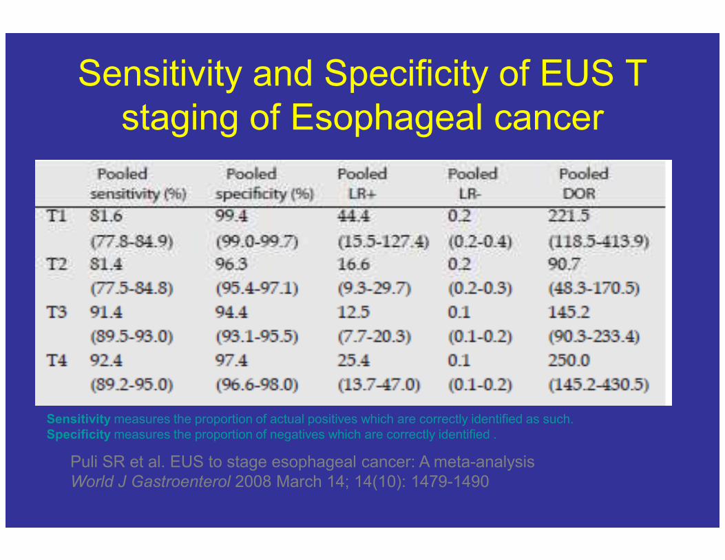
Sensitivity and Specificity of EUS T
staging of Esophageal cancer
Puli SR et al. EUS to stage esophageal cancer: A meta-analysis
World J Gastroenterol 2008 March 14; 14(10): 1479-1490
Sensitivity measures the proportion of actual positives which are correctly identified as such.
Specificity measures the proportion of negatives which are correctly identified .
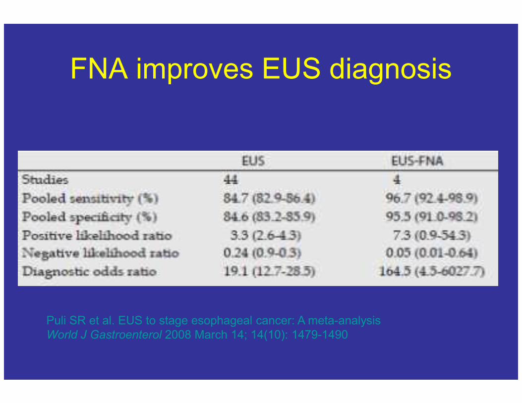
FNA improves EUS diagnosis
Puli SR et al. EUS to stage esophageal cancer: A meta-analysis
World J Gastroenterol 2008 March 14; 14(10): 1479-1490
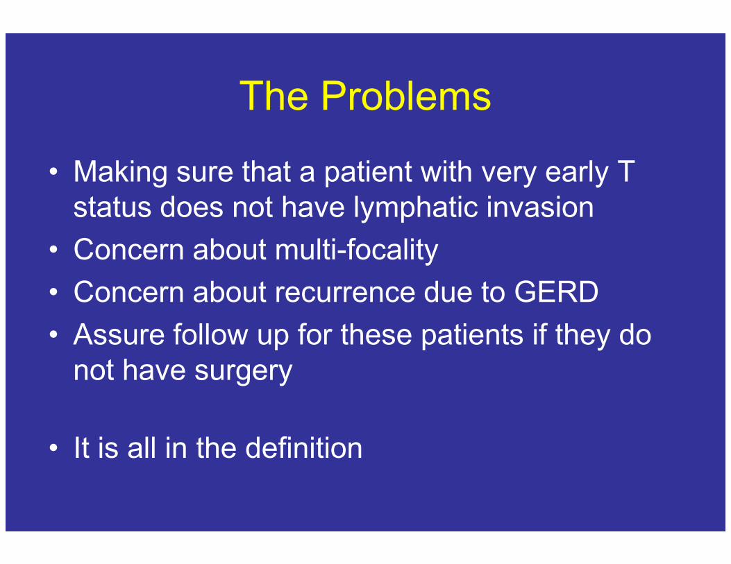
The Problems
• Making sure that a patient with very early T
status does not have lymphatic invasion
• Concern about multi-focality
• Concern about recurrence due to GERD• Concern about recurrence due to GERD
• Assure follow up for these patients if they do
not have surgery
• It is all in the definition
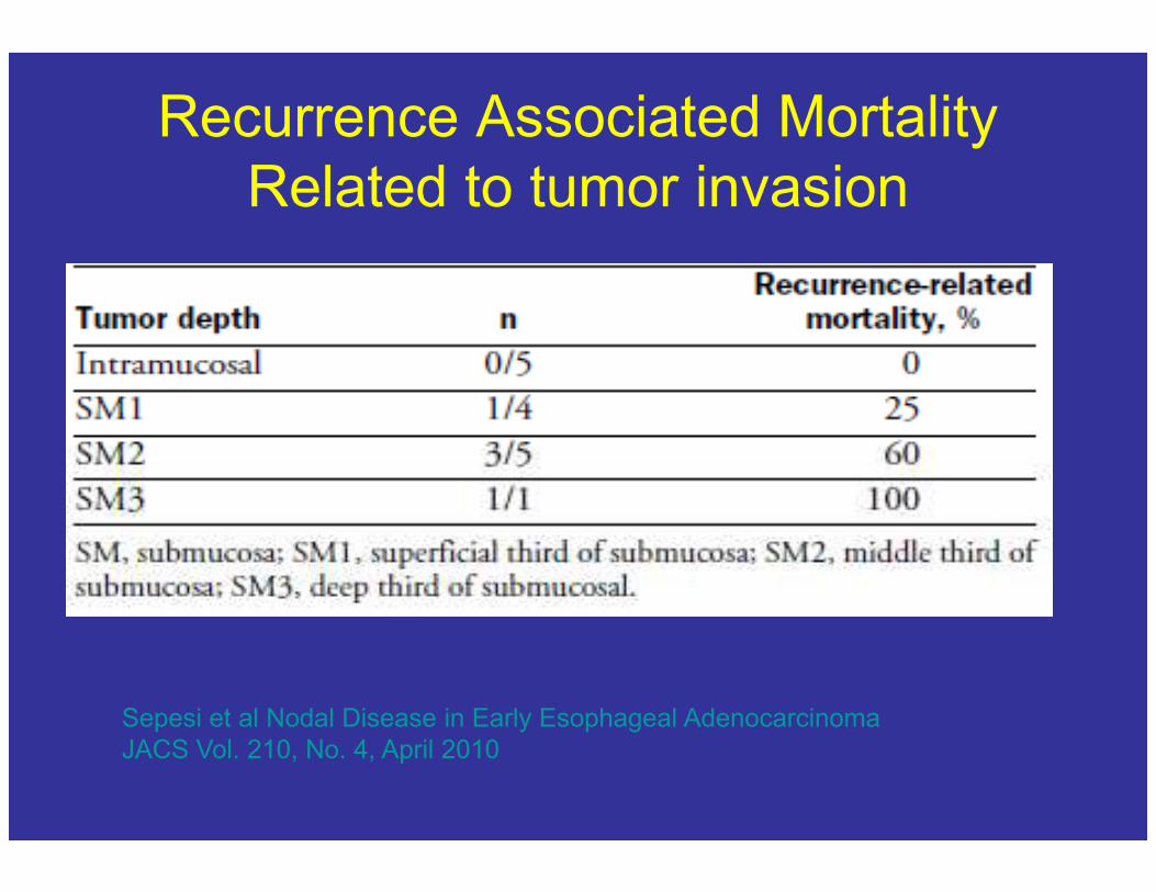
Recurrence Associated Mortality
Related to tumor invasion
Sepesi et al Nodal Disease in Early Esophageal Adenocarcinoma
JACS Vol. 210, No. 4, April 2010
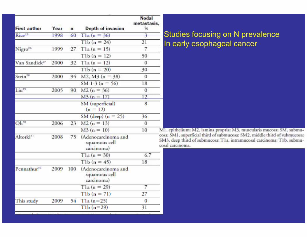
Studies focusing on N prevalence
In early esophageal cancer
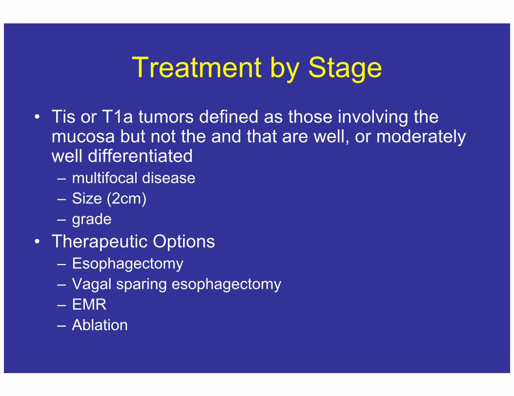
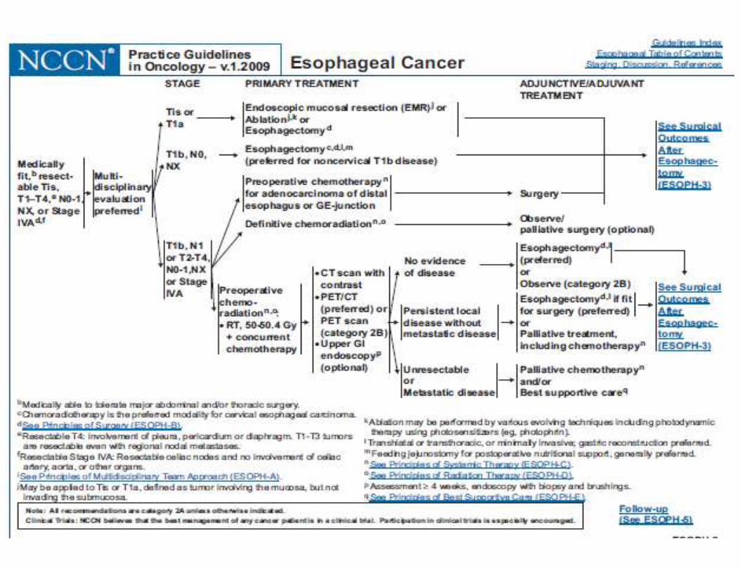
Treatment by Stage
• Tis or T1a tumors defined as those involving the mucosa but not the and that are well, or moderately well differentiated – multifocal disease
– Size (2cm)– Size (2cm)
– grade
• Therapeutic Options– Esophagectomy
– Vagal sparing esophagectomy
– EMR
– Ablation
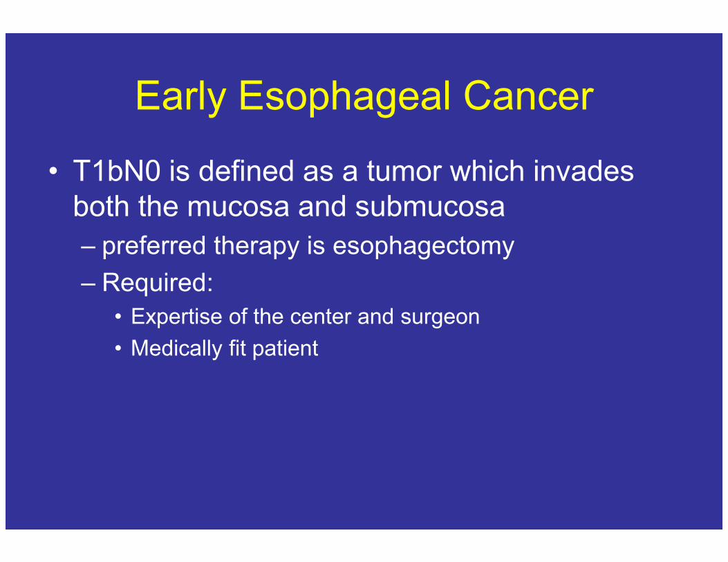
Early Esophageal Cancer
• T1bN0 is defined as a tumor which invades
both the mucosa and submucosa
– preferred therapy is esophagectomy
– Required:– Required:
• Expertise of the center and surgeon
• Medically fit patient
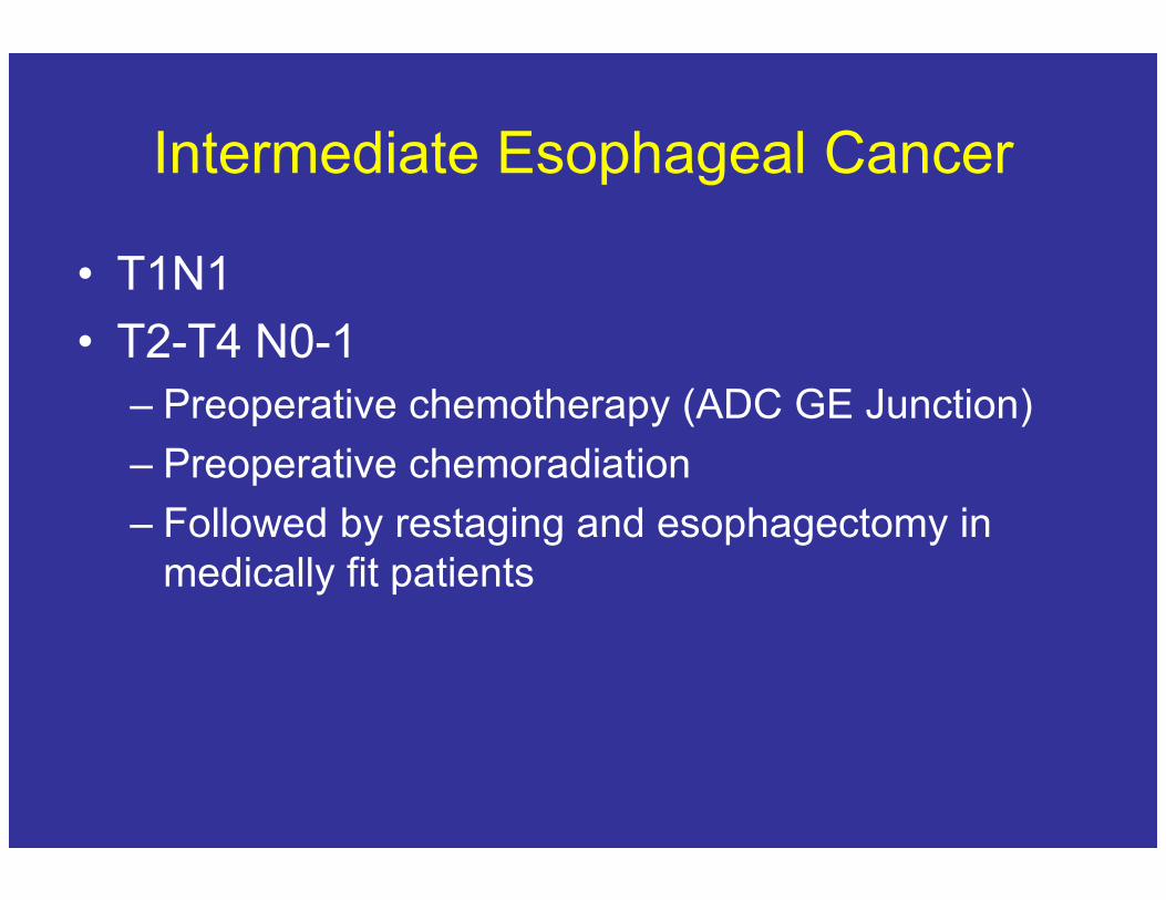
Intermediate Esophageal Cancer
• T1N1
• T2-T4 N0-1
– Preoperative chemotherapy (ADC GE Junction)
– Preoperative chemoradiation– Preoperative chemoradiation
– Followed by restaging and esophagectomy in
medically fit patients
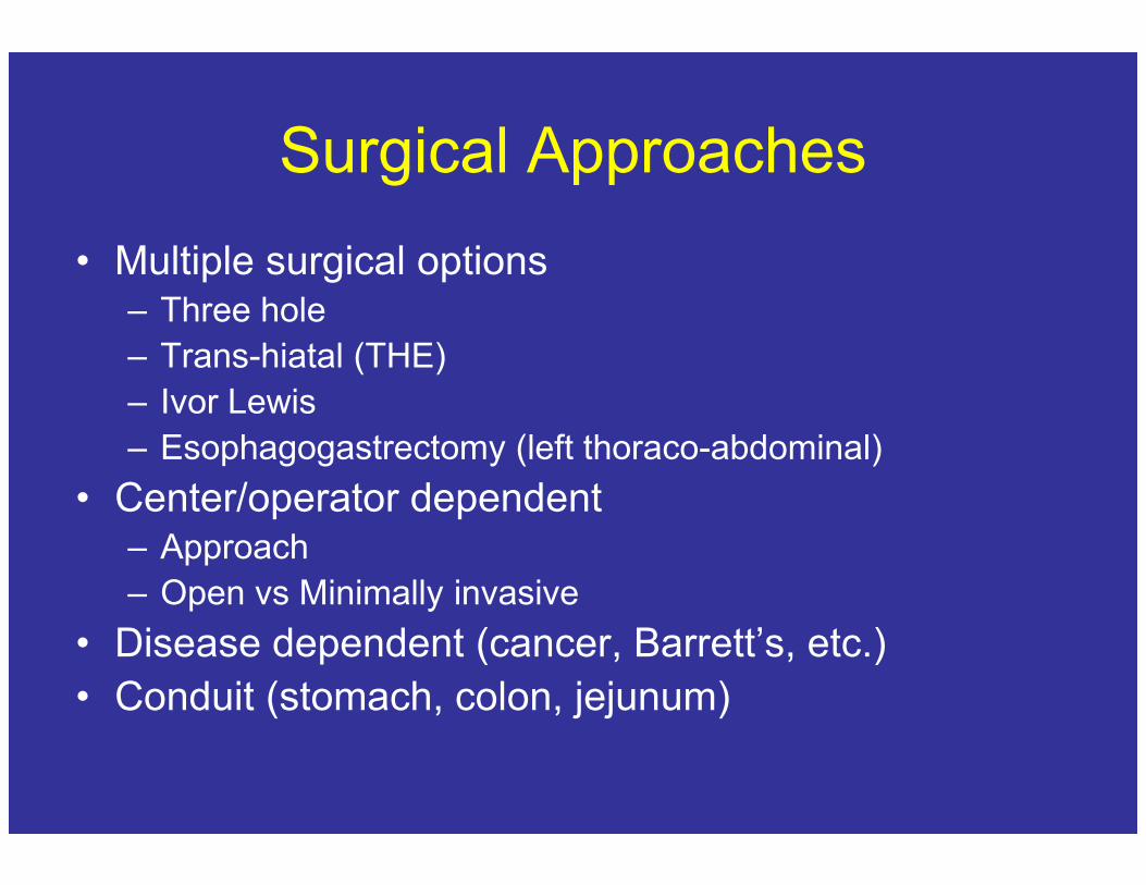
Surgical Approaches
• Multiple surgical options– Three hole
– Trans-hiatal (THE)
– Ivor Lewis
– Esophagogastrectomy (left thoraco-abdominal)– Esophagogastrectomy (left thoraco-abdominal)
• Center/operator dependent– Approach
– Open vs Minimally invasive
• Disease dependent (cancer, Barrett’s, etc.)
• Conduit (stomach, colon, jejunum)
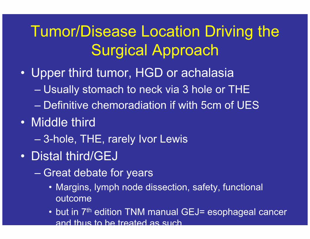
Tumor/Disease Location Driving the
Surgical Approach
• Upper third tumor, HGD or achalasia
– Usually stomach to neck via 3 hole or THE
– Definitive chemoradiation if with 5cm of UES
• Middle third• Middle third
– 3-hole, THE, rarely Ivor Lewis
• Distal third/GEJ
– Great debate for years
• Margins, lymph node dissection, safety, functional
outcome
• but in 7th edition TNM manual GEJ= esophageal cancer
and thus to be treated as such
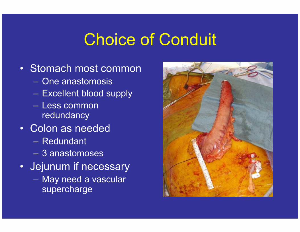
Choice of Conduit
• Stomach most common– One anastomosis
– Excellent blood supply
– Less common redundancyredundancy
• Colon as needed– Redundant
– 3 anastomoses
• Jejunum if necessary– May need a vascular
supercharge
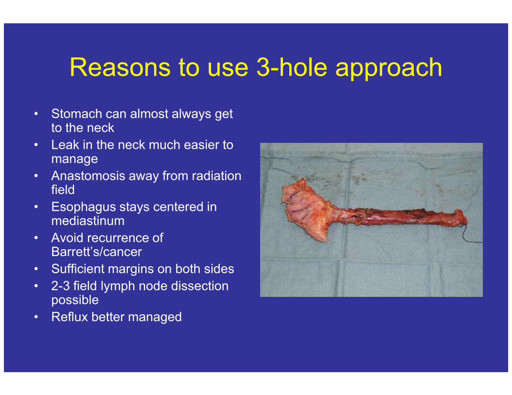
Reasons to use 3-hole approach
• Stomach can almost always get to the neck
• Leak in the neck much easier to manage
• Anastomosis away from radiation field
• Esophagus stays centered in mediastinum
• Avoid recurrence of Barrett’s/cancer
• Sufficient margins on both sides
• 2-3 field lymph node dissection possible
• Reflux better managed
Reasons given for not doing total
esophagectomy
• Swallowing problems (risk of nerve palsy)
• Concerns about tension
• Limitations of some conduits
• Ivor Lewis anastomosis can be placed quite • Ivor Lewis anastomosis can be placed quite
high
• Avoiding operating on the chest
• Make sure that there is no metastatic disease
in the peritoneum
Known Complications after
Esophagectomy• Respiratory
– ARDS
– Aspiration/pneumonia
– effusion
• Cardiac/vascular
• Anastomotic leak
• Leak from the conduit
• Conduit necrosis
• Anastomotic stricture
• Recurrent nerve palsey– MI
– AF
– CVA
• DVT/PE
• Hemorrhage
• Dehiscence
• Sepsis
• Recurrent nerve palsey
• Thoracic duct leak
• Airway injury/fistula
• Abscess infection
• Splenic injury
• Bowel injury
• Radiation pneumonitis
Reducing Mortality and Managing
Complications
• High volume centers with high volume
specialist (mortality 1-5%)
• Team experience with system approach
• Preventative measures• Preventative measures
– Pathway/orders; DVT/PE/CAD prophylaxis
– Intensive mobilization; Nutrition
• Aggressive search and early management of
complications
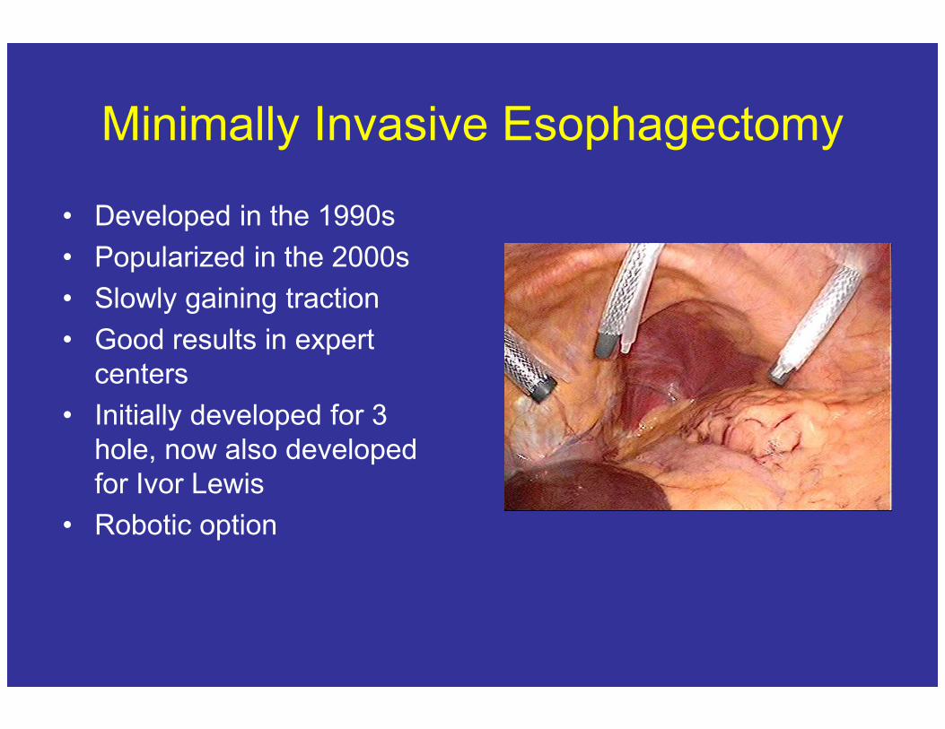
Minimally Invasive Esophagectomy
• Developed in the 1990s
• Popularized in the 2000s
• Slowly gaining traction
• Good results in expert
centerscenters
• Initially developed for 3
hole, now also developed
for Ivor Lewis
• Robotic option
Why Should Surgeons Do MIE
• Truly no randomized data
• Rationale given so far:
– Less pain
– Faster recovery– Faster recovery
– reduced blood loss
– Shorter ICU and hospital stay
– Immune response and cancer
– Better visualization
– Fewer splenectomies
How About the Patients
• Patients want it because they perceive (rightly
so) that it hurts less
• It is done well mainly in major centers of
expertise and may associated with lower expertise and may associated with lower
mortality
• Patients will go to centers that offer these
cases, even if they have to fly and pay out of
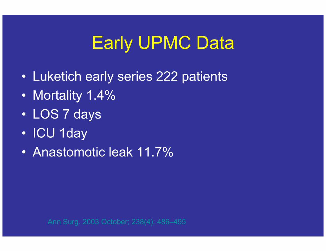
Early UPMC Data
• Luketich early series 222 patients
• Mortality 1.4%
• LOS 7 days
• ICU 1day• ICU 1day
• Anastomotic leak 11.7%
Ann Surg. 2003 October; 238(4): 486–495
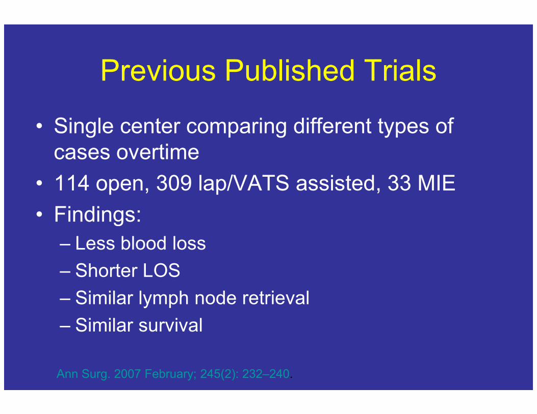
Previous Published Trials
• Single center comparing different types of
cases overtime
• 114 open, 309 lap/VATS assisted, 33 MIE
• Findings:• Findings:
– Less blood loss
– Shorter LOS
– Similar lymph node retrieval
– Similar survival
Ann Surg. 2007 February; 245(2): 232–240.
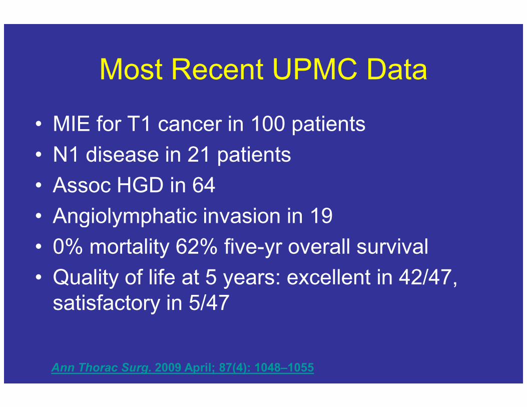
Most Recent UPMC Data
• MIE for T1 cancer in 100 patients
• N1 disease in 21 patients
• Assoc HGD in 64
• Angiolymphatic invasion in 19• Angiolymphatic invasion in 19
• 0% mortality 62% five-yr overall survival
• Quality of life at 5 years: excellent in 42/47,
satisfactory in 5/47
Ann Thorac Surg. 2009 April; 87(4): 1048–1055
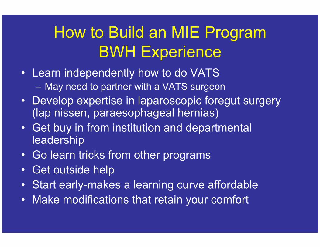
How to Build an MIE Program
BWH Experience
• Learn independently how to do VATS– May need to partner with a VATS surgeon
• Develop expertise in laparoscopic foregut surgery (lap nissen, paraesophageal hernias)
• Get buy in from institution and departmental • Get buy in from institution and departmental leadership
• Go learn tricks from other programs
• Get outside help
• Start early-makes a learning curve affordable
• Make modifications that retain your comfort
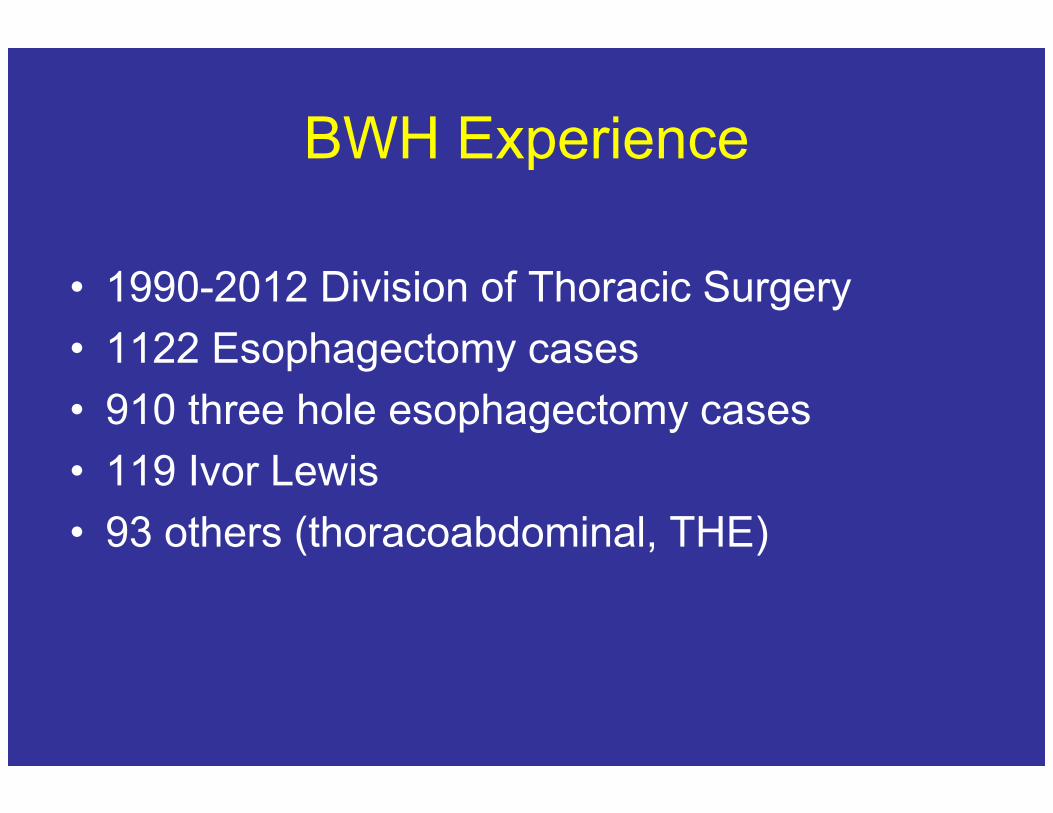
BWH Experience
• 1990-2012 Division of Thoracic Surgery
• 1122 Esophagectomy cases
• 910 three hole esophagectomy cases• 910 three hole esophagectomy cases
• 119 Ivor Lewis
• 93 others (thoracoabdominal, THE)
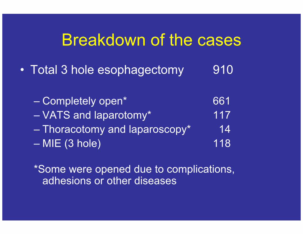
Breakdown of the cases
• Total 3 hole esophagectomy 910
– Completely open* 661
– VATS and laparotomy* 117– VATS and laparotomy* 117
– Thoracotomy and laparoscopy* 14
– MIE (3 hole) 118
*Some were opened due to complications, adhesions or other diseases
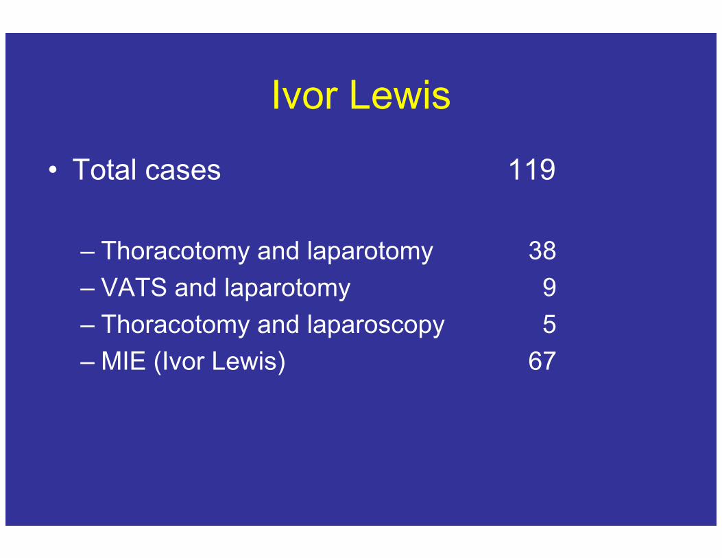
Ivor Lewis
• Total cases 119
– Thoracotomy and laparotomy 38
– VATS and laparotomy 9– VATS and laparotomy 9
– Thoracotomy and laparoscopy 5
– MIE (Ivor Lewis) 67
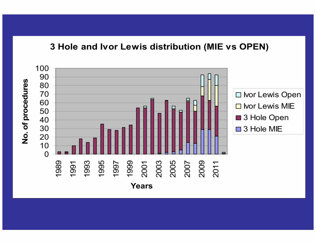
3 Hole and Ivor Lewis distribution (MIE vs OPEN)
405060708090
100
No. of procedures
Ivor Lewis Open
Ivor Lewis MIE
3 Hole Open
010203040
1989
1991
1993
1995
1997
1999
2001
2003
2005
2007
2009
2011
Years
No. of procedures
3 Hole MIE
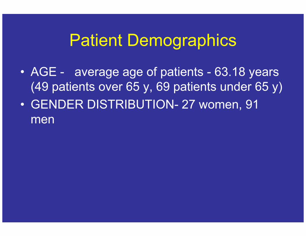
Patient Demographics
• AGE - average age of patients - 63.18 years
(49 patients over 65 y, 69 patients under 65 y)
• GENDER DISTRIBUTION- 27 women, 91
menmen
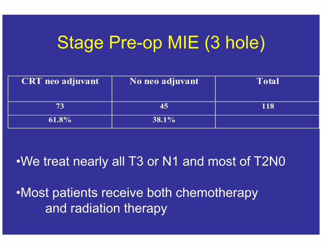
Stage Pre-op MIE (3 hole)
CRT neo adjuvant No neo adjuvant Total
73 45 118
61.8% 38.1%
•We treat nearly all T3 or N1 and most of T2N0
•Most patients receive both chemotherapy
and radiation therapy
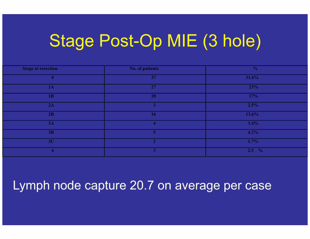
Stage Post-Op MIE (3 hole)
Stage at resection No. of patients %
0 37 31.6%
1A 27 23%
1B 20 17%
2A 3 2.5%
2B 16 13.6%
3A 4 3.4%
3B 5 4.2%
3C 2 1.7%
4 3 2.5 %
Lymph node capture 20.7 on average per case
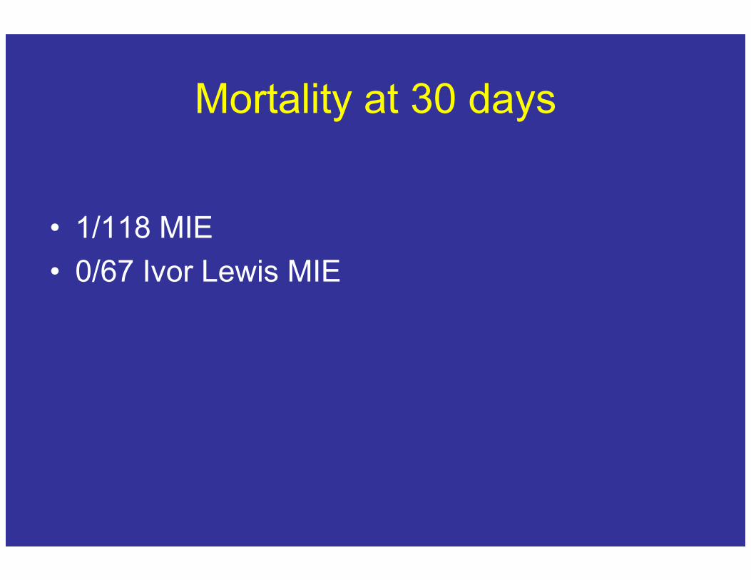
Mortality at 30 days
• 1/118 MIE
• 0/67 Ivor Lewis MIE
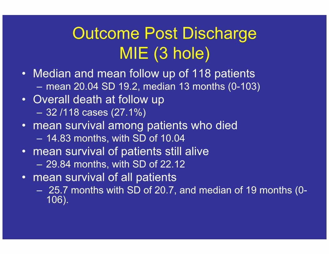
Outcome Post Discharge
MIE (3 hole)• Median and mean follow up of 118 patients
– mean 20.04 SD 19.2, median 13 months (0-103)
• Overall death at follow up – 32 /118 cases (27.1%)
• mean survival among patients who died • mean survival among patients who died – 14.83 months, with SD of 10.04
• mean survival of patients still alive – 29.84 months, with SD of 22.12
• mean survival of all patients– 25.7 months with SD of 20.7, and median of 19 months (0-
106).
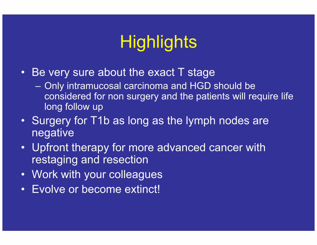
Highlights
• Be very sure about the exact T stage– Only intramucosal carcinoma and HGD should be
considered for non surgery and the patients will require life long follow up
• Surgery for T1b as long as the lymph nodes are • Surgery for T1b as long as the lymph nodes are negative
• Upfront therapy for more advanced cancer with restaging and resection
• Work with your colleagues
• Evolve or become extinct!
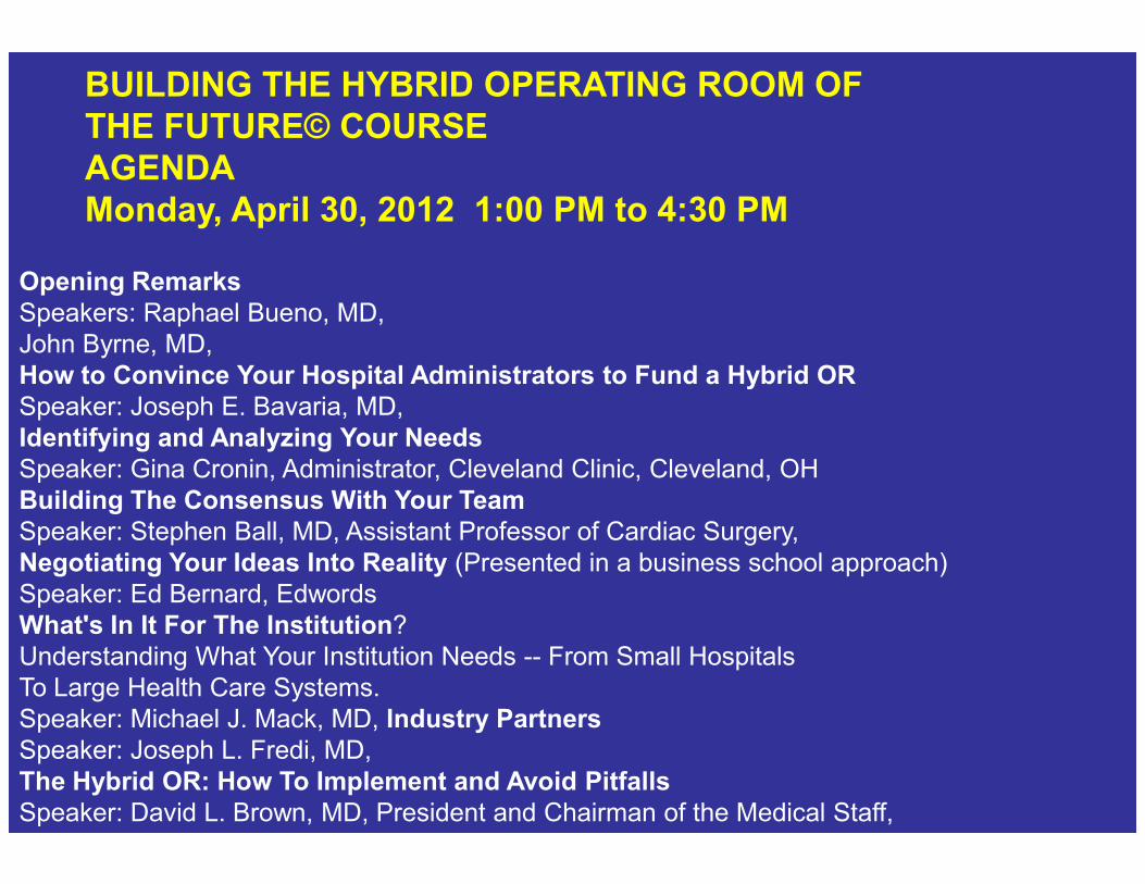
BUILDING THE HYBRID OPERATING ROOM OF
THE FUTURE© COURSE
AGENDA
Monday, April 30, 2012 1:00 PM to 4:30 PM
Opening Remarks
Speakers: Raphael Bueno, MD,
John Byrne, MD,
How to Convince Your Hospital Administrators to Fund a Hybrid OR
Speaker: Joseph E. Bavaria, MD,
Identifying and Analyzing Your NeedsIdentifying and Analyzing Your Needs
Speaker: Gina Cronin, Administrator, Cleveland Clinic, Cleveland, OH
Building The Consensus With Your Team
Speaker: Stephen Ball, MD, Assistant Professor of Cardiac Surgery,
Negotiating Your Ideas Into Reality (Presented in a business school approach)
Speaker: Ed Bernard, Edwords
What's In It For The Institution?
Understanding What Your Institution Needs -- From Small Hospitals
To Large Health Care Systems.
Speaker: Michael J. Mack, MD, Industry Partners
Speaker: Joseph L. Fredi, MD,
The Hybrid OR: How To Implement and Avoid Pitfalls
Speaker: David L. Brown, MD, President and Chairman of the Medical Staff,
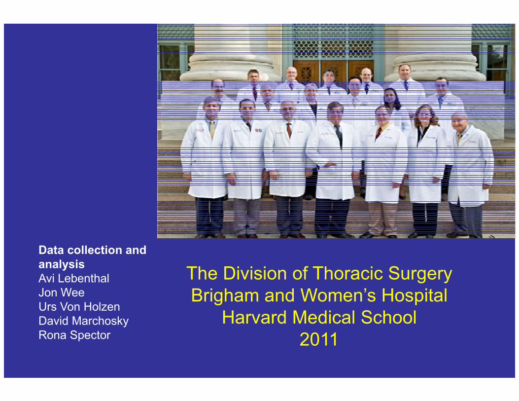
Data collection and
analysis
Avi Lebenthal
Jon Wee
Urs Von Holzen
David Marchosky
Rona Spector
The Division of Thoracic Surgery
Brigham and Women’s Hospital
Harvard Medical School
2011