Embed Size (px)
Citation preview
94 Vlaams Diergeneeskundig Tijdschrift, 2015, 84
BSTRACT
A four-year-old red Holstein Friesian cow was admitted to the clinic with fever and milk drop. Blood analysis revealed the presence of a chronic infection, and the diagnosis of pyelone-phritis of the right kidney was made after repeated ultrasound examinations. The animal was treated with procaine benzylpenicillin, sulfadoxine trimethoprim, oxytetracycline and enrofloxa-cine but this therapy was not successful. Nephrectomy was performed after the left kidney proved to have a normal function. The removed right kidney was greatly enlarged and filled with pus. Escherichia coli was isolated from the kidney. The strain was multidrug-resistant, including re-sistance to aminopenicillins, streptomycin, sulfonamides and trimethoprim. The cow was treated with amoxicillin and clavulanic acid after the operation. Postoperatively, an abscess developed and a tube drain was placed during a second surgery to enable daily rinsing with a chlorhexidine solution. After a postoperative care period of two months, the animal was sent back to the farm, where it returned to an acceptable level of milk production. This case demonstrates that with basic surgical skills, a good preparation and knowledge of anatomy, nephrectomy is attainable for a first-opinion veterinarian, with an acceptable economic prognosis for the farmer.
SAMENVATTING
Een vier jaar oude rode holstein-friesiankoe werd aangeboden met koorts en een plotse, sterke daling van de melkproductie. Het bloedonderzoek toonde een chronische infectie aan en na herhaalde echografische onderzoeken kon de diagnose van pyelonefritis van de rechternier worden gesteld. Het dier werd zonder effect met procaïne-benzylpenicilline, sulfadoxine-trimethoprim, oxytetracycline en enrofloxacine behandeld. Aangezien de linkernier onaangetast was, kwam het dier in aanmerking voor een nefrectomie van de aangetaste rechternier. De verwijderde rechternier was opvallend groot en gevuld met etter. Hieruit werden kolonies Escherichia coli geïsoleerd die resistent bleken tegen aminopenicillinen, streptomycine, sulfonamiden en trimethoprim. De koe werd verder behandeld met amoxicilline en clavulaanzuur op basis van het antibiogram. Postoperatief ontstond een retroperi- toneaal abces. Er werd een buisdrain geplaatst tijdens een tweede operatie om dagelijkse spoeling met een chloorhexidineoplossing mogelijk te maken. Hierna bleef het dier koortsvrij en herstelde goed. De koe werd uit de kliniek ontslagen en was op het bedrijf in staat om een goede melkproductie te halen. In deze casus wordt aangetoond dat nefrectomie, mits een goede voorbereiding, een haalbare ingreep is voor de eerstelijnsdierenarts in geval van unilaterale pyelonefritis, met een acceptabele economische prognose voor de landbouwer.
A
Surgical correction of pyelonephritis caused by multidrug-resistant Escherichia coli in a dairy cow
Chirurgische correctie van pyelonefritis veroorzaakt door multiresistente Escherichia coli bij een melkkoe
1E. Put, 1B. Valgaeren, 1B. Pardon, 2J. De Latthauwer, 2D. Valckenier, 1P. Deprez
1Department of Large Animal Internal Medicine, Faculty of Veterinary Medicine, Ghent University, Salisburylaan 133, B-9820 Merelbeke, Belgium
2Department of Reproduction, Obstetrics and Herd Health, Faculty of Veterinary Medicine, Ghent University, Salisburylaan 133, B-9820 Merelbeke, Belgium
94 Case report Vlaams Diergeneeskundig Tijdschrift, 2015, 84
Vlaams Diergeneeskundig Tijdschrift, 2015, 84 95
INTRODUCTION
Pyelonephritis is one of the most common renal pathologies in cattle (Divers et al., 1982; Markusfeld et al., 1989; Rosenbaum et al., 2005). It involves in-fection and inflammation of the renal sinus, with only non-specific symptoms of an acute infection. These symptoms include milk drop, anorexia, fever and depression. Pyelonephritis is most frequently due to ascending infections, but in rare cases, it may also have a hematogenous origin. Female animals are pre-disposed and most cases are related to recent calving, abortion or urinary catheterization (Yeruham et al., 2006). Corynebacterium renale (C. renale) and Esche- richia coli (E. coli) are the most frequently isolated causative agents from pyelonephritis cases in cattle (Braun et al., 2008; Moore et al., 2010). Both bacte-ria have fimbriae, which enable them to bind to the epithelial cells of the urogenital tract (Hayashi et al., 1985; Gyles and Fairbrother, 2010).
Treatment of pyelonephritis consists of fluid ther-apy, anti-inflammatory therapy and administration of antimicrobials, which are active against the involved pathogens (Van Metre, 2009). In different countries, formularies to enhance responsible antimicrobial use have recently been initiated. In the Netherlands, pro-caine benzylpenicillin or sulfadoxine trimethoprim are proposed as first-choice treatments for pyelonephritis. Second-choice drugs are ampicillin, amoxicillin and the combination of procaine benzylpenicillin and dihy-drostreptomycin (Werkgroep Veterinair Antibioticum Beleid, 2012). The antibacterial therapy should be maintained for at least three to four weeks (Kahn and Line, 2010). Whereas C. renale is commonly sus-ceptible to penicillins, E. coli is naturally resistant (Prescott, 2013). E. coli is a species which easily ac-quires resistance to various antimicrobial agents, either by mutation in chromosomal DNA or by plasmid
transfer (Okusu et al., 1996; Rogers et al., 2011). In recent years, an overall increase in (multi)resistance of E. coli, associated with antimicrobial use, has been reported worldwide (Levy and Marshall, 2004; Rog-ers et al., 2011). In this case report, a case of pyelone-phritis caused by a multidrug-resistant E. coli strain is described, in which unilateral nephrectomy resulted in clinical recovery.
CASE REPORT
A four-year-old red Holstein Friesian cow was ad-mitted to the Faculty of Veterinary Medicine of the Ghent University (Belguim) with symptoms of milk drop, fever and anorexia, one month after calving. At arrival, the cow was alert and in good body condition. No other abnormalities were found during the physi-cal examination. Also the rectal palpation and ultra-sonic examination were normal except for a slightly enlarged right kidney, visible on the transcutaneous ultrasound.
Venous blood analysis showed dehydration, caus-ing an increased blood urea nitrogen (BUN) and total protein concentration and leukocytosis (Table 1). Uri-nary analysis only revealed a slight positive protein reaction on urine stick but a negative nitric acid test. The animal was hospitalized with an acute infection of unknown origin and treated by administration of procaine benzylpenicillin (16 mg/kg IM, Duphapen®, Zoetis Belgium s.a., Belgium). Fluid therapy was per-formed by intravenous perfusion and oral drenching. The fever persisted and after four days, also sulfadox-ine trimethoprim (12.5 mg/kg and 2.5 mg/kg, respec-tively IV, Borgal® 24%, Virbac, France) and oxytet-racycline (7.2 mg/kg IM, Duphacycline®, Zoetis Bel-gium s.a., Belgium) were administered for three days each, together with flunixin meglumin (2.2 mg/kg
Table 1. Hematology, biochemistry and electrophoresis conducted on the serum of venous blood on different times during the stay in the clinic.
Day 11 Day 4 Day 8 Day 11 Day 19 Day 34 Reference values
Leukocytes 4.2 12.2 10.6 2.0 4.8 6.0 - 9.0 x 10⁹/lPacked cell volume 314 268 237 250 - 350 ml/lNeutrophils 43 40 83 20 39 15 - 45 %Lymphocytes 57 32 17 50 29 45 - 75 %Monocytes 28 25 2 - 7 %Eosinophils 1 7 0 - 10 %Thrombocytes 193 201 151 467 576 100 - 800 x 109/lTotal protein 87 69 82 87 77 60 - 80 g/lUrea (BUN) 15.7 5.3 3 - 8 mmol/lCreatinine 141 92 88 - 172 µmol/lGamma glutamyltransferase 28 33 <30 mU/mlA/G ratio 0.61 0.70 - 0.95Albumin 38 30 - 40 %Alpha globulin 18 12 - 16 %Beta globulin 10 9 - 13 %Gamma globulin 34 31 - 44 %
1 Day 1 = arrival at the clinic
96 Vlaams Diergeneeskundig Tijdschrift, 2015, 84
IV, Emdofluxin, Emdoka, Belgium) in every fever epi- sode. Repeated clinical and ultrasonographic exami-nation, 12 days later, revealed a marked dilatation of the right kidney. The enlarged right kidney consisted of several vesicles filled with hypoechogenic fluid, consistent with pyelonephritis (Figure 1). The blad-der had an irregular lining of the wall and echogenic particles swirling in the urine. Blood examination at that time showed a chronic infection (leukocytosis, neutrophilia, lymphopenia, increased total protein), but normal BUN and creatinine.
Since the additional treatment with enrofloxacin (3.1 mg/kg IV, Floxadil, Emdoka, Belgium) for three days was not successful and given that the renal val-ues were still within normal reference values, it was opted to perform a unilateral nephrectomy after 12 days of hospitalization. Before the surgical procedure, the animal received procaine benzylpenicillin com-bined with neomycin (14.0 mg/kg and 7.0 mg/kg, re-spectively IM, Neopen, Intervet, the Netherlands), to-gether with flunixin meglumin (2.2 mg/kg IV, Emdo-fluxin, Emdoka, Belgium) as part of the standard sur-gical protocol. The surgical procedure was performed standing, with epidural, distal paravertebral and linear infiltration anesthesia on the right side and flank us-
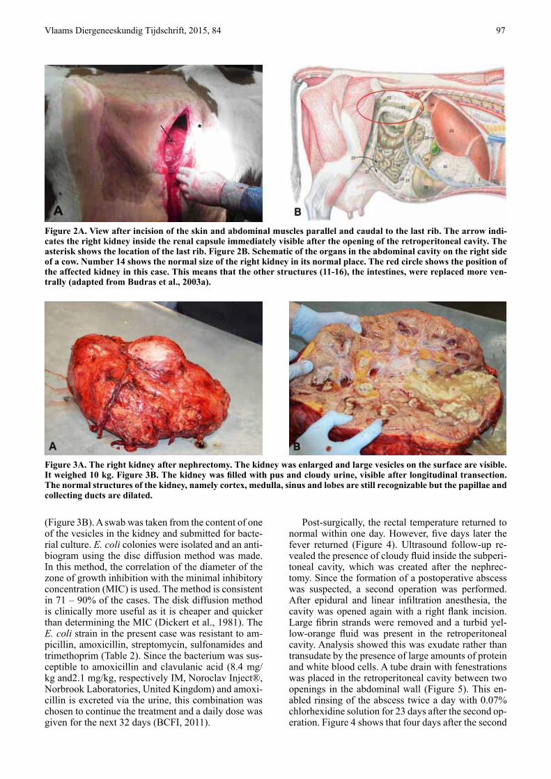
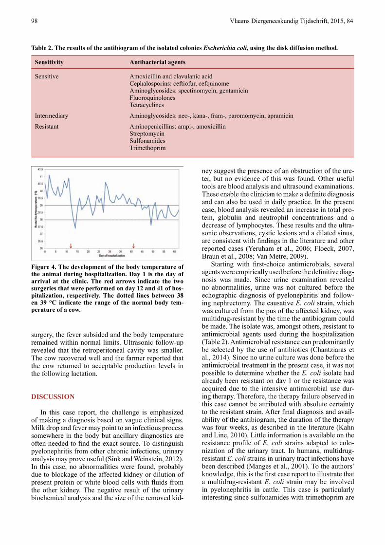
ing procaine hydrochloride (Procaïne hydrochloride 4% + Adrenaline, VMD n.v., Belgium). Additionally, the animal was sedated with xylazine hydrochloride (0.012 mg/kg IV, Xyl-M® 2%, VMD n.v., Belgium). On the right side, parallel with the last rib, an incision was made through the skin and abdominal muscles (Figure 2A). Figure 2B shows that the right kidney covered the whole of the right flank, instead of ex-panding normally from the last rib until the second or third lumbal vertebra (Budras et al., 2003b). First, the renal vascular structures and ureter were identified through blunt dissection at their site of insertion in the renal hilus. The renal artery was ligated three times, using absorbable braided multifilament suture mate-rial (Surgicryl® polyglycolic acid, SMI, Belgium). The thickened ureter was followed approximately 15 cm caudally, where it was ligated two times and cut between these ligatures to prevent leakage of fluids. The renal vein was not found before removal and rup-tured. Subsequently, the vein was clamped and ligated until there was no more visible blood loss. The affect-ed kidney weighed 10 kg instead of the normal 600 - 750 g (Budras et al., 2003b). As shown in Figure 3A, large vesicles were visible on the surface. The kidney, including the vesicles, was filled with fluid and pus
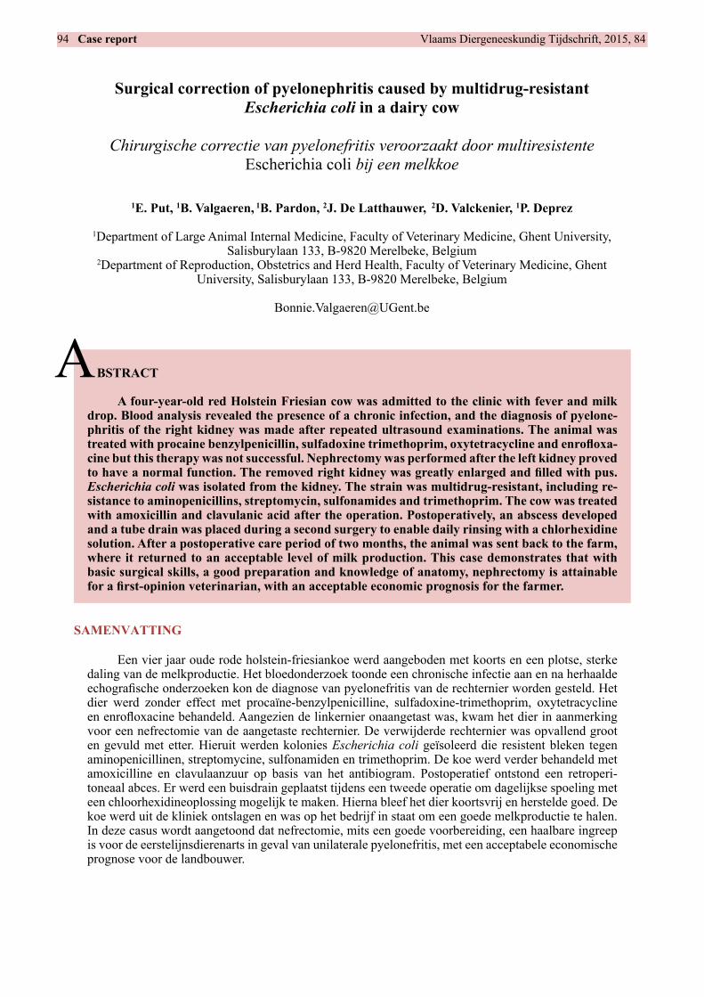
Figures 1A and B. Ultrasound images made with a 3.5 MHz sectorial probe with a depth of 27 centimeters after 12 days of hospitalization. Figures 1C and D. Transrectal ultrasound images made with a 7.5 MHz linear probe with a depth of 8 cm after 12 days of hospitalization. Figure 1A shows the normal liver and on the right side, a part of the right kidney. Figure 1B shows the right kidney with vesicles filled with fluid. The kidney is greatly enlarged. Figure 1C visualizes the normal-sized left kidney with the renal sinus in the center and medulla and cortex surrounding it. Figure 1D shows the bladder with echogenic particles swirling in the urine and irregular lining of the bladder wall on the bottom right side.
Vlaams Diergeneeskundig Tijdschrift, 2015, 84 97
(Figure 3B). A swab was taken from the content of one of the vesicles in the kidney and submitted for bacte-rial culture. E. coli colonies were isolated and an anti-biogram using the disc diffusion method was made. In this method, the correlation of the diameter of the zone of growth inhibition with the minimal inhibitory concentration (MIC) is used. The method is consistent in 71 – 90% of the cases. The disk diffusion method is clinically more useful as it is cheaper and quicker than determining the MIC (Dickert et al., 1981). The E. coli strain in the present case was resistant to am-picillin, amoxicillin, streptomycin, sulfonamides and trimethoprim (Table 2). Since the bacterium was sus-ceptible to amoxicillin and clavulanic acid (8.4 mg/kg and2.1 mg/kg, respectively IM, Noroclav Inject®, Norbrook Laboratories, United Kingdom) and amoxi-cillin is excreted via the urine, this combination was chosen to continue the treatment and a daily dose was given for the next 32 days (BCFI, 2011).
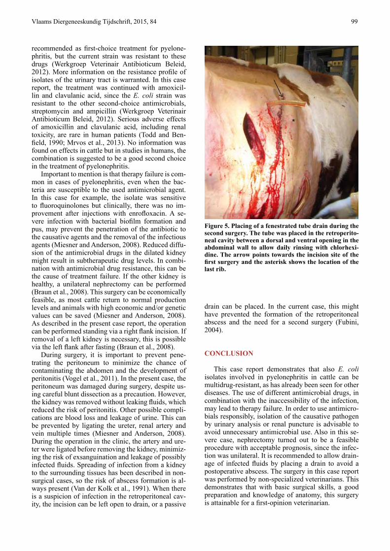
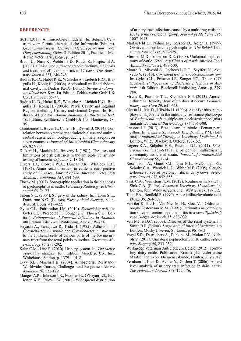
Post-surgically, the rectal temperature returned to normal within one day. However, five days later the fever returned (Figure 4). Ultrasound follow-up re-vealed the presence of cloudy fluid inside the subperi-toneal cavity, which was created after the nephrec-tomy. Since the formation of a postoperative abscess was suspected, a second operation was performed. After epidural and linear infiltration anesthesia, the cavity was opened again with a right flank incision. Large fibrin strands were removed and a turbid yel-low-orange fluid was present in the retroperitoneal cavity. Analysis showed this was exudate rather than transudate by the presence of large amounts of protein and white blood cells. A tube drain with fenestrations was placed in the retroperitoneal cavity between two openings in the abdominal wall (Figure 5). This en-abled rinsing of the abscess twice a day with 0.07% chlorhexidine solution for 23 days after the second op-eration. Figure 4 shows that four days after the second
Figure 2A. View after incision of the skin and abdominal muscles parallel and caudal to the last rib. The arrow indi-cates the right kidney inside the renal capsule immediately visible after the opening of the retroperitoneal cavity. The asterisk shows the location of the last rib. Figure 2B. Schematic of the organs in the abdominal cavity on the right side of a cow. Number 14 shows the normal size of the right kidney in its normal place. The red circle shows the position of the affected kidney in this case. This means that the other structures (11-16), the intestines, were replaced more ven-trally (adapted from Budras et al., 2003a).
Figure 3A. The right kidney after nephrectomy. The kidney was enlarged and large vesicles on the surface are visible. It weighed 10 kg. Figure 3B. The kidney was filled with pus and cloudy urine, visible after longitudinal transection. The normal structures of the kidney, namely cortex, medulla, sinus and lobes are still recognizable but the papillae and collecting ducts are dilated.
98 Vlaams Diergeneeskundig Tijdschrift, 2015, 84
surgery, the fever subsided and the body temperature remained within normal limits. Ultrasonic follow-up revealed that the retroperitoneal cavity was smaller. The cow recovered well and the farmer reported that the cow returned to acceptable production levels in the following lactation.
DISCUSSION
In this case report, the challenge is emphasized of making a diagnosis based on vague clinical signs. Milk drop and fever may point to an infectious process somewhere in the body but ancillary diagnostics are often needed to find the exact source. To distinguish pyelonephritis from other chronic infections, urinary analysis may prove useful (Sink and Weinstein, 2012). In this case, no abnormalities were found, probably due to blockage of the affected kidney or dilution of present protein or white blood cells with fluids from the other kidney. The negative result of the urinary biochemical analysis and the size of the removed kid-
ney suggest the presence of an obstruction of the ure-ter, but no evidence of this was found. Other useful tools are blood analysis and ultrasound examinations. These enable the clinician to make a definite diagnosis and can also be used in daily practice. In the present case, blood analysis revealed an increase in total pro-tein, globulin and neutrophil concentrations and a decrease of lymphocytes. These results and the ultra-sonic observations, cystic lesions and a dilated sinus, are consistent with findings in the literature and other reported cases (Yeruham et al., 2006; Floeck, 2007, Braun et al., 2008; Van Metre, 2009).
Starting with first-choice antimicrobials, several agents were empirically used before the definitive diag- nosis was made. Since urine examination revealed no abnormalities, urine was not cultured before the echographic diagnosis of pyelonephritis and follow-ing nephrectomy. The causative E. coli strain, which was cultured from the pus of the affected kidney, was multidrug-resistant by the time the antibiogram could be made. The isolate was, amongst others, resistant to antimicrobial agents used during the hospitalization (Table 2). Antimicrobial resistance can predominantly be selected by the use of antibiotics (Chantziaras et al., 2014). Since no urine culture was done before the antimicrobial treatment in the present case, it was not possible to determine whether the E. coli isolate had already been resistant on day 1 or the resistance was acquired due to the intensive antimicrobial use dur-ing therapy. Therefore, the therapy failure observed in this case cannot be attributed with absolute certainty to the resistant strain. After final diagnosis and avail-ability of the antibiogram, the duration of the therapy was four weeks, as described in the literature (Kahn and Line, 2010). Little information is available on the resistance profile of E. coli strains adapted to colo-nization of the urinary tract. In humans, multidrug-resistant E. coli strains in urinary tract infections have been described (Manges et al., 2001). To the authors’ knowledge, this is the first case report to illustrate that a multidrug-resistant E. coli strain may be involved in pyelonephritis in cattle. This case is particularly interesting since sulfonamides with trimethoprim are
Table 2. The results of the antibiogram of the isolated colonies Escherichia coli, using the disk diffusion method.
Sensitivity Antibacterial agents
Sensitive Amoxicillin and clavulanic acid Cephalosporins: ceftiofur, cefquinome Aminoglycosides: spectinomycin, gentamicin Fluoroquinolones Tetracyclines
Intermediary Aminoglycosides: neo-, kana-, fram-, paromomycin, apramicin
Resistant Aminopenicillins: ampi-, amoxicillin Streptomycin Sulfonamides Trimethoprim
Figure 4. The development of the body temperature of the animal during hospitalization. Day 1 is the day of arrival at the clinic. The red arrows indicate the two surgeries that were performed on day 12 and 41 of hos-pitalization, respectively. The dotted lines between 38 en 39 °C indicate the range of the normal body tem-perature of a cow.
Vlaams Diergeneeskundig Tijdschrift, 2015, 84 99
recommended as first-choice treatment for pyelone-phritis, but the current strain was resistant to these drugs (Werkgroep Veterinair Antibioticum Beleid, 2012). More information on the resistance profile of isolates of the urinary tract is warranted. In this case report, the treatment was continued with amoxicil-lin and clavulanic acid, since the E. coli strain was resistant to the other second-choice antimicrobials, streptomycin and ampicillin (Werkgroep Veterinair Antibioticum Beleid, 2012). Serious adverse effects of amoxicillin and clavulanic acid, including renal toxicity, are rare in human patients (Todd and Ben-field, 1990; Mrvos et al., 2013). No information was found on effects in cattle but in studies in humans, the combination is suggested to be a good second choice in the treatment of pyelonephritis.
Important to mention is that therapy failure is com-mon in cases of pyelonephritis, even when the bac-teria are susceptible to the used antimicrobial agent. In this case for example, the isolate was sensitive to fluoroquinolones but clinically, there was no im-provement after injections with enrofloxacin. A se-vere infection with bacterial biofilm formation and pus, may prevent the penetration of the antibiotic to the causative agents and the removal of the infectious agents (Miesner and Anderson, 2008). Reduced diffu-sion of the antimicrobial drugs in the dilated kidney might result in subtherapeutic drug levels. In combi-nation with antimicrobial drug resistance, this can be the cause of treatment failure. If the other kidney is healthy, a unilateral nephrectomy can be performed (Braun et al., 2008). This surgery can be economically feasible, as most cattle return to normal production levels and animals with high economic and/or genetic values can be saved (Miesner and Anderson, 2008). As described in the present case report, the operation can be performed standing via a right flank incision. If removal of a left kidney is necessary, this is possible via the left flank after fasting (Braun et al., 2008).
During surgery, it is important to prevent pene-trating the peritoneum to minimize the chance of contaminating the abdomen and the development of peritonitis (Vogel et al., 2011). In the present case, the peritoneum was damaged during surgery, despite us-ing careful blunt dissection as a precaution. However, the kidney was removed without leaking fluids, which reduced the risk of peritonitis. Other possible compli-cations are blood loss and leakage of urine. This can be prevented by ligating the ureter, renal artery and vein multiple times (Miesner and Anderson, 2008). During the operation in the clinic, the artery and ure-ter were ligated before removing the kidney, minimiz-ing the risk of exsanguination and leakage of possibly infected fluids. Spreading of infection from a kidney to the surrounding tissues has been described in non-surgical cases, so the risk of abscess formation is al-ways present (Van der Kolk et al., 1991). When there is a suspicion of infection in the retroperitoneal cav-ity, the incision can be left open to drain, or a passive
drain can be placed. In the current case, this might have prevented the formation of the retroperitoneal abscess and the need for a second surgery (Fubini, 2004).
CONCLUSION
This case report demonstrates that also E. coli isolates involved in pyelonephritis in cattle can be multidrug-resistant, as has already been seen for other diseases. The use of different antimicrobial drugs, in combination with the inaccessibility of the infection, may lead to therapy failure. In order to use antimicro-bials responsibly, isolation of the causative pathogen by urinary analysis or renal puncture is advisable to avoid unnecessary antimicrobial use. Also in this se-vere case, nephrectomy turned out to be a feasible procedure with acceptable prognosis, since the infec-tion was unilateral. It is recommended to allow drain-age of infected fluids by placing a drain to avoid a postoperative abscess. The surgery in this case report was performed by non-specialized veterinarians. This demonstrates that with basic surgical skills, a good preparation and knowledge of anatomy, this surgery is attainable for a first-opinion veterinarian.
Figure 5. Placing of a fenestrated tube drain during the second surgery. The tube was placed in the retroperito-neal cavity between a dorsal and ventral opening in the abdominal wall to allow daily rinsing with chlorhexi-dine. The arrow points towards the incision site of the first surgery and the asterisk shows the location of the last rib.
100 Vlaams Diergeneeskundig Tijdschrift, 2015, 84
REFERENCES
BCFI (2011), Antimicrobiële middelen. In: Belgisch Cen-trum voor Farmacotherapeutische Informatie (Editors). Gecommentarieerd Geneesmiddelenrepertorium voor Diergeneeskundig Gebruik. Edition 2011, Faculté de Mé-decine Vétérinarie, Liège, p. 1-53.
Braun U., Nuss K., Wehbrink D., Rauch S., Pospischil A. (2008). Clinical and ultrasonographic findings, diagnosis and treatment of pyelonophritis in 17 cows. The Veteri-nary Journal 175, 240-248.
Budras K.-D., Habel R.E., Wünsche A., Liebich H.G., Bra-gulla H., König H. (2003a). Abdominall wall and abdom-inal cavity. In: Budras K.-D. (Editor). Bovine Anatomy: An Illustrated Text. 1st Edition, Schlütersche GmbH & Co., Hannover, 66-77.
Budras K.-D., Habel R.E., Wünsche A., Liebich H.G., Bra-gulla H., König H. (2003b). Pelvic Cavity and Inguinal Region, including Urinary and Genital Organs. In: Bu-dras K.-D. (Editor). Bovine Anatomy: An Illustrated Text. 1st Edition, Schlütersche GmbH & Co., Hannover, 78-95.
Chantziaras I., Boyen F., Callens B., Dewulf J. (2014). Cor-relation between veterinary antimicrobial use and antimi-crobial resistance in food-producing animals: a report on seven countries. Journal of Antimicrobial Chemotherapy 69, 827-834.
Dickert H., Machka K., Braveny I. (1981). The uses and limitations of disk diffusion in the antibiotic sensitivity testing of bacteria. Infection 9, 18-24.
Divers T.J., Crowell W.A., Duncan J.R., Whitlock R.H. (1982). Acute renal disorders in cattle: a retrospective study of 22 cases. Journal of the American Veterinary Medical Association 181, 694-699.
Floeck M. (2007). Sonographic application in the diagnosis of pyelonephritis in cattle. Veterinary Radiology & Ultra- sound 48, 74-77.
Fubini S.L. (2004). Surgery of the kidney. In: Fubini S.L., Ducharme N.G. (Editors) Farm Animal Surgery, Saun-ders, St. Louis, 419-422.
Gyles C.L., Fairbrother J.M. (2010). Escherichia coli. In: Gyles C.L., Prescott J.F., Songer J.G., Thoen C.O. (Edi-tors). Pathogenesis of Bacterial Infections in Animals. 4th Edition, Blackwell Publishing, Ames, 279-284.
Hayashi A., Yanagawa R., Kida H. (1985). Adhesion of Corynebacterium renale and Corynebacterium pilosum to the epithelial cells of various parts of the bovine uri-nary tract from the renal pelvis to urethra. Veterinary Mi-crobiology 10, 287-292.
Kahn C.M., Line S. (2010). Urinary system. In: The Merck Veterinary Manual. 10th Edition, Merck & Co., Inc., Whitehouse Station, p. 1379 – 1418.
Levy S.B., Marshall B. (2004). Antibacterial Resistance Worldwide: Causes, Challenges and Responses. Nature Medicine 10, 122-129.
Manges A.R., Johnson J.R., Foxman B., O’Bryan T.T., Ful-lerton K.E., Riley L.W. (2001). Widespread distribution
of urinary tract infections caused by a multidrug-resistant Escherichia coli clonal group. Journal of Medicine 345, 1007-1013.
Markusfeld O., Nahari N., Kessner D., Adler H. (1989). Observations on bovine pyelonephritis. The British Vete-rinary Journal 145, 573-579.
Miesner M.D., Anderson D.E. (2008). Unilateral nephrec-tomy of cattle. Veterinary Clinics of North America Food Animal Practice 24, 497-500.
Moore R., Miyoshi A., Pacheco L.G.C., Seyffert N., Aze-vedo V. (2010). Corynebacterium and Arcanobacterium. In: Gyles C.L., Prescott J.F., Songer J.G., Thoen C.O. (Editors). Pathogenesis of Bacterial Infections in Ani-mals. 4th Edition, Blackwell Publishing, Ames, p. 279-284.
Mrvos R., Pummer T.L., Krenzelok E.P. (2013). Amoxi-cillin renal toxicity: how often does it occur? Pediatric Emergency Care 29, 641-643.
Okusu H., Ma D., Nikaido H. (1996). AcrAB efflux pump plays a major role in the antibiotic resistance phenotype of Escherichia coli multiple-antibiotic-resistance (mar) mutants. Journal of Bacteriology 178, 306-308.
Prescott J.F. (2013). Beta-lactam antibiotics: Penam peni-cillins. In: Giguère S., Prescott J.F., Dowling P.M. (Edi-tors). Antimicrobial Therapy in Veterinary Medicine. 5th Edition, Wiley Blackwell, Oxford, 133-152.
Rogers B.A., Sidjabat H.E., Paterson D.L. (2011). Esch-erichia coli O25b-ST131: a pandemic, multiresistant, community-associated strain. Journal of Antimicrobial Chemotherapy 66, 1-14.
Rosenbaum A., Guard C.L, Njaa B.L., McDonagh P.L, Schultz C.A., Warnick L.D., White M.E. (2005). Slaugh-terhouse survey of pyelonephritis in dairy cows. Veteri-nary Record 157, 652-655.
Sink C.A., Weinstein N.M. (2012). Routine urinalysis. In: Sink C.A. (Editor). Practical Veterinary Urinalysis. 1st Edition, John Wiley & Sons, Inc., West Sussex, 19-112.
Todd P.A., Benfield P. (1990). Amoxicillin/clavulanic acid. Drugs 39, 264-307.
Van der Kolk J.H., Van Niel M. H., Sloet Van Oldruiten-borgh-Oosterbaan M.M. (1991). Peritonitis as complica-tion of cysto-uretero-pyelonephritis in a cow. Tijdschrift voor Diergeneeskunde 15, 628-932.
Van Metre D.C. (2009). Diseases of the renal system. In: Smith B.P. (Editor). Large Animal Internal Medicine. 4th Edition, Mosby Elsevier, St. Louis, p. 961-963.
Vogel S.R., Desrochers A., Babkine M., Mulon P.Y., Nich-ols S. (2011). Unilateral nephrectomy in 10 cattle. Veteri-nary Surgery 40, 233-239.
Werkgroep Veterinair Antibioticum Beleid (2012). Formu-lary dairy cattle. Publication Koninklijke Nederlandse Maatschappij voor Diergeneeskunde, Houten, July 2012.
Yeruham I., Elad D., Avidar Y., Goshen T. (2006). A herd level analysis of urinary tract infection in dairy cattle. The Veterinary Journal 171, 172-176.