Embed Size (px)
Citation preview
1
Figure 1. General anatomy of a bacterium.
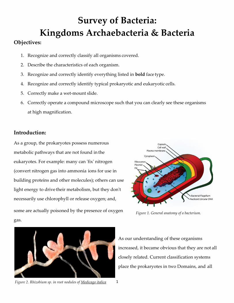
Figure 2. Rhizobium sp. in root nodules of Medicago italica
Survey of Bacteria:
Kingdoms Archaebacteria & Bacteria Objectives:
1. Recognize and correctly classify all organisms covered.
2. Describe the characteristics of each organism.
3. Recognize and correctly identify everything listed in bold face type.
4. Recognize and correctly identify typical prokaryotic and eukaryotic cells.
5. Correctly make a wet-mount slide.
6. Correctly operate a compound microscope such that you can clearly see these organisms
at high magnification.
Introduction:
As a group, the prokaryotes possess numerous
metabolic pathways that are not found in the
eukaryotes. For example: many can 'fix' nitrogen
(convert nitrogen gas into ammonia ions for use in
building proteins and other molecules); others can use
light energy to drive their metabolism, but they don't
necessarily use chlorophyll or release oxygen; and,
some are actually poisoned by the presence of oxygen
gas.
As our understanding of these organisms
increased, it became obvious that they are not all
closely related. Current classification systems
place the prokaryotes in two Domains, and all
2
of the eukaryotes in another. Again, this underscores their extreme amounts of difference, both
when compared to each other and when compared to the eukaryotes. In this lab, we will briefly
survey the structural variability and some of the metabolic diversity of these groups.
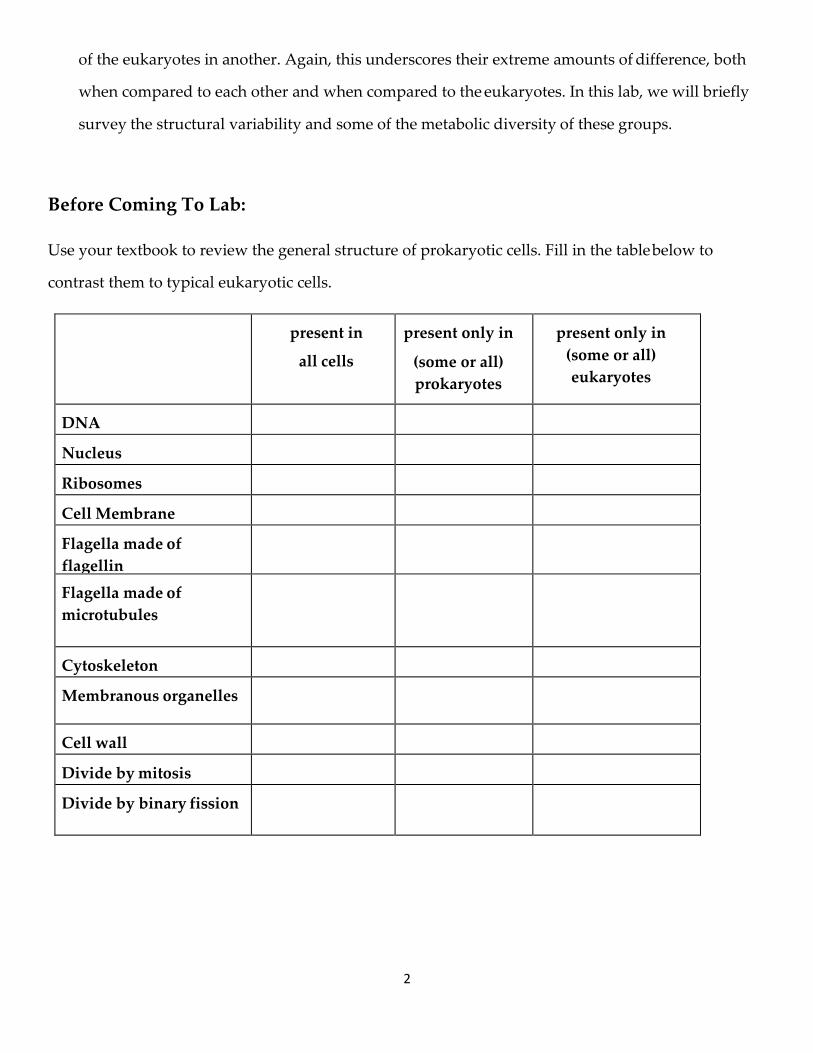
Before Coming To Lab:
Use your textbook to review the general structure of prokaryotic cells. Fill in the table below to
contrast them to typical eukaryotic cells.
present in
all cells
present only in
(some or all)
prokaryotes
present only in
(some or all)
eukaryotes
DNA
Nucleus
Ribosomes
Cell Membrane
Flagella made of
flagellin
Flagella made of
microtubules
Cytoskeleton
Membranous organelles
Cell wall
Divide by mitosis
Divide by binary fission
3
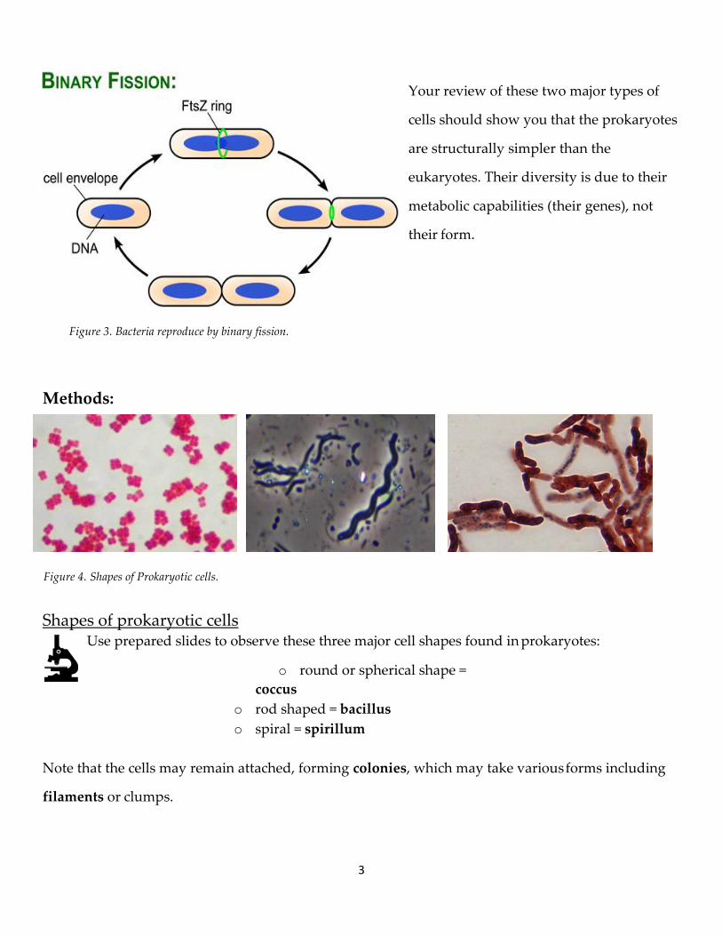
Figure 3. Bacteria reproduce by binary fission.
Your review of these two major types of
cells should show you that the prokaryotes
are structurally simpler than the
eukaryotes. Their diversity is due to their
metabolic capabilities (their genes), not
their form.
Methods:
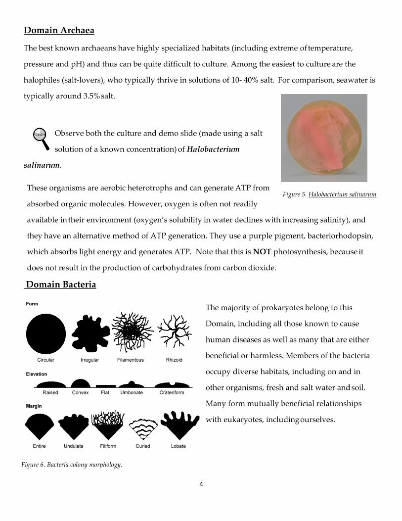
Shapes of prokaryotic cells Use prepared slides to observe these three major cell shapes found in prokaryotes:
o round or spherical shape =
coccus
o rod shaped = bacillus
o spiral = spirillum
Note that the cells may remain attached, forming colonies, which may take various forms including
filaments or clumps.
Figure 4. Shapes of Prokaryotic cells.
4
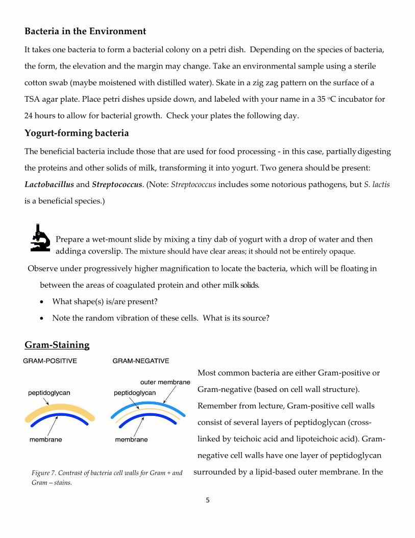
Figure 5. Halobacterium salinarum
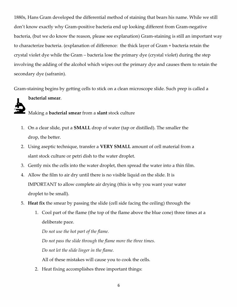
Figure 6. Bacteria colony morphology.
Domain Archaea
The best known archaeans have highly specialized habitats (including extreme of temperature,
pressure and pH) and thus can be quite difficult to culture. Among the easiest to culture are the
halophiles (salt-lovers), who typically thrive in solutions of 10- 40% salt. For comparison, seawater is
typically around 3.5% salt.
Observe both the culture and demo slide (made using a salt
solution of a known concentration) of Halobacterium
salinarum.
These organisms are aerobic heterotrophs and can generate ATP from
absorbed organic molecules. However, oxygen is often not readily
available in their environment (oxygen’s solubility in water declines with increasing salinity), and
they have an alternative method of ATP generation. They use a purple pigment, bacteriorhodopsin,
which absorbs light energy and generates ATP. Note that this is NOT photosynthesis, because it
does not result in the production of carbohydrates from carbon dioxide.
Domain Bacteria
The majority of prokaryotes belong to this
Domain, including all those known to cause
human diseases as well as many that are either
beneficial or harmless. Members of the bacteria
occupy diverse habitats, including on and in
other organisms, fresh and salt water and soil.
Many form mutually beneficial relationships
with eukaryotes, including ourselves.
5
Figure 7. Contrast of bacteria cell walls for Gram + and
Gram – stains.
Bacteria in the Environment
It takes one bacteria to form a bacterial colony on a petri dish. Depending on the species of bacteria,
the form, the elevation and the margin may change. Take an environmental sample using a sterile
cotton swab (maybe moistened with distilled water). Skate in a zig zag pattern on the surface of a
TSA agar plate. Place petri dishes upside down, and labeled with your name in a 35 oC incubator for
24 hours to allow for bacterial growth. Check your plates the following day.
Yogurt-forming bacteria
The beneficial bacteria include those that are used for food processing - in this case, partially digesting
the proteins and other solids of milk, transforming it into yogurt. Two genera should be present:
Lactobacillus and Streptococcus. (Note: Streptococcus includes some notorious pathogens, but S. lactis
is a beneficial species.)
Prepare a wet-mount slide by mixing a tiny dab of yogurt with a drop of water and then
adding a coverslip. The mixture should have clear areas; it should not be entirely opaque.
Observe under progressively higher magnification to locate the bacteria, which will be floating in
between the areas of coagulated protein and other milk solids.
What shape(s) is/are present?
Note the random vibration of these cells. What is its source?
Gram-Staining
Most common bacteria are either Gram-positive or
Gram-negative (based on cell wall structure).
Remember from lecture, Gram-positive cell walls
consist of several layers of peptidoglycan (cross-
linked by teichoic acid and lipoteichoic acid). Gram-
negative cell walls have one layer of peptidoglycan
surrounded by a lipid-based outer membrane. In the
6
1880s, Hans Gram developed the differential method of staining that bears his name. While we still
don’t know exactly why Gram-positive bacteria end up looking different from Gram-negative
bacteria, (but we do know the reason, please see explanation) Gram-staining is still an important way
to characterize bacteria. (explanation of difference: the thick layer of Gram + bacteria retain the
crystal violet dye while the Gram – bacteria lose the primary dye (crystal violet) during the step
involving the adding of the alcohol which wipes out the primary dye and causes them to retain the
secondary dye (safranin).
Gram-staining begins by getting cells to stick on a clean microscope slide. Such prep is called a
bacterial smear.
Making a bacterial smear from a slant stock culture
1. On a clear slide, put a SMALL drop of water (tap or distilled). The smaller the
drop, the better.
2. Using aseptic technique, transfer a VERY SMALL amount of cell material from a
slant stock culture or petri dish to the water droplet.
3. Gently mix the cells into the water droplet, then spread the water into a thin film.
4. Allow the film to air dry until there is no visible liquid on the slide. It is
IMPORTANT to allow complete air drying (this is why you want your water
droplet to be small).
5. Heat fix the smear by passing the slide (cell side facing the ceiling) through the
1. Cool part of the flame (the top of the flame above the blue cone) three times at a
deliberate pace.
Do not use the hot part of the flame.
Do not pass the slide through the flame more the three times.
Do not let the slide linger in the flame.
All of these mistakes will cause you to cook the cells.
2. Heat fixing accomplishes three important things:
7
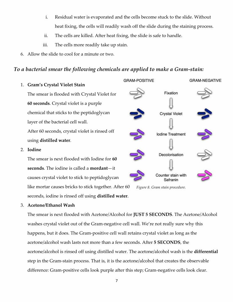
Figure 8. Gram stain procedure.
i. Residual water is evaporated and the cells become stuck to the slide. Without
heat fixing, the cells will readily wash off the slide during the staining process.
ii. The cells are killed. After heat fixing, the slide is safe to handle.
iii. The cells more readily take up stain.
6. Allow the slide to cool for a minute or two.
To a bacterial smear the following chemicals are applied to make a Gram-stain:
1. Gram’s Crystal Violet Stain
The smear is flooded with Crystal Violet for
60 seconds. Crystal violet is a purple
chemical that sticks to the peptidoglycan
layer of the bacterial cell wall.
After 60 seconds, crystal violet is rinsed off
using distilled water.
2. Iodine
The smear is next flooded with Iodine for 60
seconds. The iodine is called a mordant—it
causes crystal violet to stick to peptidoglycan
like mortar causes bricks to stick together. After 60
seconds, iodine is rinsed off using distilled water.
3. Acetone/Ethanol Wash
The smear is next flooded with Acetone/Alcohol for JUST 5 SECONDS. The Acetone/Alcohol
washes crystal violet out of the Gram-negative cell wall. We’re not really sure why this
happens, but it does. The Gram-positive cell wall retains crystal violet as long as the
acetone/alcohol wash lasts not more than a few seconds. After 5 SECONDS, the
acetone/alcohol is rinsed off using distilled water. The acetone/alcohol wash is the differential
step in the Gram-stain process. That is, it is the acetone/alcohol that creates the observable
difference: Gram-positive cells look purple after this step; Gram-negative cells look clear.
8
4. Safranin Stain
The smear is flooded with Safranin for 90 seconds to two minutes. Safranin is a pink stain that
sticks to cytoplasmic components of the cell. All cells become stained with Safranin. Gram-
positive cells are pink on the inside, but you can’t see this because they are dark purple on the
outside (kind of like a bon-bon). Gram- negative cells, which were cleared in the previous step,
end up looking pink. After 90 seconds to two minutes (the longer the better), Safranin is rinsed
off with distilled water.
5. Drying the slide
The completed Gram-stained slide is stuck into a book of bibulous paper and allowed to dry for a
minute or two. Once dry, the slide is ready for observation.
Microscopic observations of the Gram-stains
To observe cell structure and stain color, YOU MUST VIEW CELLS THROUGH THE OIL
IMMERSION LENS (total magnification = 1000x). At lower magnification, the cells will be
too small to reliably determine shape, arrangement, and color.
1. Observe the smear using the 4x, 10x, and 40x lenses. When in focus at high power, move the
40x lens out of the way.
2. Place a drop of immersion oil on the slide in the field of view.
3. Rotate the 100x (oil immersion) lens into place.
CAUTION: The 100x lens is built to be put into oil. The 40x lens is NOT. Oil will leak into the
barrel of the 40x lens and ruin it. NEVER, EVER USE THE 40X LENS WHEN OIL IS ON THE
SLIDE.
5. Use the fine focus adjustment to focus the view. Be very subtle with your focusing. Even the
slightest exaggeration of focusing will cause you to overshoot. Sometimes, focusing with the
oil immersion lens takes a great deal of patience. Remember to jiggle the slide back and forth.
6. Describe cell size, shape, arrangement, and Gram-reaction for your smears.
9
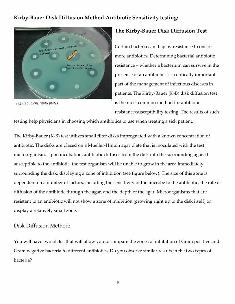
Figure 9. Sensitivity plates.
Kirby-Bauer Disk Diffusion Method-Antibiotic Sensitivity testing:
The Kirby-Bauer Disk Diffusion Test
Certain bacteria can display resistance to one or
more antibiotics. Determining bacterial antibiotic
resistance – whether a bacterium can survive in the
presence of an antibiotic - is a critically important
part of the management of infectious diseases in
patients. The Kirby-Bauer (K-B) disk diffusion test
is the most common method for antibiotic
resistance/susceptibility testing. The results of such
testing help physicians in choosing which antibiotics to use when treating a sick patient.
The Kirby-Bauer (K-B) test utilizes small filter disks impregnated with a known concentration of
antibiotic. The disks are placed on a Mueller-Hinton agar plate that is inoculated with the test
microorganism. Upon incubation, antibiotic diffuses from the disk into the surrounding agar. If
susceptible to the antibiotic, the test organism will be unable to grow in the area immediately
surrounding the disk, displaying a zone of inhibition (see figure below). The size of this zone is
dependent on a number of factors, including the sensitivity of the microbe to the antibiotic, the rate of
diffusion of the antibiotic through the agar, and the depth of the agar. Microorganisms that are
resistant to an antibiotic will not show a zone of inhibition (growing right up to the disk itself) or
display a relatively small zone.
Disk Diffusion Method:
You will have two plates that will allow you to compare the zones of inhibition of Gram positive and
Gram negative bacteria to different antibiotics. Do you observe similar results in the two types of
bacteria?
10
Typical Photosynthetic Bacteria – Cyanobacteria
These bacteria are the only prokaryotic group which does 'typical' photosynthesis, using
chlorophyll a and releasing oxygen from water. That means they do not have membrane-
bound organelles within their cells. Their physical structure is limited to the cell wall, which is
made of peptidoglycan, the plasma membrane, which has the same structure as in plants and
animals, and inclusions, a form of food storage. However, there is strong evidence to suggest that
the organelles found in plants and animals (the mitochondria and chloroplasts) are actually the
descendants of bacteria that were engulfed by other cells. Studying cyanobacteria may give us
insight into the early evolution of plants, and the photosynthetic process.
Cyanobacteria are the closest relatives of the eukaryotic chloroplast. They also include species with
some of the largest prokaryotic cells and some which show limited cooperation between cells and
specialization of function.
Representative Cyanobacteria
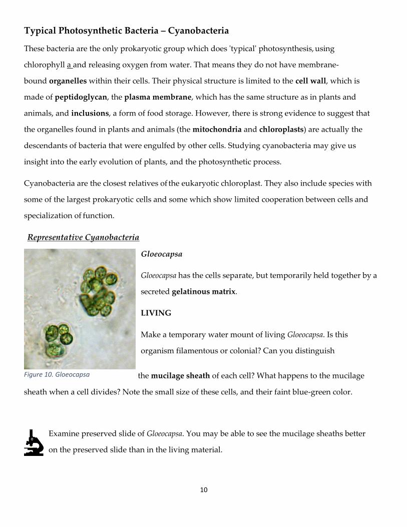
Gloeocapsa
Gloeocapsa has the cells separate, but temporarily held together by a
secreted gelatinous matrix.
LIVING
Make a temporary water mount of living Gloeocapsa. Is this
organism filamentous or colonial? Can you distinguish
the mucilage sheath of each cell? What happens to the mucilage
sheath when a cell divides? Note the small size of these cells, and their faint blue-green color.
Examine preserved slide of Gloeocapsa. You may be able to see the mucilage sheaths better
on the preserved slide than in the living material.
Figure 10. Gloeocapsa
11
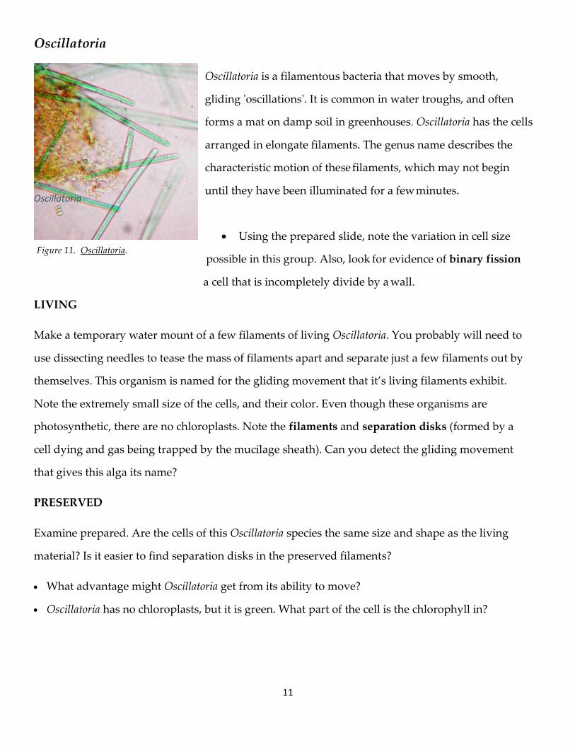
Figure 11. Oscillatoria.
Oscillatoria
Oscillatoria is a filamentous bacteria that moves by smooth,
gliding 'oscillations'. It is common in water troughs, and often
forms a mat on damp soil in greenhouses. Oscillatoria has the cells
arranged in elongate filaments. The genus name describes the
characteristic motion of these filaments, which may not begin
until they have been illuminated for a few minutes.
Using the prepared slide, note the variation in cell size
possible in this group. Also, look for evidence of binary fission
a cell that is incompletely divide by a wall.
LIVING
Make a temporary water mount of a few filaments of living Oscillatoria. You probably will need to
use dissecting needles to tease the mass of filaments apart and separate just a few filaments out by
themselves. This organism is named for the gliding movement that it’s living filaments exhibit.
Note the extremely small size of the cells, and their color. Even though these organisms are
photosynthetic, there are no chloroplasts. Note the filaments and separation disks (formed by a
cell dying and gas being trapped by the mucilage sheath). Can you detect the gliding movement
that gives this alga its name?
PRESERVED
Examine prepared. Are the cells of this Oscillatoria species the same size and shape as the living
material? Is it easier to find separation disks in the preserved filaments?
What advantage might Oscillatoria get from its ability to move?
Oscillatoria has no chloroplasts, but it is green. What part of the cell is the chlorophyll in?
Oscillatoria
12
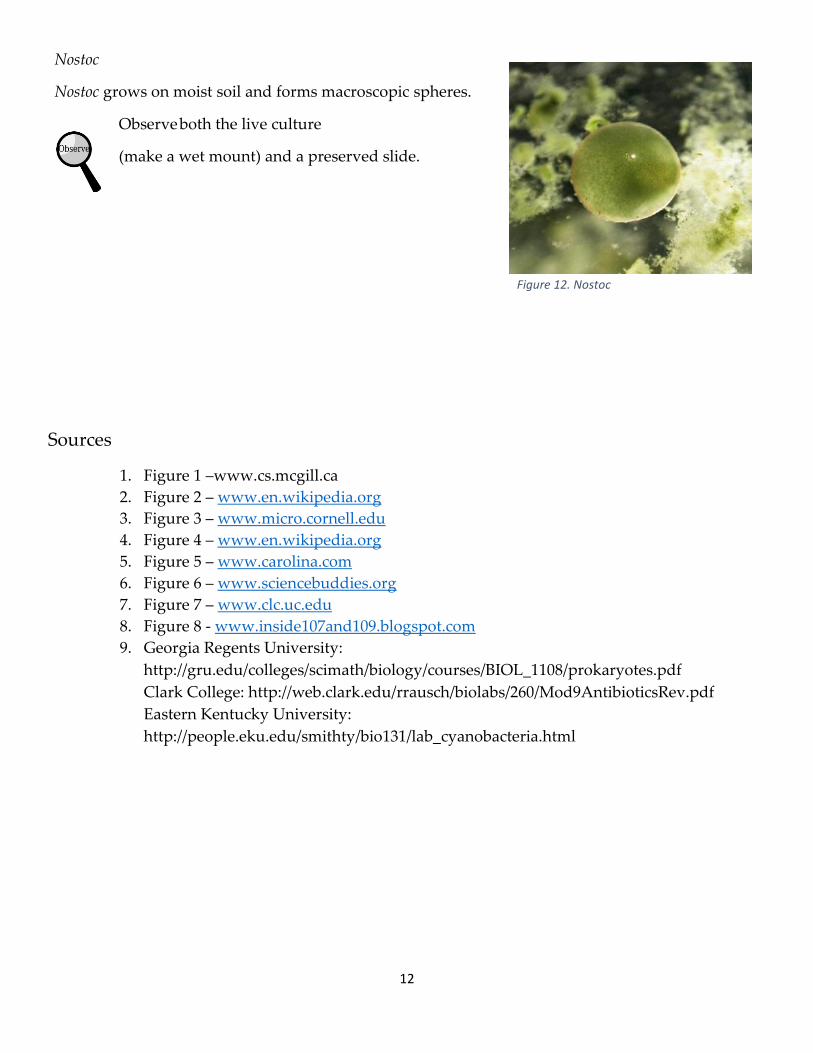
Figure 12. Nostoc
Nostoc
Nostoc grows on moist soil and forms macroscopic spheres.
Observe both the live culture
(make a wet mount) and a preserved slide.
Sources
1. Figure 1 –www.cs.mcgill.ca
2. Figure 2 – www.en.wikipedia.org
3. Figure 3 – www.micro.cornell.edu
4. Figure 4 – www.en.wikipedia.org
5. Figure 5 – www.carolina.com
6. Figure 6 – www.sciencebuddies.org
7. Figure 7 – www.clc.uc.edu
8. Figure 8 - www.inside107and109.blogspot.com
9. Georgia Regents University:
http://gru.edu/colleges/scimath/biology/courses/BIOL_1108/prokaryotes.pdf
Clark College: http://web.clark.edu/rrausch/biolabs/260/Mod9AntibioticsRev.pdf
Eastern Kentucky University:
http://people.eku.edu/smithty/bio131/lab_cyanobacteria.html