Embed Size (px)
Citation preview
1
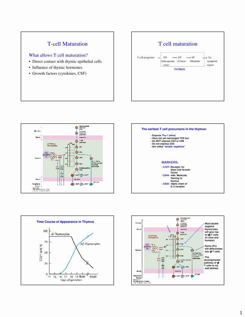
T-cell Maturation
What allows T cell maturation?
• Direct contact with thymic epithelial cells
• Influence of thymic hormones
• Growth factors (cytokines, CSF)
T cell maturation
T cell progenitor DN DP SP 2ry
(Subcapsular (Cortex) (Medulla) lymphoid
zone) organs
THYMUS
The earliest T cell precursors in the thymus:
- Express Thy-1 (mice)
- Have not yet rearranged TCR loci
- Do NOT express CD4 or CD8
- Do not express CD3
- Are called “double negatives”
MARKERS:
- C-KIT- Receptor for
Stem Cell Growth
Factor
- CD44 - Adh. Molecule.
Homing to
thymus
- CD25 - Alpha chain of
IL-2 receptor
Time Course of Appearance in Thymus- Most double
negative
thymocytes
will give rise
to αβαβαβαβ T cells
(in mice and
humans).
- Some (5%)
will differentiate
into γδγδγδγδ T cells.
- The
developmental
pathway of γδγδγδγδ
T cells is not
well defined.
*
2
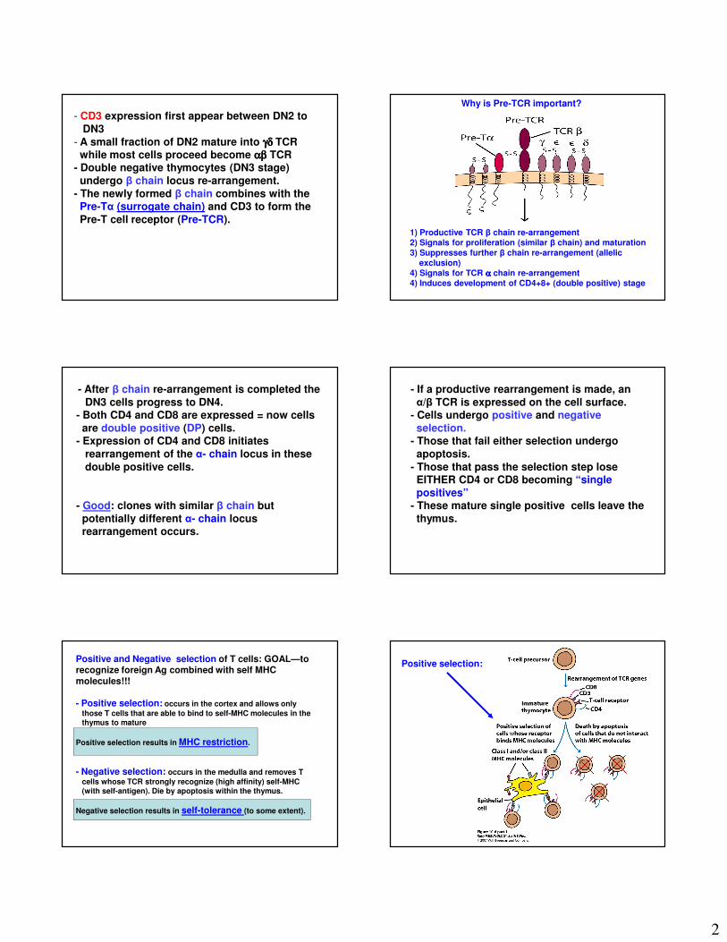
- CD3 expression first appear between DN2 to
DN3
- A small fraction of DN2 mature into γδγδγδγδ TCR while most cells proceed become αβαβαβαβ TCR
- Double negative thymocytes (DN3 stage)
undergo β chain locus re-arrangement.
- The newly formed β chain combines with the Pre-Tα (surrogate chain) and CD3 to form the
Pre-T cell receptor (Pre-TCR).1) Productive TCR β chain re-arrangement2) Signals for proliferation (similar β chain) and maturation3) Suppresses further β chain re-arrangement (allelic
exclusion)4) Signals for TCR αααα chain re-arrangement4) Induces development of CD4+8+ (double positive) stage
Why is Pre-TCR important?
- After β chain re-arrangement is completed the
DN3 cells progress to DN4.- Both CD4 and CD8 are expressed = now cells
are double positive (DP) cells.- Expression of CD4 and CD8 initiates
rearrangement of the α- chain locus in these
double positive cells.
- Good: clones with similar β chain but
potentially different α- chain locus rearrangement occurs.
- If a productive rearrangement is made, an
α/β TCR is expressed on the cell surface.- Cells undergo positive and negative
selection.- Those that fail either selection undergo
apoptosis.
- Those that pass the selection step lose EITHER CD4 or CD8 becoming “single
positives”- These mature single positive cells leave the
thymus.
Positive and Negative selection of T cells: GOAL—to recognize foreign Ag combined with self MHC molecules!!!
- Positive selection: occurs in the cortex and allows only
those T cells that are able to bind to self-MHC molecules in the
thymus to mature
Positive selection results in MHC restriction.
- Negative selection: occurs in the medulla and removes T
cells whose TCR strongly recognize (high affinity) self-MHC
(with self-antigen). Die by apoptosis within the thymus.
Negative selection results in self-tolerance (to some extent).
Positive selection:
3
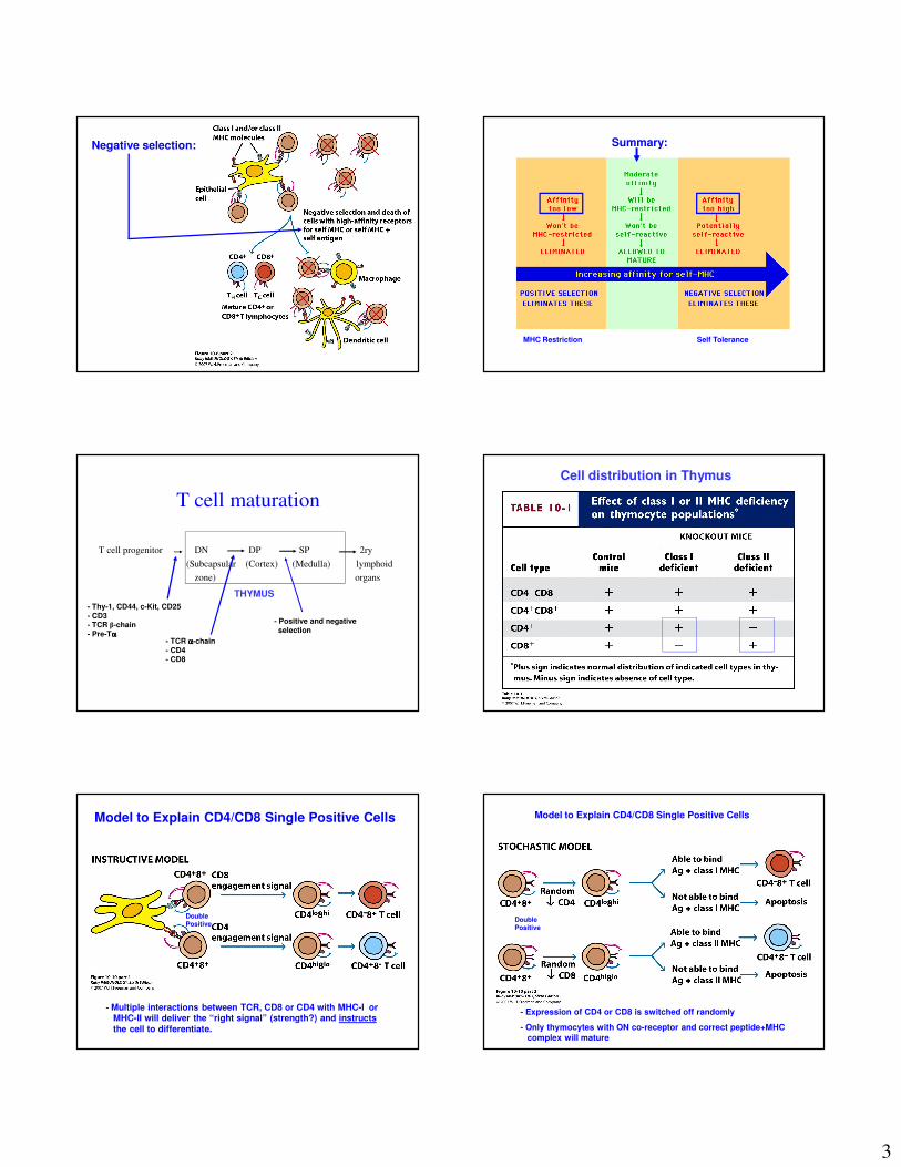
Negative selection: Summary:
MHC Restriction Self Tolerance
T cell maturation
T cell progenitor DN DP SP 2ry
(Subcapsular (Cortex) (Medulla) lymphoid
zone) organs
THYMUS
- Thy-1, CD44, c-Kit, CD25
- CD3- TCR β-chain
- Pre-Tαααα- TCR αααα-chain
- CD4- CD8
- Positive and negative
selection
Cell distribution in Thymus
Model to Explain CD4/CD8 Single Positive Cells
- Multiple interactions between TCR, CD8 or CD4 with MHC-I or MHC-II will deliver the “right signal” (strength?) and instructs
the cell to differentiate.
Double Positive
Model to Explain CD4/CD8 Single Positive Cells
- Expression of CD4 or CD8 is switched off randomly
- Only thymocytes with ON co-receptor and correct peptide+MHC
complex will mature
Double Positive
4
Summary of T cell maturation (αβαβαβαβ T cells only)
- Thymocytes enter the thymus as "double negative“ (markers?)
- Induces β-chain rearrangement (apoptosis of cells that fail to rearrange ββββ
chain correctly)
- Expression of pre-TCR (surrogate α chain)
- Proliferation of similar β-chain clones with surrogate αααα-chain
- Expression of CD4 and CD8 (to form "double positive" thymocytes)
-α-chain rearrangement (apoptosis of cells that fail to rearrange αααα correctly)
- Expression of mature αβαβαβαβ TCR
-Positive and negative selection (death of cells with too low or too high an
affinity for self MHC…>99% of thymocytes die within the thymus)
-Loss of either CD4 or CD8
-Migration to periphery of cells that successfully complete these steps
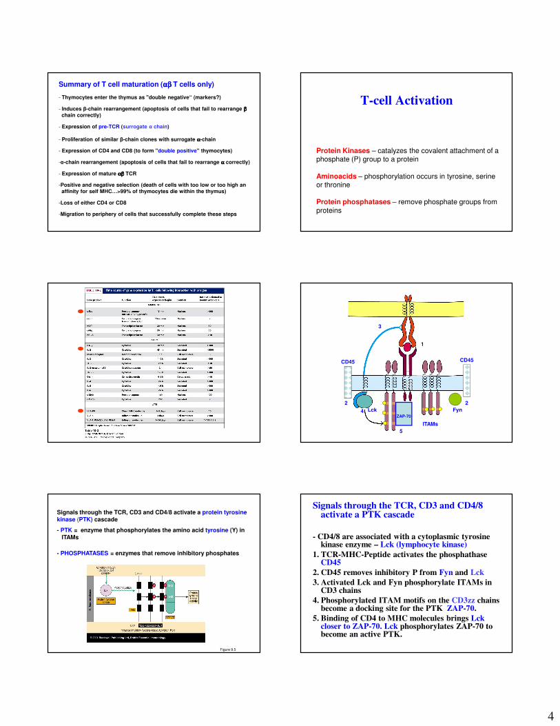
T-cell Activation
Protein Kinases – catalyzes the covalent attachment of a phosphate (P) group to a protein
Aminoacids – phosphorylation occurs in tyrosine, serine or thronine
Protein phosphatases – remove phosphate groups from
proteins
1
3
5
4 4
ITAMs
Lck FynZAP-70
CD45CD45
2 2
Figure 9.5
Signals through the TCR, CD3 and CD4/8 activate a protein tyrosine kinase (PTK) cascade
- PTK = enzyme that phosphorylates the amino acid tyrosine (Y) in
ITAMs
- PHOSPHATASES = enzymes that remove inhibitory phosphates
Signals through the TCR, CD3 and CD4/8 activate a PTK cascade
- CD4/8 are associated with a cytoplasmic tyrosine kinase enzyme – Lck (lymphocyte kinase)
1. TCR-MHC-Peptide activates the phosphathase CD45
2. CD45 removes inhibitory P from Fyn and Lck
3. Activated Lck and Fyn phosphorylate ITAMs in CD3 chains
4. Phosphorylated ITAM motifs on the CD3zz chains become a docking site for the PTK ZAP-70.
5. Binding of CD4 to MHC molecules brings Lck closer to ZAP-70. Lck phosphorylates ZAP-70 to become an active PTK.
5
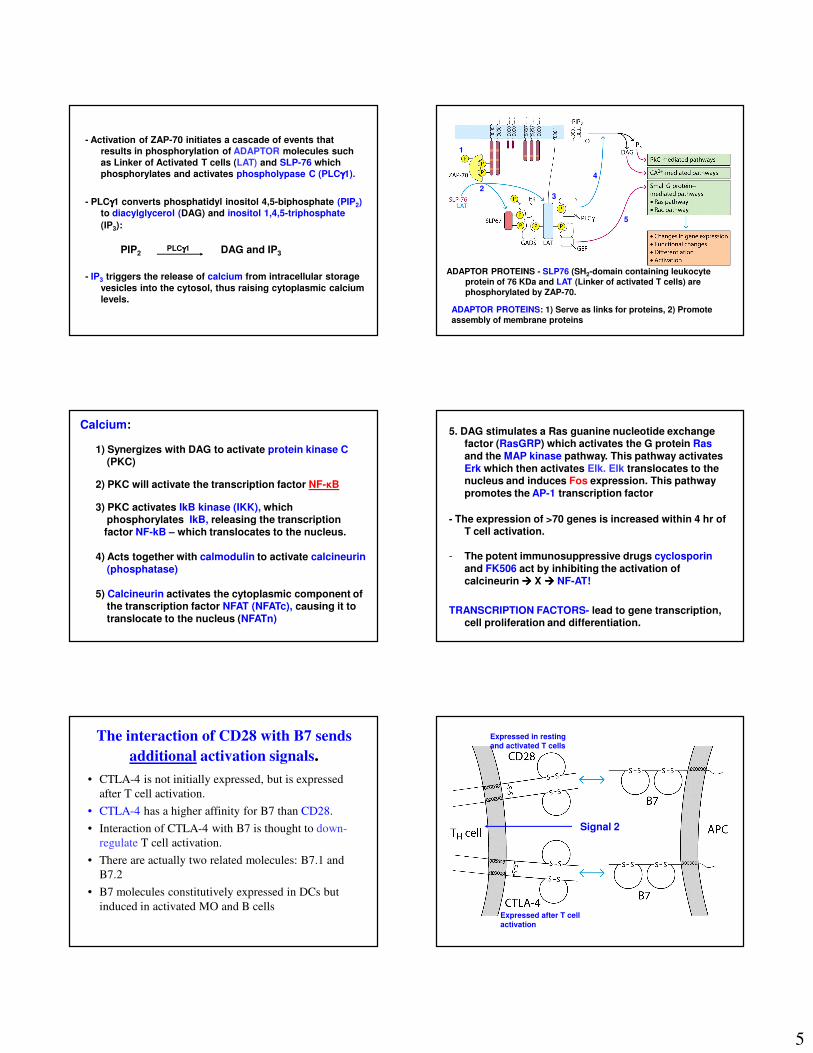
- Activation of ZAP-70 initiates a cascade of events that results in phosphorylation of ADAPTOR molecules such as Linker of Activated T cells (LAT) and SLP-76 which phosphorylates and activates phospholypase C (PLCγγγγ1).
- PLCγγγγ1 converts phosphatidyl inositol 4,5-biphosphate (PIP2)to diacylglycerol (DAG) and inositol 1,4,5-triphosphate
(IP3):
PIP2 DAG and IP3
- IP3 triggers the release of calcium from intracellular storage
vesicles into the cytosol, thus raising cytoplasmic calcium levels.
PLCγγγγ1
ADAPTOR PROTEINS: 1) Serve as links for proteins, 2) Promote assembly of membrane proteins
ADAPTOR PROTEINS - SLP76 (SH2-domain containing leukocyte protein of 76 KDa and LAT (Linker of activated T cells) are phosphorylated by ZAP-70.
2
1
3
4
5
Calcium:
1) Synergizes with DAG to activate protein kinase C(PKC)
2) PKC will activate the transcription factor NF-κB
3) PKC activates IkB kinase (IKK), which phosphorylates IkB, releasing the transcription factor NF-kB – which translocates to the nucleus.
4) Acts together with calmodulin to activate calcineurin (phosphatase)
5) Calcineurin activates the cytoplasmic component of the transcription factor NFAT (NFATc), causing it totranslocate to the nucleus (NFATn)
5. DAG stimulates a Ras guanine nucleotide exchange factor (RasGRP) which activates the G protein Rasand the MAP kinase pathway. This pathway activates Erk which then activates Elk. Elk translocates to the nucleus and induces Fos expression. This pathway promotes the AP-1 transcription factor
- The expression of >70 genes is increased within 4 hr of T cell activation.
- The potent immunosuppressive drugs cyclosporin and FK506 act by inhibiting the activation of calcineurin ���� X ���� NF-AT!
TRANSCRIPTION FACTORS- lead to gene transcription, cell proliferation and differentiation.
The interaction of CD28 with B7 sends
additional activation signals.
• CTLA-4 is not initially expressed, but is expressed
after T cell activation.
• CTLA-4 has a higher affinity for B7 than CD28.
• Interaction of CTLA-4 with B7 is thought to down-
regulate T cell activation.
• There are actually two related molecules: B7.1 and
B7.2
• B7 molecules constitutively expressed in DCs but
induced in activated MO and B cellsExpressed after T cell
activation
Expressed in resting
and activated T cells
Signal 2
6
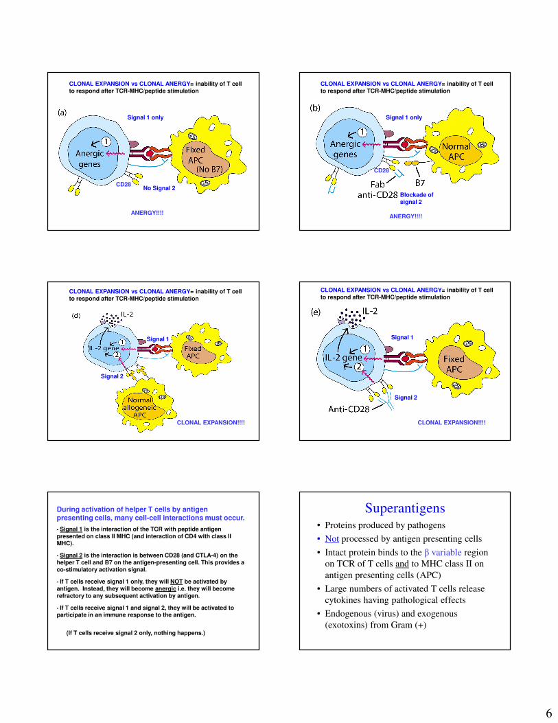
CLONAL EXPANSION vs CLONAL ANERGY= inability of T cell to respond after TCR-MHC/peptide stimulation
Signal 1 only
No Signal 2
ANERGY!!!!
CD28
Signal 1 only
Blockade of signal 2
CLONAL EXPANSION vs CLONAL ANERGY= inability of T cell to respond after TCR-MHC/peptide stimulation
ANERGY!!!!
CD28
Signal 1
Signal 2
CLONAL EXPANSION!!!!
CLONAL EXPANSION vs CLONAL ANERGY= inability of T cell to respond after TCR-MHC/peptide stimulation
Signal 1
Signal 2
CLONAL EXPANSION!!!!
CLONAL EXPANSION vs CLONAL ANERGY= inability of T cell to respond after TCR-MHC/peptide stimulation
During activation of helper T cells by antigen presenting cells, many cell-cell interactions must occur.
- Signal 1 is the interaction of the TCR with peptide antigen presented on class II MHC (and interaction of CD4 with class II MHC).
- Signal 2 is the interaction is between CD28 (and CTLA-4) on the
helper T cell and B7 on the antigen-presenting cell. This provides a co-stimulatory activation signal.
- If T cells receive signal 1 only, they will NOT be activated by antigen. Instead, they will become anergic i.e. they will become
refractory to any subsequent activation by antigen.
- If T cells receive signal 1 and signal 2, they will be activated to participate in an immune response to the antigen.
(If T cells receive signal 2 only, nothing happens.)
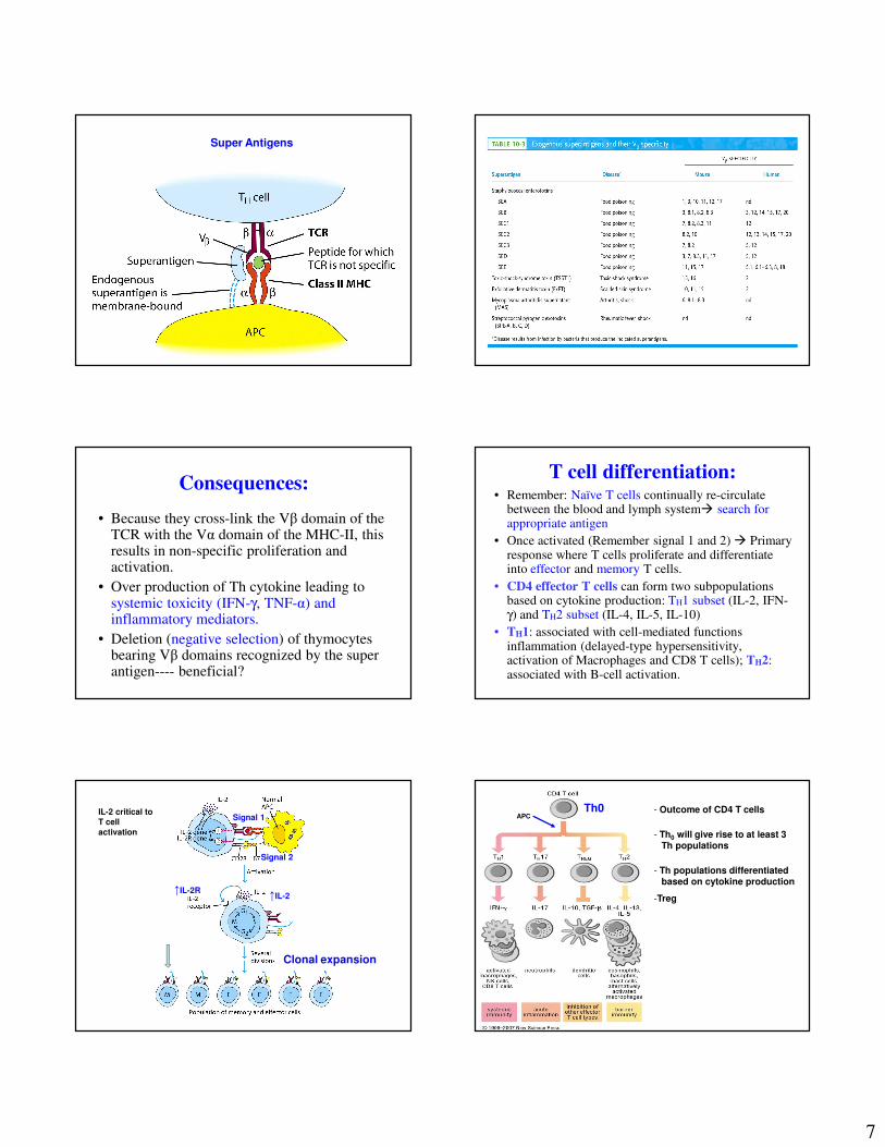
Superantigens• Proteins produced by pathogens
• Not processed by antigen presenting cells
• Intact protein binds to the β variable region
on TCR of T cells and to MHC class II on
antigen presenting cells (APC)
• Large numbers of activated T cells release
cytokines having pathological effects
• Endogenous (virus) and exogenous
(exotoxins) from Gram (+)
7
Super Antigens
Consequences:
• Because they cross-link the Vβ domain of the TCR with the Vα domain of the MHC-II, this results in non-specific proliferation and activation.
• Over production of Th cytokine leading to systemic toxicity (IFN-γ, TNF-α) and inflammatory mediators.
• Deletion (negative selection) of thymocytes bearing Vβ domains recognized by the super antigen---- beneficial?
T cell differentiation:• Remember: Naïve T cells continually re-circulate
between the blood and lymph system� search for appropriate antigen
• Once activated (Remember signal 1 and 2) � Primary response where T cells proliferate and differentiate into effector and memory T cells.
• CD4 effector T cells can form two subpopulations based on cytokine production: TH1 subset (IL-2, IFN-γ) and TH2 subset (IL-4, IL-5, IL-10)
• TH1: associated with cell-mediated functions inflammation (delayed-type hypersensitivity, activation of Macrophages and CD8 T cells); TH2: associated with B-cell activation.
Signal 1
Signal 2
↑IL-2↑IL-2R
Clonal expansion
IL-2 critical to
T cell
activation
- Outcome of CD4 T cells
- Th0 will give rise to at least 3Th populations
- Th populations differentiated based on cytokine production
-Treg
Th0APC
8
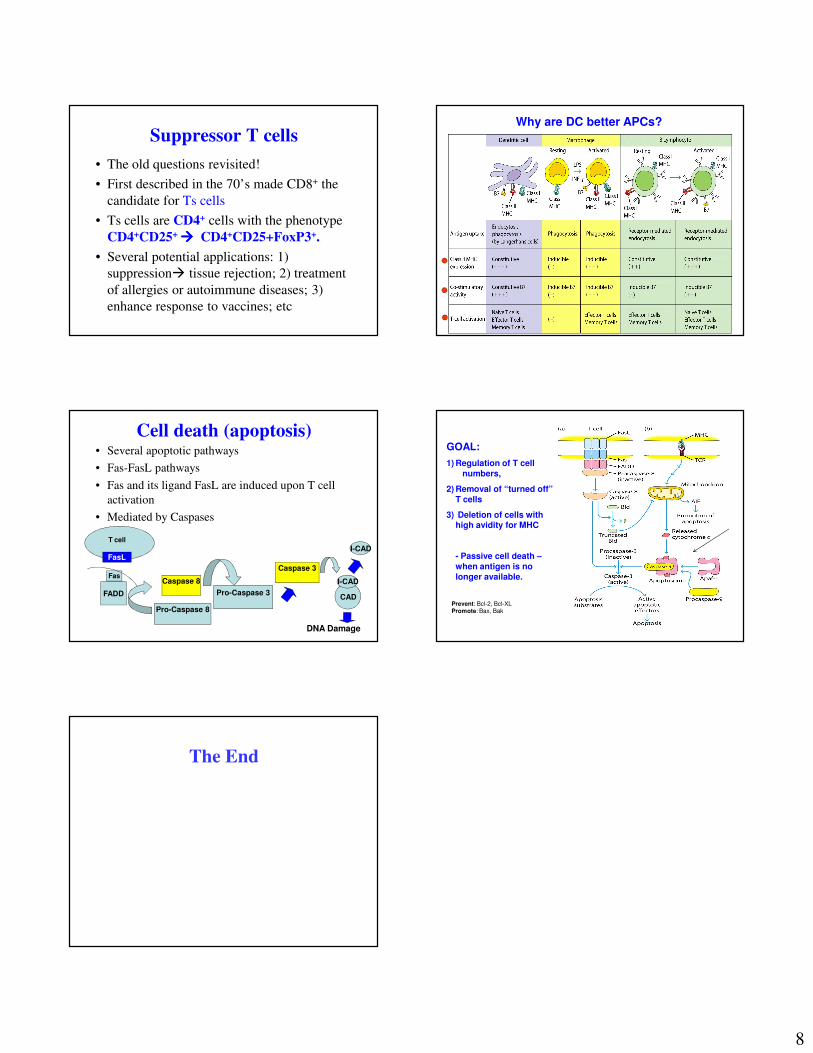
Suppressor T cells
• The old questions revisited!
• First described in the 70’s made CD8+ the
candidate for Ts cells
• Ts cells are CD4+ cells with the phenotype
CD4+CD25+ ���� CD4+CD25+FoxP3+.
• Several potential applications: 1)
suppression� tissue rejection; 2) treatment
of allergies or autoimmune diseases; 3)
enhance response to vaccines; etc
Why are DC better APCs?
Cell death (apoptosis)• Several apoptotic pathways
• Fas-FasL pathways
• Fas and its ligand FasL are induced upon T cell
activation
• Mediated by Caspases
Pro-Caspase 3
Fas
FasL
FADD
Pro-Caspase 8
Caspase 8
Caspase 3
CAD
I-CAD
I-CAD
DNA Damage
T cell
GOAL:
1) Regulation of T cell numbers,
2) Removal of “turned off” T cells
3) Deletion of cells with high avidity for MHC
- Passive cell death –when antigen is no longer available.
Prevent: Bcl-2, Bcl-XLPromote: Bax, Bak
The End