Embed Size (px)
Citation preview
Tartu 2009 I Introduction to Neuropathology
& General Aspects
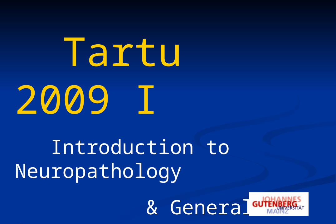
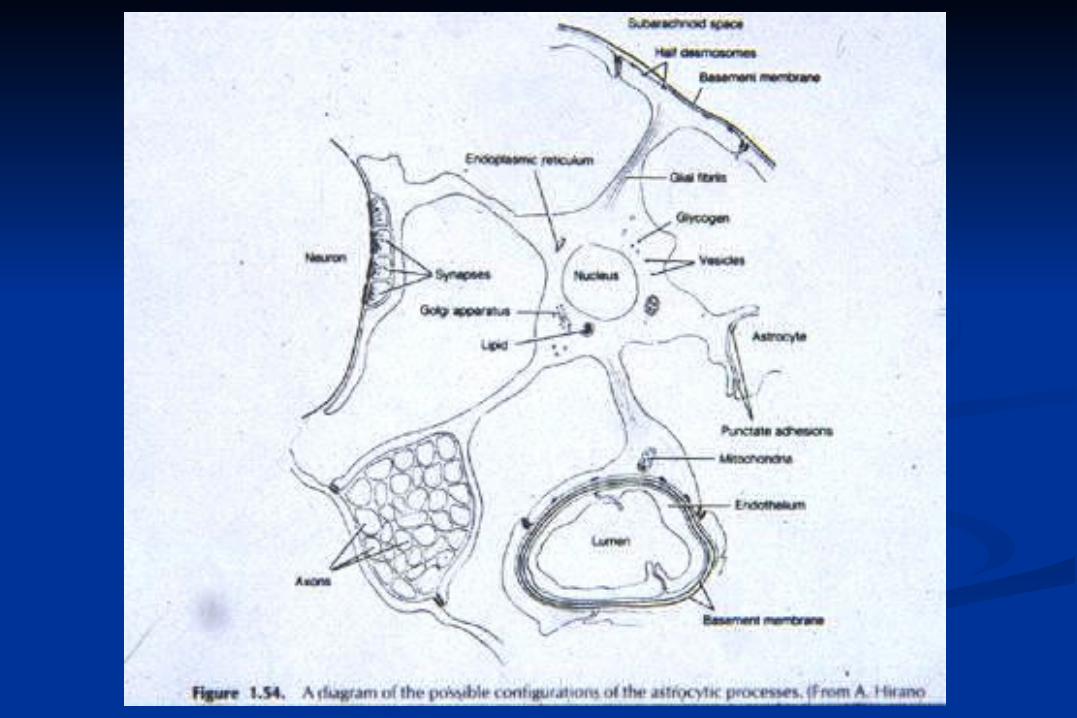
Special Features of the CNS
Complex and diverse topography
Complex and diverse cytology
Axoplasmic transport
Myelin
3 classes of intermediate filaments –
neurofilaments, glial fibrillary acidic protein, vimentin
Neurotransmitters
Separate population of interstitial cells-glia
Blood brain barrier
Cerebrospinal fluid
Absent lymphatic vessels and lymph nodes
Special aspects of cranial cavity (intracranial pressure)
Neuropathology – in a broader sense
Neurology of the
Central Nervous System (Brain and spinal cord, incl. their coverings) Peripheral Nervous System (and its coverings)
Skeletal Muscle
Neuropathology in a limited sense
Neuropathomorphology
General Neuropathology =
Neuropathology-related Special Features
Cell Pathology
General „Organ Pathology“
General Principles of neuropathologic disease groups
Neuropathology of
Nerve cells blood brain barrier
Glial cells peripheral nerves
Oligodendroglia skeletal muscle
Astroglia and Ependyma
Microglia
Keyhole Neuropathology
Brain biopsy
Nerve biopsy
extracerebral biopsy in
neurodegenerative diseasesin adultsin children
General Principles of Neuropathologic Groups of Diseases
Neurodegenerative Diseases Neurometabolic Diseases
Inflammatory Diseases Infections Autoimmune processes
Toxic Diseases
Malformations
TumorsCirculation Diseases
I. Pathologic Reactions in the CNS
II. Brain Edema
Neuronal Reactions
Pathology of Neuronal Processes
Dendrites
Axons
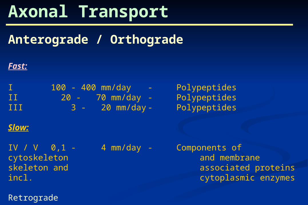
Axonal Transport
Anterograde / Orthograde
Fast:
I 100 - 400 mm/day - PolypeptidesII 20 - 70 mm/day - PolypeptidesIII 3 - 20 mm/day - Polypeptides
Slow:
IV / V 0,1 - 4 mm/day - Components of cytoskeleton and membrane skeleton and associated proteins incl. cytoplasmic enzymes
Retrograde
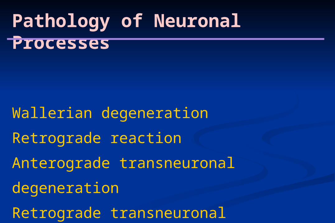
Pathology of Neuronal Processes
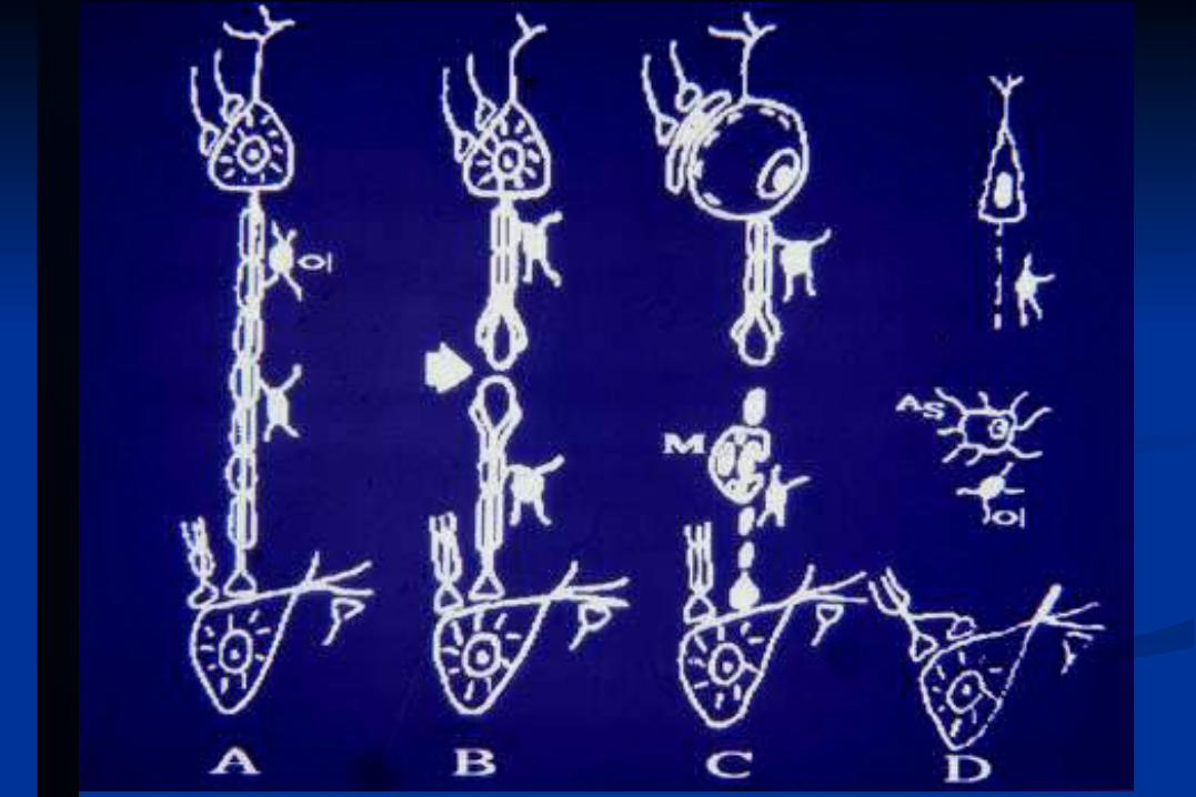
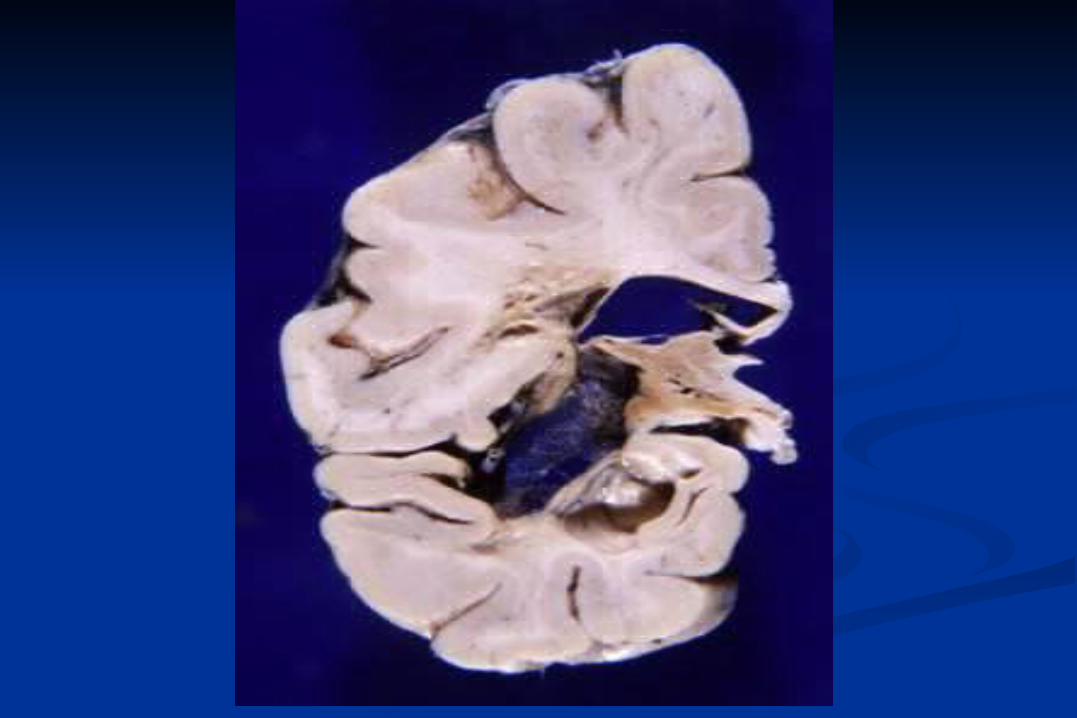
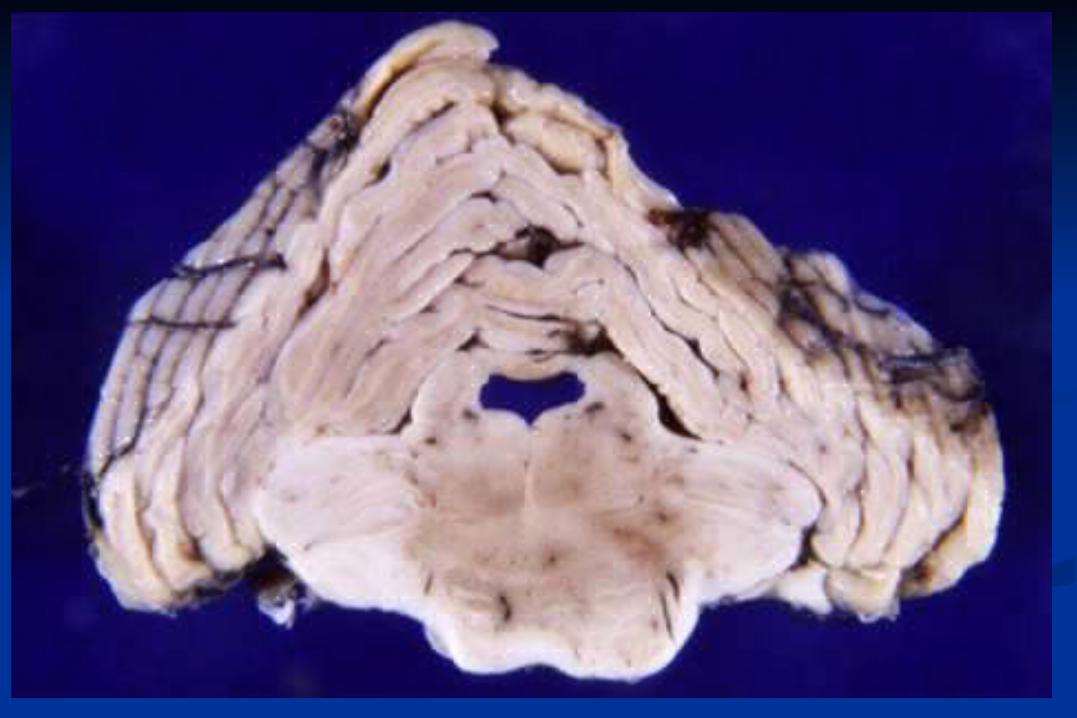
Wallerian degeneration
Retrograde reaction
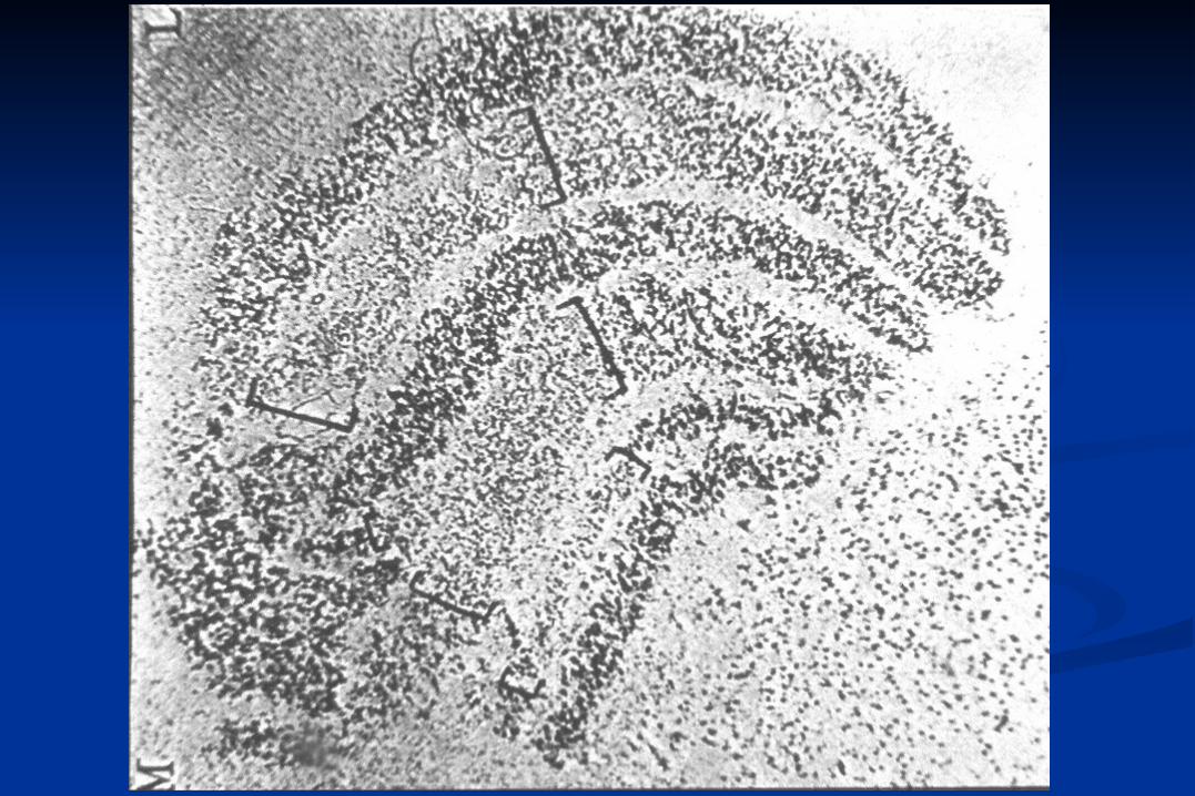
Anterograde transneuronal degeneration
Retrograde transneuronal degeneration
Wallerian Degeneration
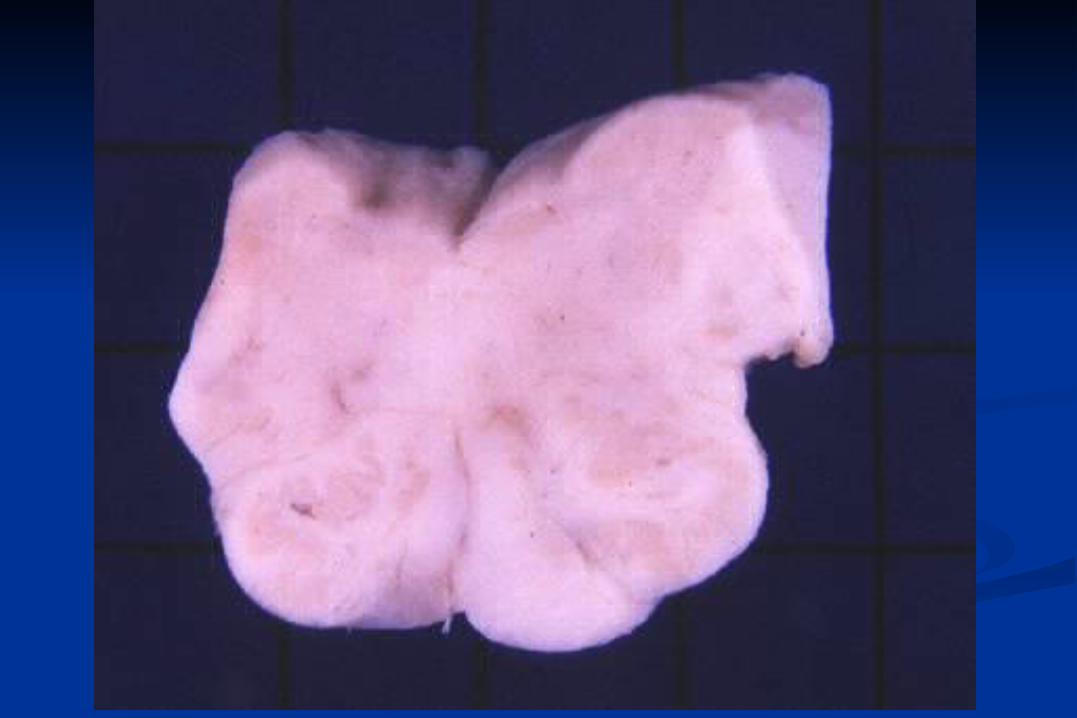
Anterogade transneuronal Degeneration
loss of eye lateral geniculate bodylesion of optic nerve
lesions of fornix mamillary bodies
loss of sensory gracilis and cuneate fibers in posterior nuclei
spinal columns
loss of cortico- pontine nucleipontine fibers
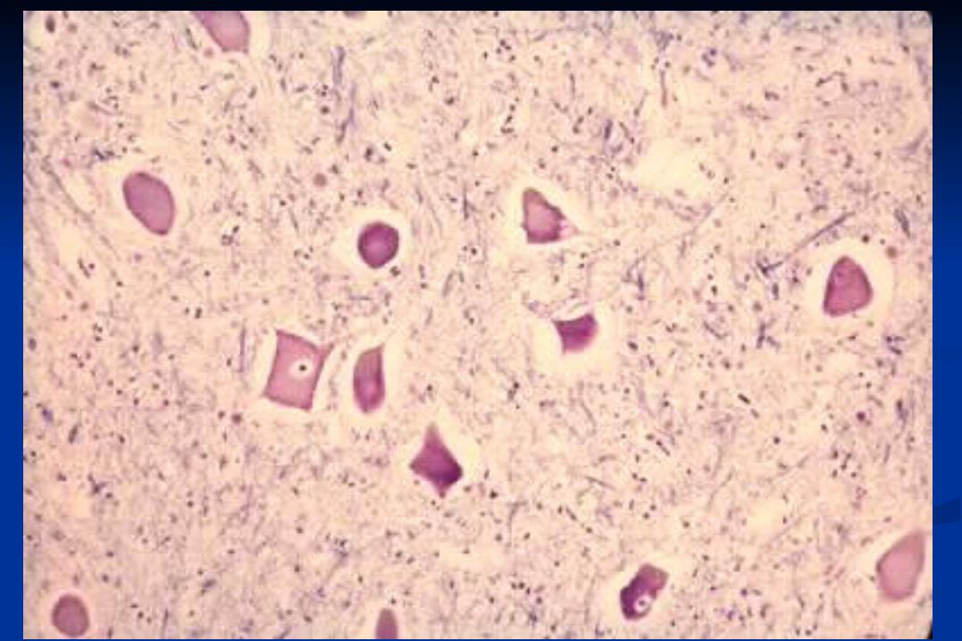
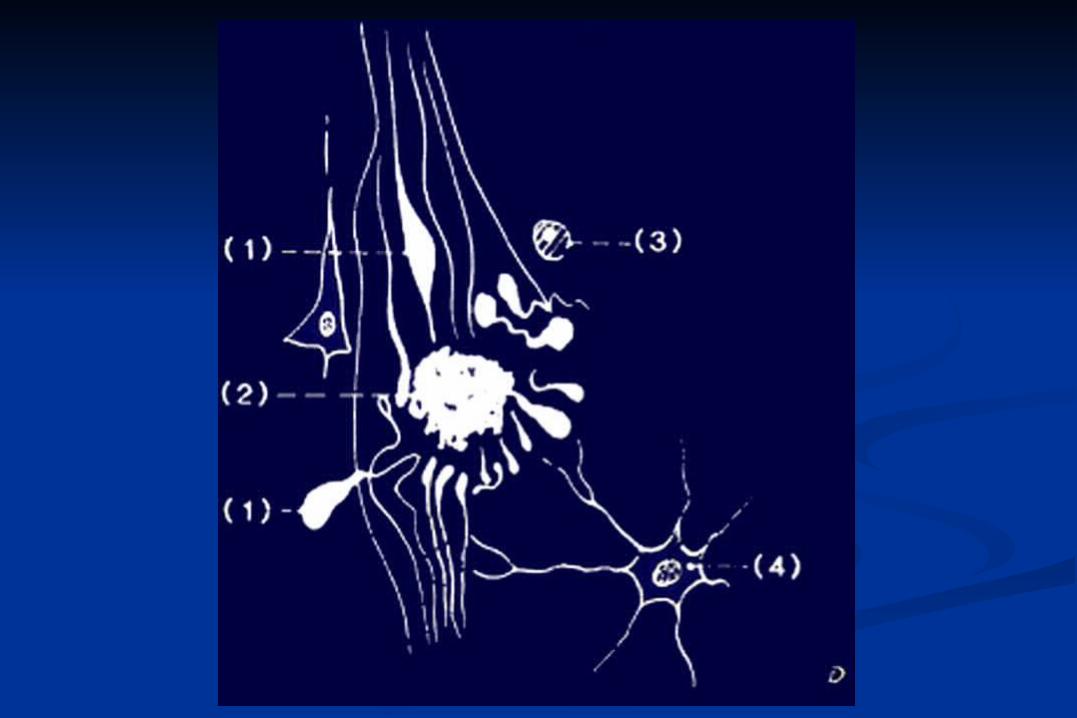
(Central) Chromatolysis – „axonal“ Reaction
rounding of perikaryon
loss of central Nissl bodies
peripheral displacement of nucleus
retraction of presynaptic terminals
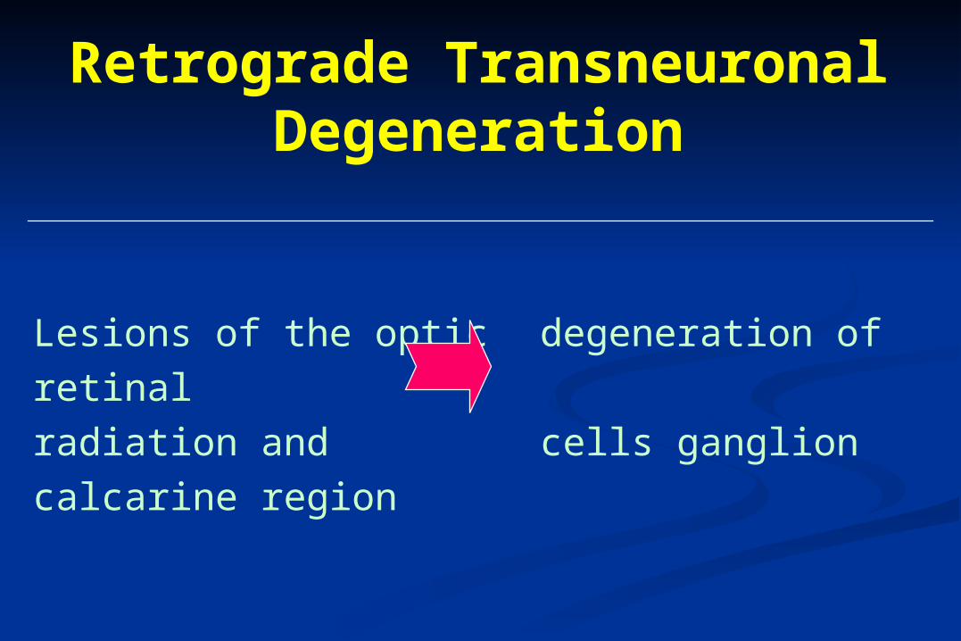
Retrograde Transneuronal Degeneration
Lesions of the optic degeneration of retinal
radiation and cells ganglion
calcarine region
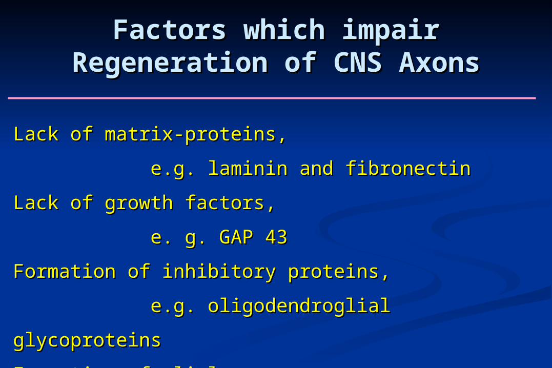
Factors which impair Regeneration of Factors which impair Regeneration of CNS AxonsCNS Axons
Lack of matrix-proteins, Lack of matrix-proteins,
e.g. laminin and fibronectine.g. laminin and fibronectin
Lack of growth factors, Lack of growth factors,
e. g. GAP 43e. g. GAP 43
Formation of inhibitory proteins, Formation of inhibitory proteins,
e.g. oligodendroglial glycoproteinse.g. oligodendroglial glycoproteins
Formation of glial scarsFormation of glial scars
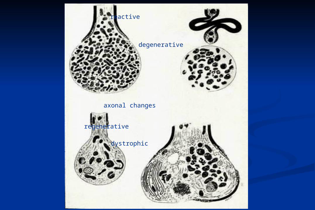
reactive
degenerative
axonal changes
regenerative
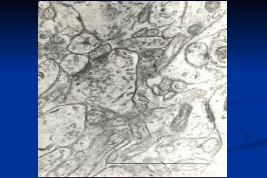
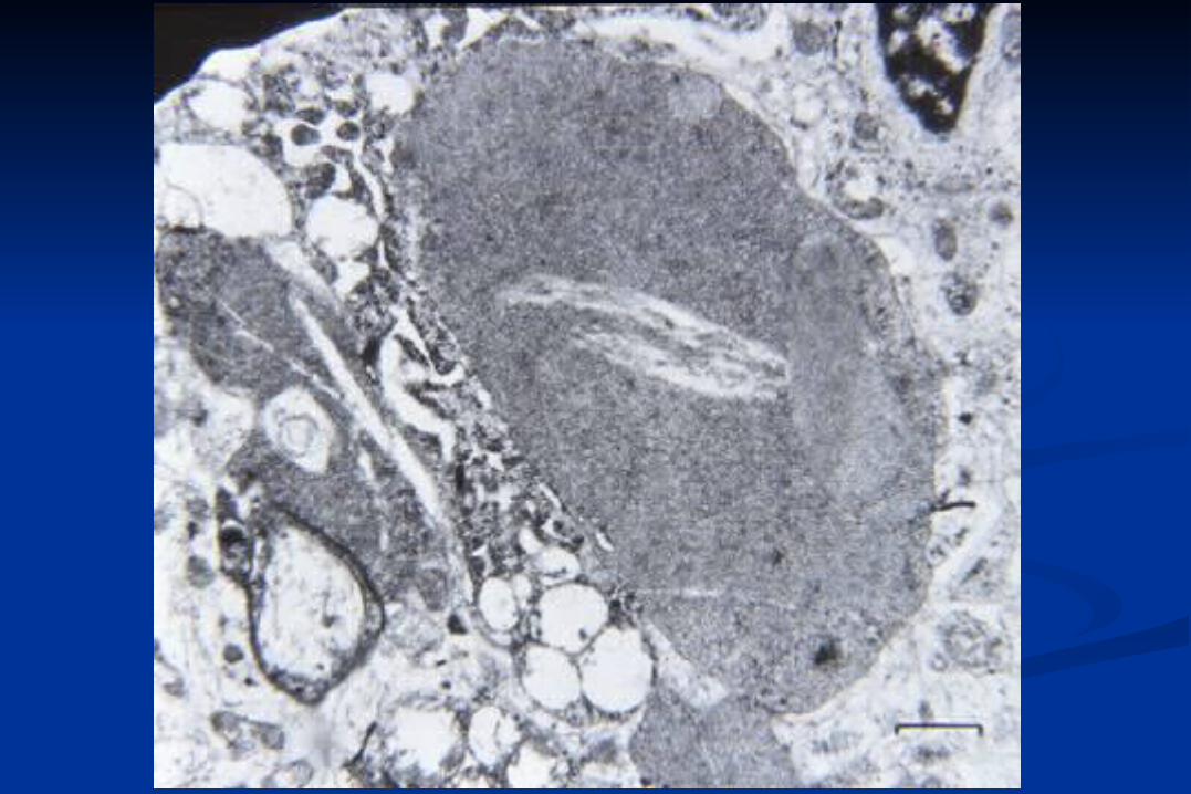
dystrophic
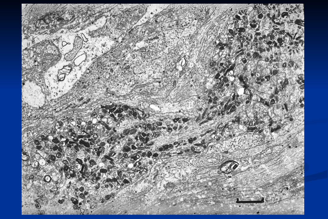
Neuroaxonal Dystrophies
Pathology of the Neuronal Perikaryon
retrograde reaction
vacuolisation
cell death
atrophy
aggregation of proteins lysosomal substrates viruses
Cell Death
Types of Cell Death
necrosis
apoptosis
autophagy
„loss“
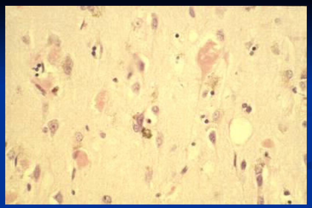
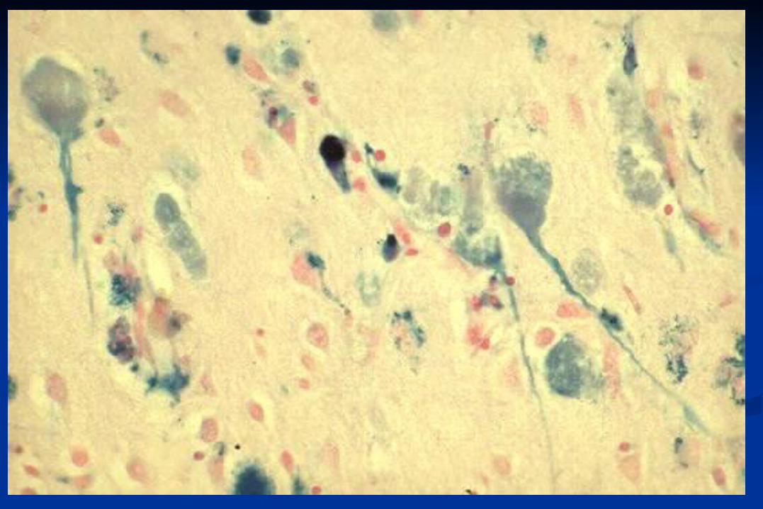
Necrophanerosis
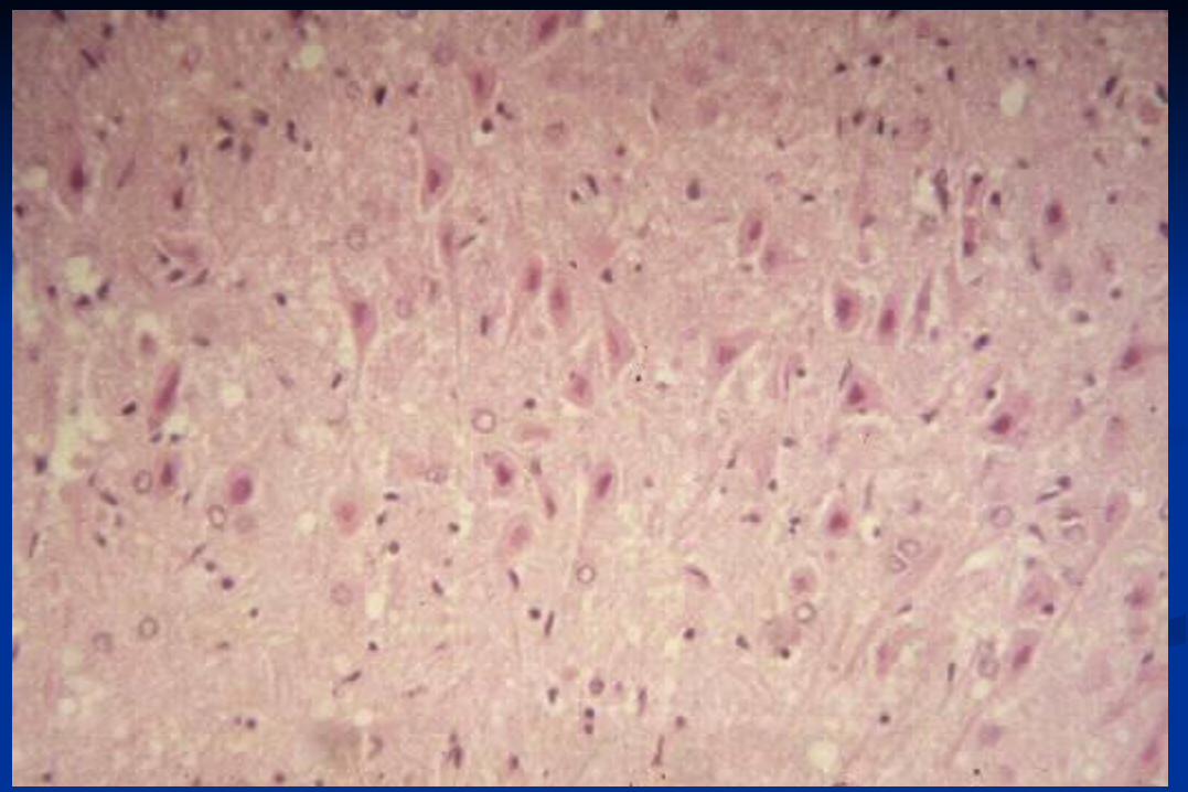
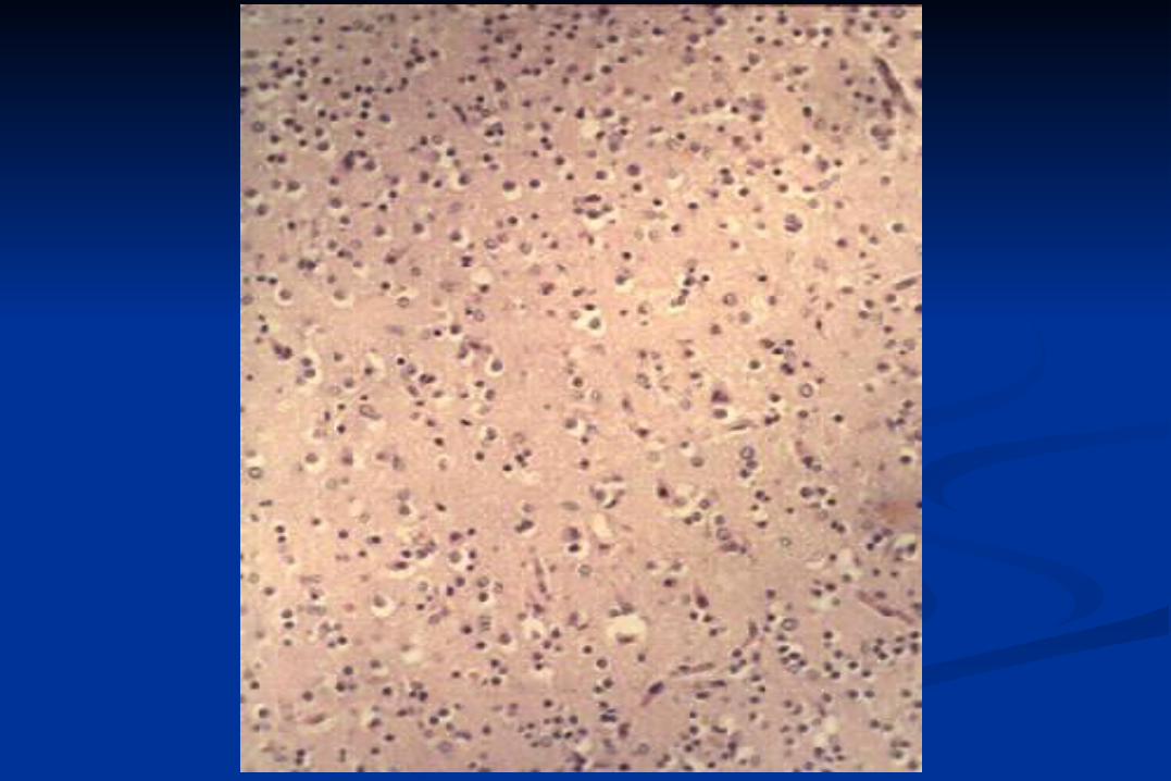
Elective Parenchymal Necrosis
=
selective neuronal necrosis
Causes of elective Causes of elective Parenchymal NecrosisParenchymal Necrosis
Anoxia / HypoxiaAnoxia / Hypoxia Cardiac arrestCardiac arrest
AnaemiaAnaemia CO intoxicationCO intoxication
Pulmonary diseasePulmonary disease HypoglycaemiaHypoglycaemia
Regions of elective Parenchymal Regions of elective Parenchymal NecrosisNecrosis
Purkinje cells (cerebellar cortex)Purkinje cells (cerebellar cortex)
Pyramidal cells of cortex, incl. Pyramidal cells of cortex, incl. hippocampus (cerebral cortex)hippocampus (cerebral cortex)
Striatal neuronsStriatal neurons
Thalamic neuronsThalamic neurons
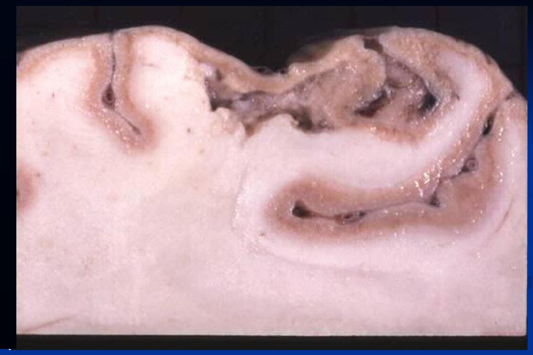
Tissue Death
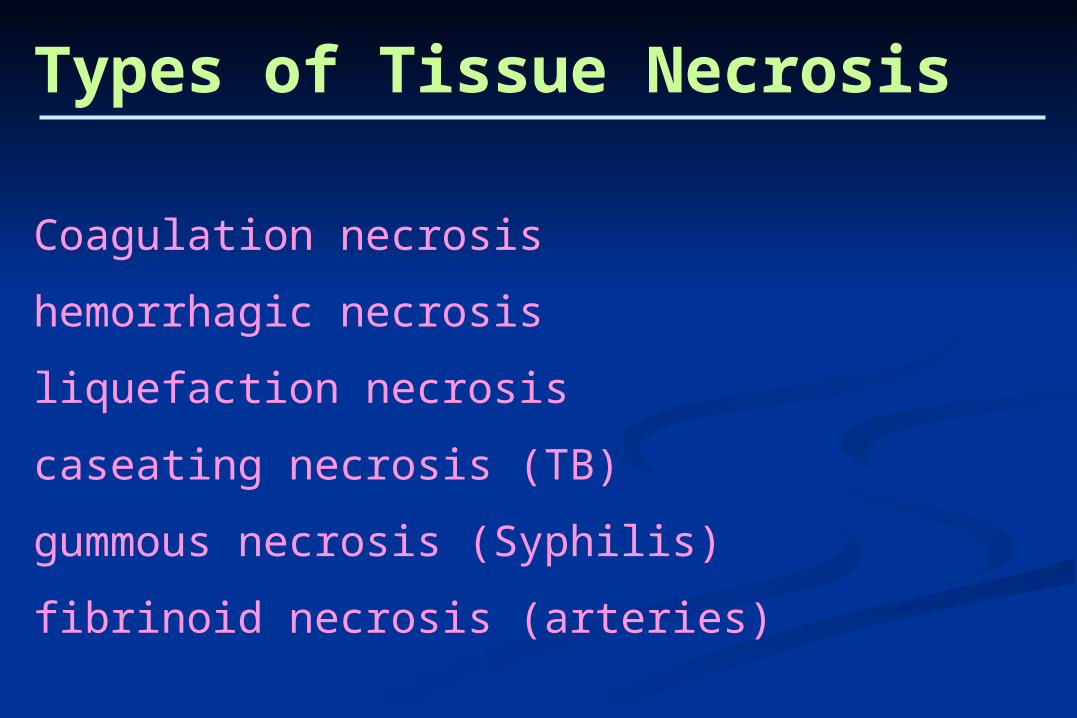
Types of Tissue Necrosis
Coagulation necrosis
hemorrhagic necrosis
liquefaction necrosis
caseating necrosis (TB)
gummous necrosis (Syphilis)
fibrinoid necrosis (arteries)
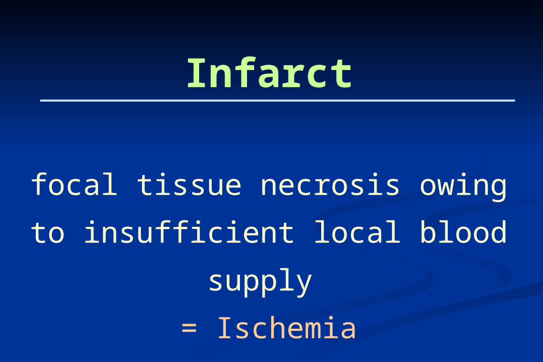
Infarct
focal tissue necrosis owing to
insufficient local blood supply
= Ischemia
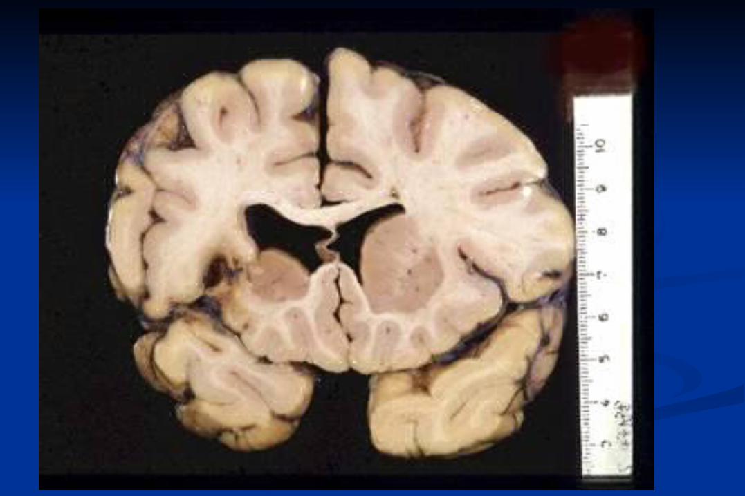
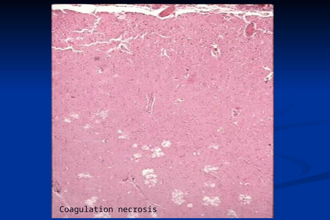
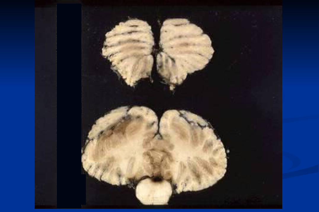
Coagulation necrosis
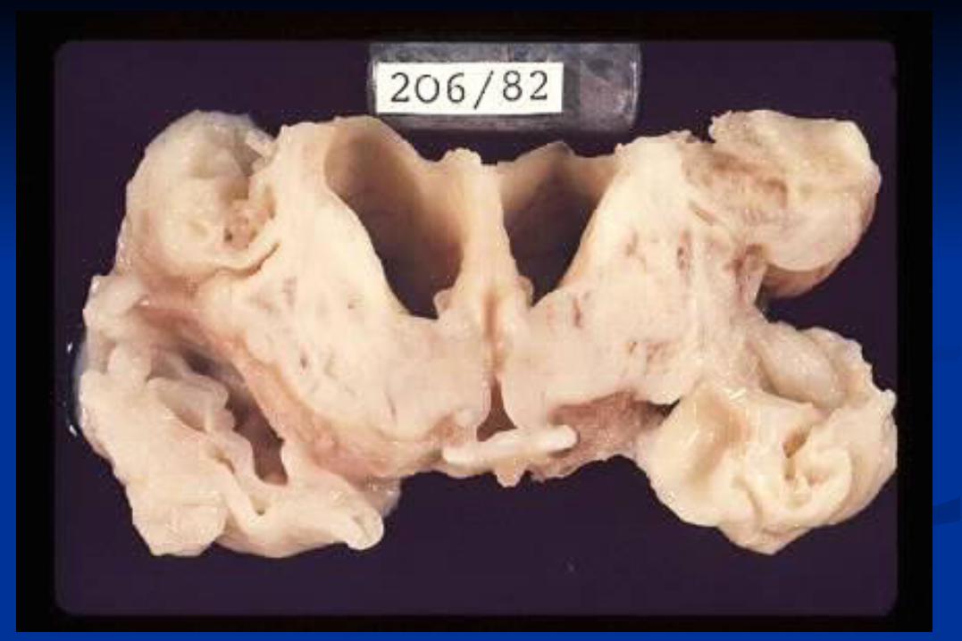
Antemortem brain deathAntemortem brain death
Dissociated (brain)deathDissociated (brain)death
Complete infarct of the brainComplete infarct of the brain
Antemortem autolysis of the brainAntemortem autolysis of the brain
Respirator brainRespirator brain
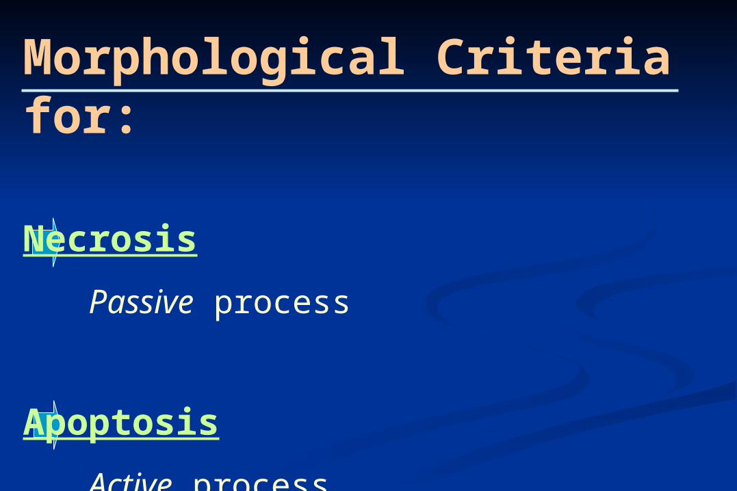
Morphological Criteria for:
Necrosis
Passive process
Apoptosis
Active process
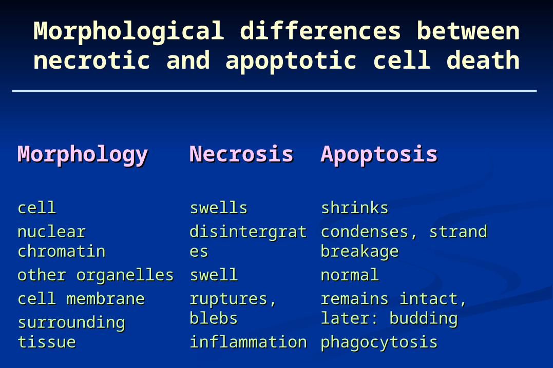
MorphologyMorphology NecrosisNecrosis ApoptosisApoptosis
cellcell
nuclear chromatinnuclear chromatin
other organelles other organelles
cell membrane cell membrane
surrounding tissuesurrounding tissue
swellsswells
disintergratesdisintergrates
swellswell
ruptures, blebsruptures, blebs
inflammationinflammation
shrinksshrinks
condenses, strand breakagecondenses, strand breakage
normalnormal
remains intact, later: buddingremains intact, later: budding
phagocytosisphagocytosis
Morphological differences between necrotic and apoptotic cell death
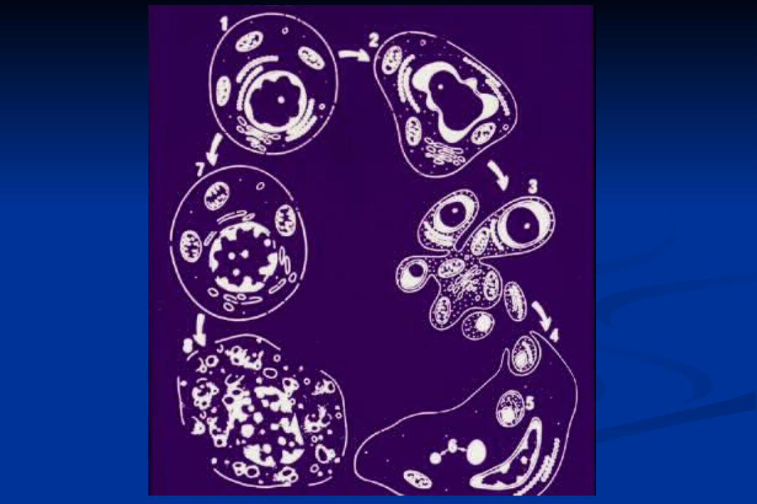
Phases of Apoptosis:
Initiation phase: different stimuli
Effector phase: common to all cells
Degradation phase: metabolic enzymes
activated
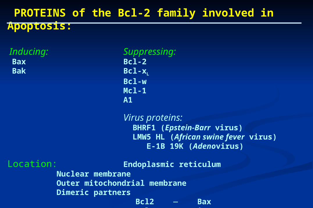
PROTEINS of the Bcl-2 family involved in Apoptosis:
Inducing: Suppressing: Bax Bcl-2 Bak Bcl-xL
Bcl-wMcl-1A1
Virus proteins: BHRF1 (Epstein-Barr virus) LMW5 HL (African swine fever virus)
E-1B 19K (Adenovirus)
Location: Endoplasmic reticulumNuclear membraneOuter mitochondrial membraneDimeric partners Bcl2 Bax Bcl-xL Bak
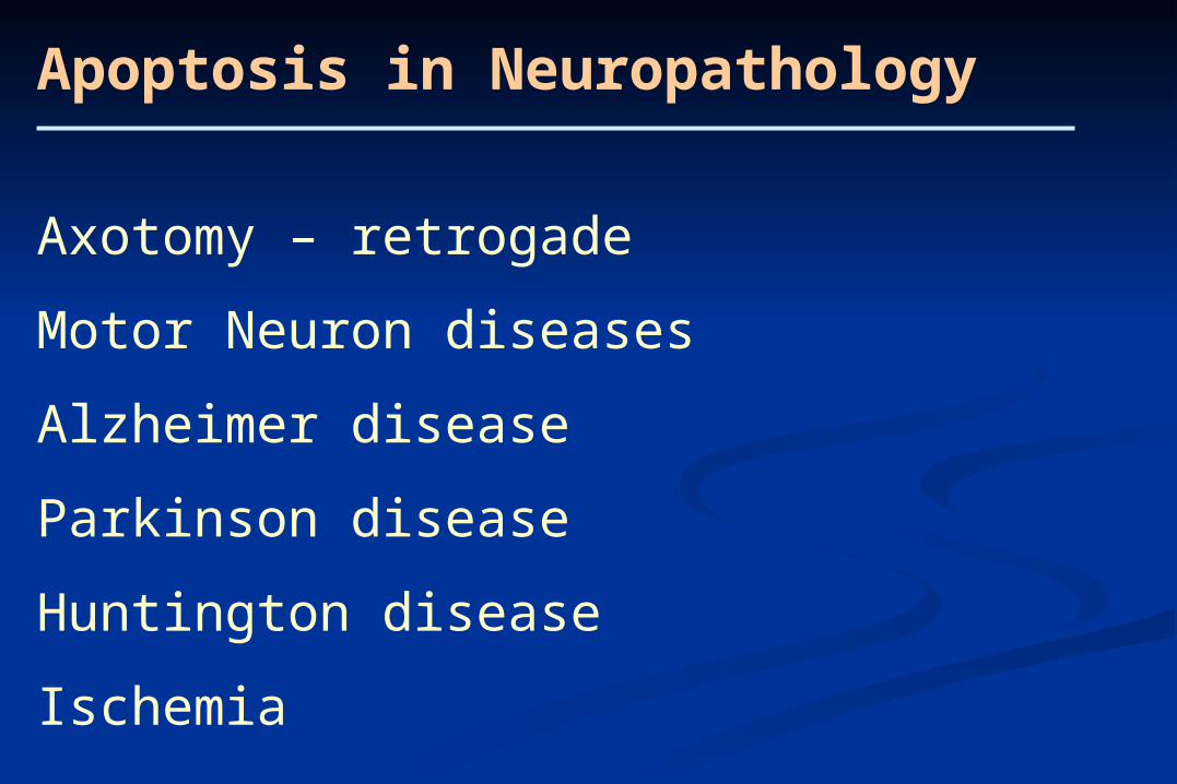
Apoptosis in Neuropathology
Axotomy – retrogade
Motor Neuron diseases
Alzheimer disease
Parkinson disease
Huntington disease
Ischemia
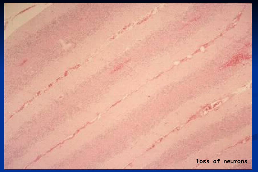
loss of neurons

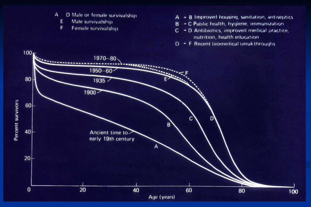
Ageing
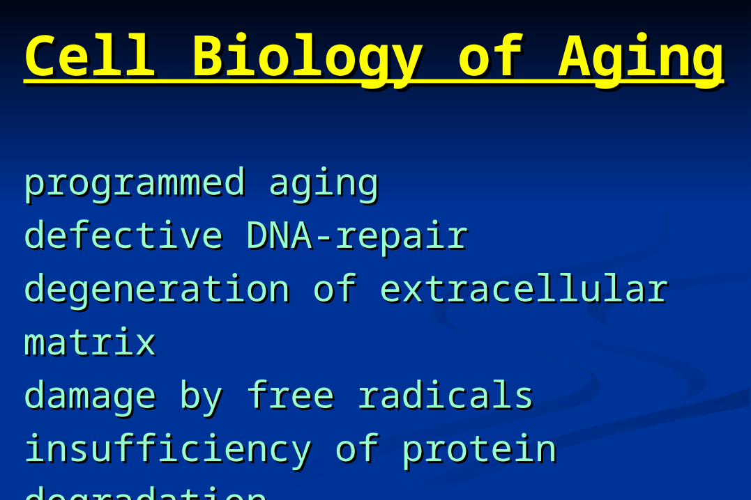
Cell Biology of AgingCell Biology of Aging
programmed agingprogrammed aging
defective DNA-repairdefective DNA-repair
degeneration of extracellular matrixdegeneration of extracellular matrix
damage by free radicalsdamage by free radicals
insufficiency of protein degradationinsufficiency of protein degradation
cumulating cell damagecumulating cell damage
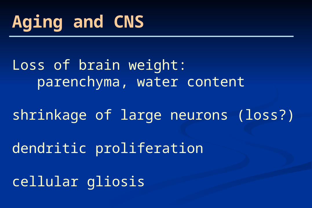
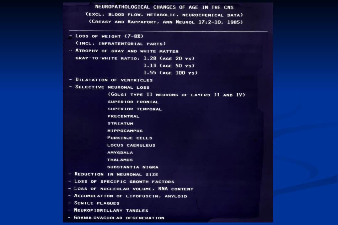
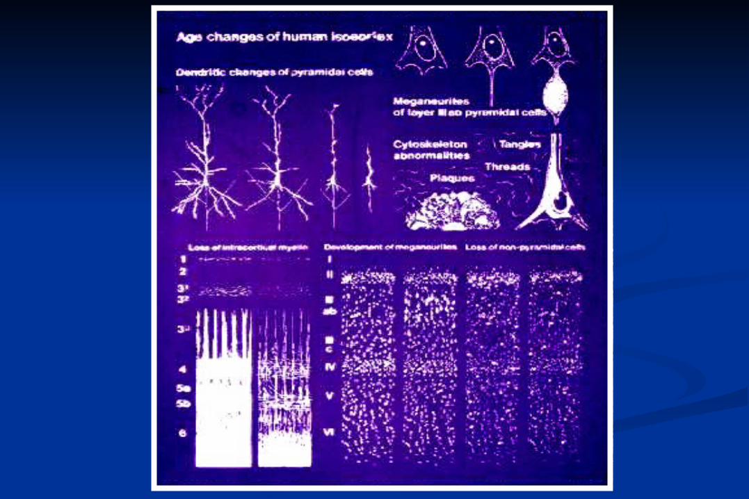
Aging and CNS
Loss of brain weight: parenchyma, water content
shrinkage of large neurons (loss?)
dendritic proliferation
cellular gliosis
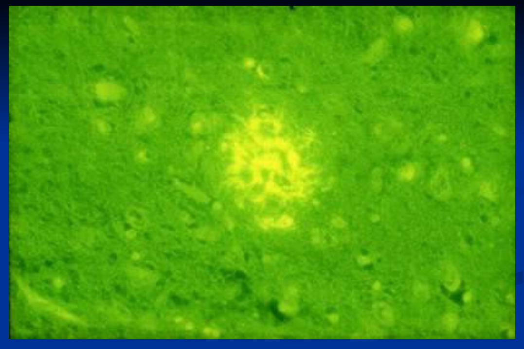
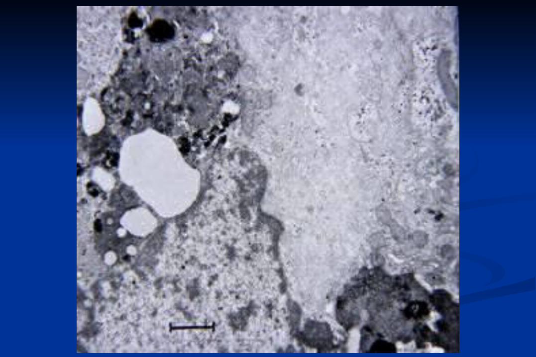
Granulovacuolar
Degeneration
Simchowicz
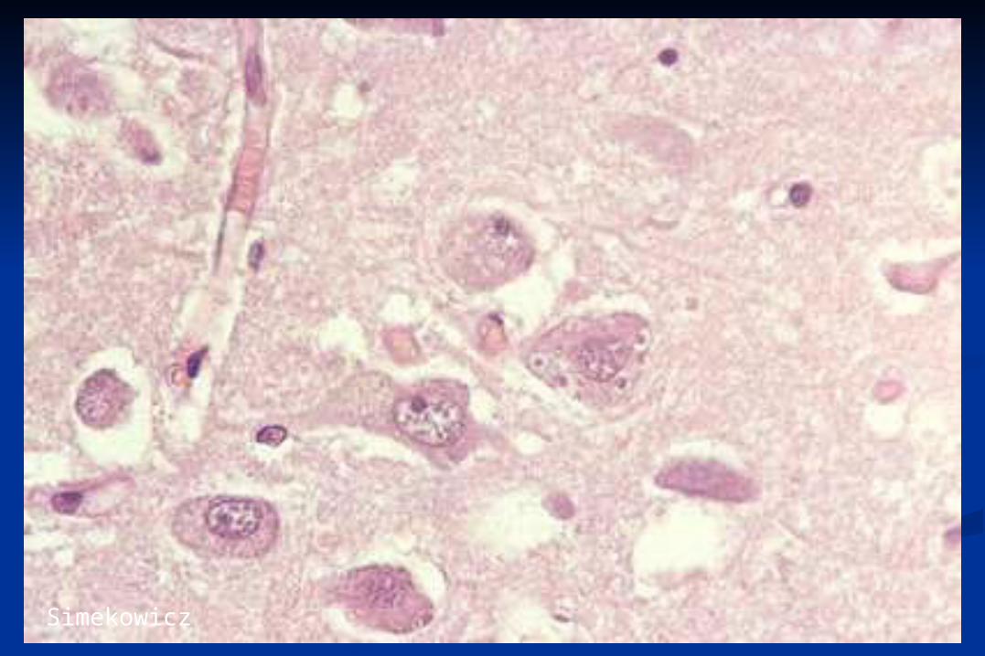
Simekowicz
Intraneuronal (intraglial) Aggegation
Proteins
Viruses
Lysosomal substrates
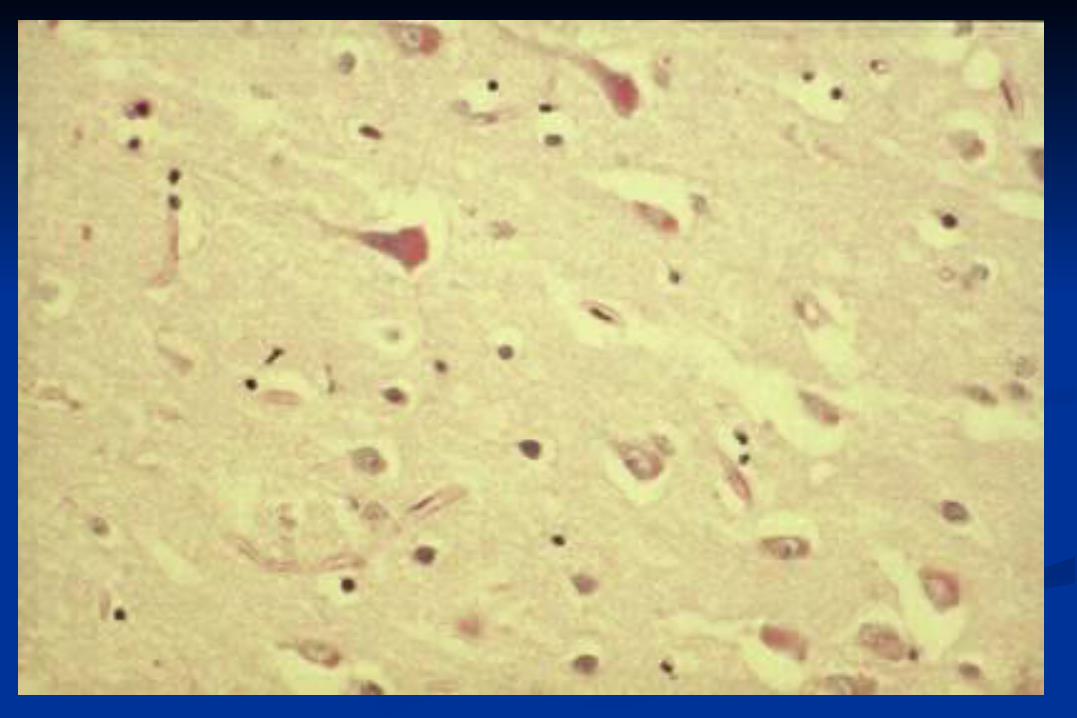
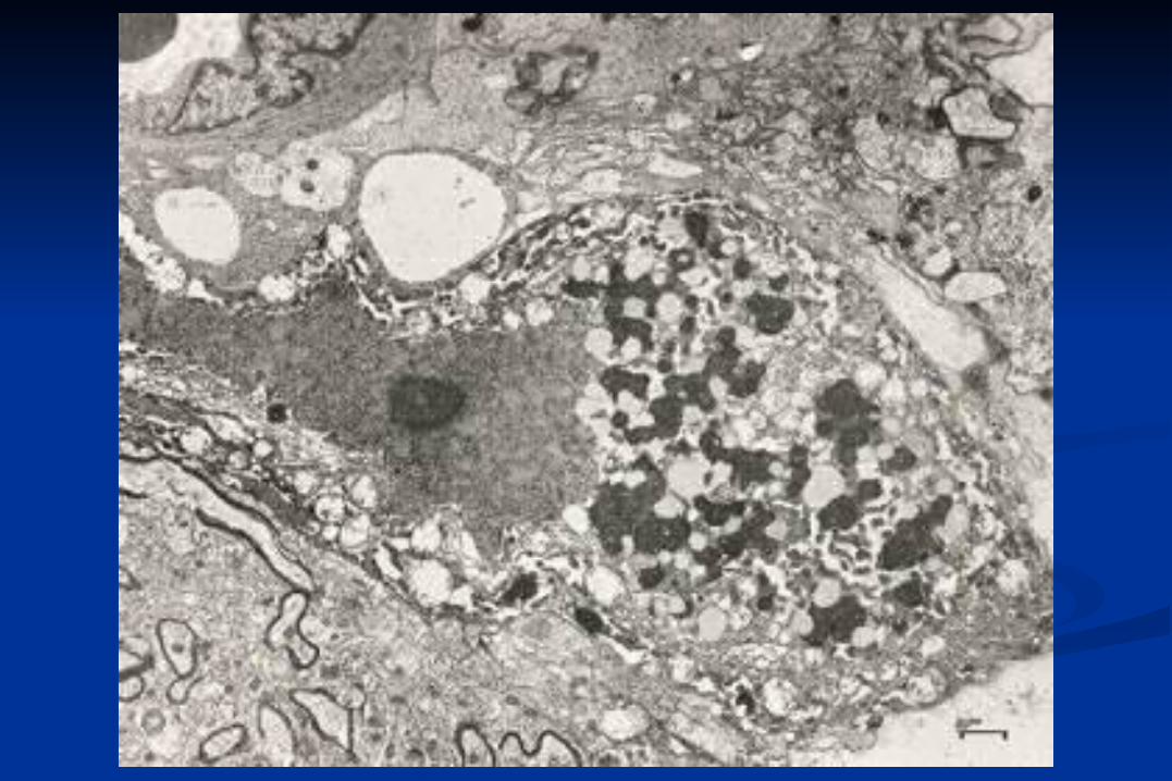
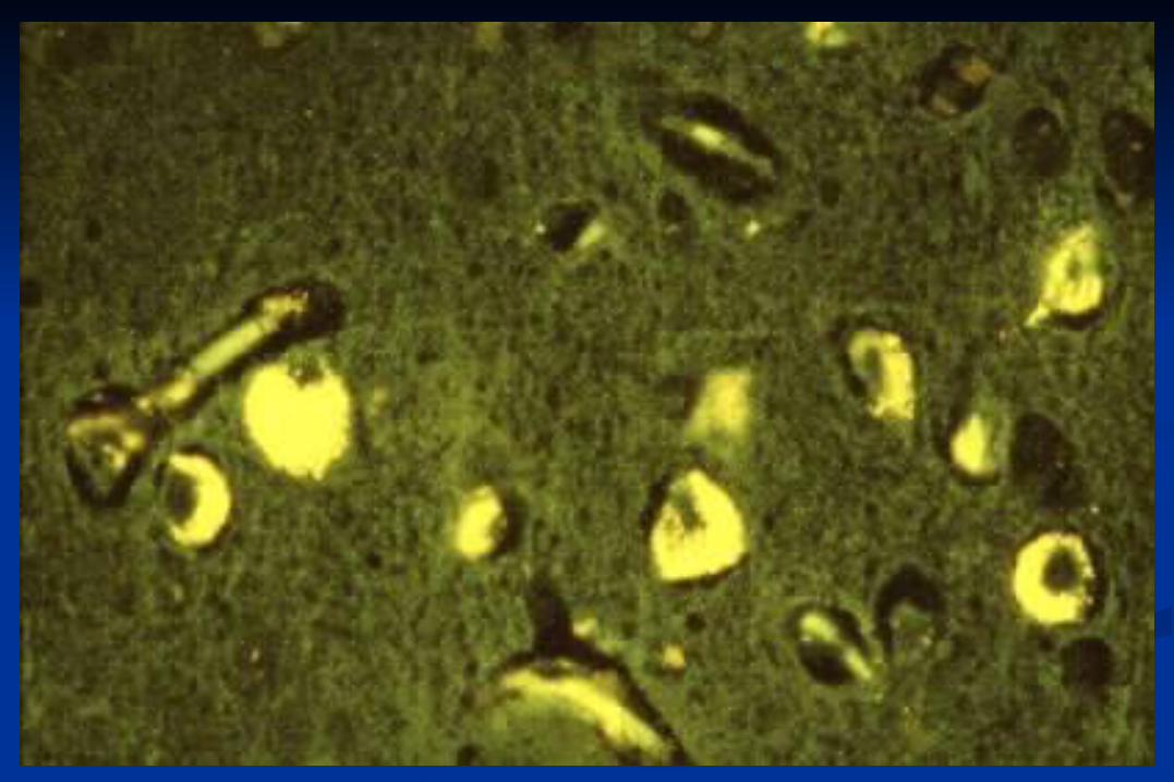
Lipofuscin (Lipopigment)
Age pigment
Wear and tear pigment
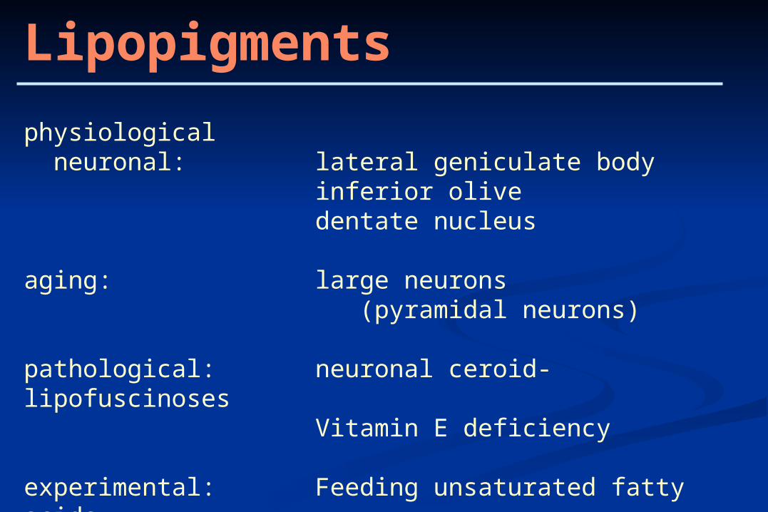
Lipopigments
physiological neuronal: lateral geniculate body
inferior olivedentate nucleus
aging: large neurons (pyramidal neurons)
pathological: neuronal ceroid-lipofuscinosesVitamin E deficiency
experimental: Feeding unsaturated fatty acids
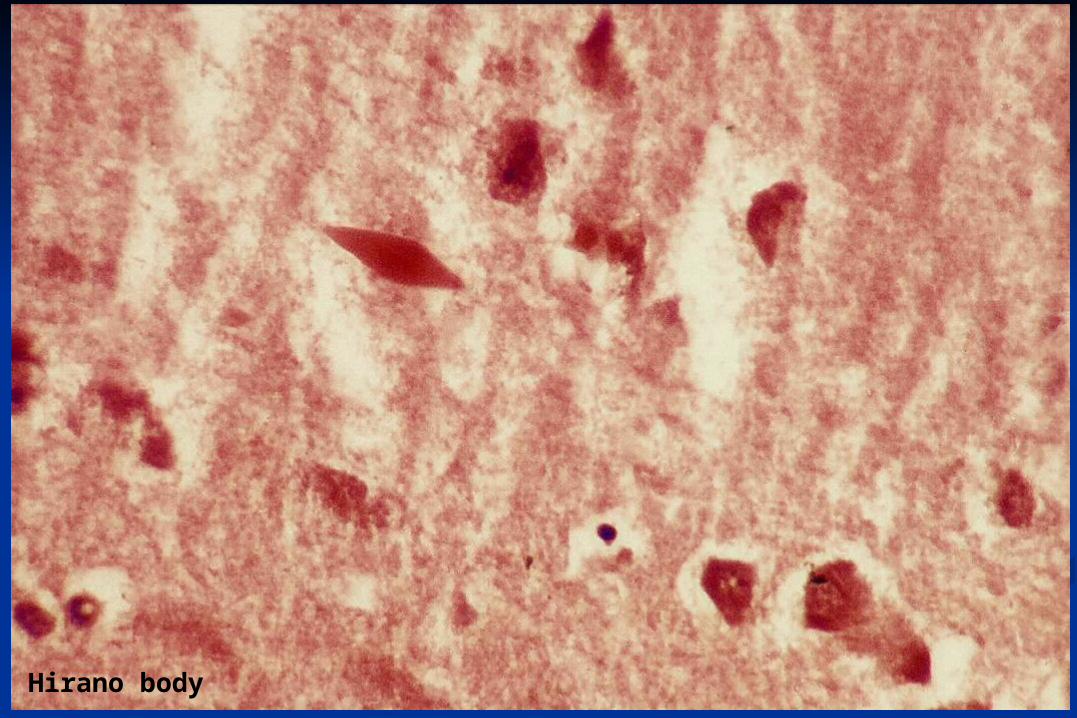
Hirano body
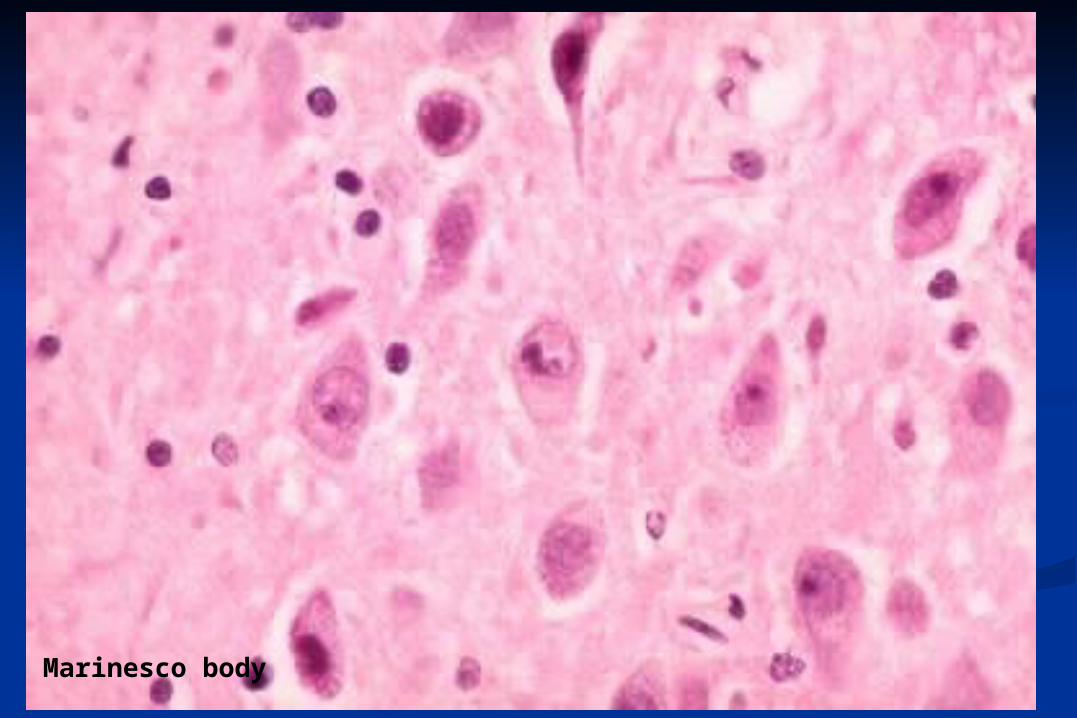
Marinesco body
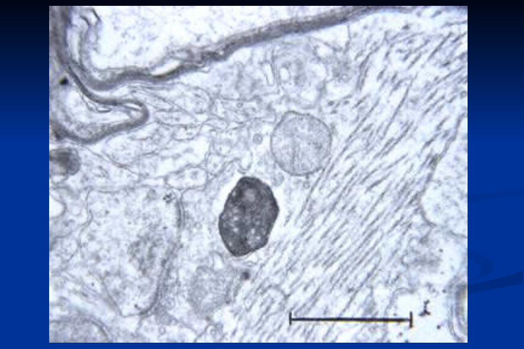
Alzheimer Disease
NFT neurofibrillary tangles
PHF paired helical filaments
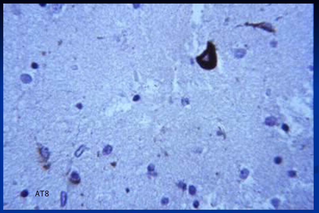
AT8
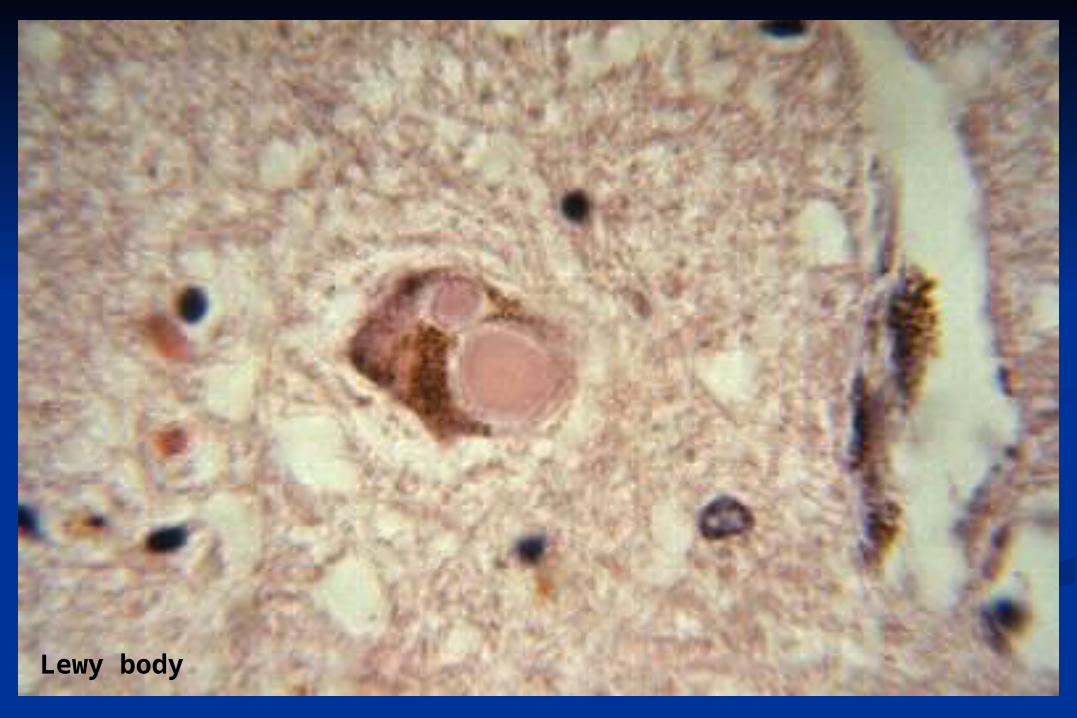
Lewy body
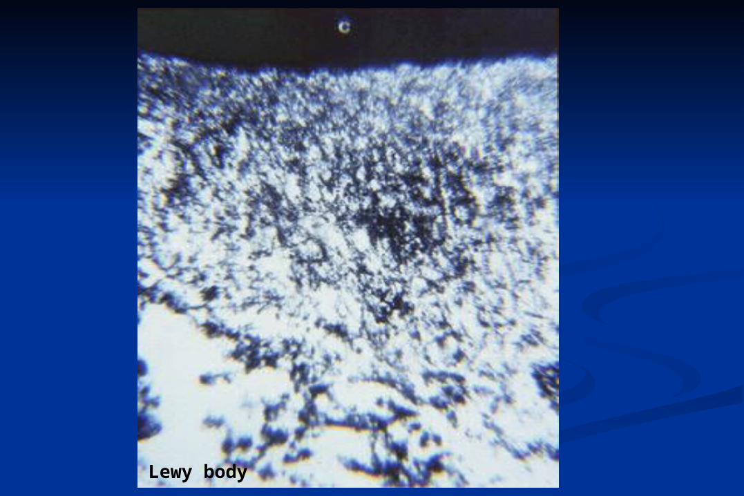
Lewy body
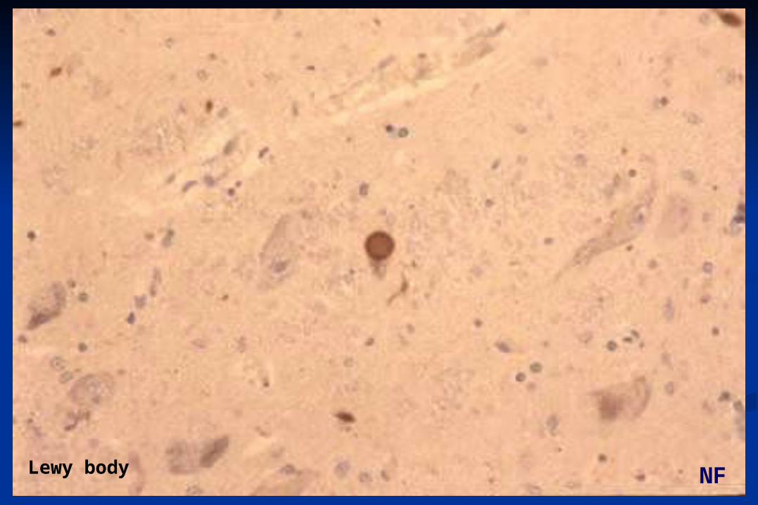
Lewy body NF
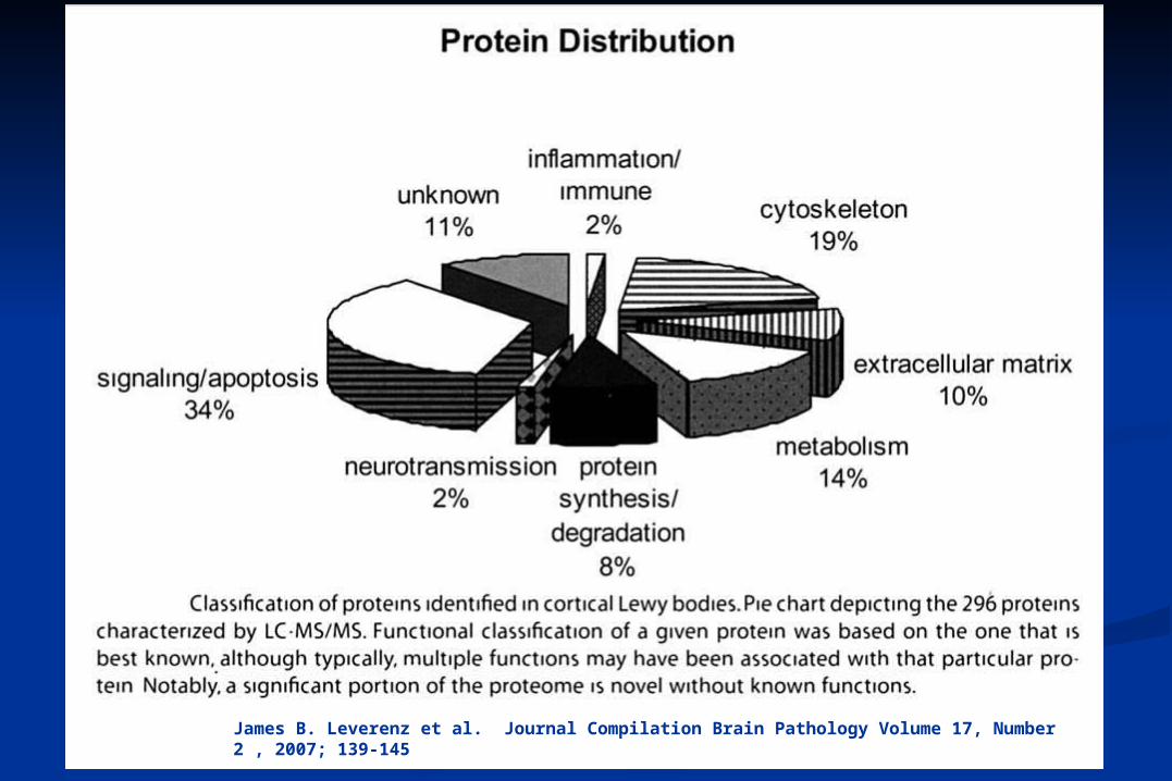
James B. Leverenz et al. Journal Compilation Brain Pathology Volume 17, Number 2 , 2007; 139-145
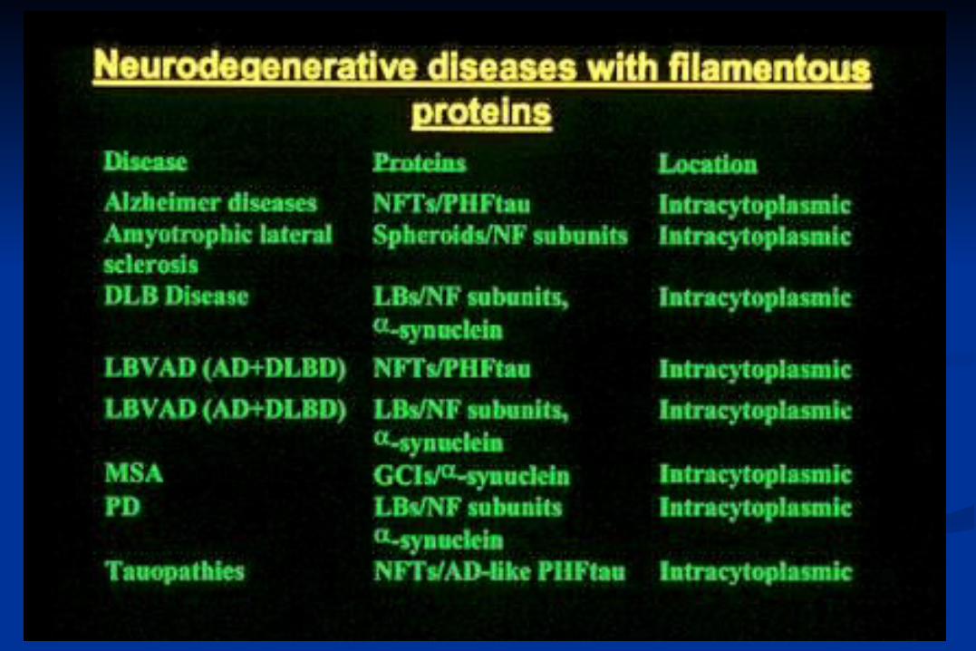
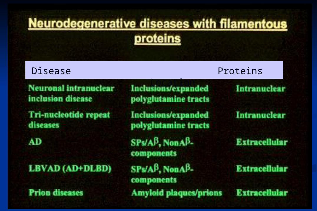
Disease Proteins Location
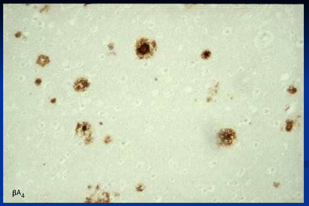
A4
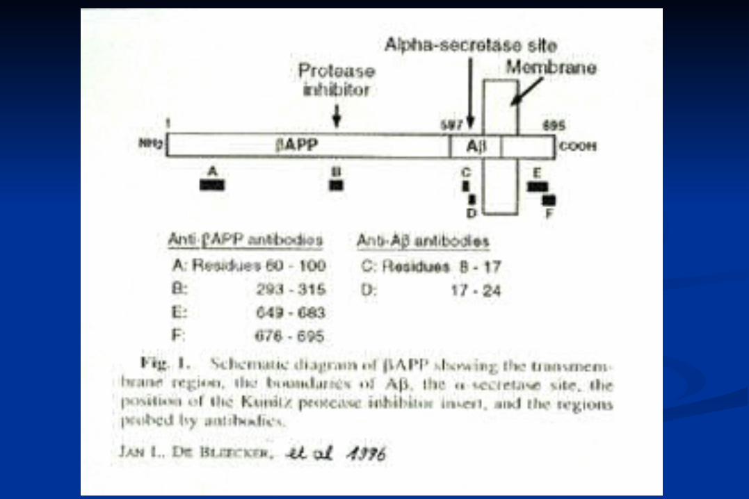
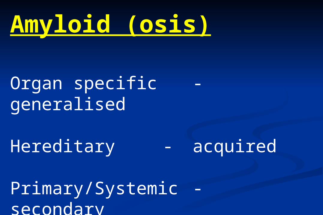
Amyloid (osis)
Organ specific - generalised
Hereditary - acquired
Primary/Systemic - secondary
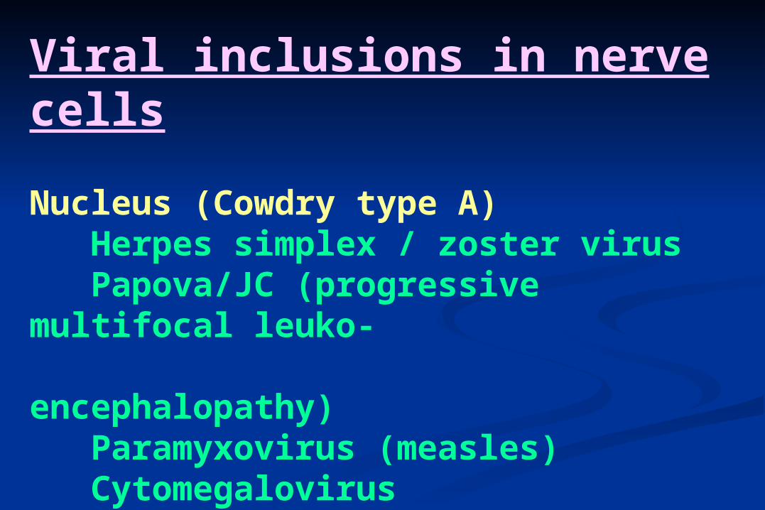
Viral inclusions in nerve cells
Nucleus (Cowdry type A) Herpes simplex / zoster virus Papova/JC (progressive multifocal leuko- encephalopathy) Paramyxovirus (measles) Cytomegalovirus
Cytoplasm Negri / Lyssa bodies (Rabies)
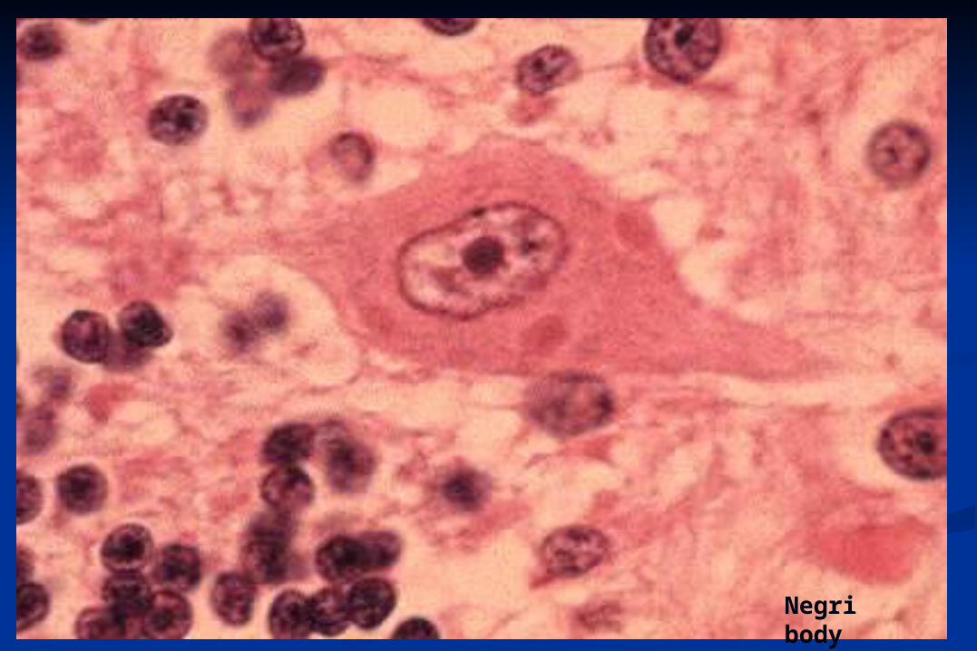
Negri body
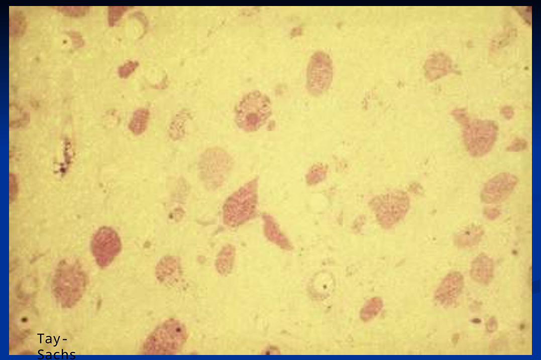
Tay-Sachs
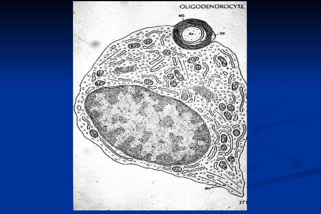
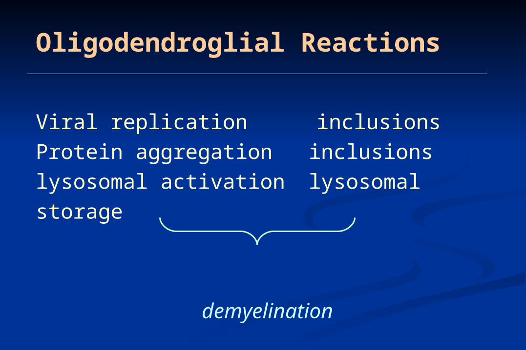
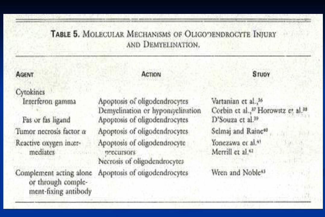
Oligodendroglial Reactions
Viral replication inclusions
Protein aggregation inclusions
lysosomal activation lysosomal storage
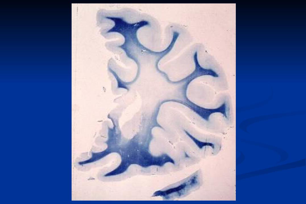
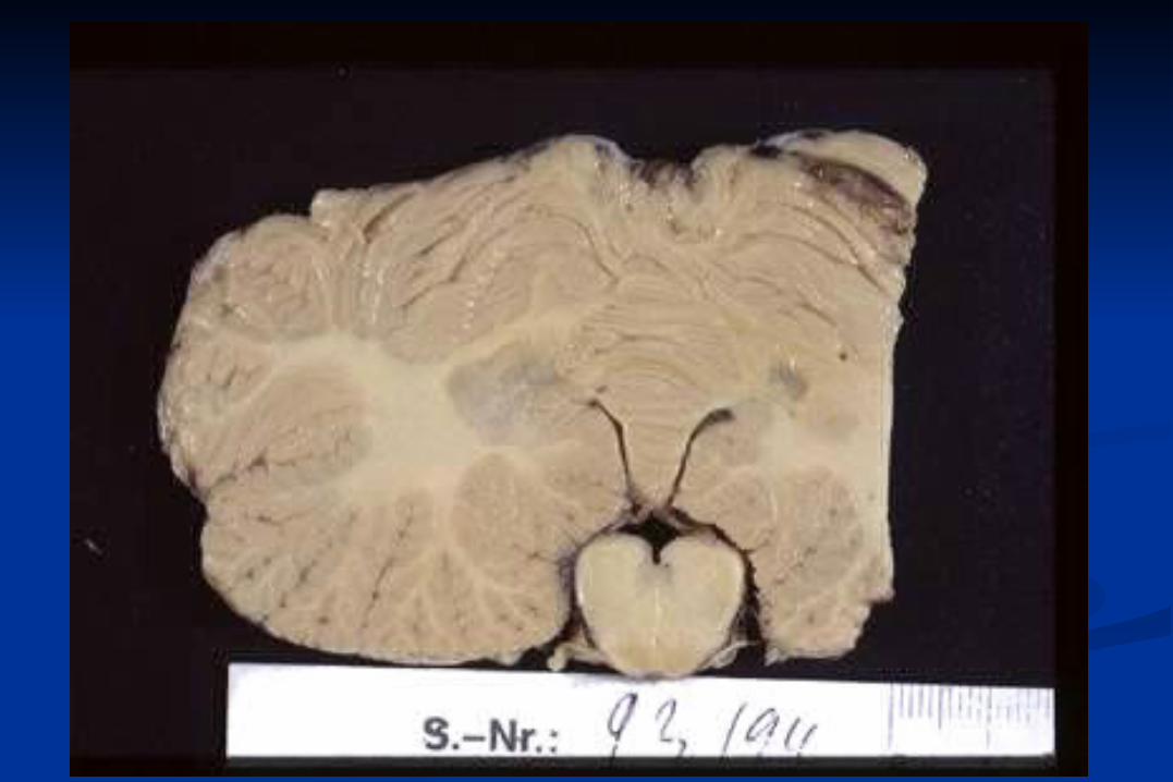
demyelination
Primary and secondary Demyelination
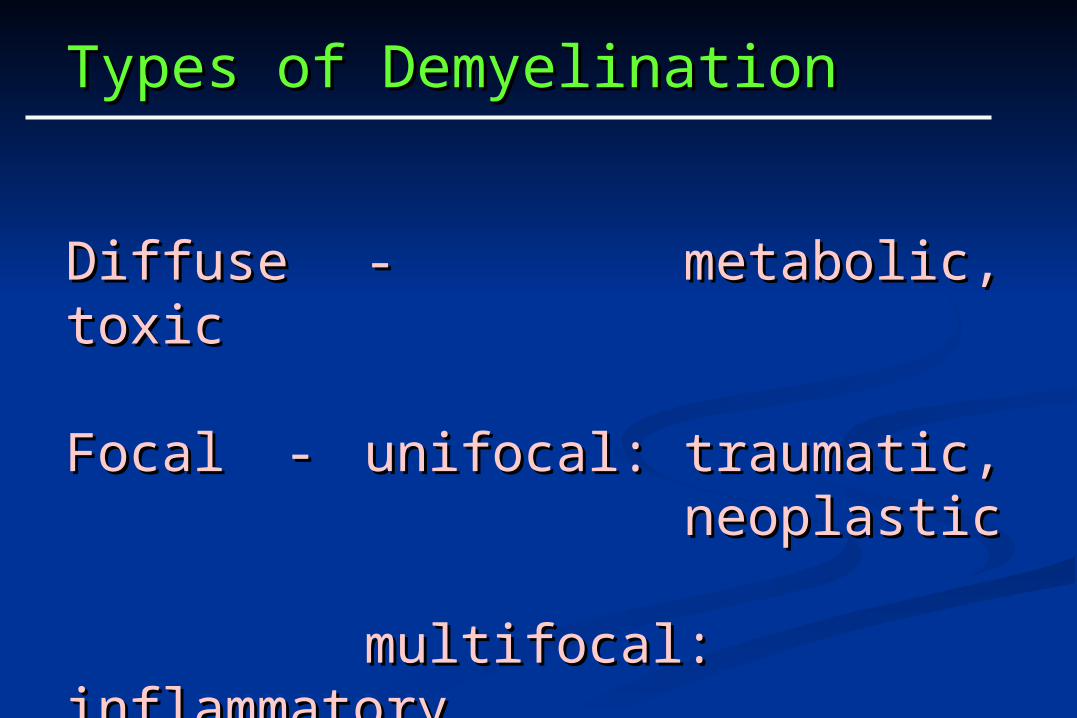
Types of DemyelinationTypes of Demyelination
DiffuseDiffuse -- metabolic, toxicmetabolic, toxic
Focal Focal -- unifocal:unifocal: traumatic,traumatic,neoplasticneoplastic
multifocal:multifocal: inflammatoryinflammatory
CLASSIFICATION OF PRIMARY CLASSIFICATION OF PRIMARY MYELIN DISEASESMYELIN DISEASES
Allergic and infectious diseases
Hereditary (metabolic) diseases
Toxic diseases
Nutritional diseases
Traumatic diseases
Vascular diseases
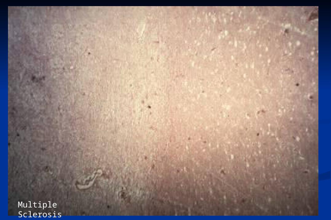
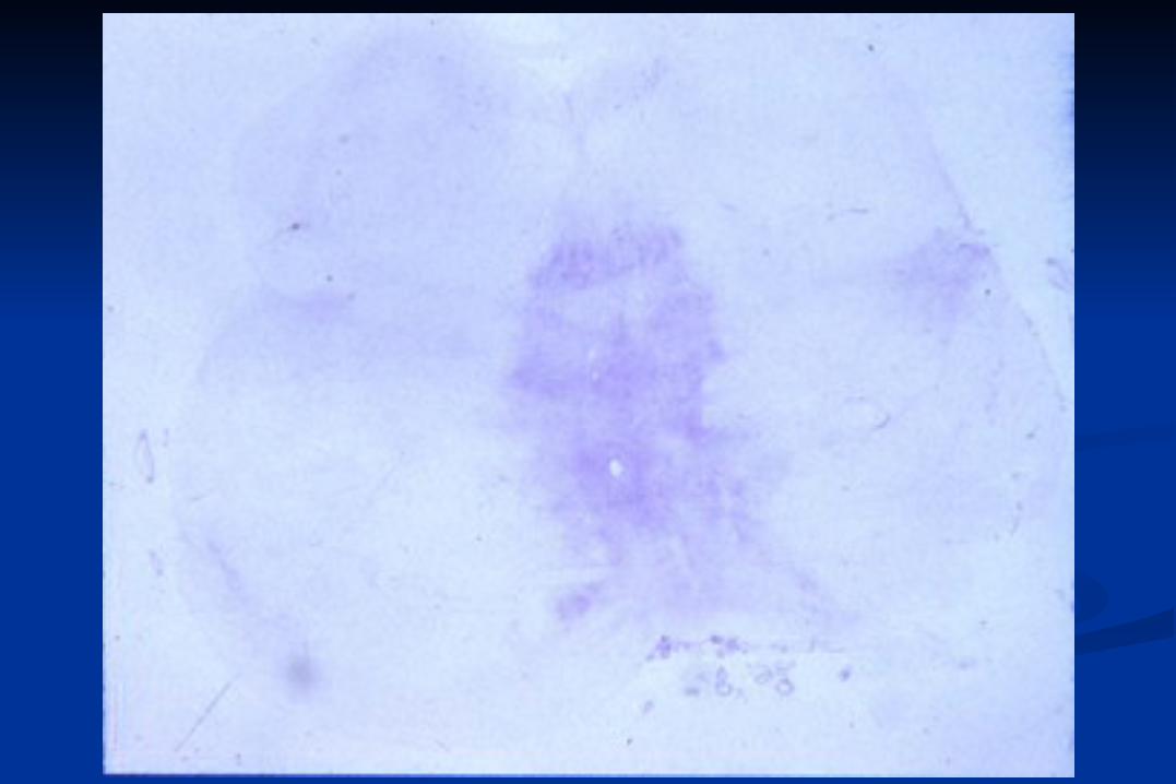
Multiple Sclerosis
Glia reactionsMicrogliaAstrocytes (Glial scar)
Mesenchymal reactionsVesselsFibroblastsScar
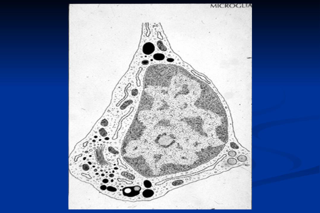
Microglia-rapid reaction
phagocytes
MHC I + II positive
express APP, complement receptor
produce cytokines, NO
present antigens
Synaptic stripping by microglia
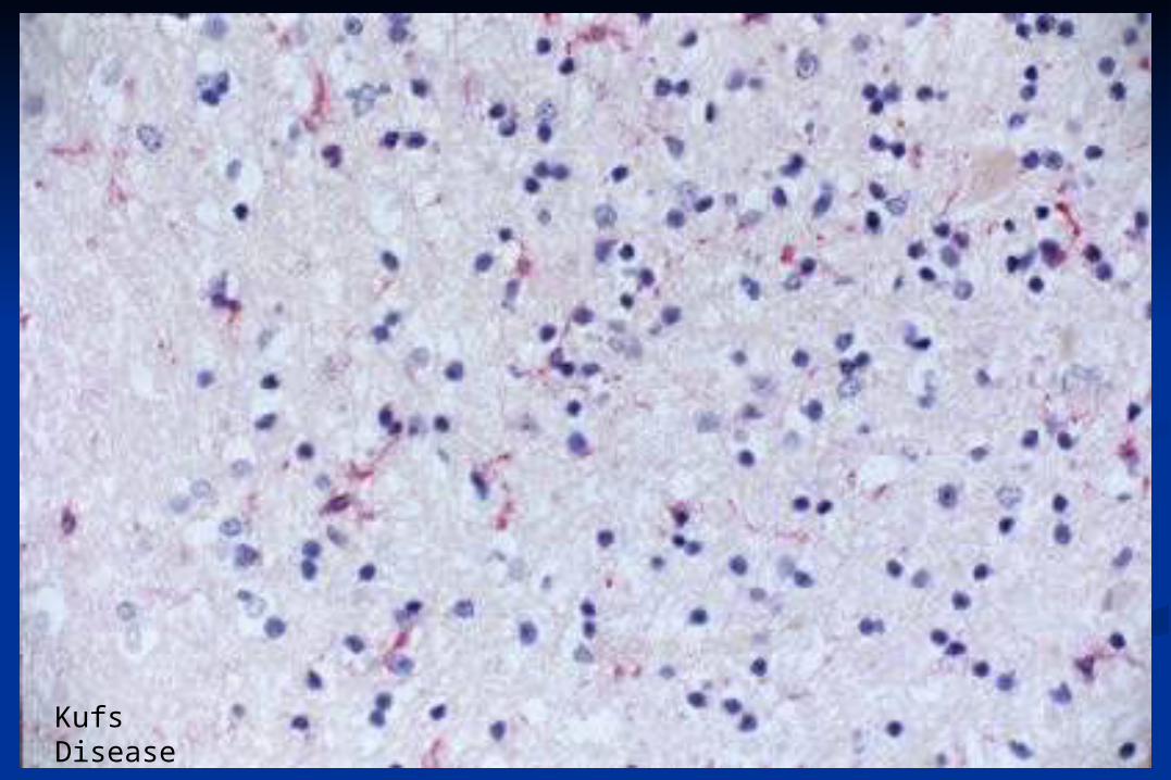
Kufs Disease
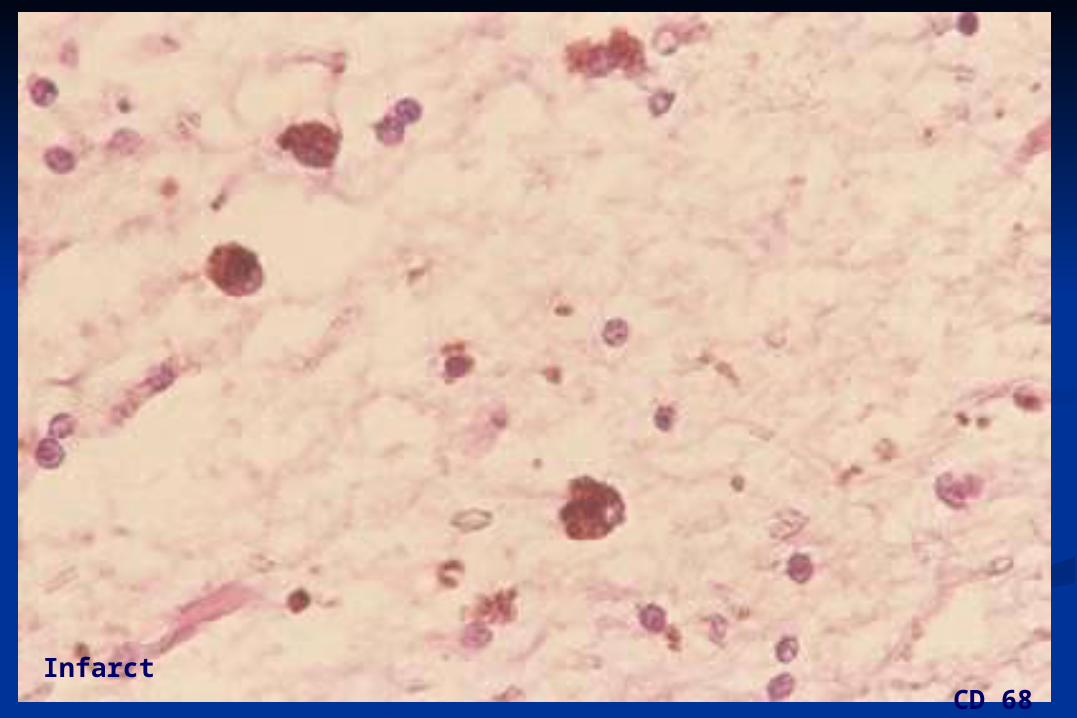
Infarct CD 68
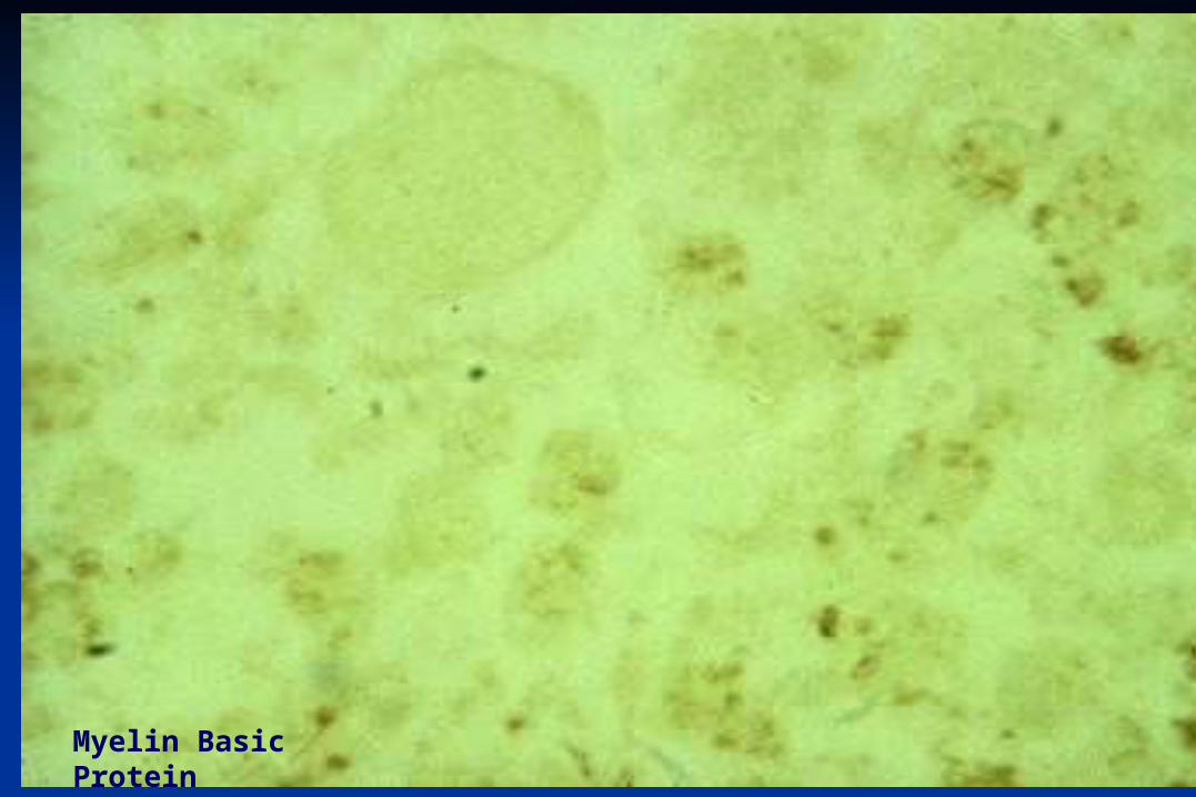
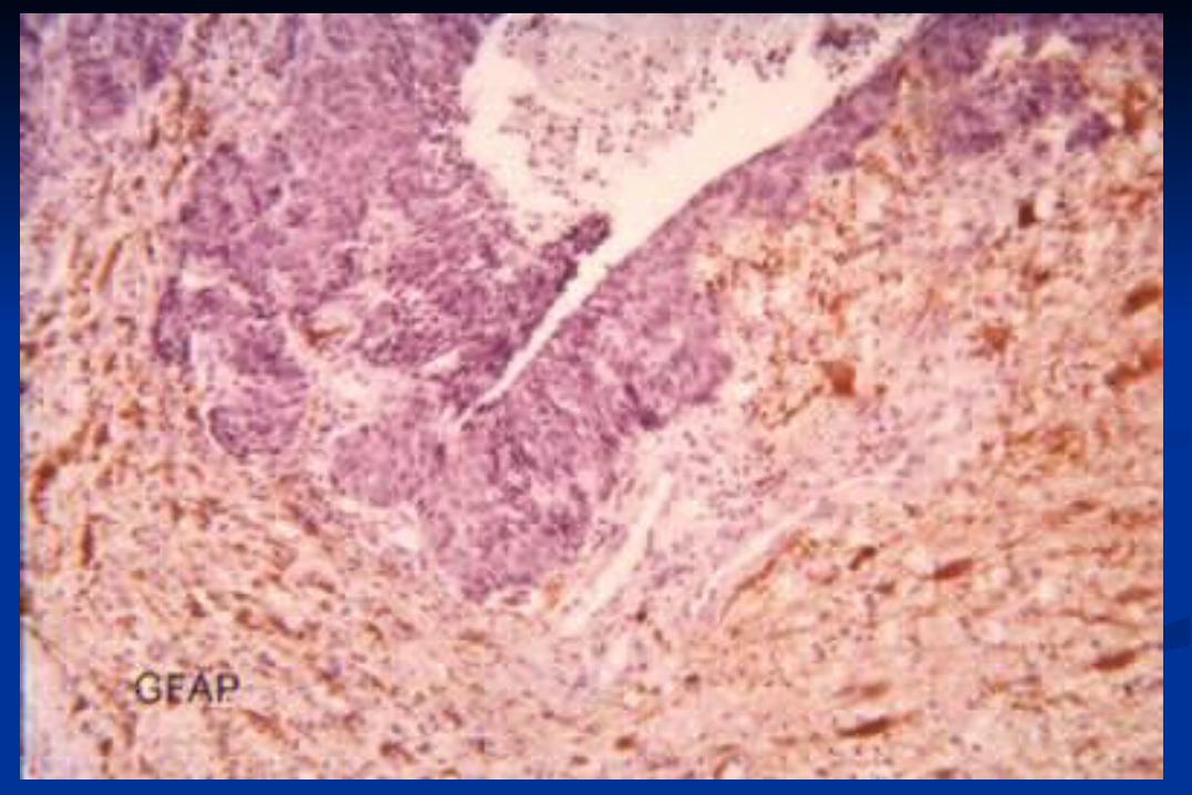
Myelin Basic Protein
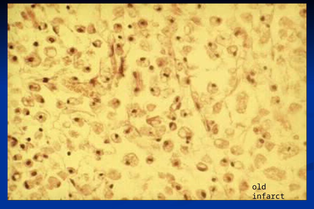
old infarct
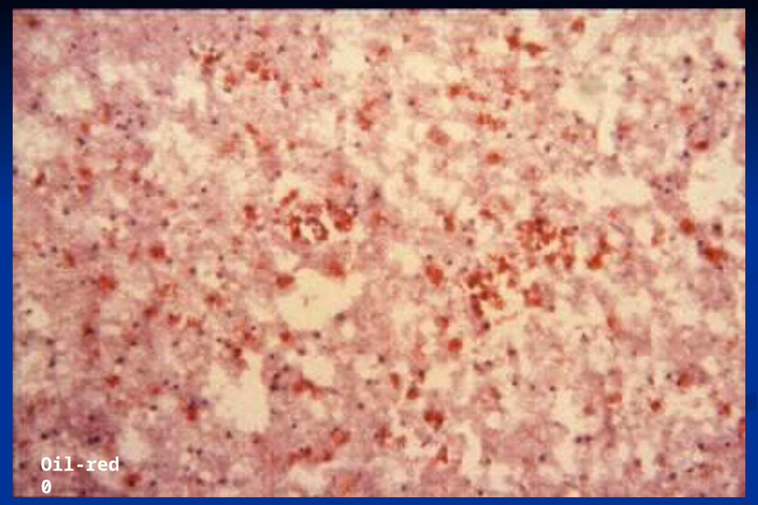
Oil-red 0
Orthochromatic(sudanophilic) Degradation
metachromatic Degradation
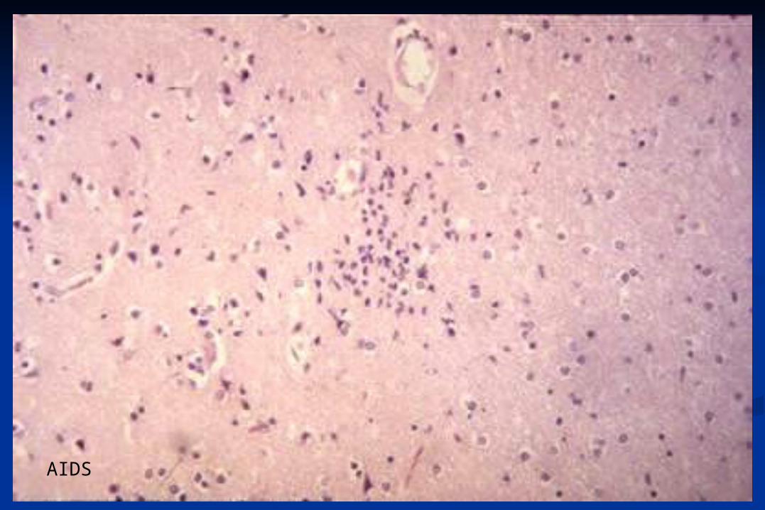
AIDS
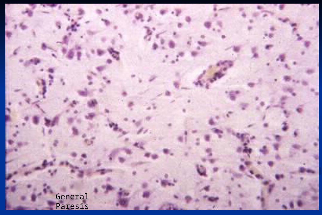
General Paresis
Hepatic Glia
Hypertrophy of astrocytic nuclei
Alzheimer type I = Wilson disease(hepato-lenticular degeneration)
Alzheimer Type II = hepatic and uremic encephalopathies
Alzheimer type I glia:
M. Wilson = hepatolenticular
degeneration
Alzheimer type II glia:
hepatic encephalopathy
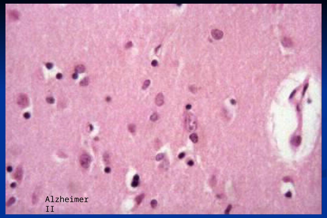
Alzheimer II
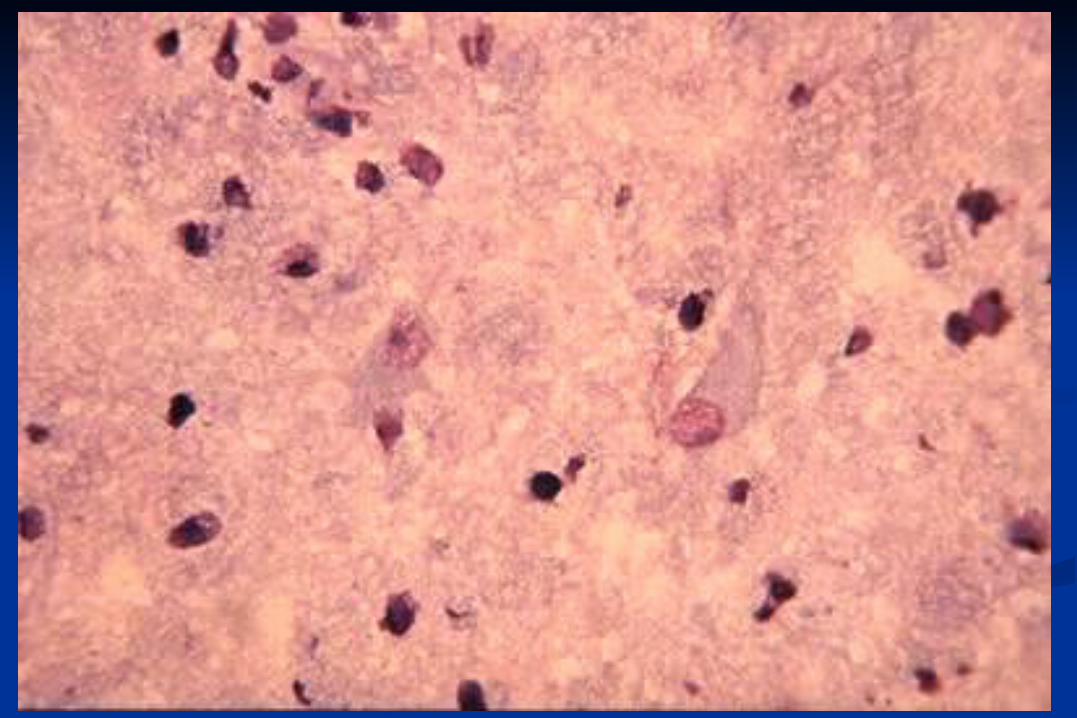
Gemistocytes
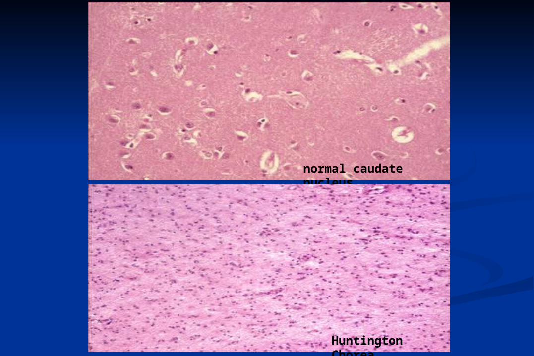
normal caudate nucleus
Huntington Chorea
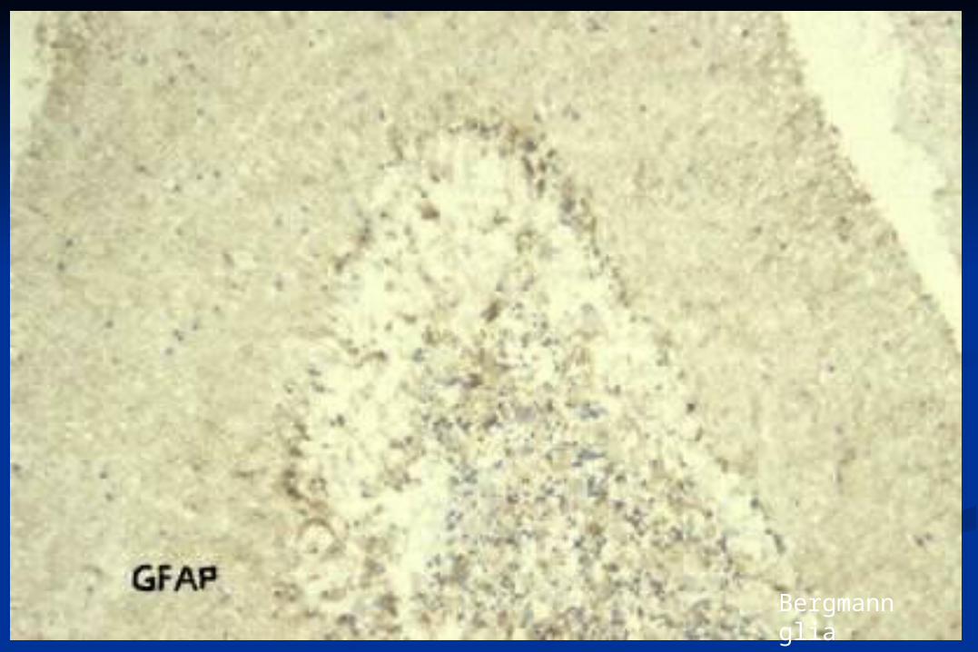
Bergmann glia
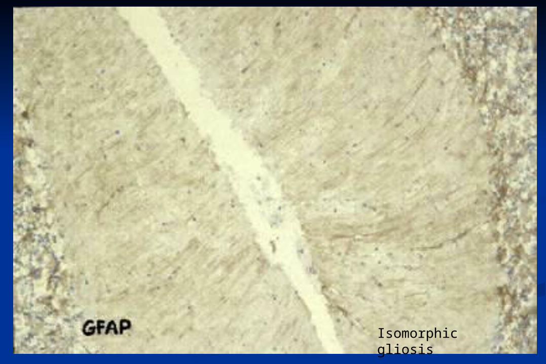
Isomorphic gliosis
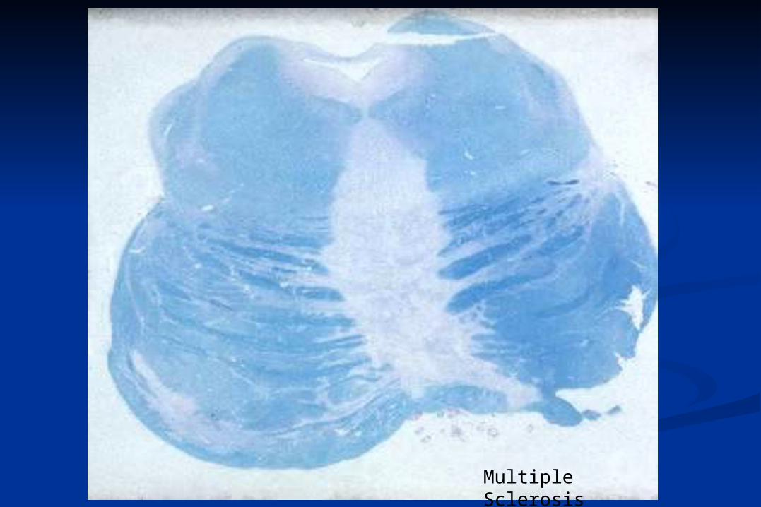
Multiple Sclerosis

Corpora amylacea
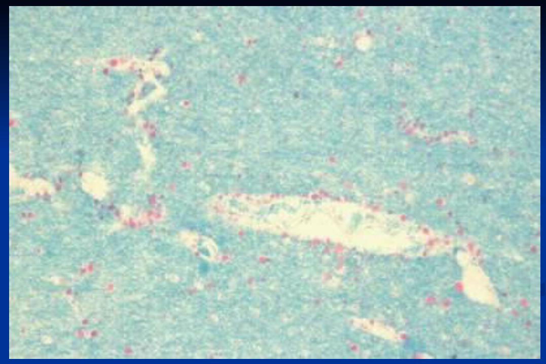
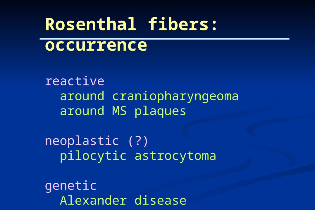
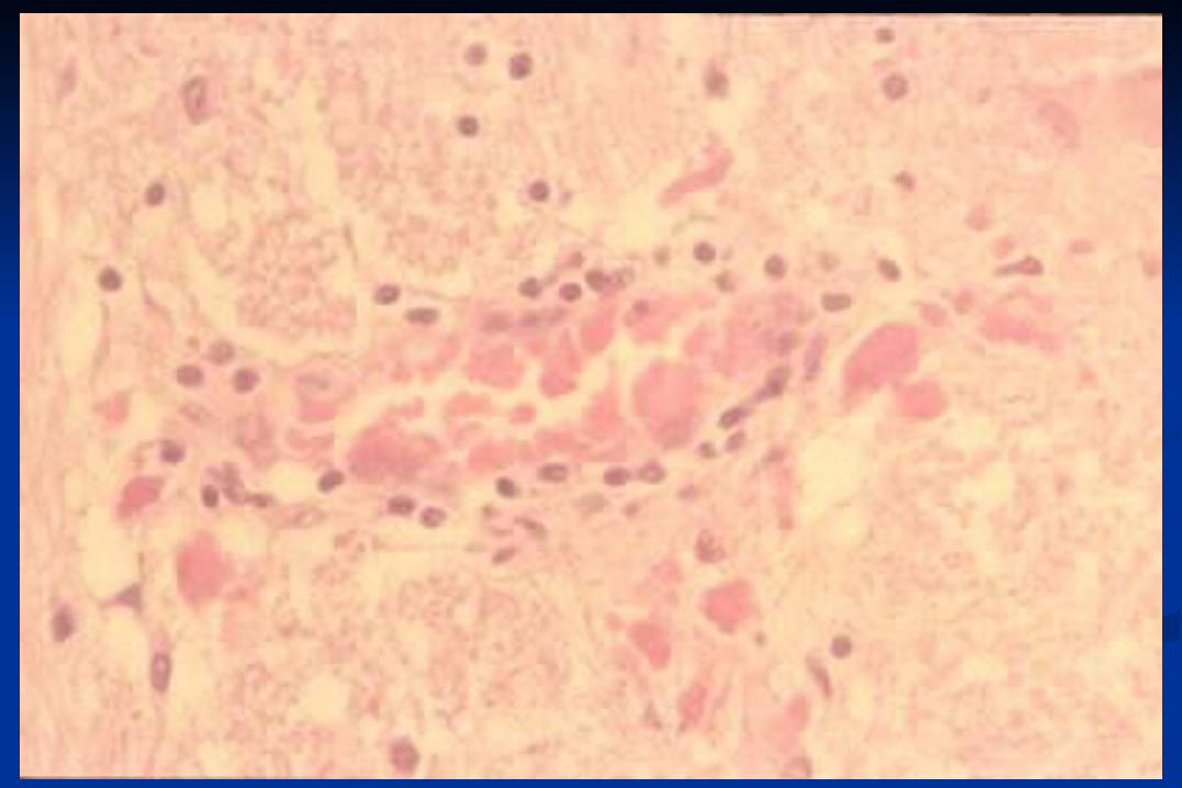
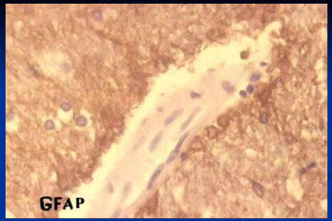
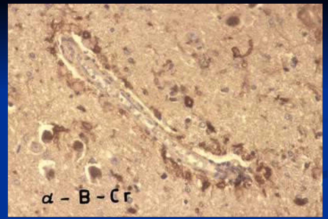
Rosenthal fibers: occurrence
reactive around craniopharyngeoma around MS plaques
neoplastic (?) pilocytic astrocytoma
genetic Alexander disease giant axonal neuropathy
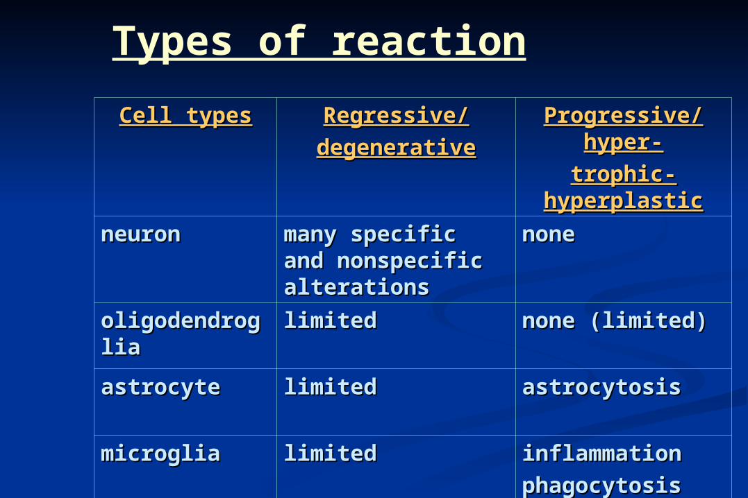
Types of reaction
Cell typesCell types Regressive/Regressive/
degenerativedegenerative
Progressive/hyper-Progressive/hyper-
trophic-trophic-hyperplastichyperplastic
neuronneuron many specific and many specific and nonspecific nonspecific alterationsalterations
nonenone
oligodendrogliaoligodendroglia limitedlimited none (limited)none (limited)
astrocyteastrocyte limitedlimited astrocytosisastrocytosis
microgliamicroglia limitedlimited inflammationinflammation
phagocytosisphagocytosis
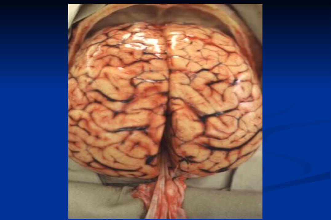
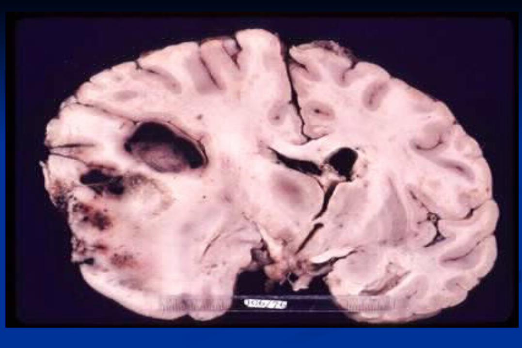
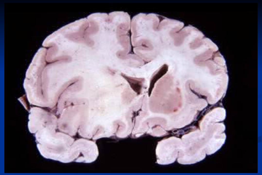
Space occupying Lesions
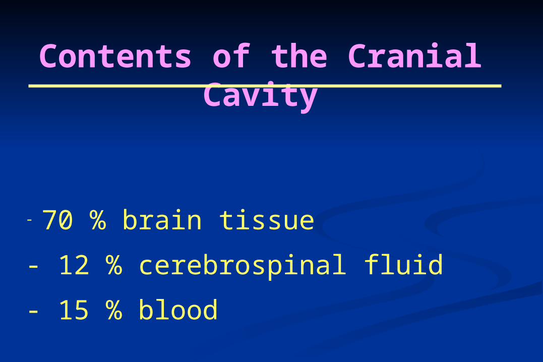
Contents of the Cranial Cavity
- 70 % brain tissue
- 12 % cerebrospinal fluid
- 15 % blood
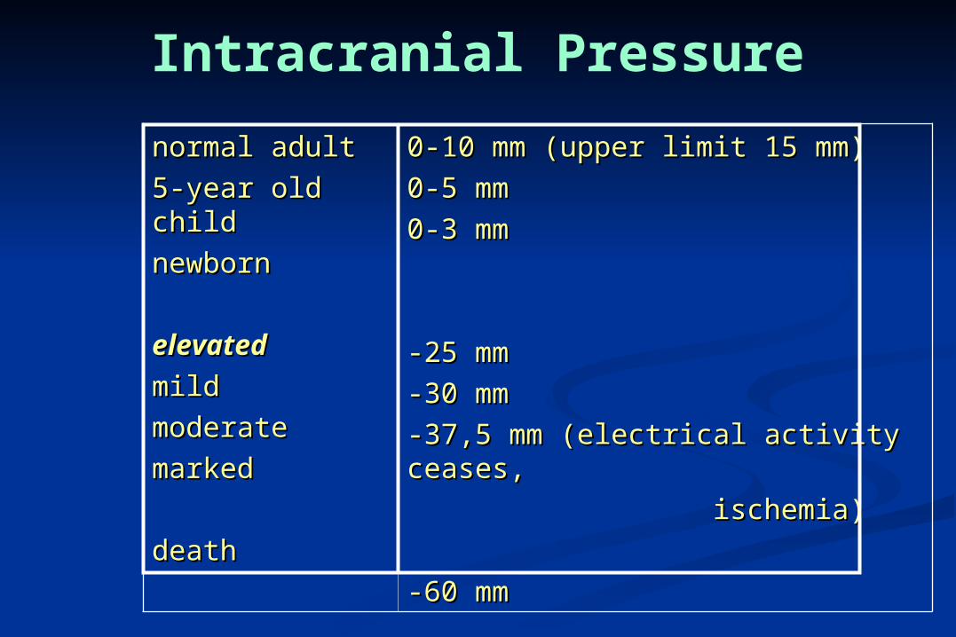
normal adultnormal adult
5-year old child5-year old child
newbornnewborn
elevatedelevated
mildmild
moderatemoderate
markedmarked
deathdeath
0-10 mm (upper limit 15 mm)0-10 mm (upper limit 15 mm)
0-5 mm0-5 mm
0-3 mm0-3 mm
-25 mm-25 mm
-30 mm-30 mm
-37,5 mm (electrical activity ceases, -37,5 mm (electrical activity ceases,
ischemia)ischemia)
-60 mm-60 mm
Intracranial Pressure
Causes of space-occupying lesions
tubors
haemorrhages
inflammatory processes
blockage of CSF (hydrocephalus)
brain edema
trauma
ischemia / anoxia

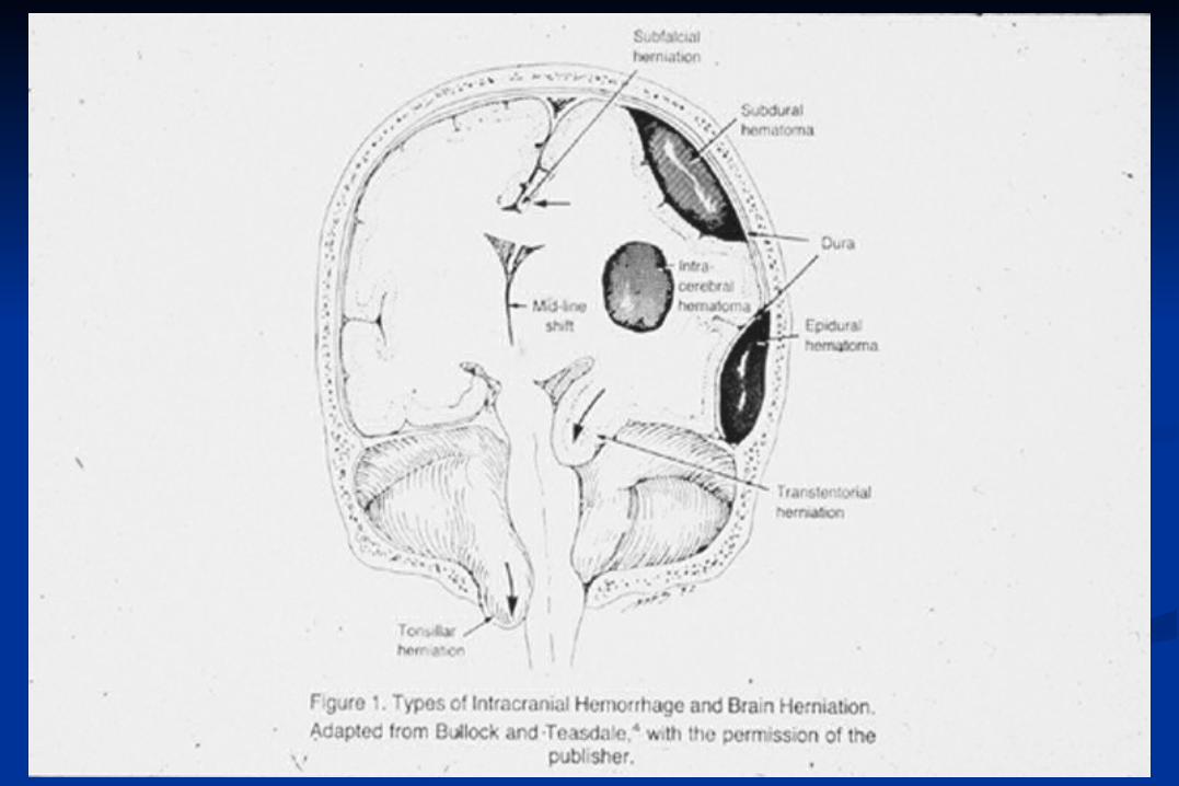
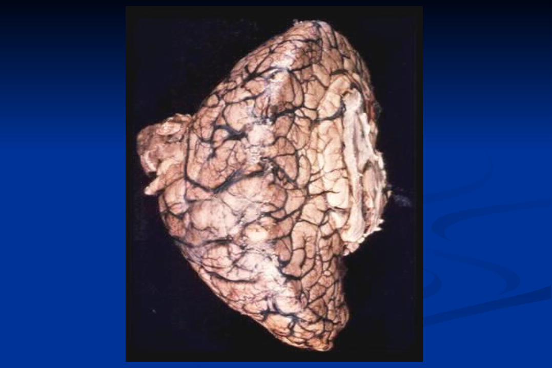
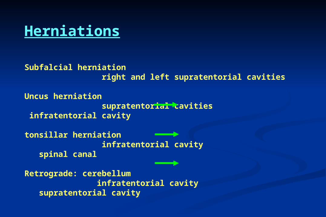
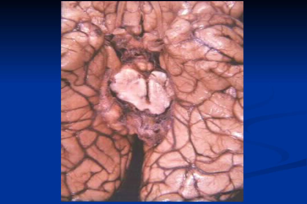
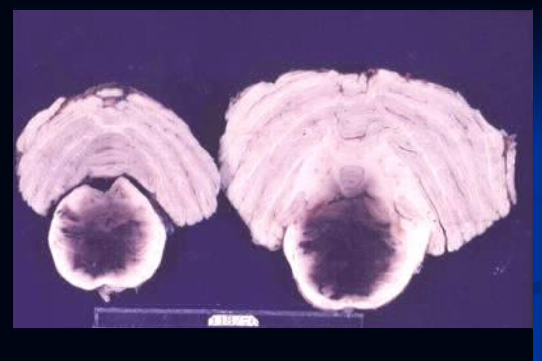
Herniations
Subfalcial herniation right and left supratentorial cavities
Uncus herniation supratentorial cavities infratentorial cavity
tonsillar herniation infratentorial cavity spinal canal
Retrograde: cerebellum infratentorial cavity supratentorial cavity
MAINZ
Walther Wagner