Embed Size (px)
Citation preview
1
Tetralogy of Fallot Epidemiology: Tetralogy of Fallot (TOF) is the most common cyanotic congenital heart disease in all age groups, constituting approximately 8% of congenital heart disease overall. TOF occurs in approximately 0.19‐0.26/1,000 live births. In the United States, the prevalence of TOF is approximately 3.9 per 10,000 live births. Definition: Tetralogy of Fallot is characterized by the presence of four anatomical findings:
1. Ventricular septal defect 2. Pulmonary stenosis (right ventricular outflow obstruction) 3. Dextroposition of the aorta (overriding aorta) 4. Right ventricular hypertrophy
Pathophysiology: The figure below compares the normal anatomy and blood flow of the heart to that found in Tetralogy of Fallot. The initial defect in TOF is a narrowing of the right ventricular outflow tract into the pulmonary artery. This prevents deoxygenated blood from entering the pulmonary circuit. In response to this outflow obstruction, the myocardium of the right ventricle hypertrophies in order to contract forcefully enough to push blood past the stenosis. Additionally, patients have a large ventricular septal defect which allows shunting of blood between the ventricles. In a patient with an isolated VSD, the blood flow is shunted initially from left‐to‐right. However, in TOF, the right ventricular outflow obstruction may impede the normal blood flow so significantly that the left side of the heart becomes the path of least resistance. Blood from the right ventricle is then forced into the left ventricle, creating a right‐to‐left shunt and subsequent cyanosis. Finally, the aorta overrides the ventricular septal defect, straddling the VSD. This allows deoxygenated blood shunted from the right ventricle to immediately exit the heart mixed with blood from the left ventricle.
The most important determinant of the severity and clinical consequences of TOF is the degree of right ventricular outflow obstruction. With a lesser obstruction, blood is shunted from left‐to‐right and permitted to preferentially enter the pulmonary circulation, allowing for oxygenation. With a greater degree of obstruction, however, blood is forced in the opposite direction, away from the pulmonary circulation, leftward across the VSD and ultimately blood exits the heart before being oxygenated. Patients will present with differing degrees of outflow obstruction, and this may fluctuate throughout the course of the illness.
2
Other Associated Abnormalities: Of note, approximately 40% of patients with TOF have additional congenital heart defects. This includes frank pulmonic stenosis, right aortic arch, abnormalities of the coronary arteries, collateral vessels supplying the pulmonary arteries, patent ductus arteriosus or other defects. It is important to evaluate the patient for all associated heart defects as this may affect surgical intervention or medical therapy. Additionally, clinicians should recall that TOF is associated with a number of genetic syndromes. This includes Trisomy 21 (Down Syndrome) as well as DiGeorge Syndrome and velocardiofacial syndromes. Presenting Signs and Symptoms: The timing and features of presentation depend on the degree of right ventricular outflow obstruction. Patients with more severe obstruction will present earlier due to cyanosis. This may be as early as the immediate newborn period. For patients with more moderate disease, the presenting sign may be a heart murmur (see below). Finally, for patients with mild disease, with so‐called “pink tetralogy” due to the lack of cyanosis, their presentation may consist of signs and symptoms of congestive heart failure due to the left‐to‐right shunting across the VSD and subsequent pulmonary overcirculation. Ultimately, most patients with mild disease will become cyanotic as the degree of outflow obstruction increases over time. Clinical Features: Patients with TOF have a number of distinguishing signs and symptoms that can be found on physical exam and elucidated with a detailed history.
• Cardiac exam: Most importantly, the heart murmur heart in TOF is not due to the VSD! It is in fact due to the right ventricular outflow obstruction. The murmur is typically crescendo‐decrescendo with a harsh systolic ejection quality; it is appreciated best along the left mid to upper sternal border with radiation posteriorly. (Remember, an isolated VSD murmur is a holosystolic murmur, best heard in the tricuspid area. It may radiate to the right lower sternal border.) Patients will have a normal S1 and possibly a single S2 due to diminished P2 component.
• Cyanosis: If patients are cyanotic, this is most commonly seen on the lips or nail beds. • Tet spells: Tet spells are hypercyanotic episodes precipitated by a sudden increase in right‐to‐
left shunting of blood. They can be elicited by activity (e.g. feeding, crying), or they may occur without warning. The classic description is of a patient who becomes cyanotic and then assumes a squatting position to relieve the cyanosis and hypoxia. Squatting serves to increase peripheral vascular resistance, thereby increasing the pressure in the left heart, and subsequently forcing blood back into the pulmonary circulation.
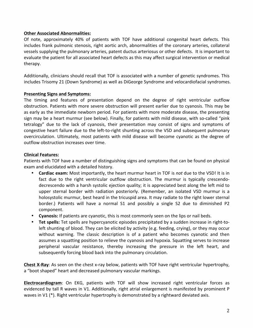
Chest X‐Ray: As seen on the chest x‐ray below, patients with TOF have right ventricular hypertrophy, a “boot shaped” heart and decreased pulmonary vascular markings. Electrocardiogram: On EKG, patients with TOF will show increased right ventricular forces as evidenced by tall R waves in V1. Additionally, right atrial enlargement is manifested by prominent P waves in V1 (*). Right ventricular hypertrophy is demonstrated by a rightward deviated axis.
3
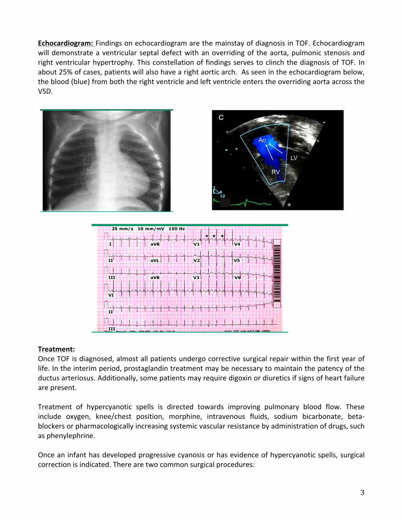
Echocardiogram: Findings on echocardiogram are the mainstay of diagnosis in TOF. Echocardiogram will demonstrate a ventricular septal defect with an overriding of the aorta, pulmonic stenosis and right ventricular hypertrophy. This constellation of findings serves to clinch the diagnosis of TOF. In about 25% of cases, patients will also have a right aortic arch. As seen in the echocardiogram below, the blood (blue) from both the right ventricle and left ventricle enters the overriding aorta across the VSD.
Treatment: Once TOF is diagnosed, almost all patients undergo corrective surgical repair within the first year of life. In the interim period, prostaglandin treatment may be necessary to maintain the patency of the ductus arteriosus. Additionally, some patients may require digoxin or diuretics if signs of heart failure are present. Treatment of hypercyanotic spells is directed towards improving pulmonary blood flow. These include oxygen, knee/chest position, morphine, intravenous fluids, sodium bicarbonate, beta‐blockers or pharmacologically increasing systemic vascular resistance by administration of drugs, such as phenylephrine. Once an infant has developed progressive cyanosis or has evidence of hypercyanotic spells, surgical correction is indicated. There are two common surgical procedures:
4
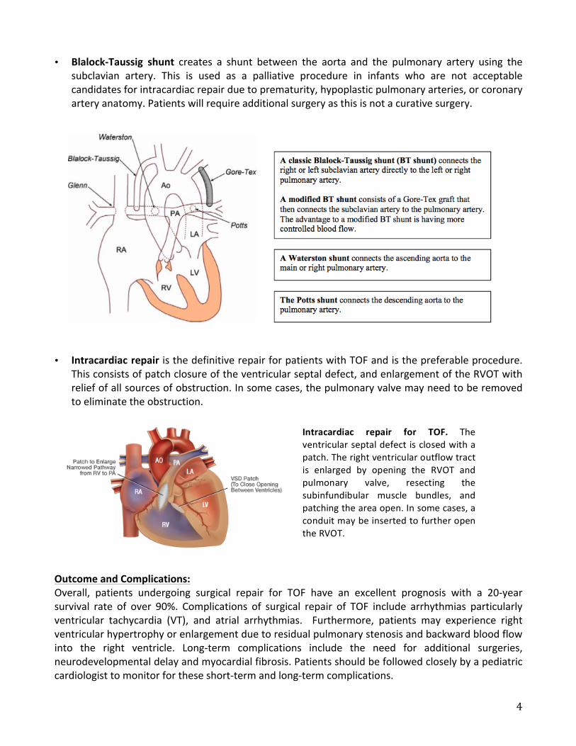
• Blalock‐Taussig shunt creates a shunt between the aorta and the pulmonary artery using the subclavian artery. This is used as a palliative procedure in infants who are not acceptable candidates for intracardiac repair due to prematurity, hypoplastic pulmonary arteries, or coronary artery anatomy. Patients will require additional surgery as this is not a curative surgery.
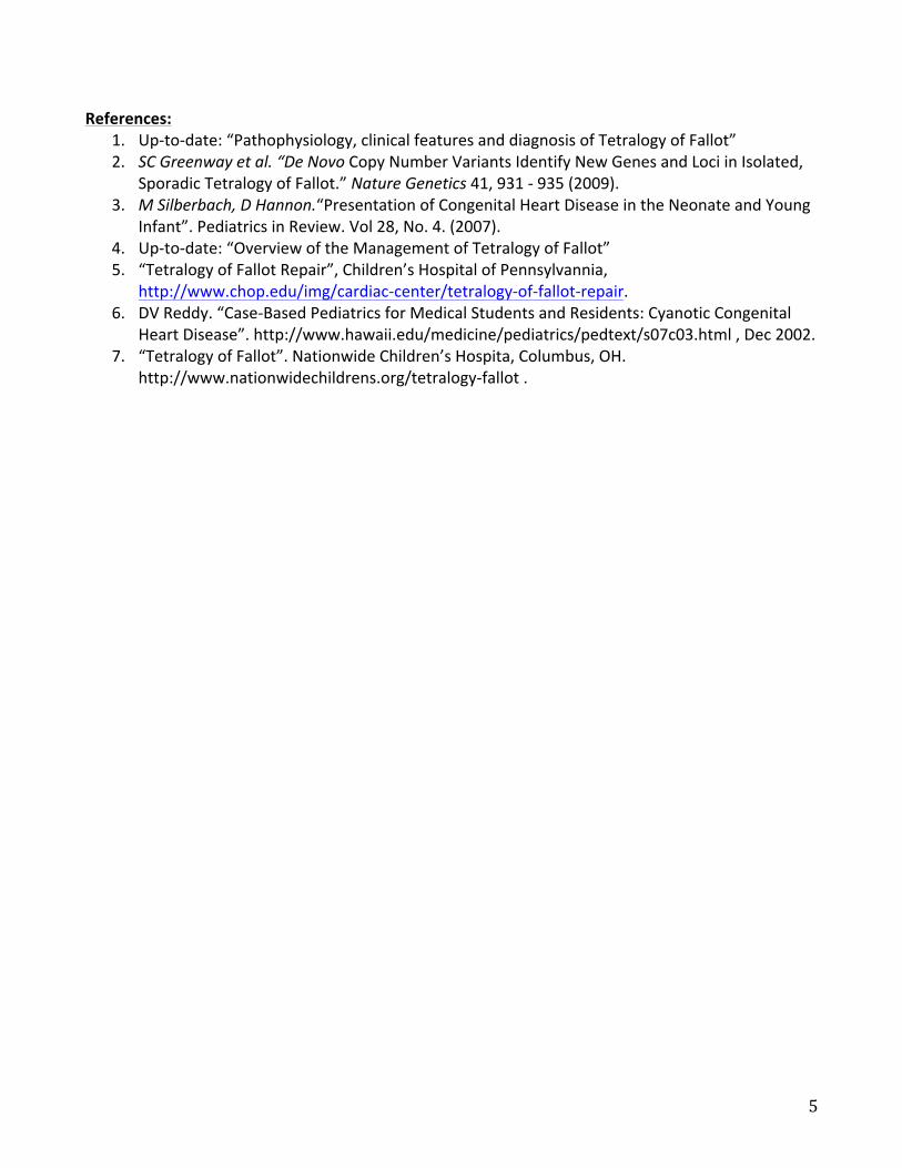
• Intracardiac repair is the definitive repair for patients with TOF and is the preferable procedure.
This consists of patch closure of the ventricular septal defect, and enlargement of the RVOT with relief of all sources of obstruction. In some cases, the pulmonary valve may need to be removed to eliminate the obstruction.
Outcome and Complications: Overall, patients undergoing surgical repair for TOF have an excellent prognosis with a 20‐year survival rate of over 90%. Complications of surgical repair of TOF include arrhythmias particularly ventricular tachycardia (VT), and atrial arrhythmias. Furthermore, patients may experience right ventricular hypertrophy or enlargement due to residual pulmonary stenosis and backward blood flow into the right ventricle. Long‐term complications include the need for additional surgeries, neurodevelopmental delay and myocardial fibrosis. Patients should be followed closely by a pediatric cardiologist to monitor for these short‐term and long‐term complications.
Intracardiac repair for TOF. The ventricular septal defect is closed with a patch. The right ventricular outflow tract is enlarged by opening the RVOT and pulmonary valve, resecting the subinfundibular muscle bundles, and patching the area open. In some cases, a conduit may be inserted to further open the RVOT.
5
References:
1. Up‐to‐date: “Pathophysiology, clinical features and diagnosis of Tetralogy of Fallot” 2. SC Greenway et al. “De Novo Copy Number Variants Identify New Genes and Loci in Isolated,
Sporadic Tetralogy of Fallot.” Nature Genetics 41, 931 ‐ 935 (2009). 3. M Silberbach, D Hannon.“Presentation of Congenital Heart Disease in the Neonate and Young
Infant”. Pediatrics in Review. Vol 28, No. 4. (2007). 4. Up‐to‐date: “Overview of the Management of Tetralogy of Fallot” 5. “Tetralogy of Fallot Repair”, Children’s Hospital of Pennsylvannia,
http://www.chop.edu/img/cardiac‐center/tetralogy‐of‐fallot‐repair. 6. DV Reddy. “Case‐Based Pediatrics for Medical Students and Residents: Cyanotic Congenital
Heart Disease”. http://www.hawaii.edu/medicine/pediatrics/pedtext/s07c03.html , Dec 2002. 7. “Tetralogy of Fallot”. Nationwide Children’s Hospita, Columbus, OH.
http://www.nationwidechildrens.org/tetralogy‐fallot .