Embed Size (px)
Citation preview
© The Ulster Medical Society, 2013. www.ums.ac.uk
Ulster Med J 2013;82(3):179-187
Dept of Radiology, Royal Victoria Hospital, Grosvenor Road, Belfast BT12 6BA
Correspondence to Dr James
Email: [email protected]
Pictorial Review
The Abdominal RadiographBarry James, Barry Kelly
Accepted 8 August 2013
The abdominal radiograph can often be a daunting prospect to the uninitiated, but by following some basic principles of image interpretation, even those with little clinical experience can almost always reach a correct diagnosis.
Technique:
The abdominal radiograph (AXR) is performed almost exclusively in the supine position and in the AP (anteroposterior) projection, i.e. the x-ray beam passes through the patient from front to back. Historically the abdominal radiograph was performed in both supine and erect postures, but this practice has been discontinued due to concerns over often excessive radiation dose. The Royal College of Radiologists recommends that when a patient presents with an acute abdomen, a single supine abdominal radiograph is performed. If there is the clinical suspicion of an associated intra-abdominal perforation, then an erect chest radiograph should also be performed. These guidelines and many others can be found on the Royal College’s latest edition of iRefer1.
The standard abdominal radiograph should extend from the diaphragm to the inferior pubic rami, and include the lateral abdominal wall musculature. This results in a standard ‘portrait’ radiograph. Failure to image the entire abdomen can lead to a misdiagnosis. For example, an obstructing inguinal hernia can be missed on an abdominal radiograph which does not extend below the level of the inguinal ligament. The associated delay in diagnosis can increase the risk of incarceration and perforation.
Radiation dose is an important consideration when ordering any imaging investigation. An abdominal radiograph in an average patient incurs a radiation dose of approximately1.5 mSv. This equates to seventy-five chest radiographs (0.02mSv) or approximately one sixth of the dose incurred by a standard CT of the abdomen (9-10mSv). The abdominal radiograph, like all ionizing radiation procedures, should therefore be used appropriately.
inTerpreTaTion
As with all radiographs, a logical repeatable step-by-step approach should be employed to examine all aspects of the film. This enables the reader to take an overview of the radiograph and allow all the information to be collated in order to arrive at the most accurate diagnosis. A classic example that highlights the benefits of such an approach is
the patient with unknown advanced colorectal carcinoma. Such a film typically shows large bowel obstruction (which is a reasonably straightforward diagnosis to make), but as the reader progresses through the film review, the detection of metastases at the lung bases can help confirm the aetiology is almost certainly malignant obstruction.
An algorithm outlining for interpreting an abdominal radiograph is presented below.
Algorithm for assessing an abdominal radiograph
The acuTe abdomen
There are a number of significant abdominal conditions that should not be missed and these are discussed below.
bowel Gas paTTerns
1. Free Air
© The Ulster Medical Society, 2013.
180 The Ulster Medical Journal
www.ums.ac.uk
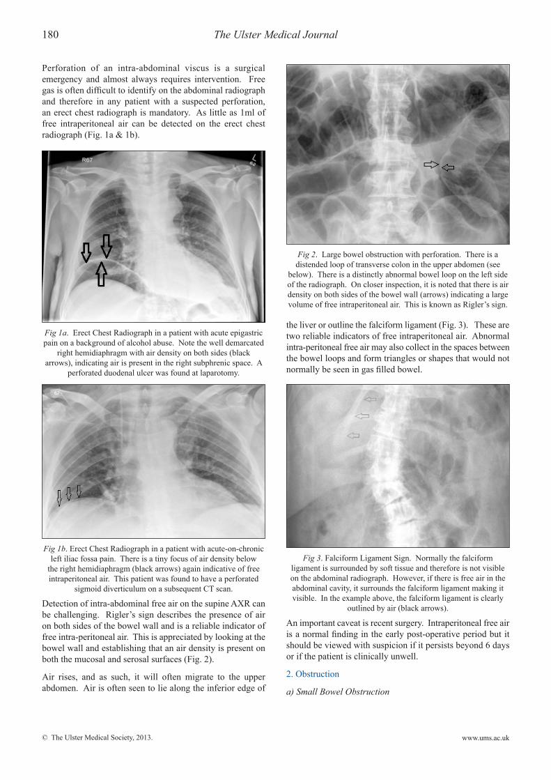
Perforation of an intra-abdominal viscus is a surgical emergency and almost always requires intervention. Free gas is often difficult to identify on the abdominal radiograph and therefore in any patient with a suspected perforation, an erect chest radiograph is mandatory. As little as 1ml of free intraperitoneal air can be detected on the erect chest radiograph (Fig. 1a & 1b).
Fig 1a. Erect Chest Radiograph in a patient with acute epigastric pain on a background of alcohol abuse. Note the well demarcated
right hemidiaphragm with air density on both sides (black arrows), indicating air is present in the right subphrenic space. A
perforated duodenal ulcer was found at laparotomy.
Fig 1b. Erect Chest Radiograph in a patient with acute-on-chronic left iliac fossa pain. There is a tiny focus of air density below
the right hemidiaphragm (black arrows) again indicative of free intraperitoneal air. This patient was found to have a perforated
sigmoid diverticulum on a subsequent CT scan.
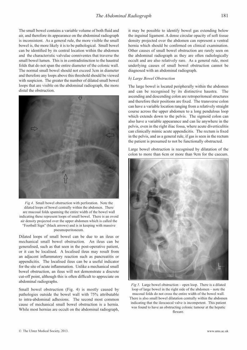
Detection of intra-abdominal free air on the supine AXR can be challenging. Rigler’s sign describes the presence of air on both sides of the bowel wall and is a reliable indicator of free intra-peritoneal air. This is appreciated by looking at the bowel wall and establishing that an air density is present on both the mucosal and serosal surfaces (Fig. 2).
Air rises, and as such, it will often migrate to the upper abdomen. Air is often seen to lie along the inferior edge of
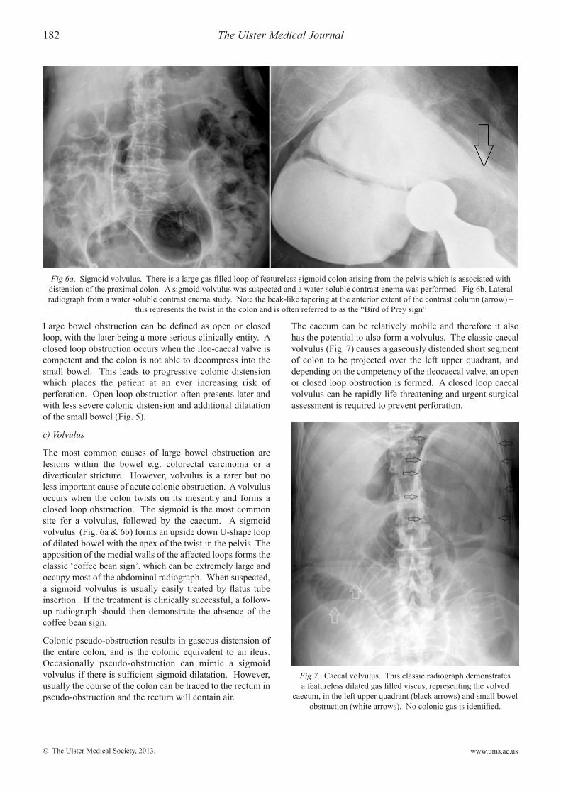
the liver or outline the falciform ligament (Fig. 3). These are two reliable indicators of free intraperitoneal air. Abnormal intra-peritoneal free air may also collect in the spaces between the bowel loops and form triangles or shapes that would not normally be seen in gas filled bowel.
Fig 3. Falciform Ligament Sign. Normally the falciform ligament is surrounded by soft tissue and therefore is not visible on the abdominal radiograph. However, if there is free air in the abdominal cavity, it surrounds the falciform ligament making it visible. In the example above, the falciform ligament is clearly
outlined by air (black arrows).
An important caveat is recent surgery. Intraperitoneal free air is a normal finding in the early post-operative period but it should be viewed with suspicion if it persists beyond 6 days or if the patient is clinically unwell.
2. Obstruction
a) Small Bowel Obstruction
Fig 2. Large bowel obstruction with perforation. There is a distended loop of transverse colon in the upper abdomen (see
below). There is a distinctly abnormal bowel loop on the left side of the radiograph. On closer inspection, it is noted that there is air density on both sides of the bowel wall (arrows) indicating a large volume of free intraperitoneal air. This is known as Rigler’s sign.
© The Ulster Medical Society, 2013.
The Abdominal Radiograph 181
www.ums.ac.uk
The small bowel contains a variable volume of both fluid and air, and therefore its appearance on the abdominal radiograph is inconsistent. As a general rule, the more visible the small bowel is, the more likely it is to be pathological. Small bowel can be identified by its central location within the abdomen and the characteristic valvulae conniventes that traverse the small bowel lumen. This is in contradistinction to the haustral folds that do not span the entire diameter of the colonic wall. The normal small bowel should not exceed 3cm in diameter and therefore any loops above this threshold should be viewed with suspicion. The greater the number of dilated small bowel loops that are visible on the abdominal radiograph, the more distal the obstruction.
Fig 4. Small bowel obstruction with perforation. Note the dilated loops of bowel centrally within the abdomen. There
are mucosal folds spanning the entire width of the bowel wall indicating these represent loops of small bowel. There is an ovoid air density projected over the upper abdomen which is called the “Football Sign” (black arrows) and is in keeping with massive
pneumoperitoneum.
Dilated loops of small bowel can be due to an ileus or mechanical small bowel obstruction. An ileus can be generalised, such as that seen in the post-operative patient, or it can be localised. A localised ileus may result from an adjacent inflammatory reaction such as pancreatitis or appendicitis. The localised ileus can be a useful indicator for the site of acute inflammation. Unlike a mechanical small bowel obstruction, an ileus will not demonstrate a discrete cut-off point, although this is often difficult to appreciate on abdominal radiographs.
Small bowel obstruction (Fig. 4) is mostly caused by pathologies outside the bowel wall with 75% attributable to intra-abdominal adhesions. The second most common cause of mechanical small bowel obstruction is a hernia. While most hernias are occult on the abdominal radiograph,
it may be possible to identify bowel gas extending below the inguinal ligament. A dense circular opacity of soft tissue density projected over the abdomen can represent a ventral hernia which should be confirmed on clinical examination. Other causes of small bowel obstruction are rarely seen on the abdominal radiograph as they are often radiologically occult and are also relatively rare. As a general rule, most underlying causes of small bowel obstruction cannot be diagnosed with an abdominal radiograph.
b) Large Bowel Obstruction
The large bowel is located peripherally within the abdomen and can be recognised by its distinctive haustra. The ascending and descending colon are retroperitoneal structures and therefore their positions are fixed. The transverse colon can have a variable location ranging from a relatively straight course across the upper abdomen to a long pendulous loop which extends down to the pelvis. The sigmoid colon can also have a variable appearance and can lie anywhere in the pelvis, even in the right iliac fossa, where acute diverticulitis can clinically mimic acute appendicitis. The rectum is fixed in the pelvis, and as a general rule, if gas is seen in the rectum the patient is presumed to not be functionally obstructed.
Large bowel obstruction is recognised by dilatation of the colon to more than 6cm or more than 9cm for the caecum.
Fig 5. Large bowel obstruction – open loop. There is a dilated loop of large bowel in the right side of the abdomen – note the mucosal folds do not cross the entire width of the bowel wall.
There is also small bowel dilatation centrally within the abdomen indicating that the ileocaecal valve is incompetent. This patient was found to have an obstructing colonic tumour at the hepatic
flexure.
© The Ulster Medical Society, 2013.
182 The Ulster Medical Journal
www.ums.ac.uk
Large bowel obstruction can be defined as open or closed loop, with the later being a more serious clinically entity. A closed loop obstruction occurs when the ileo-caecal valve is competent and the colon is not able to decompress into the small bowel. This leads to progressive colonic distension which places the patient at an ever increasing risk of perforation. Open loop obstruction often presents later and with less severe colonic distension and additional dilatation of the small bowel (Fig. 5).
c) Volvulus
The most common causes of large bowel obstruction are lesions within the bowel e.g. colorectal carcinoma or a diverticular stricture. However, volvulus is a rarer but no less important cause of acute colonic obstruction. A volvulus occurs when the colon twists on its mesentry and forms a closed loop obstruction. The sigmoid is the most common site for a volvulus, followed by the caecum. A sigmoid volvulus (Fig. 6a & 6b) forms an upside down U-shape loop of dilated bowel with the apex of the twist in the pelvis. The apposition of the medial walls of the affected loops forms the classic ‘coffee bean sign’, which can be extremely large and occupy most of the abdominal radiograph. When suspected, a sigmoid volvulus is usually easily treated by flatus tube insertion. If the treatment is clinically successful, a follow-up radiograph should then demonstrate the absence of the coffee bean sign.
Colonic pseudo-obstruction results in gaseous distension of the entire colon, and is the colonic equivalent to an ileus. Occasionally pseudo-obstruction can mimic a sigmoid volvulus if there is sufficient sigmoid dilatation. However, usually the course of the colon can be traced to the rectum in pseudo-obstruction and the rectum will contain air.
The caecum can be relatively mobile and therefore it also has the potential to also form a volvulus. The classic caecal volvulus (Fig. 7) causes a gaseously distended short segment of colon to be projected over the left upper quadrant, and depending on the competency of the ileocaecal valve, an open or closed loop obstruction is formed. A closed loop caecal volvulus can be rapidly life-threatening and urgent surgical assessment is required to prevent perforation.
Fig 7. Caecal volvulus. This classic radiograph demonstrates a featureless dilated gas filled viscus, representing the volved
caecum, in the left upper quadrant (black arrows) and small bowel obstruction (white arrows). No colonic gas is identified.
Fig 6a. Sigmoid volvulus. There is a large gas filled loop of featureless sigmoid colon arising from the pelvis which is associated with distension of the proximal colon. A sigmoid volvulus was suspected and a water-soluble contrast enema was performed. Fig 6b. Lateral radiograph from a water soluble contrast enema study. Note the beak-like tapering at the anterior extent of the contrast column (arrow) –
this represents the twist in the colon and is often referred to as the “Bird of Prey sign”
© The Ulster Medical Society, 2013.
The Abdominal Radiograph 183
www.ums.ac.uk
coliTis
Colonic “thumb printing” is an indicator of colitis, but unfortunately it is a non-specific sign with many causes. Thumb-printing represents oedematous mucosal folds that look like thumb prints along the wall of the gas-filled colon (Fig.8a & 8b).
Fig 8a. Colitis. Note the thickened mucosal folds of the left hemicolon, often referred to as “thumbprinting”. Fig 8b. The transverse colon is featureless in keeping with chronic colitis.
Toxic megacolon can develop in patients with colitis. The classic radiographic finding in toxic megacolon is dilatation of the transverse colon to 6cm or more but it can also be indicated by progressive colonic distension on serial radiographs. Toxic megacolon is a surgical emergency and prompt assessment is required. It is also worth noting that water soluble/barium enemas are contraindicated in this condition due to the high risk of perforation.
Chronic colitis causes atrophy and scarring of the colonic mucosa which will result in what is known as a ‘lead-pipe’ colon on the abdominal radiograph (Fig. 8b). This is recognised by an absence of the normal colonic haustra and the formation of two featureless parallel colonic walls. This is most commonly seen in the descending and sigmoid colon due to the increased prevalence of colitis at these sites.
air in The liver
This can present as two clinical entities: pneumobilia or portal venous gas.
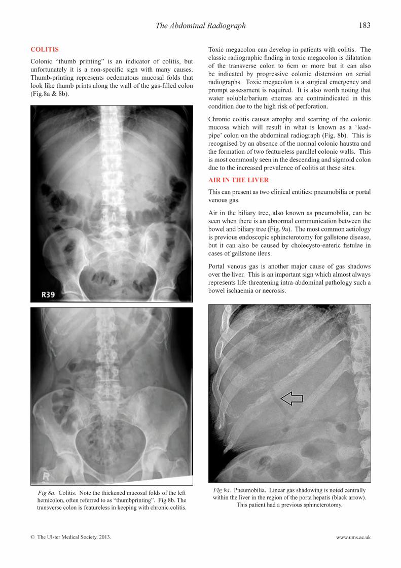
Air in the biliary tree, also known as pneumobilia, can be seen when there is an abnormal communication between the bowel and biliary tree (Fig. 9a). The most common aetiology is previous endoscopic sphincterotomy for gallstone disease, but it can also be caused by cholecysto-enteric fistulae in cases of gallstone ileus.
Portal venous gas is another major cause of gas shadows over the liver. This is an important sign which almost always represents life-threatening intra-abdominal pathology such a bowel ischaemia or necrosis.
Fig 9a. Pneumobilia. Linear gas shadowing is noted centrally within the liver in the region of the porta hepatis (black arrow).
This patient had a previous sphincterotomy.
© The Ulster Medical Society, 2013.
184 The Ulster Medical Journal
www.ums.ac.uk
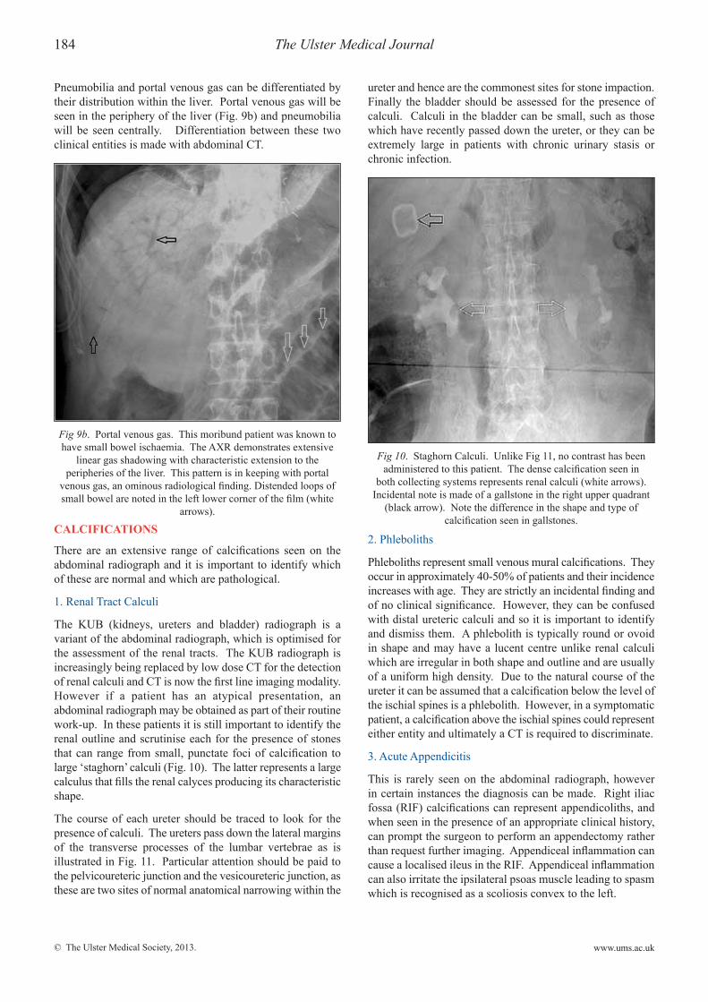
Pneumobilia and portal venous gas can be differentiated by their distribution within the liver. Portal venous gas will be seen in the periphery of the liver (Fig. 9b) and pneumobilia will be seen centrally. Differentiation between these two clinical entities is made with abdominal CT.
Fig 9b. Portal venous gas. This moribund patient was known to have small bowel ischaemia. The AXR demonstrates extensive
linear gas shadowing with characteristic extension to the peripheries of the liver. This pattern is in keeping with portal
venous gas, an ominous radiological finding. Distended loops of small bowel are noted in the left lower corner of the film (white
arrows).
calcificaTions
There are an extensive range of calcifications seen on the abdominal radiograph and it is important to identify which of these are normal and which are pathological.
1. Renal Tract Calculi
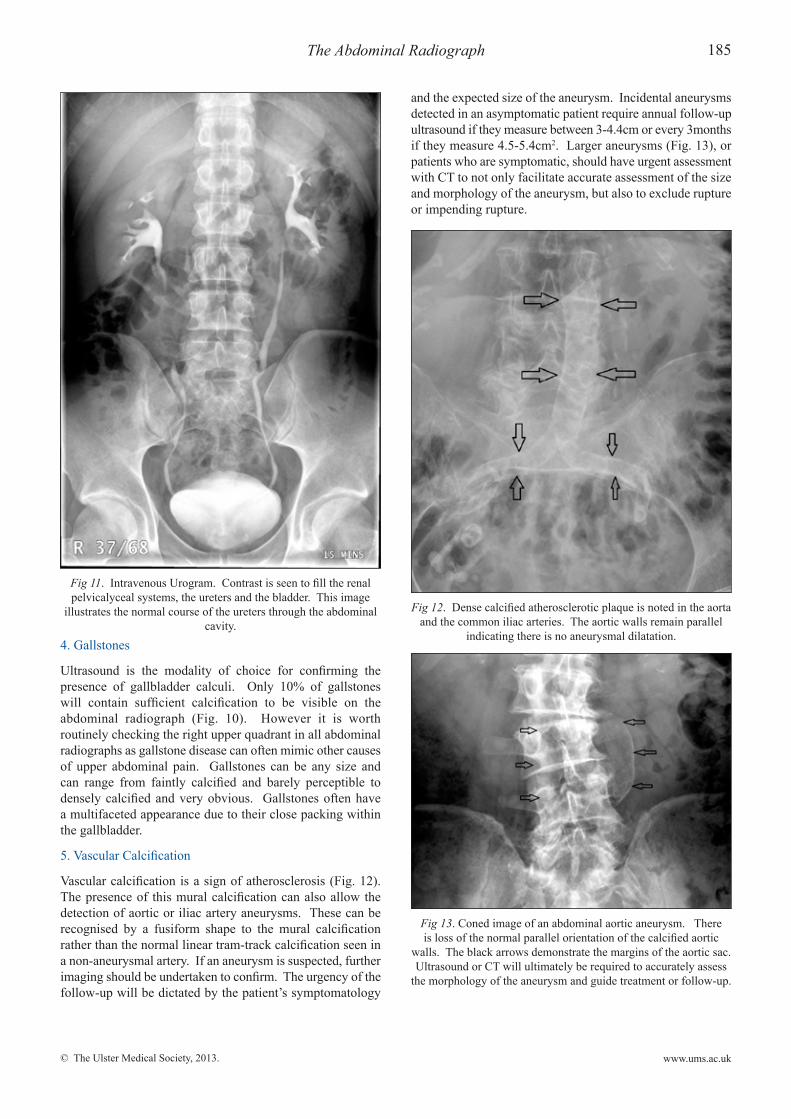
The KUB (kidneys, ureters and bladder) radiograph is a variant of the abdominal radiograph, which is optimised for the assessment of the renal tracts. The KUB radiograph is increasingly being replaced by low dose CT for the detection of renal calculi and CT is now the first line imaging modality. However if a patient has an atypical presentation, an abdominal radiograph may be obtained as part of their routine work-up. In these patients it is still important to identify the renal outline and scrutinise each for the presence of stones that can range from small, punctate foci of calcification to large ‘staghorn’ calculi (Fig. 10). The latter represents a large calculus that fills the renal calyces producing its characteristic shape.
The course of each ureter should be traced to look for the presence of calculi. The ureters pass down the lateral margins of the transverse processes of the lumbar vertebrae as is illustrated in Fig. 11. Particular attention should be paid to the pelvicoureteric junction and the vesicoureteric junction, as these are two sites of normal anatomical narrowing within the
ureter and hence are the commonest sites for stone impaction. Finally the bladder should be assessed for the presence of calculi. Calculi in the bladder can be small, such as those which have recently passed down the ureter, or they can be extremely large in patients with chronic urinary stasis or chronic infection.
Fig 10. Staghorn Calculi. Unlike Fig 11, no contrast has been administered to this patient. The dense calcification seen in
both collecting systems represents renal calculi (white arrows). Incidental note is made of a gallstone in the right upper quadrant
(black arrow). Note the difference in the shape and type of calcification seen in gallstones.
2. Phleboliths
Phleboliths represent small venous mural calcifications. They occur in approximately 40-50% of patients and their incidence increases with age. They are strictly an incidental finding and of no clinical significance. However, they can be confused with distal ureteric calculi and so it is important to identify and dismiss them. A phlebolith is typically round or ovoid in shape and may have a lucent centre unlike renal calculi which are irregular in both shape and outline and are usually of a uniform high density. Due to the natural course of the ureter it can be assumed that a calcification below the level of the ischial spines is a phlebolith. However, in a symptomatic patient, a calcification above the ischial spines could represent either entity and ultimately a CT is required to discriminate.
3. Acute Appendicitis
This is rarely seen on the abdominal radiograph, however in certain instances the diagnosis can be made. Right iliac fossa (RIF) calcifications can represent appendicoliths, and when seen in the presence of an appropriate clinical history, can prompt the surgeon to perform an appendectomy rather than request further imaging. Appendiceal inflammation can cause a localised ileus in the RIF. Appendiceal inflammation can also irritate the ipsilateral psoas muscle leading to spasm which is recognised as a scoliosis convex to the left.
© The Ulster Medical Society, 2013.
The Abdominal Radiograph 185
www.ums.ac.uk
4. Gallstones
Ultrasound is the modality of choice for confirming the presence of gallbladder calculi. Only 10% of gallstones will contain sufficient calcification to be visible on the abdominal radiograph (Fig. 10). However it is worth routinely checking the right upper quadrant in all abdominal radiographs as gallstone disease can often mimic other causes of upper abdominal pain. Gallstones can be any size and can range from faintly calcified and barely perceptible to densely calcified and very obvious. Gallstones often have a multifaceted appearance due to their close packing within the gallbladder.
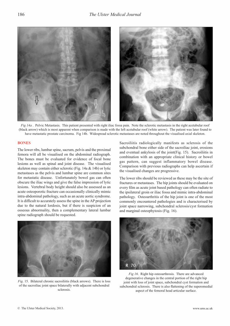
5. Vascular Calcification
Vascular calcification is a sign of atherosclerosis (Fig. 12). The presence of this mural calcification can also allow the detection of aortic or iliac artery aneurysms. These can be recognised by a fusiform shape to the mural calcification rather than the normal linear tram-track calcification seen in a non-aneurysmal artery. If an aneurysm is suspected, further imaging should be undertaken to confirm. The urgency of the follow-up will be dictated by the patient’s symptomatology
and the expected size of the aneurysm. Incidental aneurysms detected in an asymptomatic patient require annual follow-up ultrasound if they measure between 3-4.4cm or every 3months if they measure 4.5-5.4cm2. Larger aneurysms (Fig. 13), or patients who are symptomatic, should have urgent assessment with CT to not only facilitate accurate assessment of the size and morphology of the aneurysm, but also to exclude rupture or impending rupture.
Fig 12. Dense calcified atherosclerotic plaque is noted in the aorta and the common iliac arteries. The aortic walls remain parallel
indicating there is no aneurysmal dilatation.
Fig 13. Coned image of an abdominal aortic aneurysm. There is loss of the normal parallel orientation of the calcified aortic
walls. The black arrows demonstrate the margins of the aortic sac. Ultrasound or CT will ultimately be required to accurately assess
the morphology of the aneurysm and guide treatment or follow-up.
Fig 11. Intravenous Urogram. Contrast is seen to fill the renal pelvicalyceal systems, the ureters and the bladder. This image
illustrates the normal course of the ureters through the abdominal cavity.
© The Ulster Medical Society, 2013.
186 The Ulster Medical Journal
www.ums.ac.uk
bones
The lower ribs, lumbar spine, sacrum, pelvis and the proximal femora will all be visualised on the abdominal radiograph. The bones must be evaluated for evidence of focal bone lesions as well as spinal and joint disease. The visualised skeleton may contain either sclerotic (Fig. 14a & 14b) or lytic metastases as the pelvis and lumbar spine are common sites for metastatic disease. Unfortunately bowel gas can often obscure the iliac wings and give the false impression of lytic lesions. Vertebral body height should also be assessed as an acute osteoporotic fracture can occasionally clinically mimic intra-abdominal pathology, such as an acute aortic syndrome. It is difficult to accurately assess the spine in the AP projection due to the natural lordosis, but if there is suspicion of an osseous abnormality, then a complementary lateral lumbar spine radiograph should be requested.
Sacroiliitis radiologically manifests as sclerosis of the subchondral bone either side of the sacroiliac joint, erosions and eventual ankylosis of the joint(Fig. 15). Sacroiliitis in combination with an appropriate clinical history or bowel gas pattern, can suggest inflammatory bowel disease. Comparison with previous radiographs can help ascertain if the visualised changes are progressive.
The lower ribs should be reviewed as these may be the site of fractures or metastases. The hip joints should be evaluated on every film as acute joint based pathology can often radiate to the ipsilateral groin or iliac fossa and mimic intra-abdominal pathology. Osteoarthritis of the hip joint is one of the most commonly encountered pathologies and is characterised by joint space narrowing, subchondral sclerosis/cyst formation and marginal osteophytosis (Fig. 16).
Fig 16. Right hip osteoarthrosis. There are advanced degenerative changes in the central portion of the right hip
joint with loss of joint space, subchondral cyst formation and subchondral sclerosis. There is also flattening of the superomedial
aspect of the femoral head articular surface.
Fig 14a . Pelvic Metastasis. This patient presented with right iliac fossa pain. Note the sclerotic metastasis in the right acetabular roof (black arrow) which is most apparent when comparison is made with the left acetabular roof (white arrow). The patient was later found to
have metastatic prostate carcinoma. Fig 14b. Widespread sclerotic metastases are noted throughout the visualised axial skeleton.
Fig. 15. Bilateral chronic sacroiliitis (black arrows). There is loss of the sacroiliac joint space bilaterally with adjacent subchondral
sclerosis.
© The Ulster Medical Society, 2013.
The Abdominal Radiograph 187
www.ums.ac.uk
The lunG bases
A portion of the lung bases will be visible on most technically satisfactory abdominal radiographs. A basal pneumonia can cause acute epigastric pain and this should be visible on the abdominal radiograph as loss of the ipsilateral hemidiaphragm. Lung metastases can also occasionally be seen at the lung bases.
conclusion
The abdominal radiograph provides a wealth of clinical
information that can help guide patient management. It is important to recognise that it incurs a relatively high radiation dose, but when used in the appropriate clinical setting it can facilitate a rapid and confident diagnosis.
references:
1. www.irefer.org.uk iRefer – making the best use of clinical radiology. Royal College of Radiologists, 2013.
2. http://aaa.screening.nhs.uk/cms.php?folder=2454 NHS Abdominal Aortic Aneurysm Screening Programme, 2009.