Embed Size (px)
Citation preview
The Added Diagnostic Value of LiquidGastric Emptying Compared with SolidEmptying Alone
Harvey A. Ziessman1, Ankit Chander1, John O. Clarke2, Alison Ramos1, and Richard L.Wahl1
1Division of Nuclear Medicine, Russell H. Morgan Department of Radiology and Radiological Sciences, Johns Hopkins MedicalInstitutions, Baltimore, Maryland; and 2Division of Gastroenterology, Department of Medicine, Johns Hopkins Medical Institutions,Baltimore, Maryland
The medical literature states that solid gastric-emptying studiesare more sensitive for the detection of gastroparesis than areliquid studies; thus, liquid studies are rarely required. However,we have seen patients with normal solid but delayed liquid emp-tying. The purpose of this investigation was to determine whethera study of clear liquid gastric empting has added value for the di-agnosis of gastroparesis over a study of solid emptying alone.Methods: A total of 101 patients underwent both solid and liquidgastric-emptying studies, acquired sequentially on the sameday. A 30-min (1-min frames) liquid study (300 mL of water with7.4 MBq [0.2 mCi] of 111In-diethylenetriaminepentaacetic acid)was followed by a standardized 4-h solid-meal study (a 99mTc-sulfur colloid–labeled egg-substitute sandwich meal). Emptyingwas quantified as a best-fit exponential emptying rate (T1/2) forliquids and percentage emptying at 4 h for solid empting. Thirtyhealthy volunteers underwent a study of clear liquid emptyingto establish normal values. The results of the liquid and solidstudies were compared. 111In liquid downscatter into the subse-quent 99mTc solid meal results was analyzed. Results: The upperrange of normal for clear liquid emptying (T1/2) for healthy volun-teers was 22 min (mean 6 3 SDs) and 19 min (mean 6 2 SDs). Of101 patients, delayed emptying was found in 36% of liquid and16% of solid studies. Of all patients with normal solid emptying,32% had delayed liquid emptying. 111In downscatter into the99mTc window was not generally significant. Conclusion: Forthe detection of gastroparesis, a 30-min study of clear liquid gas-tric-emptying has considerable added diagnostic value over astudy of solid emptying alone.
Key Words: gastric emptying; gastroparesis; stomach
J Nucl Med 2009; 50:726–731DOI: 10.2967/jnumed.108.059790
The radionuclide gastric-emptying study has long beenthe standard clinical diagnostic test for the detection ofgastroparesis. Both solid and liquid studies have been usedover the years, either as individual or as dual-phase studies,
for investigative and clinical purposes. Standard teachinghas been that only a solid study is needed for clinical pur-poses because the liquid study is less sensitive for the detec-tion of gastroparesis, and liquid studies should be reservedfor patients who cannot tolerate solids (1–6).
However, we have observed patients who have haddelayed liquid but normal solid emptying (7). The primarypurpose of this investigation was to determine, in a largepatient population, whether liquid gastric emptying pro-vided added diagnostic value over solid emptying alone forthe diagnosis of gastroparesis.
We chose to perform the studies on the same day to avoidpotential problems (e.g., different fasting conditions, dif-ferent medications, and possible intercurrent clinical con-ditions that might affect emptying) that might arise fromseparate-day studies. Thus, the clear liquid and solid gastric-emptying studies were performed sequentially; that is, a 30-min water study was performed, followed by the 4-hsimplified and standardized solid protocol recommendedby recently published consensus recommendations (8). A si-multaneous dual-isotope dual-phase study was consideredbut not used for several reasons. First, the liquid-only studyhad been our routine liquid methodology, and the cases ofdiscordance that we observed between solid and liquidstudies had been noted using the clear liquid–only study.Additionally, there were no published normal values forliquid emptying performed in conjunction with the stan-dardized solid gastric-emptying protocol reported by Tougaset al. (9).
As part of this investigation, we also sought to establishnormal values for clear liquid emptying in 30 healthy vol-unteers. Previously, only a few subjects had been reported.Finally, we analyzed whether 111In downscatter from theliquid study into the subsequent solid study 99mTc windowcould affect the solid emptying results.
MATERIALS AND METHODS
Patient StudiesBetween December 2007 and October 2008, sequential liquid
and solid radionuclide gastric-emptying studies were performed in
Received Nov. 3, 2008; revision accepted Jan. 7, 2009.For correspondence or reprints contact: Harvey A. Ziessman, Johns
Hopkins Outpatient Center, 601 N. Caroline St., Suite 3231, Baltimore,MD 21278.
E-mail: [email protected] ª 2009 by the Society of Nuclear Medicine, Inc.
726 THE JOURNAL OF NUCLEAR MEDICINE • Vol. 50 • No. 5 • May 2009
by on April 11, 2019. For personal use only. jnm.snmjournals.org Downloaded from
101 patients (24 men, 77 women; age range, 17–77 y; mean 6 SD,48 6 15 y). The patients were referred because of symptomssuggestive of gastroparesis. Symptoms included postprandial full-ness, epigastric discomfort or pain, nausea, and vomiting. No patientshad undergone prior gastric surgery. Seven patients had diabetesmellitus. Patients were taking various medications as ordered by theirreferring physicians, and the referring physicians decided whetherthe medications should be discontinued. Patients were instructed notto eat after dinner the night before, to fast overnight, and to havenothing by mouth the morning of the study. The sequential liquidand solid studies were performed in the early morning as the firststudy of the day. A dual-head large-field-of-view g-camera withmedium-energy collimators was used for both studies. Patientsingested 300 mL of water from a commercial water cooler, with7.4 MBq (0.2 mCi) of 111In-diethylenetriaminepentaacetic acid(DTPA), while lying semiupright (45�) on a hospital gurney. Theliquid study was acquired using only 1 of the detector headspositioned in the left anterior oblique projection so that thestomach and upper abdomen were in the field of view and yetthe patient could drink the liquid without difficulty. Imagingstarted immediately after the ingestion of the water. The patientscould more easily ingest the liquid in the semiupright positionthan they could in the supine position, and imaging could bestarted more promptly after ingestion. Images were acquired as1-min frames · 30 (128 · 128 matrix) using a 20% windowaround the 111In photopeaks (171 and 247 keV).
Immediately after the 30-min liquid study was performed, thepatient ingested the solid meal. The methodology and mealrecommended in a recently published consensus report (8) wereclosely followed. The meal consisted of a commercially availableegg substitute (equivalent to 2 large eggs), mixed with 74 MBq(2 mCi) of 99mTc-sulfur colloid and cooked in a microwave oven for2 min; 2 slices of toast; strawberry jam; and 120 mL of water (9).Patients ingested the solid meal within 10–15 min while sitting inan upright position. Patients then lay supine on the imaging table.Simultaneous anterior and posterior images were acquired for 1 mineach (256 · 256 matrix), with a 20% window centered around the99mTc photopeak (140 keV). Images were acquired immediately(time 0) and at 1, 2, 3, and 4 h after the meal was ingested.
Both studies were processed on a Xeleris (GE Healthcare)workstation. Regions of interest were drawn around the stomachusing a computer, and time–activity curves were generated. Forliquid emptying, a half-emptying time (time required for theemptying of half the meal) and a best-fit exponential emptying rate(T1/2) were calculated. Liquid gastric-emptying studies were notattenuation-corrected; prior investigations have shown that correc-tion is not necessary for liquid gastric emptying (10,11). Solidstudies were corrected for attenuation and radioactive decay. Thegeometric mean method (square root of the product of the anteriorand posterior counts) was used at each imaging time. The percentagesolid gastric emptying was quantified at 1, 2, 3, and 4 h. Normalvalues for solid emptying were based on the published results ofTougas et al. (9). Data from our study are reported as the percentageemptying rather than as the percentage retention used by Tougas etal. Emptying of less than 90% at 4 h was considered delayed. If thepatient reached greater than 90% gastric emptying (the 4-h normalvalue) at any imaging time, the study was concluded.
Healthy VolunteersTo establish normal values for clear liquid emptying (water), 30
healthy volunteers were recruited (17 women, 13 men; age range,
24–57 y; mean age 6 SD, 41 6 12 y). The volunteers’ bodyweight ranged from 49.5 to 117 kg (110–260 lbs) (mean 6 SD,73.8 6 21.15 kg [164 6 47 lbs]); 15 volunteers were AfricanAmerican, 12 were Caucasian, and 3 were Asian. The volunteerswere judged to be healthy if they had no recent, recurrent, orchronic symptoms or diseases; had not undergone prior abdominalsurgery; did not have diabetes mellitus, gastrointestinal symptoms,or chronic illnesses; and were not taking medications (2 subjectswere taking a daily low-dose oral contraceptive and several pa-tients were taking over-the-counter vitamins). The study acquisition,processing, and quantification were identical to those describedfor the patient study, except that 37 MBq (1 mCi) of 99mTc-DTPA(rather than 7.4 MBq [0.2 mCi] of 111In-DTPA) in water wereused because of the lower radiation dose to the volunteers from the99mTc-DTPA.
Investigation of Potential 111In DownscatterUsing our sequential combination liquid–solid gastric-emptying
studies, we determined that residual 111In-DTPA from the liquidemptying study might still be present in the stomach or clearedinto the adjacent small bowel during the 99mTc-sulfur colloid solidemptying study, depending on the degree of gastric emptying.111In downscatters into the 99mTc window, potentially altering theresults of the 99mTc study. We sought to determine and quantifythe effects of 111In-DTPA, as observed in real data, and predictwhen the presence of 111In would have the greatest effect.
True solid emptying is calculated by determining the percent-age of 99mTc emptying, not the percentage of measured activityemptying. That is, at time zero (T 5 0 h), immediately afteringestion of the solid meal, 99mTc activity in the entire abdomen isthe measured activity in the entire abdomen minus any residual111In activity in the abdomen from the prior 111In liquid emptyingstudy. The 111In activity was measured in the 99mTc windowbefore administration of 99mTc, in both anterior and posteriorprojections before the calculation of the solid emptying geometricmean. At 4 h, 99mTc activity in the stomach is the measuredactivity in the stomach minus the residual 111In activity in thestomach. Although the amount of 111In in the stomach was notknown, it was modeled as either zero or the maximum amount of111In originally in the stomach. At 4 h, the image processing withour software automatically performed decay correction for 99mTc(T1/2 5 6 h), resulting in an artificial decay correction for 111In(T1/2 5 67 h) activity scattered into the 99mTc window. Thisartificial increase was rectified by the following equation: 99mTc inthe stomach at 4 h 5 measured activity in the stomach minus 111Inactivity in the stomach at 4 h, where 111In activity in the stomachat 4 h can be modeled as zero or as no emptying (111In in thestomach at 0 h) · 1.5 (1.5 is the 99mTc decay data–correctionfactor at 4 h).
RESULTS
Data are mean 6 SD.
Healthy Volunteers
For the 30 healthy subjects, the liquid water half-empty-ing time ranged from 6 to 20 min (14.3 6 4.1 min). TheT1/2 exponential-fit clearance rate ranged from 7 to 19 min(12.4 6 3.1 min). All healthy subjects had an exponentialemptying pattern. A delay before emptying began, rangingfrom 1 to 10 min (1.9 6 2.6 min), was seen in 12 of 30
LIQUID GASTRIC EMPTYING • Ziessman et al. 727
by on April 11, 2019. For personal use only. jnm.snmjournals.org Downloaded from
subjects. Because of the more variable and longer half-emptying time (compared with T1/2 exponential-fit clear-ance rate) due to this delay, the exponential-fit value wasused to determine normal. For the mean plus 2 SDs, theupper range of normal T1/2 was 18.6, or less than 19 min;for the mean plus 3 SDs, the upper range of normal was21.8 min, or less than 22 min.
Of 7 diabetic patients in this study, 4 had normal solid andliquid emptying, 2 had delayed solid and liquid emptying,and 1 had normal solid but delayed liquid emptying. Theserum blood glucose was available for 1 patient with normalsolid and liquid emptying (185 dL/mL) and for 1 patientwith delayed solid and liquid emptying (145 dL/mL).
Patient Studies
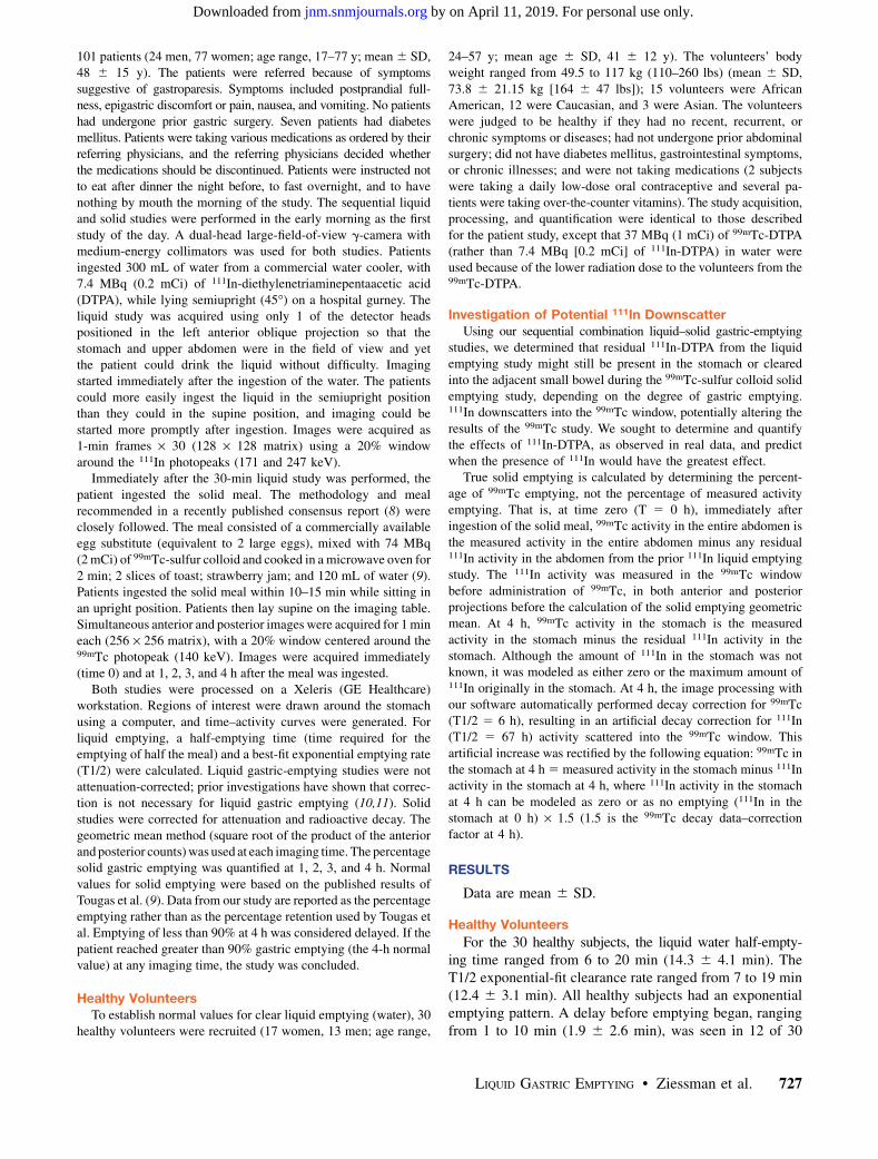
Figures 1A and 1B show the distribution of normal andabnormal liquid and solid studies based on normal values ofthe mean plus 2 and 3 SDs. Of the 101 patient studies, solidemptying was delayed in 16. Liquid emptying was delayedin 39 (2 SDs) and 36 (3 SDs), respectively. Thus, liquidemptying was delayed more than twice as frequently as wassolid emptying. Both solid and liquid emptying studies weredelayed in 12 patients, and solid emptying was delayed butliquid was normal in 4 patients. A total of 27 (2 SDs) and24 (3 SDs) had delayed liquid but normal solid emptying.
Of 85 patients with normal solid emptying, 27 (2 SDs;32%) and 24 (3SDs; 28%) had delayed liquid emptying.
Not all patients ingested the entire solid meal. Of 101patients, 13 ingested only 50%290% of the meal. Five ofthe 13 patients had prolonged liquid emptying with anexponential T1/2 of 28, 35, 40, 41, and 284 min. All hadnormal solid emptying values; however, because they didnot ingest the entire meal, the normal values may not apply.
Patients with a normal T1/2 for liquid emptying had anexponential clearance pattern in 61 of 65 studies (94%). Ofthe 36 patients with a delayed T1/2 (mean 6 3 SDs), 56%had an exponential pattern of emptying; 44% did not.None of the patients with a T1/2 greater than 42 min hadexponential clearance. A delay before emptying began wasseen in 41 of 65 patients (63%) with a normal T1/2 (2.7 6
3.4 min) and in 29 of 36 (81%) of patients with anabnormally prolonged T1/2 (8.9 6 9.5 min). The distribu-tion of delayed T1/2 liquid emptying was 22–24 min (5patients), 25–30 min (11 patients), 31–58 min (12 patients),and 122–526 min (8 patients).
Downscatter Analysis
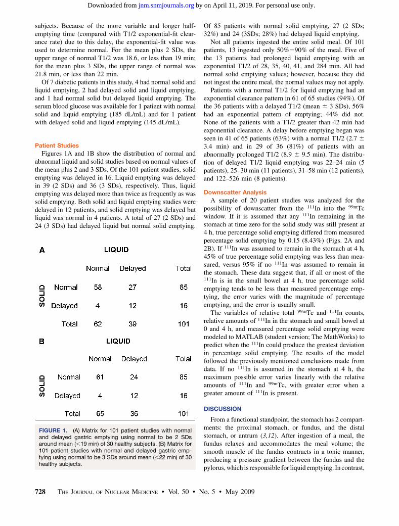
A sample of 20 patient studies was analyzed for thepossibility of downscatter from the 111In into the 99mTcwindow. If it is assumed that any 111In remaining in thestomach at time zero for the solid study was still present at4 h, true percentage solid emptying differed from measuredpercentage solid emptying by 0.15 (8.43%) (Figs. 2A and2B). If 111In was assumed to remain in the stomach at 4 h,45% of true percentage solid emptying was less than mea-sured, versus 95% if no 111In was assumed to remain inthe stomach. These data suggest that, if all or most of the111In is in the small bowel at 4 h, true percentage solidemptying tends to be less than measured percentage emp-tying, the error varies with the magnitude of percentageemptying, and the error is usually small.
The variables of relative total 99mTc and 111In counts,relative amounts of 111In in the stomach and small bowel at0 and 4 h, and measured percentage solid emptying weremodeled to MATLAB (student version; The MathWorks) topredict when the 111In could produce the greatest deviationin percentage solid emptying. The results of the modelfollowed the previously mentioned conclusions made fromdata. If no 111In is assumed in the stomach at 4 h, themaximum possible error varies linearly with the relativeamounts of 111In and 99mTc, with greater error when agreater amount of 111In is present.
DISCUSSION
From a functional standpoint, the stomach has 2 compart-ments: the proximal stomach, or fundus, and the distalstomach, or antrum (3,12). After ingestion of a meal, thefundus relaxes and accommodates the meal volume; thesmooth muscle of the fundus contracts in a tonic manner,producing a pressure gradient between the fundus and thepylorus, which is responsible for liquid emptying. In contrast,
FIGURE 1. (A) Matrix for 101 patient studies with normaland delayed gastric emptying using normal to be 2 SDsaround mean (,19 min) of 30 healthy subjects. (B) Matrix for101 patient studies with normal and delayed gastric emp-tying using normal to be 3 SDs around mean (,22 min) of 30healthy subjects.
728 THE JOURNAL OF NUCLEAR MEDICINE • Vol. 50 • No. 5 • May 2009
by on April 11, 2019. For personal use only. jnm.snmjournals.org Downloaded from
the antrum produces strong phasic muscular contractions thatgrind up solid food into small particles that can pass throughthe pylorus; the antrum is responsible for solid emptying.
Standard teaching has long been that solid radionuclidegastric emptying is more sensitive for the detection ofgastroparesis than is liquid emptying. This is stated ingastrointestinal (4–6) and nuclear medicine review articles(13–15), textbooks (1,16), and Society of Nuclear MedicineProcedure Guidelines (17). This consensus seems to havebeen based on observations made from dual-phase, dual-isotope solid–liquid gastric-emptying studies (18–23). As aresult, only solid gastric-emptying studies are recommendedfor clinical purposes; liquid studies are needed only in spe-cial situations, for example, for patients who cannot toleratesolid meals, patients who are postoperative, or patients withsuspected dumping syndrome (4,5,16).
A potential problem with accepting the results from dual-phase studies is that the emptying of the liquid phase isdirectly affected by the solid-phase meal. For example, theaddition of calories in the form of 10% dextrose to a clear
liquid meal slows the emptying rate, although the exponentialpattern of emptying is maintained. However, with increasingcaloric content (e.g., 25% dextrose) the emptying rate isslowed further and the pattern of clearance changes, fromexponential to linear, similar to that seen in a solid study (20).Thus, the solid phase has a definite effect on the rate of liquidemptying, which may explain the discrepancy between ourresults and prior reports derived from dual-phase studies.
The Tougas et al. (9) standardized and simplified solid-meal protocol has been recommended in a joint report of theSociety of Nuclear Medicine and the American Neurogas-troenterology and Motility Society (8). One criticism of thismethod is that infrequent imaging does not allow for theanalysis of the pattern of gastric contractility. The presentinvestigation is the second time that we have integrated aresearch study and the 4-h Tougas protocol (24). In the priorstudy, we investigated whether the lag phase could predictdelayed emptying. We acquired images every 10 min duringthe first hour of the 4-h study. The lag phase did not prove tobe a predictor of overall emptying (24). However, in that samestudy, we found that a solid study length of 4 h, comparedwith the more traditional 2-h study, increased the number ofpatients diagnosed with gastroparesis by 32%. In the presentinvestigation, we have integrated a 30-min clear liquid studywith the Tougas protocol and have found that the clear liquidstudy detected gastroparesis in approximately 30% of pa-tients with normal solid empting studies. The diagnosis ofgastroparesis increased significantly, from 16% of patientsfor the solid-only 4-h study to an additional 28%232% ofpatients who had a normal solid study but abnormal liquidemptying. Thus, the addition of the liquid study to the solidstudy has considerable added diagnostic value.
Past investigations have found a poor correlation betweenpatient symptomatology and the results of gastric-emptyingstudies. Abnormal emptying has been found in a relativelylow percentage of patients with symptoms suggestive ofgastroparesis, who have a clinical diagnosis of nonulcerdyspepsia, idiopathic gastroparesis, and diabetes mellitus(5,25–27). One important reason may be that we have beenstudying only antral contraction and not fundal contraction.
Previously published data on normal values for clearliquid gastric emptying are limited. In 1974, Chaudhurireported on noncaloric liquid (saline) gastric emptying in 8healthy subjects (28). Each subject underwent 3 studies,and the study was shown to have good reproducibility. Anormal T1/2 value based on these 24 total studies (8 · 3)was 6–18 min (mean 6 SD, 12 6 3 min). Two other reportsalso used a saline clear liquid meal. In 1 study with 7 healthysubjects, the upper level of normal values was less than20 min (29), and in another report with an uncertainnumber of subjects, the upper level was less than 25 min(30). Our investigation of 30 healthy subjects found normalemptying to be less than 19 min (mean 6 2 SDs) and lessthan 22 min (mean 6 3 SDs). The rate of liquid emptyingis, to some extent, position-dependent; the rate is slowerwith the patient in a supine rather than sitting position (29).
FIGURE 2. (A) Absolute difference from measured per-centage solid gastric emptying vs. true percentage emptyingat 4 h, assuming maximum 111In in stomach at 4 h. (B) Ab-solute difference from measured percentage solid gastricemptying vs. true percentage emptying, assuming no 111In instomach at 4 h.
LIQUID GASTRIC EMPTYING • Ziessman et al. 729
by on April 11, 2019. For personal use only. jnm.snmjournals.org Downloaded from
Unlike solids, clear liquids have been reported to emptyin a monoexponential pattern, with no delay before emp-tying begins (31). However, our study has shown a delay of1–10 min (1.9 6 2.6 min) in 13 of 30 healthy subjects. Allhealthy subjects in our study had an exponential pattern ofemptying. In our study, the patients with a moderate delayin liquid emptying maintained an exponential emptyingpattern; however, those with more delayed emptying oftendid not empty exponentially.
There has long been concern about 111In downscatter intothe 99mTc window in dual-isotope studies. Some investi-gations have used scatter-correction methods to minimizethis problem (20); other studies, however, have shown that,if the administered dose of 99mTc is at least 5–6 times thatof 111In, dose downscatter is not a significant problem(32,33). With the sequential methodology described in thisarticle, there was a concern that residual liquid 111In mightaffect the results of the 99mTc solid study. Using a 10:1administered dose ratio of 99mTc to 111In keVin our study, ouranalysis showed that the problem of residual liquid is gen-erally not significant using the methodology described.
There have been anecdotal reports of patients who haddelayed liquid but normal solid emptying (34). A singlepublished investigation of 85 patients with diabetes reporteddelayed liquid but normal solid emptying in 24% of patients(35). This finding was said by the authors to be specific todiabetic patients, and this observation has not been followedup. Our study clearly demonstrates that abnormal liquid butnormal solid gastric emptying is not an uncommon finding ina general referral population of patients with symptomssuggestive of gastroparesis. Only 7 of our patients werediabetic but none had delayed liquid/normal solid emptying.
Patients with delayed liquid emptying but normal solidemptying likely have dysfunction of the gastric fundus.Further investigation is needed to determine whether thishypothesis is correct. Several techniques (e.g., barostaticmanometry, ultrasonography, MRI, and SPECT) have beendescribed that might help evaluate gastric fundal function(36). If this hypothesis is true, this might lead to new methodsof therapy that are more physiologic and might allow forindividualized therapy of gastroparesis, depending on whetherthe patients have antral or fundal dysfunction or both.
CONCLUSION
We defined the reference range for clear liquid gastricemptying. In a sequential 2-phase protocol, we found that a30-min clear liquid gastric-emptying study performed im-mediately before a 4-h standardized solid emptying study isoften abnormal when the solid study is normal, and thegastric-emptying study added considerable diagnostic valuefor the detection of gastroparesis over solid emptying alone.
REFERENCES
1. Parkman HP, Fisher RS. Disorders of gastric emptying. In: Yamada T, Alpers DH,
Kaplowitz N, Laine L, Owyang C, Powell DW, eds. Textbook of Gastroenterology.
4th ed. Philadelphia, PA: Lippincott Williams & Wilkins; 2003:1292–1310.
2. Lin HC, Prather C, Fisher RS, et al. AMS Task Force Committee on gastro-
intestinal transit: measurement of gastrointestinal transit. Dig Dis Sci. 2005;50:
989–1004.
3. Hasler WL. Disorders of gastric emptying. In: Yamada T, Alpers DH, Kaplowitz
N, Laine L, Owyang C, Powell DW, eds. Textbook of Gastroenterology. 3rd ed.
Philadelphia, PA: Lippincott Williams & Wilkins; 1999:1341–1369.
4. Camilleri M, Hasler WL, Parkman HP, Quigley EM, Soffer E. Measurement of
gastrointestinal motility in the GI laboratory. Gastroenterology. 1998;115:747–762.
5. Parkman HP, Hasler WL, Fisher RS. American Gastroenterological Association
technical review on the diagnosis and treatment of gastroparesis. Prepared by the
American Gastroenterological Association Clinical Practice Committee. Gas-
troenterology. 2004;127:1592–1622.
6. Hornbuckle K, Barnett JL. The diagnosis and work-up of the patient with
gastroparesis. J Clin Gastroenterol. 2000;30:117–124.
7. Ziessman HA, Okolo P, Mullin G, Chander A. The added diagnostic value of
liquid gastric emptying when solid emptying is normal [abstract]. J Nucl Med.
2008;49(suppl 1):39P.
8. Abell TL, Camilleri M, Donohoe K, et al. Consensus recommendations for
gastric emptying scintigraphy: a joint report of the Society of Nuclear Medicine
and the American Neurogastroenterology and Motility Society. J Nucl Med
Technol. 2008;36:44–54.
9. Tougas G, Eaker EY, Abell TL, et al. Assessment of gastric emptying using a
low fat meal: establishment of international control values. Am J Gastroenterol.
2000;95:1456–1462.
10. Collins PJ, Horowitz M, Cook DJ, et al. Gastric emptying in normal subjects: a
reproducible technique using a single scintillation camera and computer system.
Gut. 1983;24:1117–1125.
11. Christian PE, Moore JG, Sorenson JA, et al. Effects of meal size and correction
technique on gastric emptying time: studies with two tracers and opposed
detectors. J Nucl Med. 1980;21:883–885.
12. Kelly KA. Gastric emptying of liquids and solids: roles of proximal and distal
stomach. Am J Physiol. 1980;239:G71–G76.
13. Maurer AH, Parkman HP. Update on gastrointestinal scintigraphy. Semin Nucl
Med. 2006;36:110–118.
14. Datz FL. Considerations for accurately measuring gastric emptying. J Nucl Med.
1991;32:881–884.
15. Mariani G, Boni G, Barreca M, et al. Radionuclide gastroesophageal motor
studies. J Nucl Med. 2004;45:1004–1028.
16. Ziessman HA, O’Malley J, Thrall JH. Gastrointestinal system. In: Ziessman HA,
O’Malley J, Thrall JH, eds. Nuclear Medicine: The Requisites. Philadelphia, PA:
Mosby; 2006.
17. Donohoe KJ, Maurer AH, Ziessman HA, et al. Procedure guideline for gastric
emptying and motility. J Nucl Med. 1999;40:1236–1239.
18. Couturier O, Bodet-Milin C, Querellou S, et al. Gastric scintigraphy with a
liquid-solid radiolabelled meal: performances of solid and liquid parameters.
Nucl Med Commun. 2004;25:1143–1150.
19. Heading RC, Tothill P, McLoughlin GP, Shearman DJC. Gastric emptying role
measurement in man: a double isotope scanning technique for simultaneous study
of liquid and solid components of a meal. Gastroenterology. 1976;71:45–50.
20. Fisher RS, Malmud LS, Bandid P, et al. Gastric empting of a physiologic mixed
solid-liquid meal. Clin Nucl Med. 1982;7:215–221.
21. Collins PJ, Houghton LA, Read NW, et al. Role of the proximal and distal
stomach in mixed solid and liquid meal emptying. Gut. 1991;32:615–619.
22. Wright RA, Thompson D, Syed I. Simultaneous markers for fluid and solid
gastric emptying: new variations on an old theme—concise communication.
J Nucl Med. 1981;22:772–776.
23. Moore JG, Christian PE, Coleman RE. Gastric empting of varying meal weight
and composition in man. Dig Dis Sci. 1981;26:16–22.
24. Ziessman HA, Bonta DV, Goetze S, Ravich WJ. Experience with a simplified,
standardized 4-hour gastric-emptying protocol. J Nucl Med. 2007;48:568–572.
25. Horowitz M, Harding PE, Maddox AF, et al. Gastric and oesophageal emptying
in patients with type 2 (non-insulin-dependent) diabetes mellitus. Diabetologia.
1989;32:151–159.
26. Talley NJ, Shuter B, McCrudden G, et al. Lack of association between gastric
emptying of solids and symptoms in nonulcer dyspepsia. J Clin Gastroenterol.
1989;11:625–630.
27. Koch KL, Stern RM, Stewart WR, Vasey MW. Gastric emptying and gastric
myoelectrical activity in patients with diabetic gastroparesis: effect of long-term
domperidone treatment. Am J Gastroenterol. 1989;84:1069–1075.
28. Chaudhuri TK. Use of 99Tc-DTPA for measuring gastric empting time. J Nucl
Med. 1974;15:391–395.
29. Anvari M, Horowitz M, Fraser R, et al. Effects of posture on gastric emptying of
nonnutrient liquids and antropyloroduodenal motility. Am J Physiol Gastrointest
Liver Physiol. 1995;31:G868–G871.
730 THE JOURNAL OF NUCLEAR MEDICINE • Vol. 50 • No. 5 • May 2009
by on April 11, 2019. For personal use only. jnm.snmjournals.org Downloaded from
30. Tamas S, Dumitrascu DL, Andreica V, Cotul S. Gastric sequential scintigraphy:
methodology with liquid isotonic meal. Physiologie. 1988;25:47–51.
31. Hunt JN, Spurrell WR. The pattern of emptying of the human stomach.
J Physiol. 1951;113:157–168.
32. Christian PE, Datz FL, Sorenson JA, et al. Technical factors in gastric emptying
studies. J Nucl Med. 1983;24:264–267.
33. Ziessman HA, Fahey FH, Collen MJ. Biphasic solid and liquid gastric empting
in normal controls and diabetics using continuous acquisition in LAO view. Dig
Dis Sci. 1992;37:744–750.
34. Lin HC, Hasler WJ. Disorders of gastric emptying. In: Yamada T, Alpers DH,
Kaplowitz N, Laine L, Owyang C, Powell DW, eds. Textbook of Gastroenterology.
2nd ed. Philadelphia, PA: JB Lippincott Company, 1995:1318–1346.
35. Horowitz M, Maddox AF, Wishart JM, Harding PE, Chatterton BE, Shearman
DJ. Relationships between oesophageal transit and solid and liquid gastric
emptying in diabetes mellitus. Eur J Nucl Med. 1990;18:229–234.
36. Kim D-Y, Myung S-J, Camilleri M. Novel testing of human gastric motor and
sensory functions: rationale, methods, and potential applications in clinical
practice. Am J Gastroenterol. 2000;95:3365–3373.
LIQUID GASTRIC EMPTYING • Ziessman et al. 731
by on April 11, 2019. For personal use only. jnm.snmjournals.org Downloaded from
Doi: 10.2967/jnumed.108.059790Published online: April 16, 2009.
2009;50:726-731.J Nucl Med. Harvey A. Ziessman, Ankit Chander, John O. Clarke, Alison Ramos and Richard L.Wahl Emptying AloneThe Added Diagnostic Value of Liquid Gastric Emptying Compared with Solid
http://jnm.snmjournals.org/content/50/5/726This article and updated information are available at:
http://jnm.snmjournals.org/site/subscriptions/online.xhtml
Information about subscriptions to JNM can be found at:
http://jnm.snmjournals.org/site/misc/permission.xhtmlInformation about reproducing figures, tables, or other portions of this article can be found online at:
(Print ISSN: 0161-5505, Online ISSN: 2159-662X)1850 Samuel Morse Drive, Reston, VA 20190.SNMMI | Society of Nuclear Medicine and Molecular Imaging
is published monthly.The Journal of Nuclear Medicine
© Copyright 2009 SNMMI; all rights reserved.
by on April 11, 2019. For personal use only. jnm.snmjournals.org Downloaded from