Embed Size (px)
Citation preview
Harmful Algae 4 (2005) 33–48
The alternation of different morphotypes in the seasonal cycleof the toxic diatomPseudo-nitzschia galaxiae
Federica Cerinoa, Luisa Orsinia,1, Diana Sarnoa, Carmela Dell’Aversanob,Luciana Tartaglioneb, Adriana Zingonea,∗
a Stazione Zoologica ‘Anton Dohrn’, Villa Comunale, Naples 80121, Italyb Dipartimento di Chimica delle Sostanze Naturali, Università degli Studi di Napoli “Federico II”,
Via D. Montesano 49, 80131 Naples, Italy
Received 18 August 2003; accepted 20 October 2003
Abstract
The marine diatomPseudo-nitzschia galaxiaeLundholm et Moestrup has been recently described from Mexican andAustralian plankton. In this paper, we illustrate the considerable morphological variability of the species in the MediterraneanSea and present first evidence of its toxicity. In addition to lanceolate cells 25–41�m long, which fit the original descriptionof the species, markedly larger (<82�m) and smaller (>10�m) specimens are commonly recorded. Cells of the largest sizehave almost parallel valve margins, while smaller specimens have extremely short rostrate ends and do not form colonies.Despite remarkable differences in shape and size, the typical ultrastructure of the species was observed for the differentsize classes in culture and in natural samples. In culture, cell length decreased at a rate of 1.1–2.1�m per month. Liquidchromatography–mass spectrometry (LC–MS) analyses revealed the presence of domoic acid (DA) at very low levels in twoof seven strains analyzed. LSU rDNA analysis confirmed the identity of the species and showed a very low genetic variabilityfor the strains from the Gulf of Naples, with no relationships with size and overall shape of the cells. A relatively high number(53) ofPseudo-nitzschiasequences were considered in the phylogenetic analysis, yet the relationships among species remainunclear, probably in relation with a recent speciation process in the genus. In natural samples,P. galaxiaepopulations ofdifferent cell sizes occurred at different times over the year, with smaller cells found in winter and early spring, and mediumand larger cells peaking in late spring–summer. The maximum concentration value in the Gulf of Naples was recorded inMay 1985 (9.4 × 10⁶ cells l−1). From the analysis of a high number of both natural and culture samples, it is concluded thatsize and shape variations are indicative of different stages of the life cycle ofP. galaxiae, which exhibit a synchronized andseasonal occurrence at the interannual scale.© 2004 Elsevier B.V. All rights reserved.
Keywords:Diatoms; Domoic acid; Mediterranean Sea; Phylogeny;Pseudo-nitzschia galaxiae; Taxonomy
∗ Corresponding author. Tel.:+39-081-5833295;fax: +39-081-7641355.
E-mail address:[email protected] (A. Zingone).1 Present address: Institut für Tierzucht und Genetik, Veter-
inärmedizinische Universität Wien, Josef Baumann Gasse 1, 1210Wien, Austria.
1. Introduction
The genusPseudo-nitzschiaH. Peragallo includesabout 25 species of pennate, colonial marine diatoms.The genus was originally considered as a section ofNitzschia americanaH. Hassall (Hasle, 1965), fromwhich it was subsequently separated based on the
1568-9883/$ – see front matter © 2004 Elsevier B.V. All rights reserved.doi:10.1016/j.hal.2003.10.005
34 F. Cerino et al. / Harmful Algae 4 (2005) 33–48
colonial habit and on some ultrastructural features(Hasle, 1995). In recent years, morphological andmolecular investigations onPseudo-nitzschiainten-sified following the discovery of domoic acid (DA)(Bates et al., 1989), a potent neurotoxic aminoacidthat can be accumulated through the trophic web andcause damage to humans, marine mammals and birds(Scholin et al., 2000; Shumway et al., 2003). NinePseudo-nitzschiaspecies are currently known to pro-duce DA (Moestrup et al., 2002). Information on thegeographic distribution of the genusPseudo-nitzschiahas also grown in recent years. The genus has aworldwide distribution and includes cosmopolitan,temperate and tropical species (Hasle, 2002). Phy-logenetic analyses conducted on two different par-tial LSU rDNA sequence datasets (Lundholm andMoestrup, 2002; Lundholm et al., 2002; Orsini et al.,2002) have shown that the genusPseudo-nitzschiaisparaphyletic (Lundholm et al., 2002), with phyloge-netic relationships well resolved at the species levelbut not at the supraspecific level.
In the Gulf of Naples,Pseudo-nitzschiaspecieswere reported, as ‘colonialNitzschia’, since the firstphytoplankton observations in the area (Schröder,1901; Issel, 1934). More recently, seven differentPseudo-nitzschiaspecies have been identified in thecourse of a 15-year sampling programme at a coastalstation of the city of Naples (Zingone et al., 2002;Zingone et al., 2003). Among these, very thin mor-photypes with a slight central swelling were attributedto Pseudo-nitzschiacf. prolongatoides(Hasle) Hasle.In May 2001, specimens with this lanceolate shapewere brought into culture and showed ultrastructuralfeatures which were clearly distinct fromP. prolonga-toidesand from any otherPseudo-nitzschiaspecies,prompting molecular analysis to clarify phylogeneticrelationships. At the same time, morphotypes simi-lar to P. cf. prolongatoidesfrom the Gulf of Napleswere described as a new species under the name ofPseudo-nitzschia galaxiae(Lundholm and Moestrup,2002). P. galaxiaehas a valve outline slightly swollenin the middle of the cell and tapering towards itsends. The valve ultrastructure is clearly distinctive,due to the lack of poroids that are typical for otherPseudo-nitzschiaspecies, and to the presence ofminute pores scattered over the frustule.
In this paper, a wider range of shape and size vari-ability is reported from cultured and field material of
P. galaxiaefrom the Mediterranean Sea, as comparedto the original description. The identity of the species,and its substantial genetic homogeneity is confirmedby LSU analysis. The production of domoic acid isdemonstrated for the first time in this species. Therole of the life cycle in the seasonal occurrence of thespecies in the Gulf of Naples is discussed based onthe succession of different size classes over the year.
2. Materials and methods
2.1. Cultures
Six of the seven strains ofP. galaxiaewere obtainedfrom serial dilution cultures (SDC) of natural seawa-ter samples, collected in May 2002 from surface wa-ters at station MC, 2 nautical miles offshore Naples(Table 1). The strain SZN-B58 was isolated from asurface net sample collected at the same station MCin July 2001. In all cases, unicellular cultures were es-tablished from a single cell or a single chain of cellsand grown inf/2 growth medium, with silica added(Guillard, 1983), prepared with oligotrophic seawa-ter (36 psu) and maintained at a temperature of 15◦C,with a photon irradiance of 70–80�E m−2 s−1, and ina 12:12 light–dark regime.
For cell enumeration, natural samples were col-lected fortnightly from 1984 and weekly as of 1995at the surface in the Gulf of Naples (St. MC), in theframework of a long-term plankton monitoring pro-gramme. Samples were fixed with CaCO3–neutralizedformaldehyde 0.8% final concentration and enumer-ated in the light microscope (Utermöhl, 1958). SmallP. galaxiaecells (10–25�m) were enumerated sep-arately from larger cells. Additional Mediterranean
Table 1List of P. galaxiaestrains analysed for morphology, domoic acidcontent and phylogeny
Strain Collection data Isolation data Sample
SZN-B54 29 May 2001 13 July 2001 SDC -4IISZN-B55 29 May 2001 13 July 2001 SDC -4IISZN-B56 29 May 2001 13 July 2001 SDC -4IISZN-B57 29 May 2001 13 July 2001 SDC -4IISZN-B58 18 July 2001 18 July 2001 Net sampleSZN-P1 29 May 2001 10 January 2002 SDC -4 VSZN-P5 29 May 2001 10 January 2002 SDC -4 V
F. Cerino et al. / Harmful Algae 4 (2005) 33–48 35
Sea samples from the Open Sicily Channel (October1991), the Balearic Sea (March 2002), Olbia (Sardinia,Tyrrhenian Sea, June 2002) and Chioggia (Venice,Adriatic Sea, May 2002) were examined. The lattertwo samples were kindly provided by P. Di Dato (Uni-versity of Rome, Italy) and R. Casotti (Stazione Zoo-logica, Naples).
For ultrastructural observation of the frustules, or-ganic matter was eliminated from culture samplesusing a mixture of 10% HNO3 and 40% H2SO4, fol-lowed by rinsing steps with distilled water until allthe acid was removed. A drop of the cleaned materialwas placed on a Formvar-coated grid and observedwith a Philips EM 400 microscope. For natural sam-ples, a droplet of fixed uncleaned material was placedon the same kind of grid, dried, rinsed with distilledwater and observed as above.
For length measurements, aliquots of cultures ofthe strains SZN-B54, SZN-B56, SZN-B58, SZN-P1and SZN-P5 were fixed with CaCO3–neutralizedformaldehyde to a final concentration of 0.8%. Asubsample of 50 cells per clone were measuredmonthly over 13 months using a ZEISS Axiophotphase contrast microscope, at a magnification of400×. Measurements of SZN-P1 and SZN-P5 strainsended beforehand when these cultures died. For thecalculations of cell size reduction, measurements ofthe apical axis were plotted versus time (as juliandays). The slope of the straight line that best fits thedata, calculated using the least squares method, repre-sents the decrease per day; this value was multipliedby 30 to obtain the monthly decrease value. Lengthmeasurements were also taken on subsamples of 50P. galaxiaecells from each of 11 field samples fromthe St. MC, corresponding to peak phases of earlyspring 1996 and 2002 (two samples), mid-spring of1996–1998 and 2001 (four samples), and summer1998–2002 (five samples).
2.2. Toxin analysis
2.2.1. Sample extractionThe cultures examined (Table 2) were concentrated
and the resultant pellet was frozen at−80◦C until theanalysis. The pellet was extracted with a solution ofmethanol–water 1:1 (3× 250�l) and filtered throughan Ultrafree-MC 0.45�m membrane (Millipore Ltd.,Bedford, MA, USA) at 6000 rpm for 10 min. The
Table 2P. galaxiaematerial used for domoic acid analysis
Strain Centrifugedvolume (ml)
Number of cells(108 ml−1)
SZN-B54 600 11.0SZN-B55 609 2.1SZN-B56 595 6.0SZN-B57 595 1.7SZN-B58 600 6.5SZN-P1 400 7.0SZN-P5 400 1.6
volume of the filtrate was adjusted to 900�l withextracting solvent and analyzed directly by Liquidchromatography–mass spectrometry (LC–MS). A450�l aliquot of the extract was evaporated to dry-ness and subsequently subjected to a SPE clean upusing the procedure suggested byQuilliam et al.(1995). Eluates were analyzed by LC–MS.
2.2.2. Liquid chromatography–mass spectrometryanalyses
High-pressure pump SP model P 4000 (Ther-moFinnigan Separation Products, San Jose, CA,USA) coupled to an Applied Biosystem API-2000triple quadrupole mass spectrometer equipped with aturbo-ionspray source (Thornhill, Ont., Canada), wasused for LC–MS experiments. LC separations wereperformed by using a 5�m TosoHaas TSK-GELAmide-80, 250 mm× 2 mm, column, isocraticallyeluted with a 71% acetonitrile–water solution con-taining 2 mM ammonium formate and 3.5 mM formicacid, as suggested byQuilliam et al. (2001). The flowrate was 200�l min−1 and a sample injection volumeof 10�l was used. The protonated ion atm/z312.5 andthe sodium adduct ion atm/z334.5 were monitored inpositive selected ion monitoring (SIM) experiments,while the [M–H]− ion at m/z310.5 was observed innegative SIM. The following groups of six transitionsm/z 312/294, 312/266, 312/248, 312/220, 312/193,312/175 (collision energy, 30 eV) andm/z 310/266,310/248, 310/222, 310/204, 310/160, 310/82 (colli-sion energy,−25 eV) were monitored in positive andnegative multiple reaction monitoring (MRM) exper-iments, respectively. The most abundant transitions(m/z310/266 and 310/222, negative ion mode) wereused for quantitative studies. Direct comparison tostandard solutions of domoic acid (Sigma–Aldrich,
36 F. Cerino et al. / Harmful Algae 4 (2005) 33–48
Steinheim, Germany) at similar concentrations in-jected in the same experimental conditions allowed todetermine DA content in the crude extracts.
2.3. Molecular analysis
2.3.1. DNA extraction and amplificationGenomic DNA was extracted from 150 to 200 ml
of exponentially growing cultures, using the DNAeasyplant minikit (Qiagen, Genomics, Bothell, WA) fol-lowing the manufacturer instructions. Amplificationcondition for genomic DNA and cloning strategy forPCR fragments are the same applied inOrsini et al.,2002). Sequences for the LSU rDNA were obtainedwith a Beckman Ceq 2000, using Dye-Terminator cy-cle sequencing kit (Beckman).
2.3.2. Phylogenetic analysisP. galaxiaeLSU sequences were aligned with LSU
Pseudo-nitzschiasequences available in GenBank(Table 3). Cylindrotheca closteriumand Nitzschiafrustulum were used as outgroup and ingroup, re-
Table 3List of diatom strains used for the LSU rDNA phylogeny withaccession number to the informatic database GenBank
Species GenBankaccessionnumber
Cylindrotheca closterium(Ehrenberg) Lewin &Reimann
M87326
Nitzschia frustulum(Kutzing) Grunow AF417671
P. americana(Hasle) Fryxell U41390
P. australisFrenguelli U41393P. australis U40850P. australis(OM1) AF417651
P. delicatissima(Cleve) Heiden AF416748P. delicatissima AF416749P. delicatissima AF416758P. delicatissima U41391P. delicatissima(1001 2b) AF417645
P. fraudulenta(Cleve) Hasle AF416750P. fraudulenta AF416751P. fraudulenta AF416762P. fraudulenta(Limens1) AF417647
P. inflatula (Hasle) Hasle (No7) AF417639
P. microporaPriisholm, Moestrup & Lundholm(VPB-B3)
AF417649
Table 3 (Continued)
Species GenBankaccessionnumber
P. multiseries(Hasle) Hasle (OFPm984) AF417655P. multiseries U41389
P. multistriata (Takano) Takano AF416753P. multistriata AF416754P. multistriata AF416756P. multistriata AF416757P. multistriata (Korea A) AF417654
P. pseudodelicatissima(Hasle) Hasle AF416747P. pseudodelicatissima AF416752P. pseudodelicatissima AF416755P. pseudodelicatissima AF416759P. pseudodelicatissima AF416760P.cf. pseudodelicatissima(Hobart 5) AF417641P. pseudodelicatissima(P-11) AF417640
P. pseudodelicatissimaSZN-B109AY550126P. pseudodelicatissimaSZN-B111AY550128P. pseudodelicatissimaSZN-B112AY550127P. pseudodelicatissimaSZN-B113AY550129
P. pungens(Grunow ex Cleve) Hasle U41392P. pungens U41262P. pungens(KBH2) AF417650P. pungens(P-24) AF417648
P. seriata(Cleve) Peragallo (Lynaes 8) AF417653P. seriata(Nissum 3) AF417652
P. subfraudulenta(Hasle) Hasle AF416761P. subfraudulentaRensubfrau AF417646
P. cf. subpacifica(Hasle) Hasle (Zhenbo 7B) AF417644P. cf. subpacifica(P-28) AF417643P. cf. subpacifica(RdA8) AF417642
P. galaxiae(Sydney) Lundholm & Moestrup a
P. galaxiae(Mexico) a
P. galaxiaeSZN-B54AY544786P. galaxiaeSZN-B55AY544791P. galaxiaeSZN-B56AY544790P. galaxiaeSZN-B57AY544789P. galaxiaeSZN-B58AY544788P. galaxiaeSZN-P1AY544787P. galaxiaeSZN-P5AY544792
SZN: strains isolated in the Gulf of Naples.a Sequences kindly provided by N. Lundholm.
spectively. Both species are pennate diatoms and,according to the LSU rDNA phylogeny includingPseudo-nitzschiaand close genera (Lundholm et al.,2002), the former is at the base of the clade group-ing Pseudo-nitzschiaspecies, while the latter clusters
F. Cerino et al. / Harmful Algae 4 (2005) 33–48 37
immediately out of theNitzschia–Pseudo-nitzschiaclade. The alignment was obtained using Clustal W(Thompson et al., 1994) in the Bioedit 4.5.8 computerpackage (Hall, 1999).
Distance analysis was performed using Bioedit4.5.8 computer package (Hall, 1999); nucleotide poly-morphism was calculated using DNAsp 3.0 version(Rozas and Rozas, 1999); phylogenetic relationshipswere inferred using both distance and parsimony anal-yses. The Neighbor-Joining (NJ) tree (Saitou and Nei,1987) was assembled using the Kimura 2 parameterdistance and getting the Neighbor option from MEGA2.1 computer package (Kumar et al., 2001); the max-imum parsimony trees were performed using theparsimony option in MEGA 2.1 (Kumar et al., 2001).
3. Results
3.1. Morphology
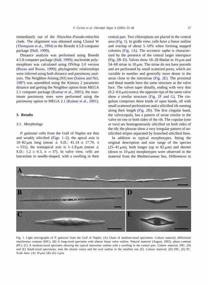
P. galaxiaecells from the Gulf of Naples are thinand weakly silicified (Figs. 1–2), the apical axis is10–82�m long (mean± S.D.: 41.14 ± 17.70, n= 551), the transapical axis is 1–1.8�m (mean±S.D.: 1.2 ± 0.3, n = 37). In valve view, cells arelanceolate to needle-shaped, with a swelling in their
Fig. 1. Light micrographs ofP. galaxiaefrom the Gulf of Naples. (A) Chain of medium-sized specimens. Culture material, differentialinterference contrast (DIC). (B) A long-sized specimen with almost linear valve outline. Natural material (August, 2002), phase contrast(PC). (C) A medium-sized specimen showing the typical lanceolate outline with a swelling in the central part. Culture material, DIC. (D)and (E) Small-sized specimens; note the shorter rostra and the oval outline in the smallest one (E). Culture material, (D) DIC, (E) PC.Scale bars: (A) 10�m; (B)–(E) 5�m.
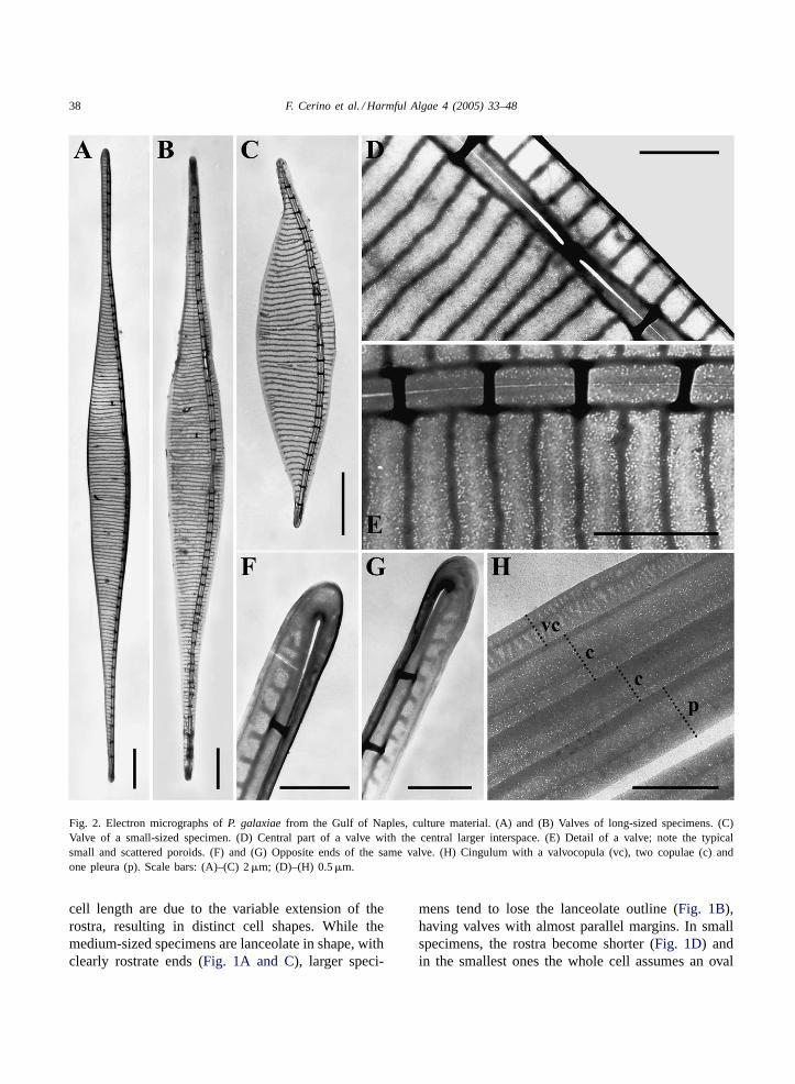
central part. Two chloroplasts are placed in the centralarea (Fig. 1). In girdle view, cells have a linear outlineand overlap of about 5–10% when forming steppedcolonies (Fig. 1A). The eccentric raphe is character-ized by the presence of the central larger interspace(Fig. 2B–D). Valves show 18–28 fibulae in 10�m and54–68 striae in 10�m. The striae do not have poroidsand are perforated by small scattered pores, which arevariable in number and generally more dense in theareas close to the interstriae (Fig. 2E). The proximaland distal mantle have the same structure as the valveface. The valves taper distally, ending with very thin(0.2–0.6�m) rostra; the opposite tips of the same valveshow a similar structure (Fig. 2F and G). The cin-gulum comprises three kinds of open bands, all withsmall scattered perforations and a silicified rib runningalong their length (Fig. 2H). The first cingular band,the valvocopula, has a pattern of striae similar to thevalve on one or both sides of the rib. The copulae (oneor two) are homogeneously silicified on both sides ofthe rib; the pleurae show a very irregular pattern of un-silicified stripes separated by branched silicified lines.
In addition to typical morphotypes, fitting theoriginal description and size range of the species(25–41�m), both longer (up to 82�m) and shorter(down to 10�m) morphotypes were observed in thematerial from the Mediterranean Sea. Differences in
38 F. Cerino et al. / Harmful Algae 4 (2005) 33–48
Fig. 2. Electron micrographs ofP. galaxiaefrom the Gulf of Naples, culture material. (A) and (B) Valves of long-sized specimens. (C)Valve of a small-sized specimen. (D) Central part of a valve with the central larger interspace. (E) Detail of a valve; note the typicalsmall and scattered poroids. (F) and (G) Opposite ends of the same valve. (H) Cingulum with a valvocopula (vc), two copulae (c) andone pleura (p). Scale bars: (A)–(C) 2�m; (D)–(H) 0.5�m.
cell length are due to the variable extension of therostra, resulting in distinct cell shapes. While themedium-sized specimens are lanceolate in shape, withclearly rostrate ends (Fig. 1A and C), larger speci-
mens tend to lose the lanceolate outline (Fig. 1B),having valves with almost parallel margins. In smallspecimens, the rostra become shorter (Fig. 1D) andin the smallest ones the whole cell assumes an oval
F. Cerino et al. / Harmful Algae 4 (2005) 33–48 39
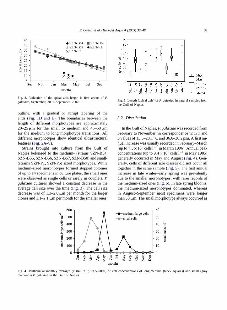
Fig. 3. Reduction of the apical axis length in five strains ofP.galaxiae, September, 2001–September, 2002.
outline, with a gradual or abrupt tapering of theends (Fig. 1D and E). The boundaries between thelength of different morphotypes are approximately20–25�m for the small to medium and 45–50�mfor the medium to long morphotype transitions. Alldifferent morphotypes show identical ultrastructuralfeatures (Fig. 2A-C).
Strains brought into culture from the Gulf ofNaples belonged to the medium- (strains SZN-B54,SZN-B55, SZN-B56, SZN-B57, SZN-B58) and small-(strains SZN-P1, SZN-P5) sized morphotypes. Whilemedium-sized morphotypes formed stepped coloniesof up to 14 specimens in culture plates, the small oneswere observed as single cells or rarely in couplets.P.galaxiaecultures showed a constant decrease in theaverage cell size over the time (Fig. 3). The cell sizedecrease was of 1.3–2.0�m per month for the largerclones and 1.1–2.1�m per month for the smaller ones.
Fig. 4. Multiannual monthly averages (1984–1991; 1995–2002) of cell concentrations of long-medium (black squares) and small (graydiamonds)P. galaxiaein the Gulf of Naples.
Fig. 5. Length (apical axis) ofP. galaxiaein natural samples fromthe Gulf of Naples.
3.2. Distribution
In the Gulf of Naples,P. galaxiaewas recorded fromFebruary to November, in correspondence withT andSvalues of 13.3–28.1◦C and 36.6–38.2 psu. A first an-nual increase was usually recorded in February–March(up to 7.3×10⁵ cells l−1 in March 1996). Annual peakconcentrations (up to 9.4×10⁶ cells l−1 in May 1985)generally occurred in May and August (Fig. 4). Gen-erally, cells of different size classes did not occur alltogether in the same sample (Fig. 5). The first annualincrease in late winter–early spring was prevalentlydue to the smaller morphotypes, with rarer records ofthe medium-sized ones (Fig. 6). In late spring blooms,the medium-sized morphotypes dominated, whereasin August–September most specimens were longerthan 50�m. The small morphotype always occurred as
40 F. Cerino et al. / Harmful Algae 4 (2005) 33–48
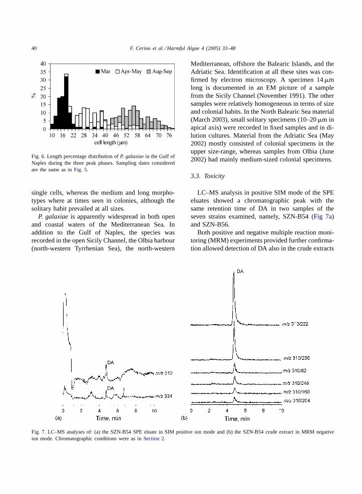
Fig. 6. Length percentage distribution ofP. galaxiaein the Gulf ofNaples during the three peak phases. Sampling dates consideredare the same as inFig. 5.
single cells, whereas the medium and long morpho-types where at times seen in colonies, although thesolitary habit prevailed at all sizes.
P. galaxiaeis apparently widespread in both openand coastal waters of the Mediterranean Sea. Inaddition to the Gulf of Naples, the species wasrecorded in the open Sicily Channel, the Olbia harbour(north-western Tyrrhenian Sea), the north-western
Fig. 7. LC–MS analyses of: (a) the SZN-B54 SPE eluate in SIM positive ion mode and (b) the SZN-B54 crude extract in MRM negativeion mode. Chromatographic conditions were as inSection 2.
Mediterranean, offshore the Balearic Islands, and theAdriatic Sea. Identification at all these sites was con-firmed by electron microscopy. A specimen 14�mlong is documented in an EM picture of a samplefrom the Sicily Channel (November 1991). The othersamples were relatively homogeneous in terms of sizeand colonial habits. In the North Balearic Sea material(March 2003), small solitary specimens (10–20�m inapical axis) were recorded in fixed samples and in di-lution cultures. Material from the Adriatic Sea (May2002) mostly consisted of colonial specimens in theupper size-range, whereas samples from Olbia (June2002) had mainly medium-sized colonial specimens.
3.3. Toxicity
LC–MS analysis in positive SIM mode of the SPEeluates showed a chromatographic peak with thesame retention time of DA in two samples of theseven strains examined, namely, SZN-B54 (Fig 7a)and SZN-B56.
Both positive and negative multiple reaction moni-toring (MRM) experiments provided further confirma-tion allowed detection of DA also in the crude extracts
F. Cerino et al. / Harmful Algae 4 (2005) 33–48 41
of the two positive strains (Fig. 7b). The retention time(4.80 min), the presence of six diagnostic fragmentsfor DA in both ionization modes and the ion ratioswere fully consistent with the presence of DA in thetwo positive samples.
The comparison with a DA standard revealed con-centration values of 3.6×10⁻⁴ and 7.8×10⁻⁷ pg percell in SZN-B54 and SZN-B56 samples, respectively.
3.4. LSU rDNA phylogeny
The alignment of the sequences did not present dif-ficulties along the 868 bp of the D1–D3 domains of theLSU rDNA. A very low level of polymorphism wasfound within the sevenP. galaxiaefrom the Gulf ofNaples, with only three mutations detected. The per-centage of polymorphic sites was 0.1; any site wasparsimony informative. The strains ofP. galaxiaefromthe Gulf of Naples were almost identical (0.0001 ge-netic distance). When the strains ofP. galaxiaefromSydney and Mexico were added to the Neapolitanones, the percentage of polymorphic sites increasedto 0.4.
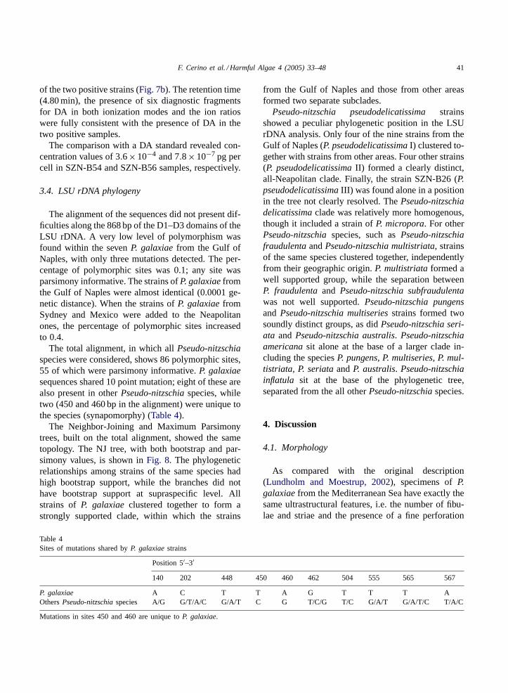
The total alignment, in which allPseudo-nitzschiaspecies were considered, shows 86 polymorphic sites,55 of which were parsimony informative.P. galaxiaesequences shared 10 point mutation; eight of these arealso present in otherPseudo-nitzschiaspecies, whiletwo (450 and 460 bp in the alignment) were unique tothe species (synapomorphy) (Table 4).
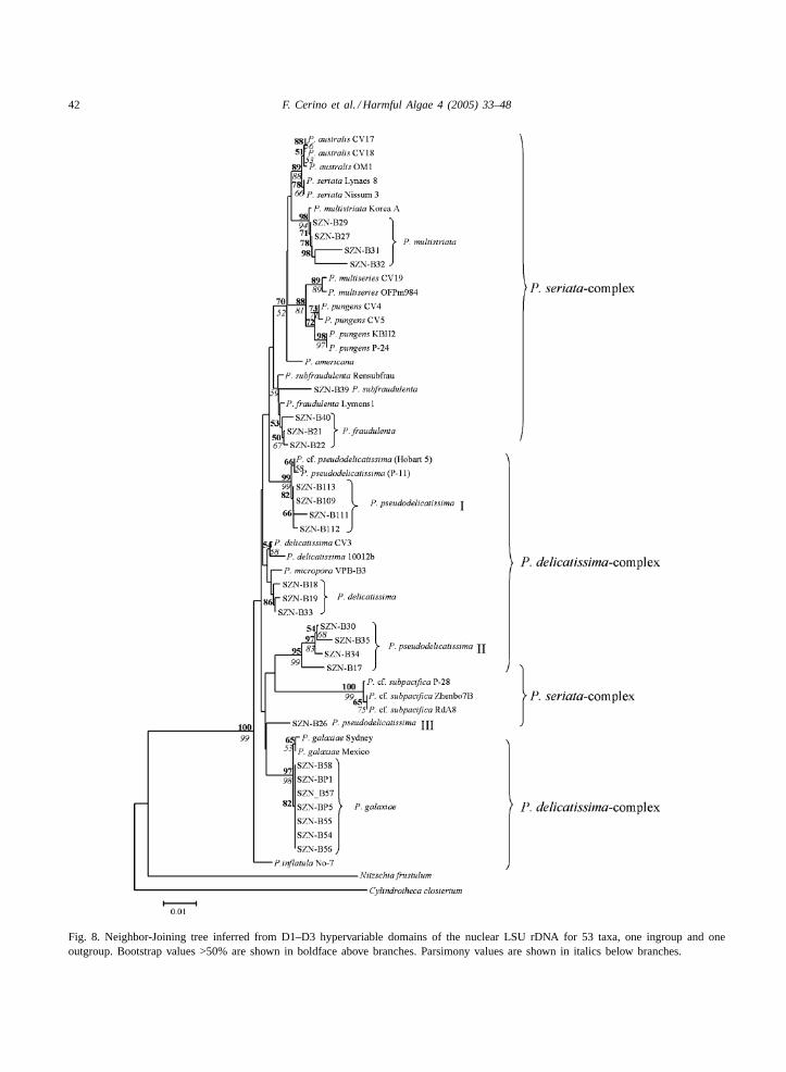
The Neighbor-Joining and Maximum Parsimonytrees, built on the total alignment, showed the sametopology. The NJ tree, with both bootstrap and par-simony values, is shown inFig. 8. The phylogeneticrelationships among strains of the same species hadhigh bootstrap support, while the branches did nothave bootstrap support at supraspecific level. Allstrains of P. galaxiaeclustered together to form astrongly supported clade, within which the strains
Table 4Sites of mutations shared byP. galaxiaestrains
Position 5′–3′
140 202 448 450 460 462 504 555 565 567
P. galaxiae A C T T A G T T T AOthersPseudo-nitzschiaspecies A/G G/T/A/C G/A/T C G T/C/G T/C G/A/T G/A/T/C T/A/C
Mutations in sites 450 and 460 are unique toP. galaxiae.
from the Gulf of Naples and those from other areasformed two separate subclades.
Pseudo-nitzschia pseudodelicatissimastrainsshowed a peculiar phylogenetic position in the LSUrDNA analysis. Only four of the nine strains from theGulf of Naples (P. pseudodelicatissimaI) clustered to-gether with strains from other areas. Four other strains(P. pseudodelicatissimaII) formed a clearly distinct,all-Neapolitan clade. Finally, the strain SZN-B26 (P.pseudodelicatissimaIII) was found alone in a positionin the tree not clearly resolved. ThePseudo-nitzschiadelicatissimaclade was relatively more homogenous,though it included a strain ofP. micropora. For otherPseudo-nitzschiaspecies, such asPseudo-nitzschiafraudulentaandPseudo-nitzschia multistriata, strainsof the same species clustered together, independentlyfrom their geographic origin.P. multistriataformed awell supported group, while the separation betweenP. fraudulentaand Pseudo-nitzschia subfraudulentawas not well supported.Pseudo-nitzschia pungensand Pseudo-nitzschia multiseriesstrains formed twosoundly distinct groups, as didPseudo-nitzschia seri-ata and Pseudo-nitzschia australis. Pseudo-nitzschiaamericanasit alone at the base of a larger clade in-cluding the speciesP. pungens, P. multiseries, P. mul-tistriata, P. seriataandP. australis. Pseudo-nitzschiainflatula sit at the base of the phylogenetic tree,separated from the all otherPseudo-nitzschiaspecies.
4. Discussion
4.1. Morphology
As compared with the original description(Lundholm and Moestrup, 2002), specimens ofP.galaxiaefrom the Mediterranean Sea have exactly thesame ultrastructural features, i.e. the number of fibu-lae and striae and the presence of a fine perforation
42 F. Cerino et al. / Harmful Algae 4 (2005) 33–48
Fig. 8. Neighbor-Joining tree inferred from D1–D3 hypervariable domains of the nuclear LSU rDNA for 53 taxa, one ingroup and oneoutgroup. Bootstrap values >50% are shown in boldface above branches. Parsimony values are shown in italics below branches.
F. Cerino et al. / Harmful Algae 4 (2005) 33–48 43
on the valve and bands. However, the MediterraneanP. galaxiaeshowed a very wide range of size andshape variability. A change in shape due to the short-ening of the rostrate ends had already been noticedin cells over the size range of 25–41�m (Lundholmand Moestrup, 2002). In the Mediterranean material,the shape change was so extreme that the smallestindividuals (10–25�m) could hardly be identified asPseudo-nitzschia.
The observed morphological variability was strictlyrelated to size reduction, which in diatoms is asso-ciated with vegetative cell divisions. Variations inshape with reducing size are not uncommon in pen-nate diatoms, where the transapical axis decreasesproportionally much more than the apical axis. Thisphenomenon is more pronounced in cells with rostrateends (Round et al., 1990), which could explain why itis much more remarkable inP. galaxiaeas compared tootherPseudo-nitzschiaspecies with linear valve out-lines. Among centric diatoms, the bipolar Cymatosir-aceae represent one of the most striking cases of shapevariability, whereby size reduction involves the changeof valve outline from a lanceolate, pennate-type to asubcircular, centric-type shape, which involves both areduction in the transapical axis and an increase in theapical axis. The variation in valve size and shape inCymatosiraceae would not allow to attribute differentmorphotypes to the same species if they had not beenstudied in cultures (Hasle et al., 1983). The same canbe said forP. galaxiae, with a further complicationdue to the fact that in natural samples the differentmorphotypes do not occur at the same time.
Chain formation was not observed in cultures ofsmaller specimens ofP. galaxiae. The cell extremitiesare probably too short to allow a suitable overlap fora stable colony formation. Intraspecific variability incolony formation also occurs in other diatoms, e.g.Cerataulina pelagica(Cleve) Hendy,LeptocylindrusdanicusCleve andChaetoceros socialisLauder. Inthe Cymatosiraceae, variability in colony formationis generally not related to size, except forMinu-tocellus Hasle, von Stosch and Syvertsen species,where, differently fromP. galaxiae, only cells with asmaller transapical axis form colonies (Hargraves andGuillard, 1974).
P. galaxiaerepresents the extreme case of intraspe-cific size, shape and colony-formation variabilityamong all thePseudo-nitzschiaspecies. InP. deli-
catissima, which is another abundant species in theGulf of Naples, transapical length values comparableto the minimum ones ofP. galaxiaecan be attainedin cultured strains (D’Alelio and Montresor, unpub-lished data). However, such small specimens ofP.delicatissimahave never been found in the Gulf ofNaples, nor reported from elsewhere, which meansthat they either cannot survive or they do not reachdetectable concentrations in the natural environment.
As discussed byLundholm and Moestrup (2002),the thin, lanceolate valve shape ofP. galaxiaeis alsofound in otherPseudo-nitzschiaspecies. In samplesfrom the Gulf of Naples, long and medium-sized spec-imens ofP. galaxiaewere conventionally identified asP. cf. prolongatoideson the basis on their shape. Thelatter species, which can form stellate colonies be-sides typical stepped colonies (Hasle, 1965) has neverbeen reported from anywhere else other than Antarc-tic waters.P. granii (Hasle) Hasle also has a lanceo-late shape with a central swelling. In some samplesfrom the Adriatic Sea, thin and delicateP. delicatis-sima specimens were found which were difficult todistinguish from long-sizedP. galaxiae. However, inall these congeneric species the frustule has poroidsand not the fine perforations typical ofP. galaxiae.As compared with otherPseudo-nitzschia, P. galax-iae is also extremely thin and delicate, due to a veryweak silicification. These features, coupled with thetendency to be solitary or to form very short colonies,could imply a lower cost for buoyancy and be an ad-vantage in stratified waters such as those of the Gulf ofNaples in late spring–summer (Ribera d’Alcalà et al.,2004).
The wide size and shape variability ofP. galaxiaein natural samples can be a source of misidentifica-tion even at the genus level. Before this investigation,the smallest specimens blooming in early spring inthe Gulf of Naples had been tentatively classified asPhaeodactylum tricornutumBohlin, and indeed thepennate stages of the latter species are very similarto smallP. galaxiae,although they possess only onechloroplast.P. tricornutumis one of the most widelyused diatoms in laboratory experiments, yet its dis-tribution in the natural environment is not known. Inthe Mediterranean Sea, the species is reported fromseveral areas, including the Catalan Sea (Estrada,1980; Margalef, 1995), the Gulf of Marseille (Travers,1975), the Tyrrhenian Sea (Puddu et al., 1983), the
44 F. Cerino et al. / Harmful Algae 4 (2005) 33–48
Ionian Sea (Rabitti et al., 1994) and the Lebanesecoasts (Abboud-Abi Saab, 1985). However, the re-semblance withP. galaxiaethrows doubt on the valueof those identifications that have been based on lightmicroscopy observations. On the other hand, longspecimens ofP. galaxiaeare not easily distinguishedfrom C. closterium(Ehrenberg) Lewin & Reimann,which at times is found with straight instead of typ-ically curved ends. The two species tend to occurtogether and at comparable concentrations in summersamples of the Gulf of Naples (unpublished data),making their classification rather troublesome. Allthese identification problems could be the reason forthe lack of previous records ofP. galaxiae, whichotherwise appears rather widely distributed in theMediterranean Sea, based on our data.
4.2. Toxicity
Domoic acid was detected in two out of the sevencultures examined, at extremely low concentrationvalues. These values were much lower than thosefound in the only other species from the Gulf ofNaples found to be toxic,P. multistriata(Orsini et al.,2002), which were already among the lowest recordedin the literature (Bates, 1998). However, the presentresults indicate for the first timeP. galaxiae as atoxic species, potentially responsible for harmful al-gal blooms, which is in contrast with previous resultsobtained on other strains (Lundholm and Moestrup,2002). Likely, this difference could be due to the highsensitivity of the LC–MS method employed and par-ticularly to the high selectivity of MRM technique,due to elimination of signals from other co-extractives.However, variations for toxin productivity in differentP. galaxiaestrains or at different physiological condi-tions cannot be ruled out. In fact some of the cloneswhich were found non toxic in this analysis couldhave been genetically identical to those revealed astoxic, since they derived from the same dilution cul-ture tubes from which toxic strains had been isolated.
In the analysis of potentially toxic species, partic-ular attention must be paid to the analytical methodemployed in order to avoid false negative due to thelow instrument sensitivity or high detection limits ofthe method rather than to a real non-toxicity of thespecies. The LC–MS method employed has great po-tential for further investigation of domoic acid in other
Pseudo-nitzschiaspp. so far thought to be non-toxic.In particular, MRM acquisition mode is recommendedbecause it is highly selective, very sensitive andpresents almost zero background signal in the chro-matograms. The potential to produce DA even at verylow levels is important information for managementpurposes, especially considering that the rate of DAproduction can vary with the physiological state of thecells and can be strongly affected by environmentalconditions (Pan et al., 1998; Maldonado et al., 2002).
4.3. Phylogeny
The LSU phylogeny confirms that the speciesfound in the Gulf of Naples is indeedP. galaxiaeand that, independent of size and shape, all strainsexamined are strictly related. The overall intraspecificgenetic diversity inP. galaxiaeis lower than in otherPseudo-nitzschiaspecies (Orsini et al., 2002). TheNeapolitan strains could be so similar because most ofthem derive from the same sample; however, the lowlevel of polymorphism shown by the non Neapolitanstrains suggests thatP. galaxiaehas a lower intraspe-cific diversity as compared to otherPseudo-nitzschiaspecies (Orsini et al., 2002).
All strains of P. galaxiae, independent of their ge-ographic origin, share some characteristic sites alongthe alignment of the LSU rDNA (Table 4). Two siteswere unique toP. galaxiaeand close enough eachother to allow the design of a species-specific molec-ular probe (Scholin et al., 1996). Probes could be auseful tool in this species given its high morphologicalvariability and the likelihood of misidentifications.
When moving from the species level to the wholephylogeny of the genusPseudo-nitzschia, a lack ofresolution is evident. In previous studies (Lundholmet al., 2002; Orsini et al., 2002), the lack of resolutionwas attributed to the limited number of sequences con-sidered. However, the use of more species and strainsin the present analysis did not improve the resolutionat the supraspecific level, rather it produced some ad-ditional ambiguous results. In particular, the unclearphylogenetic position ofP. pseudodelicatissimawasevident, with strains occurring in three distinct clades(P. pseudodelicatissimaI, II and III). P. pseudodel-icatissimahas been described as non-homogeneousfrom the morphological standpoint, with at least twomorphotypes which are distinguished on the base of
F. Cerino et al. / Harmful Algae 4 (2005) 33–48 45
the pattern of the poroids within striae (Hasle, 1965;Hallegraeff, 1994). Poroids can either be tetra- orhexa-partited, forming a kind of rose-window pattern,or bipartited. Interestingly, the poroids ofP. pseu-dodelicatissimaI from the Gulf of Naples were allbipartited, whereas those of clade II and III showeda typical rose-window pattern. This indicates that atleast two but possibly three distinct species could behidden inP. pseudodelicatissima, as already pointedout byHasle (2002). The use of multiple and/or moresensitive molecular markers coupled with a moredetailed morphological analysis are required to clar-ify the taxonomy of this taxon. The lack of clearlydistinctive features and the ultrastructural variabilityin some taxa could also be an indication of recentspeciation in these planktonic pennates that should beinvestigated also using cross-fertilization experiments.
4.4. Seasonal size distribution
Mean cell size in diatoms generally decreases ateach cell division during the phase of vegetativegrowth. When a critical minimum size is reached,the maximum cell dimensions are restored throughauxosporulation (Mann, 1988; Round et al., 1990).Auxospore formation is rarely observed in situ (Mann,1988), therefore size variations over time are oftenthe only hint of sexual reproduction (Mann, 2002).Information on diatom life cycles in the natural envi-ronment is very poor, being mainly limited to a fewpennate (Mann, 1988) or centric species (Rojo et al.,1999) from ponds or small lakes. These investigationshave shown that, within a population, sexual reproduc-tion is a nearly synchronous event which occurs withina restricted size window, with a periodicity varyingfrom 2 to 40 years (Mann, 1988). Frequent samplingand a huge number of size measurements over severalyears are hence required in order to detect sexual re-production. However, tracking size variations is a par-ticularly difficult task in the case of planktonic species,due to the spatial and temporal overlapping of distinctcohorts in the natural environment (Mann, 1988).
The peculiar seasonal size distribution recurrentlyobserved forP. galaxiaeoffers the basis for some hy-potheses on the life cycle of this species in the Gulfof Naples. The early-spring peak of theP. galaxiaemorphotypes that attain the minimum cell size isparticularly interesting. These populations, which are
presumably the oldest ones, are expected to reproducesexually to restore the maximum size. However, nolarge-sized cells that could be the product of sexualreproduction were observed together with small cellsin early spring. This could mean that sexual repro-duction occurs at extremely low rate in this season,possibly because cells are too small, most of thembeing 12–24% of the known maximum size. InP. mul-tiseries, the lowest limit of the reproductive size win-dow is 23% of the maximum size (Hiltz et al., 2000). Ifsexual reproduction does not occur, cells would growvegetatively until death, as observed in culture. Alter-natively, sexual reproduction could occur somewhereelse, e.g. in deeper, thin water layers, but this seemsless probable, since the water column in the samplingarea is thoroughly mixed in winter–early spring.
Another hypothesis could be that sexual reproduc-tion occurs over a wide size range and does not alwaysrestore the maximum size for the species. This oc-curs in other pennate species, for which a correlationbetween the size of the parent cells and that of theauxospores and daughter cells has been demonstrated(Davidovich, 1994). The few medium-sizedP. galax-iae cells observed in early spring, and possibly thoseblooming in May could derive from the small-sizedpopulations of early spring. In turn, the medium-sizedmorphotypes that dominate the late-spring bloomscould undergo sexual reproduction and originatecells of large size that bloom in August–September.Since the upper limit of the reproductive size windowhas been found to be much higher than previouslythought, up to 70% of the maximum size inP. multi-series(Hiltz et al., 2000), sexual reproduction couldalso occur in a wide size range inP. galaxiae.
The relationships between theP. galaxiaepopula-tions of one year with those of the next year are un-clear. Based on an average size decrease of 2�m permonth, it would take several years for the late-summer,maximum-size population of 70–80�m to reach theearly-spring size of 10–15�m. This size reductionrate was observed at a growth rate of ca. 0.9 divisionsper day (A. Amato and M. Montresor, unpublisheddata), which was obtained at moderate light inten-sities and 12:12 photoperiod. However, growth andsize-reduction rates could fluctuate over the year inthe natural environment, but in any case the small-cellpopulation is presumably at least 2-years old. Thesevalues for size reduction rates and maximum age are
46 F. Cerino et al. / Harmful Algae 4 (2005) 33–48
comparable to those of ca. 3 years calculated, for ex-ample inP. multiseries(Davidovich and Bates, 1998).
All these hypotheses require support from soundinformation on sexual reproduction, reproductive sizewindows, maximal cell size attained, reduction ratesunder different conditions and all other aspects of thelife cycle of P. galaxiae. The relationship existing be-tween size and life cycle highlights the interest of gath-ering data on cell size distribution in natural samplesto keep trace of the life cycle.
Marked changes in size and shape distribution wereobserved among the three peak periods ofP. galaxiae,i.e. early spring, late spring and summer. In the Gulfof Naples, these periods are characterized by verydistinct conditions of temperature, water column sta-bility, nutrient and light availability, and by differentphyto- and zooplankton populations (Ribera d’Alcalàet al., 2004). Hence, significant physiological andecological differences probably exist amongP. galax-iae morphotypes. In another diatom,ChaetoceroscurvisetusCleve, cells of different size show dis-tinct physiological responses to temperature (Furnas,1978), which may lead to blooms of selected sizeclasses under changing environmental conditions.Physiological differences, as well as different envi-ronmental conditions, could also have implicationsfor DA production in natural populations ofP. galax-iae (Bates, 1998), with consequent variations of theirtoxicity over the year.
From an evolutionary perspective, the variability insize, shape and colony formation throughout the lifecycles can be seen as an optimal strategy for diatomsto colonize a wide range of ecological niches. On theother hand, the discontinuity observed between theblooms dominated by distinct morphotypes could beinterpreted as a temporal segregation of demes thatwould lead to speciation. Based on strains isolatedin a restricted period of the year, we demonstratedthat there is molecular homogeneity and morphologi-cal continuity between medium and small-sized mor-photypes. However, we cannot exclude that moleculardifferences and reproductive isolation exist or will ex-ist among populations blooming in different periodsof the year. Molecular investigations extended over theyear, coupled with laboratory studies of the physiol-ogy and life cycle are required to shed light on therelationships between morphology, ecology and phys-iology of this very interesting diatom species.
Acknowledgements
The toxin analysis is part of a research supportedby MURST PRIN, Rome, Italy.
References
Abboud-Abi Saab, M., 1985. Étude quantitative et qualitative duphytoplancton des eaux cotières libanaises. Lebanese Sci. Bull.1, 197–222.
Bates, S.S., 1998. Ecophysiology and metabolism of ASP toxinproduction. In: Anderson, D.M., Cembella, A.D., Hallegraeff,G.M. (Eds.), Physiological Ecology of Harmful Algal Blooms.Springer–Verlag, Berlin, pp. 405–426.
Bates, S.S., Bird, C.J., Freitas, A.S.W.D., Foxall, R., Gilgan,M.W., Hanic, L.A., Johnson, J.E., McCulloch, A.W., Odense,P., Pocklington, R., Quilliam, M.A., Sim, P.G., Smith, J.C.,Rao, D.V.S., Todd, E.C.D., Walter, J.A., Wright, J.L.C., 1989.Pennate diatomNitzschia pungensas the primary source ofdomoic acid, a toxin in shellfish from eastern Prince EdwardIsland. Canadian J. Fish. Aquat. Sci. 46, 1203–1215.
Davidovich, N.A., 1994. Factors controlling the size of initial cellsin diatoms. Russian J. Plant Physiol. 41, 220–224.
Davidovich, N.A., Bates, S., 1998. Sexual reproduction in thepennate diatomPseudo-nitzschia multiseriesand P. pseudo-delicatissima(Bacillariophyceae). J. Phycol. 34, 126–137.
Estrada, M., 1980. Composición taxonómica del fitoplanctonen una zona próxima a la desembocadura del rı́o Besós(Barcelona), de octubre de 1978 a marzo de 1979. Invest. Pesq.44, 275–289.
Furnas, M., 1978. Influence of temperature and cell size on thedivision rate and chemical content of the diatomChaetoceroscurvisetumCleve. J. Exp. Mar. Biol. Ecol. 34, 97–109.
Guillard, R.R.L., 1983. Culture of phytoplankton for feedingmarine invertebrates. In: Berg, C.J.J. (Ed.), Culture of MarineInvertebrates Selected Readings. Hutchinson Ross PublishingCo., Stroudsbero, PA, pp. 108–132.
Hall, T.A., 1999. BioEdit: a user-friendly biological sequencealignment editor and analysis program for Windows 95/98/NT.Nucl. Acids Symp. Ser. 41, 95–98.
Hallegraeff, G.M., 1994. Species of the diatom genusPseudo-nitzschiain Australian waters. Bot. Mar. 37, 397–411.
Hargraves, P.E., Guillard, R.R.L., 1974. Structural and physio-logical observations on some small marine diatoms. Phycologia13, 163–172.
Hasle, G.R., 1965.Nitzschia and Fragilariopsis species studiedin the light and electron microscopes. Part II. The groupPseudonitzschia. Skr. Nor. Vidensk-Akad. Oslo Part I.Mat-Naturvidensk. Kl. 18, 1–45.
Hasle, G.R., 1995.Pseudo-nitzschia pungensand P. multiseries(Bacillariophyceae): nomenclatural history, morphology anddistribution. J. Phycol. 31, 428–435.
Hasle, G.R., 2002. Are most of the domoic acid-producing speciesof the diatom genusPseudo-nitzschiacosmopolites. HarmfulAlgae 1, 137–146.
F. Cerino et al. / Harmful Algae 4 (2005) 33–48 47
Hasle, G.R., von Stosch, H.A., Syvertsen, E.E., 1983.Cymatosiraceae, a new diatom family. Bacillaria 6, 9–156.
Hiltz, M., Bates, S.S., Kaczmarska, I., 2000. Effect oflight:dark cycles and cell apical length on the sexualreproduction of the pennate diatomPseudo-nitzschia multiseries(Bacillariophyceae) in culture. Phycologia 39, 59–66.
Issel, R., 1934. Ciclo annuale del microplancton di superficienel golfo di Napoli (golfo interno) (Introduzione illustrataall’indagine ecologica). Pubbl. Stn. Zool. Napoli 14, 1–50.
Kumar, S., Tamura, K., Jakobsen, I.B., Nei, M., 2001. MEGA2:Molecular Evolutionary Genetics Analysis software. ArizonaState University, Tempe, AZ, USA.
Lundholm, N., Moestrup, Ø., 2002. The marine diatomPseudo-nitzschia galaxiaesp. nov. (Bacillariophyceae), morphology andphylogenetic relationships. Phycologia 41, 594–605.
Lundholm, N., Daugbjerg, N., Moestrup, O., 2002. Phylogeny ofthe Bacillariaceae with emphasis on the genusPseudo-nitzschia(Bacillariophyceae) based on partial LSU rDNA. Eur. J. Phycol.37, 115–134.
Maldonado, M.T., Highes, M.P., Rue, E.L., 2002. The effectof Fe and Cu on growth and domoic acid production byPseudo-nitzschia multiseriesand Pseudo-nitzschia australis.Limnol. Oceanogr. 47, 515–526.
Mann, D.G., 1988. Why didn’t Lund see sex in Asterionella? Adiscussion of the diatom life cycle in nature. In: Round, F.E.(Ed.), Algae and the Aquatic Environment, vol. 29. Biopress,Bristol, pp. 385–412.
Mann, D.G., 2002. Diatom life cycles. In: Garcés, E., Zingone,A., Montresor, M., Reguera, B., Dale, B. (Eds.), LIFEHAB:Life History of Microalgal Species Causing Harmful Blooms.European Commission, Brussels, pp. 13–17.
Margalef, R., 1995. Fitoplancton del NW del Mediteraneo (MarCatalan) en junio del 1993, y factores que condicionan suproducción y distribucción. Mem. R. Acad. Ciencias ArtesBarcelona 60, 3–56.
Moestrup, Ø., Codd, G.A., Elbrächter, M., Faust, M.A., Fraga,S., Fukuyo, Y., Cronberg, G., Halim, Y., Taylor, F.J.R.,Zingone, A., 2002. IOC taxonomic reference list of toxic algae.Intergovernmental Oceanographic Commission of UNESCO,http://www.ioc.unesco.org/hab/data4taxlist.
Orsini, L., Sarno, D., Procaccini, G., Poletti, R., Dahlmann,J., Montresor, M., 2002. ToxicPseudo-nitzschia multistriata(Bacillariophyceae) from the Gulf of Naples: morphology,toxin analysis and phylogenetic relationships with otherPseudo-nitzschiaspecies. Eur. J. Phycol. 37, 247–257.
Pan, Y., Bates, S.S., Cembella, A.D., 1998. Environmental stressand domoic acid production byPseudo-nitzschia: a physio-logical perspective. Nat. Toxins 6, 127–135.
Puddu, A., Lombardi, F., Sequi, R., 1983. Distribuzione edevoluzione delle comunità planctoniche. Quad. Ist. Ric. Acque66, 169–199.
Quilliam, M.A., Hess, P., Dell’Aversano, C., 2001. Recentdevelopments in the analysis of phycotoxins by liquidchromatography–mass spectrometry. In: deKoe, W.J., Samson,R.A., van Egmond, H.P., Gilbert, J., Sabino, M. (Eds.),Mycotoxins and Phycotoxins in Perspective at the Turn of theMillenium. Wageningen, The Neatherlands, pp. 383–391.
Quilliam, M.A., Xie, M., Hardstaff, W.R., 1995. Rapid extractionand cleanup for liquid chromatographic determination ofdomoic acid in unsalted seafood. J. AOAC Int. 78, 543–554.
Rabitti, S., Bianchi, F., Boldrin, A., Ros, L.D., Socal, G., Totti,C., 1994. Particulate matter and phytoplankton in the IonianSea. Oceanol. Acta 17, 297–307.
Ribera d’Alcalà, M., Conversano, F., Corato, F., Licandro, P.,Mangoni, O., Marino, D., Mazzocchi, M.G., Modigh, M.,Montresor, M., Nardella, M., Saggiomo, V., Sarno, D., Zingone,A., 2004. Seasonal patterns in plankton communities in apluriannual time series at a coastal Mediterranean site (Gulfof Naples): an attempt to discern recurrences and trends. Sci.Mar. 68, 65–83.
Rojo, C., Kiss, K.T., Alvarez-Cobelas, M., Rodrigo, M.A., 1999.Population dynamics ofCyclotella ocellata(Bacillariophyceae):endogenous and exogenous factors. Arch. Hydrobiol. 145, 479–495.
Round, F.E., Crawford, R.M., Mann, D.G., 1990. TheDiatoms—Biology & Morphology of the Genera. CambridgeUniversity Press, Cambridge.
Rozas, J., Rozas, R., 1999. DnaSP version 3: an integrated programfor molecular population genetics and molecular evolutionanalysis. Bioinformatics 15, 174–175.
Saitou, N., Nei, M., 1987. The Neighbor-Joining method: a newmethod for reconstructing phylogenetic trees. Mol. Biol. Evol.4, 406–425.
Scholin, C.A., Buck, K.R., Britschgi, T., Cangelosi, G.,Chavez, F.P., 1996. Identification ofPseudo-nitzschia australis(Bacillariophyceae) using rRNA-targeted probes in whole cellsand sandwich hybridization formats. Phycologia 35, 190–197.
Scholin, C.A., Gulland, F., Doucette, G.J., Benson, S., Busman,M., Chavez, F.P., Cordaro, J., DeLong, R., Vogetaere, A.D.,Harvey, J., Haulena, M., Lefebvre, K., Lipscomb, T., Loscutoff,S., Lowenstine, L.J., Marin III, R., Miller, P.E., McLellan,W.A., Moeller, P.D.R., Powell, C.L., Rowles, T., Silvagni, P.,Silver, M., Spraker, T., Trainer, V., Van Dolah, F.M., 2000.Mortality of sea lions along the central California coast linkedto a toxic diatom bloom. Nature 403, 80–84.
Schröder, B., 1901. Das Phytoplankton des Golfes von Neapelnebst vergleichenden Ausblicken auf das des atlantischenOzeans. Mitth. Zool. Stat. Neapel 14, 1–38.
Shumway, S.E., Allen, S.M., Dee Boersma, P., 2003. Marine birdsand harmful algal blooms: sporadic victims or under-reportedevents. Harmful Algae 2, 1–17.
Thompson, J.D., Higgins, D.G., Gibbson, T.J., 1994. Clustal W:improving the sensitivity of progressive multiple sequencealignment through sequence weighting, position-specific gappenalities and weight matrix choice. Nucl. Acids Res. 22, 4673–4680.
Travers, M., 1975. Inventaire des protistes du Golfe de Marseilleet de ses parages. Ann. Inst. Oceanogr. Paris 51, 51–75.
Utermöhl, H., 1958. Zur vervollkommnung der quantitativenphytoplankton-methodik. Mitt. Int. Ver. Theor. Angew. Limnol.9, 1–38.
Zingone, A., Licandro, P., Nardella, M., Sarno, D., 2002.Seasonality and interannual variation in the occurrence of

48 F. Cerino et al. / Harmful Algae 4 (2005) 33–48
species of the genusPseudo-nitzschiain the Gulf of Naples(Mediterranean Sea). In: Abstract Book of the 10th InternationalConference on Harmful Algae, St. Pete Beach, FL, US, 21–25October 2002, p. 315.
Zingone, A., Licandro, P., Sarno, D., 2003. Revising paradigms andmyths of phytoplankton ecology using biological time series.In: Briand, F. (Ed.), Mediterranean Biological Time Series.CIESM Workshop Monographs no. 22, Monaco, pp. 109–114.