Embed Size (px)
Citation preview
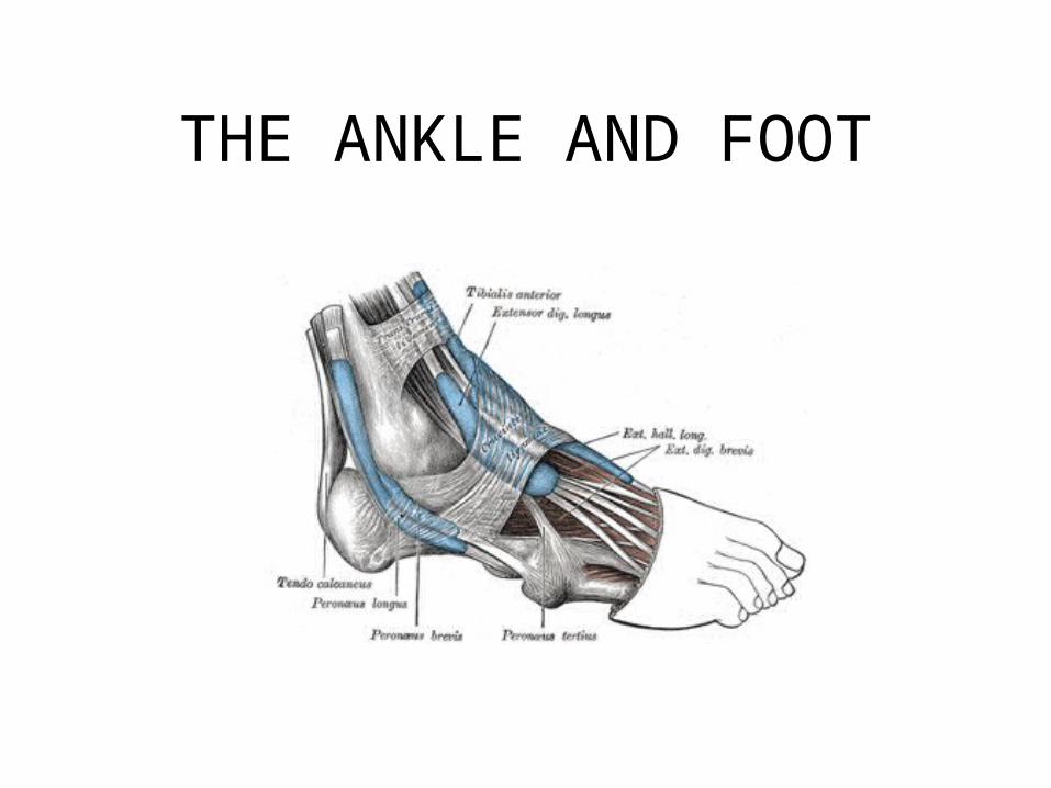
BONES
• A. Tibia– 1. condyle (lateral and medial)
– 2. tibial tuberosity
– 3. medial malleolus • B. Fibula
– 1. head
– 2. lateral malleolus
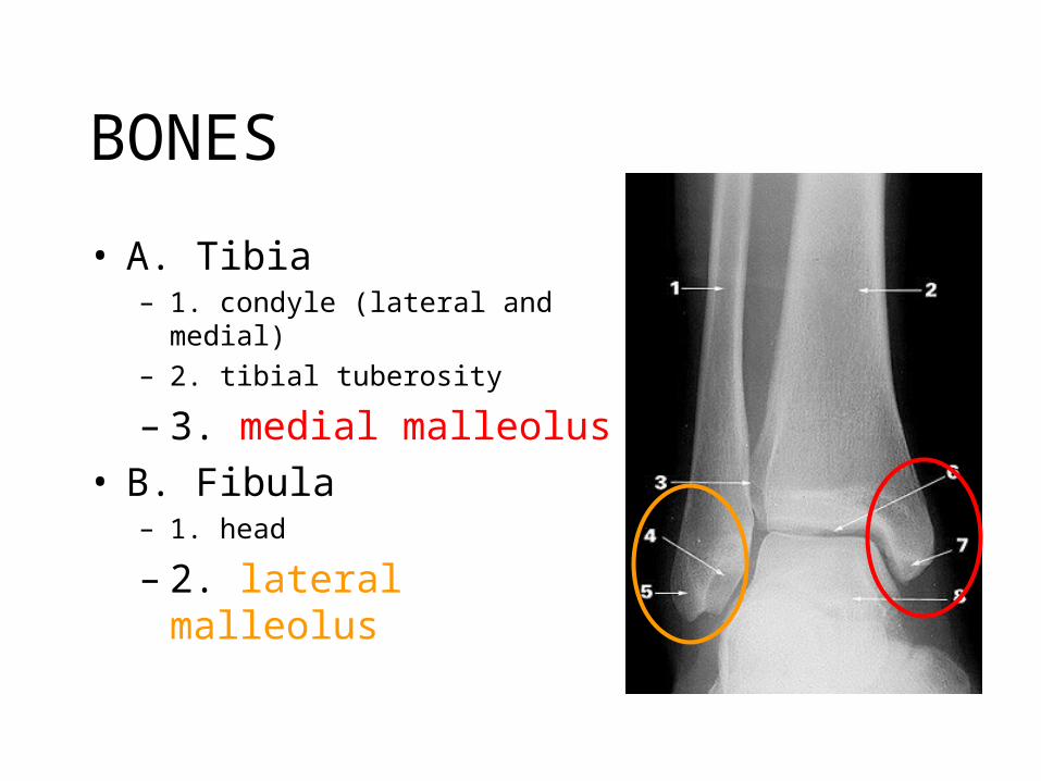
Tibia
• Medial Malleolus
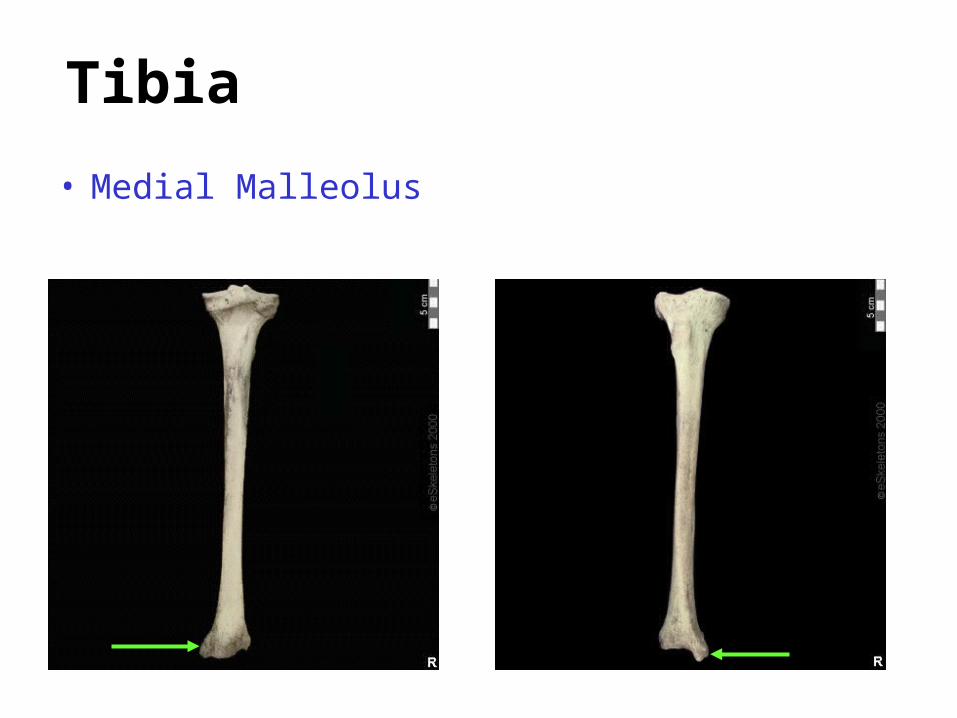
Tibia
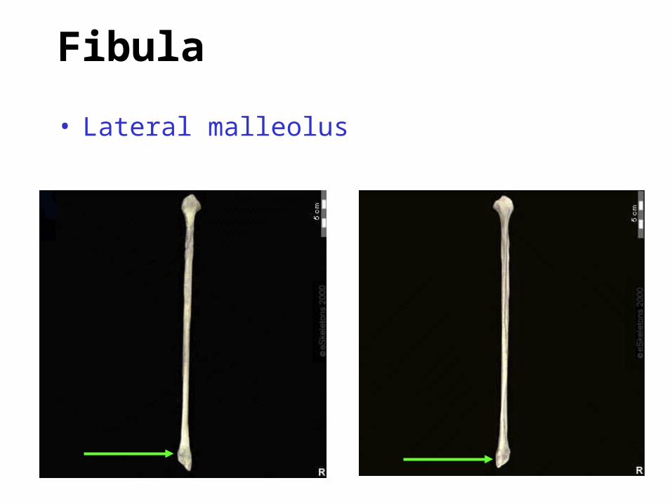
Fibula
• Lateral malleolus
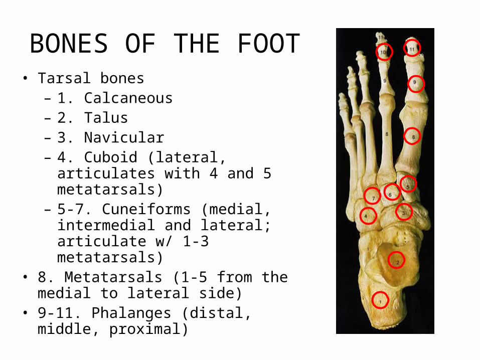
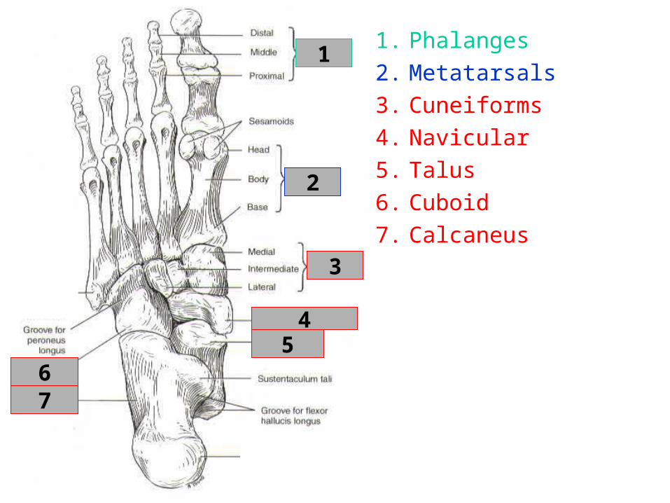
BONES OF THE FOOT• Tarsal bones
– 1. Calcaneous– 2. Talus– 3. Navicular– 4. Cuboid (lateral, articulates with 4
and 5 metatarsals)– 5-7. Cuneiforms (medial, intermedial
and lateral; articulate w/ 1-3 metatarsals)
• 8. Metatarsals (1-5 from the medial to lateral side)
• 9-11. Phalanges (distal, middle, proximal)
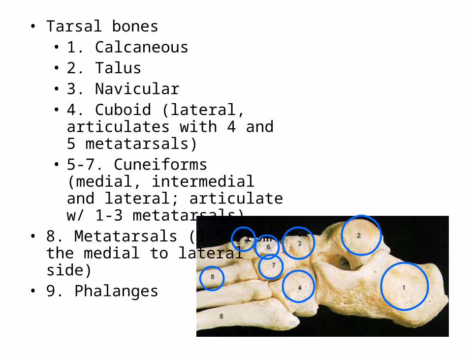
• Tarsal bones• 1. Calcaneous• 2. Talus• 3. Navicular• 4. Cuboid (lateral, articulates with
4 and 5 metatarsals)• 5-7. Cuneiforms (medial,
intermedial and lateral; articulate w/ 1-3 metatarsals)
• 8. Metatarsals (1-5 from the medial to lateral side)
• 9. Phalanges
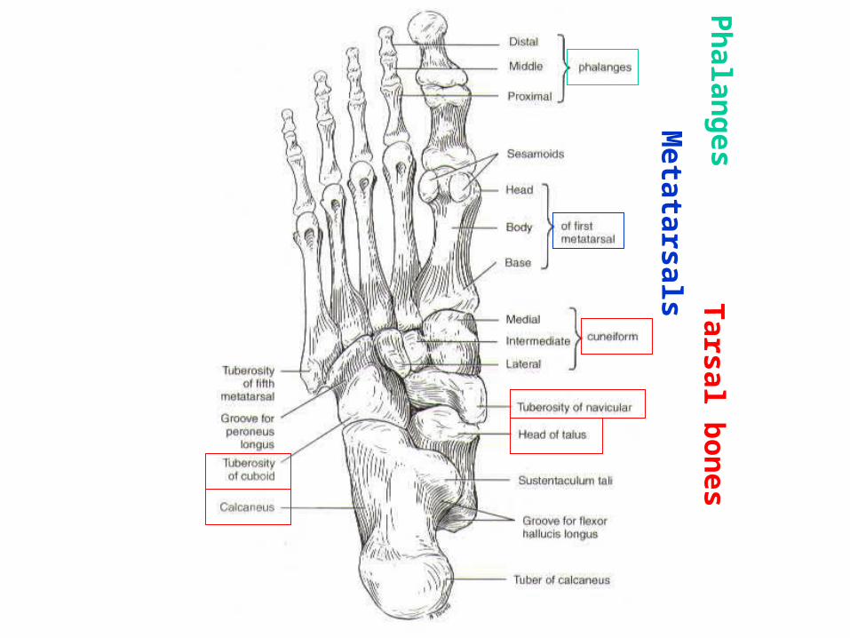
Tarsal b
ones
Metatarsals
Ph
alanges
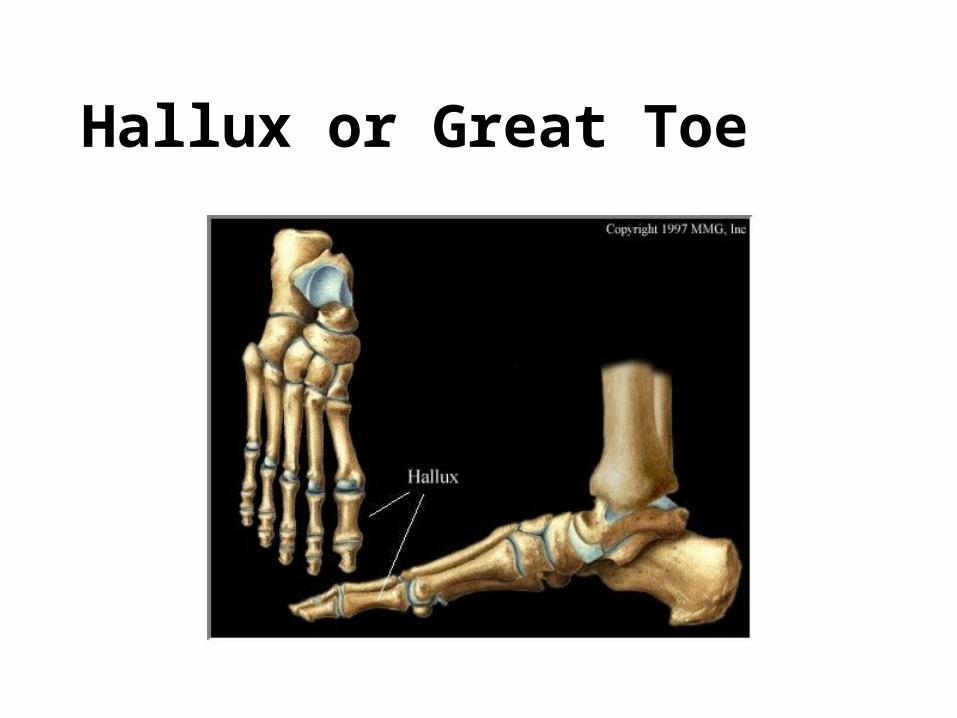
Hallux or Great Toe
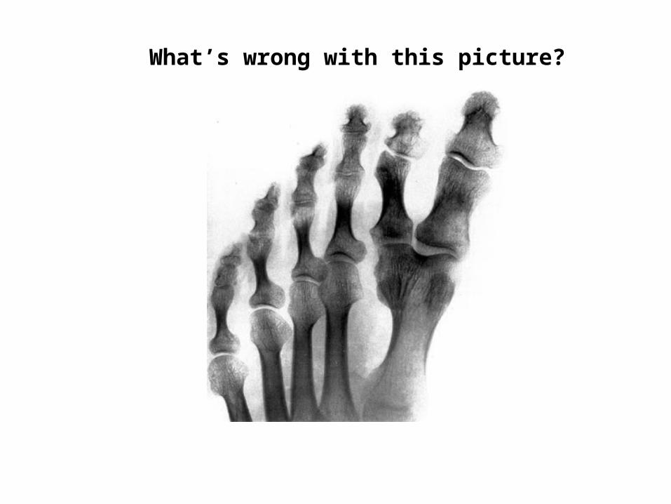
What’s wrong with this picture?
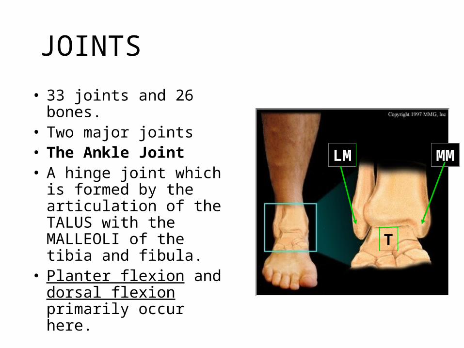
JOINTS
• 33 joints and 26 bones.• Two major joints• The Ankle Joint• A hinge joint which is
formed by the articulation of the TALUS with the MALLEOLI of the tibia and fibula.
• Planter flexion and dorsal flexion primarily occur here.
T
MMLM
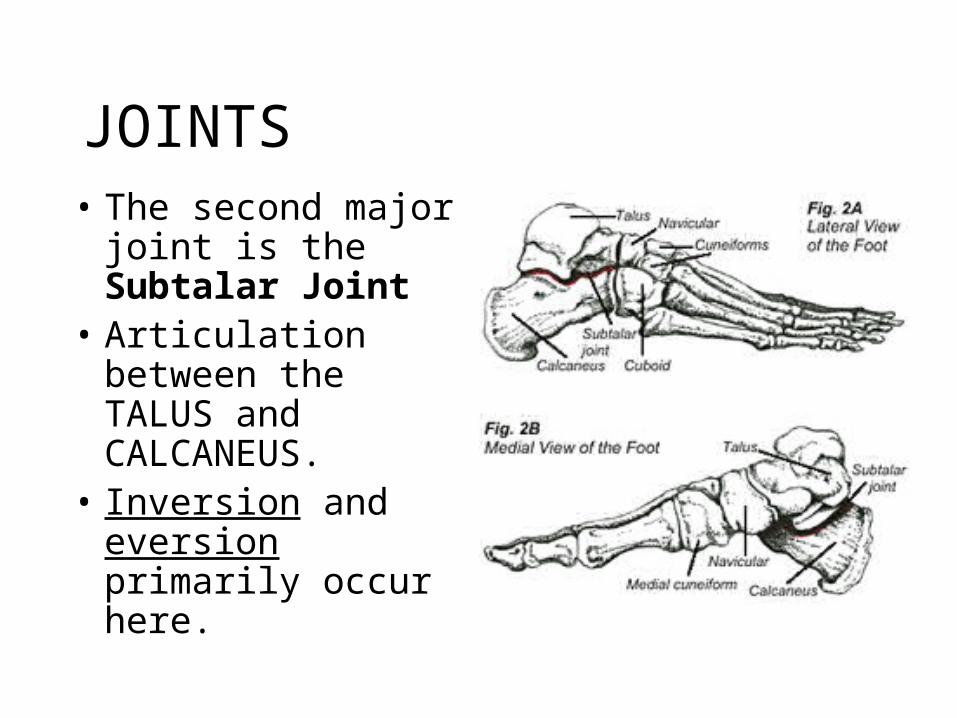
JOINTS• The second major
joint is the Subtalar Joint
• Articulation between the TALUS and CALCANEUS.
• Inversion and eversion primarily occur here.
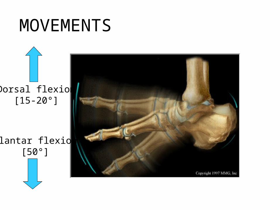
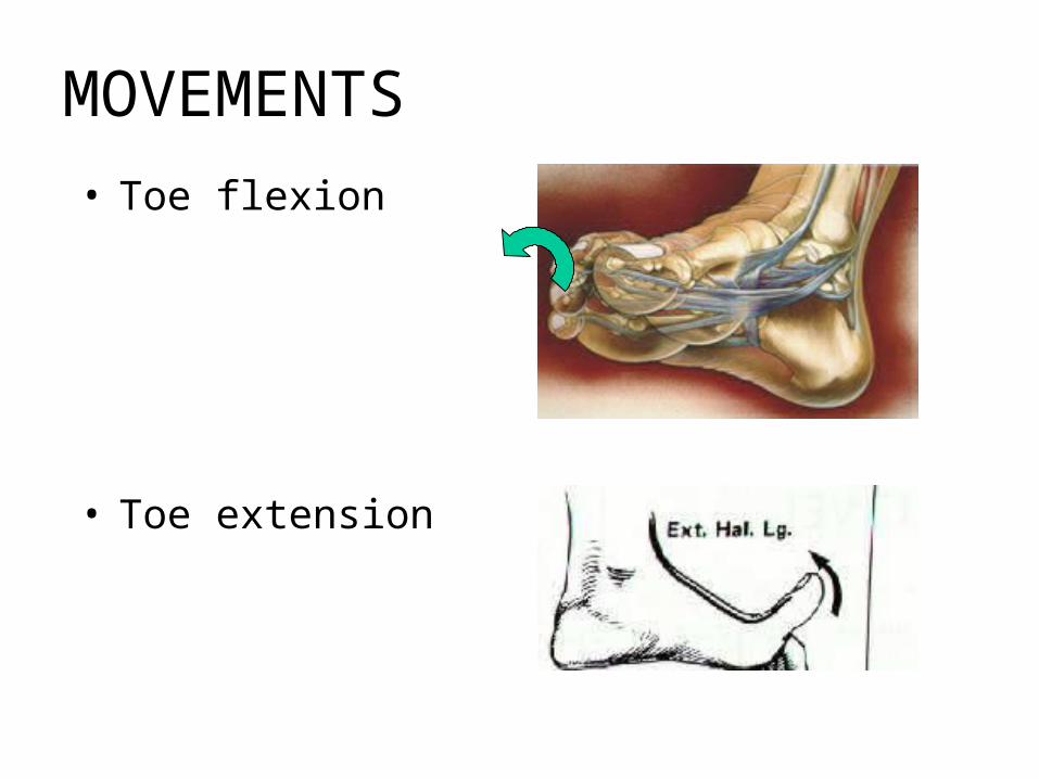
MOVEMENTS
Dorsal flexion[15-20°]
Plantar flexion[50°]
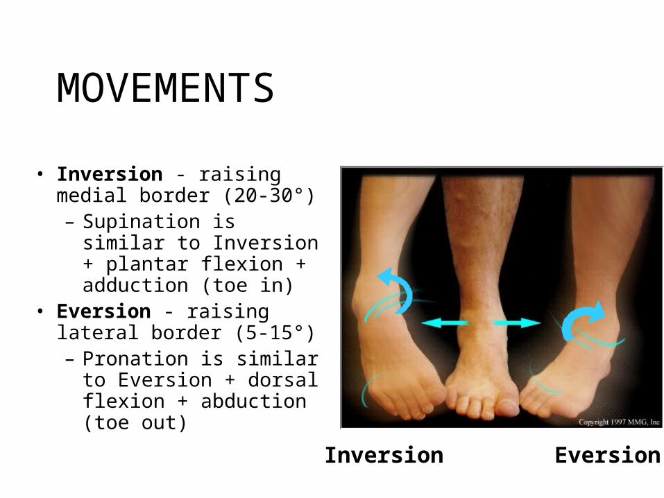
MOVEMENTS
• Inversion - raising medial border (20-30°)– Supination is similar to
Inversion + plantar flexion + adduction (toe in)
• Eversion - raising lateral border (5-15°)– Pronation is similar to
Eversion + dorsal flexion + abduction (toe out)
Inversion Eversion
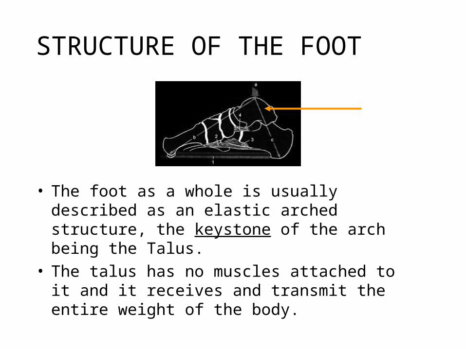
STRUCTURE OF THE FOOT
• The foot as a whole is usually described as an elastic arched structure, the keystone of the arch being the Talus.
• The talus has no muscles attached to it and it receives and transmit the entire weight of the body.
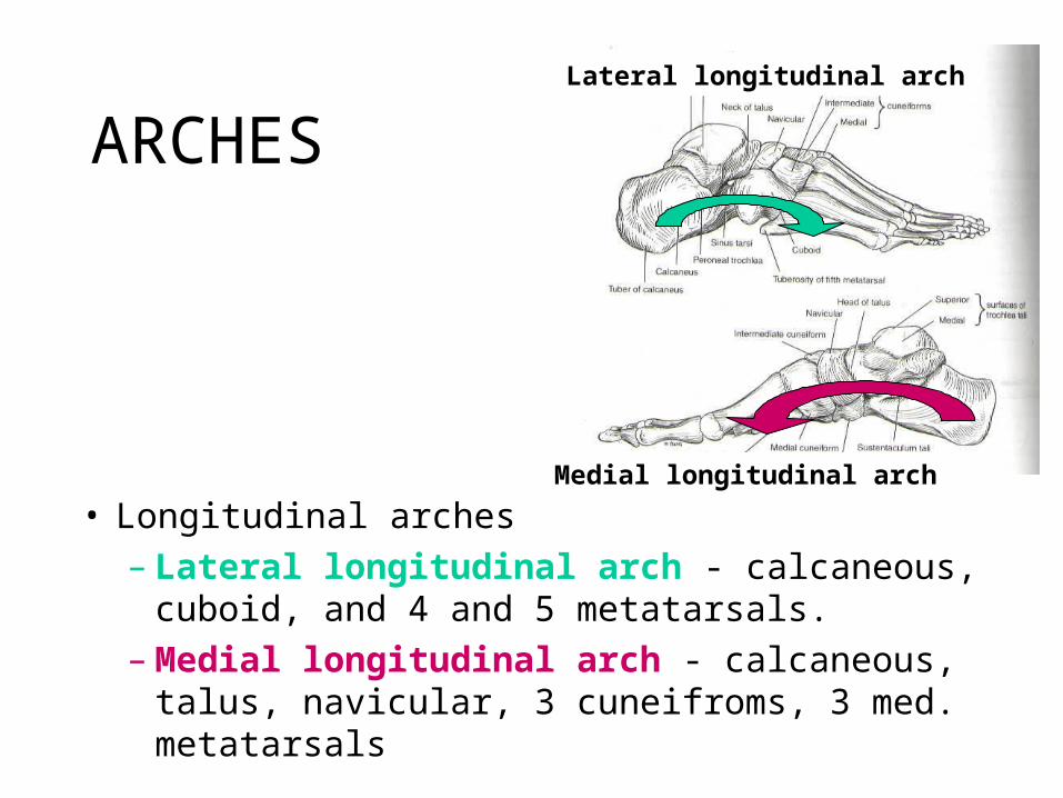
ARCHES
• Longitudinal arches – Lateral longitudinal arch - calcaneous, cuboid,
and 4 and 5 metatarsals.– Medial longitudinal arch - calcaneous, talus,
navicular, 3 cuneifroms, 3 med. metatarsals
Lateral longitudinal arch
Medial longitudinal arch
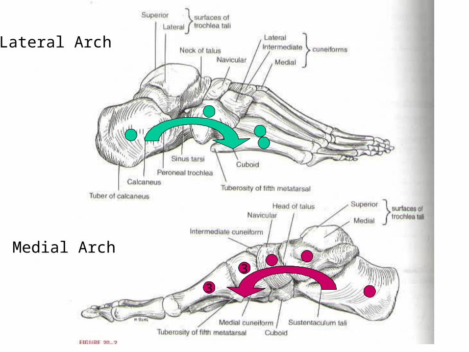
Lateral Arch
Medial Arch3
3
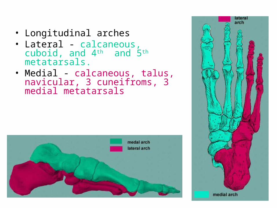
• Longitudinal arches• Lateral - calcaneous, cuboid, and 4th and
5th metatarsals.• Medial - calcaneous, talus, navicular, 3
cuneifroms, 3 medial metatarsals
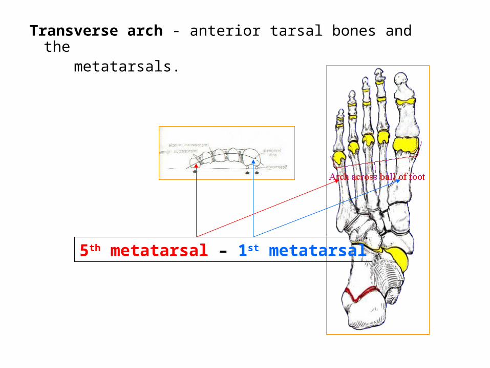
Transverse arch - anterior tarsal bones and the metatarsals.
5th metatarsal – 1st metatarsal
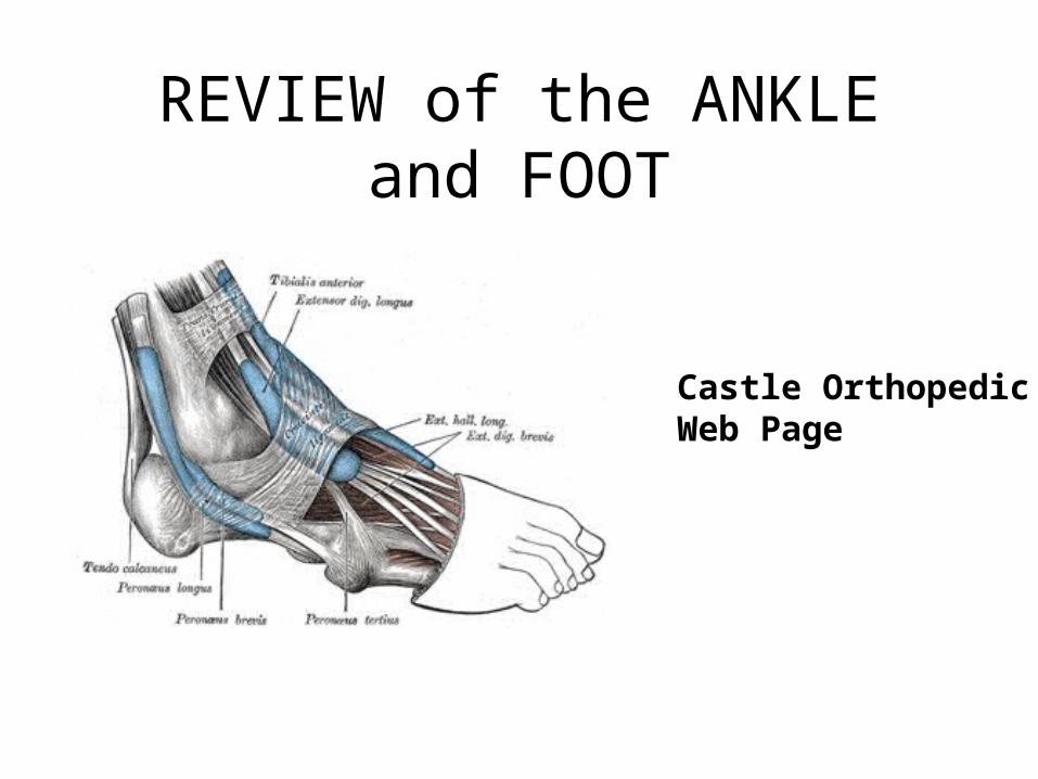
REVIEW of the ANKLE and FOOT
Castle OrthopedicWeb Page
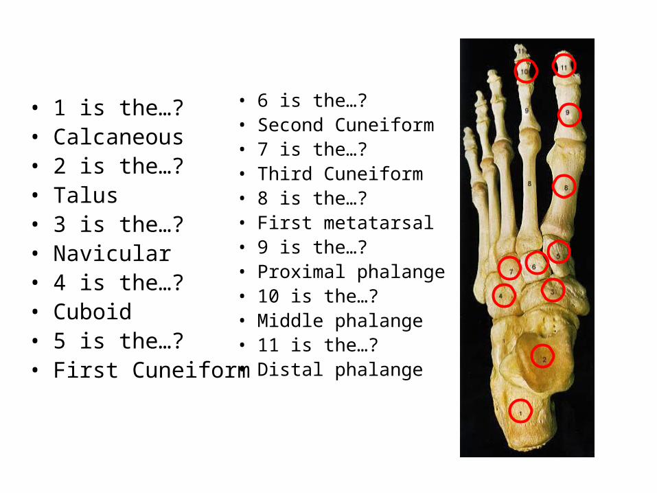
• 1 is the…?• Calcaneous• 2 is the…?• Talus• 3 is the…?• Navicular• 4 is the…? • Cuboid• 5 is the…? • First Cuneiform
• 6 is the…? • Second Cuneiform• 7 is the…? • Third Cuneiform• 8 is the…? • First metatarsal• 9 is the…?• Proximal phalange• 10 is the…?• Middle phalange• 11 is the…?• Distal phalange
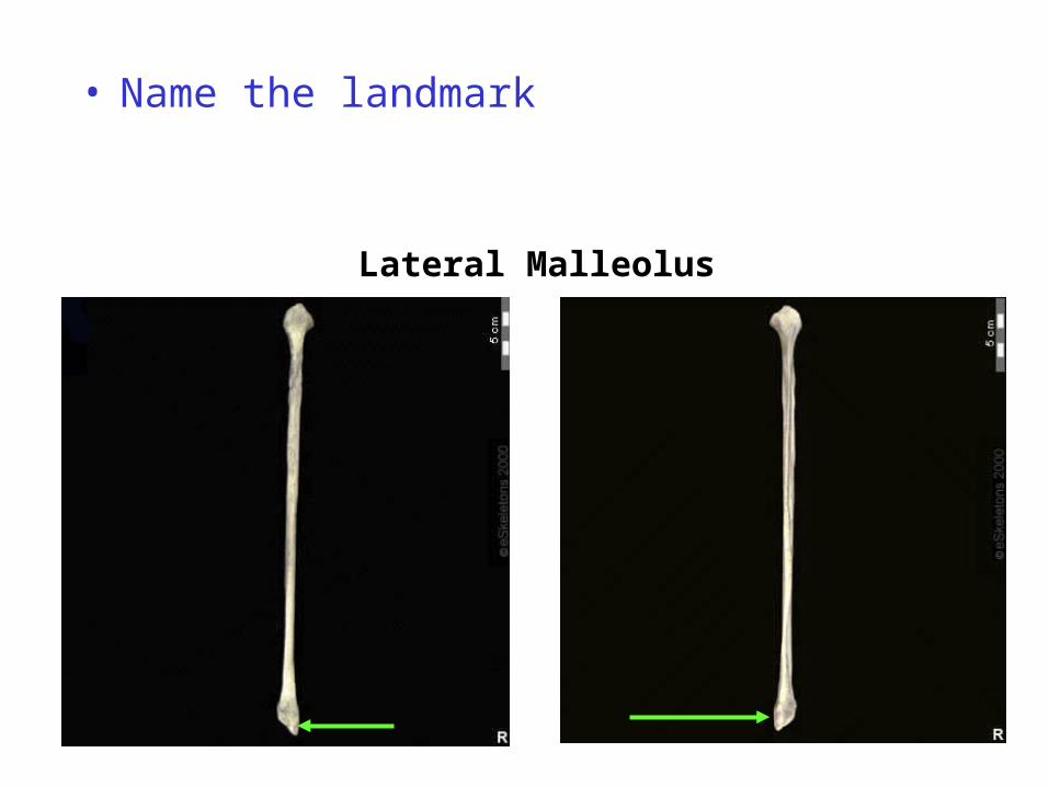
• Name the landmark
Lateral Malleolus
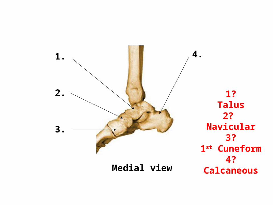
1.
2.
3.
4.
1?Talus
2? Navicular
3?1st Cuneform
4?CalcaneousMedial view
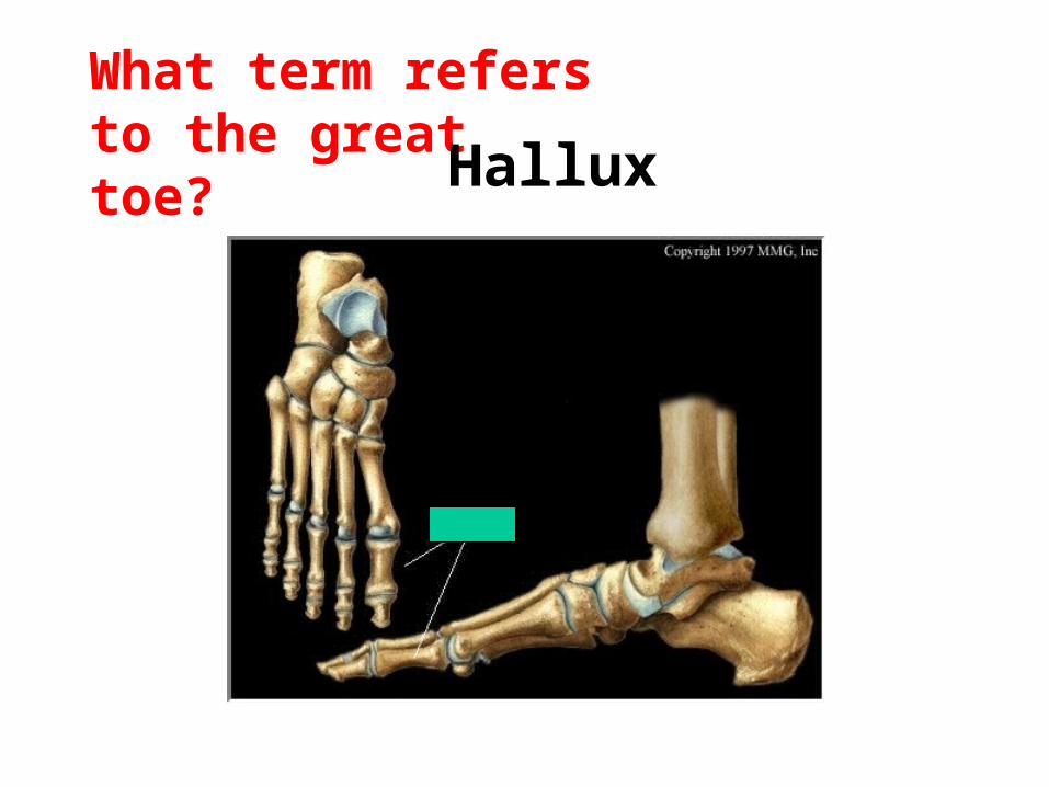
What term refers to the great toe? Hallux
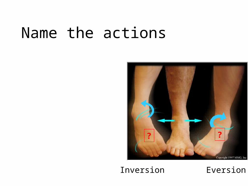
Name the actions
Inversion Eversion
? ?
Name the two movements at the toes
• Toe flexion • Toe extension
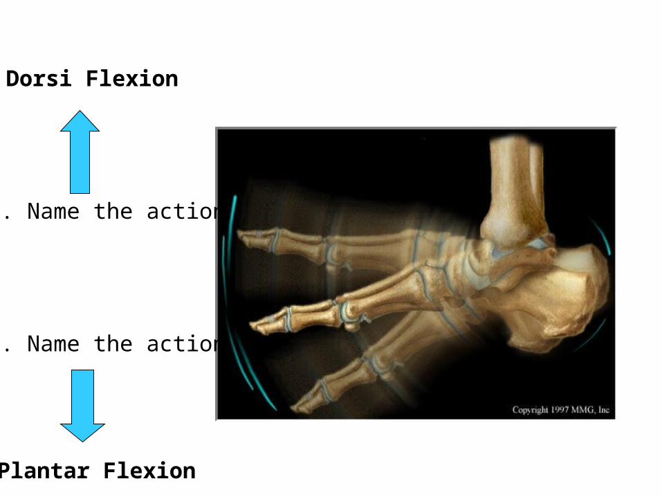
1. Name the action
2. Name the action
Dorsi Flexion
Plantar Flexion
45
7
3
6
2
11. Phalanges
2. Metatarsals
3. Cuneiforms
4. Navicular
5. Talus
6. Cuboid
7. Calcaneus
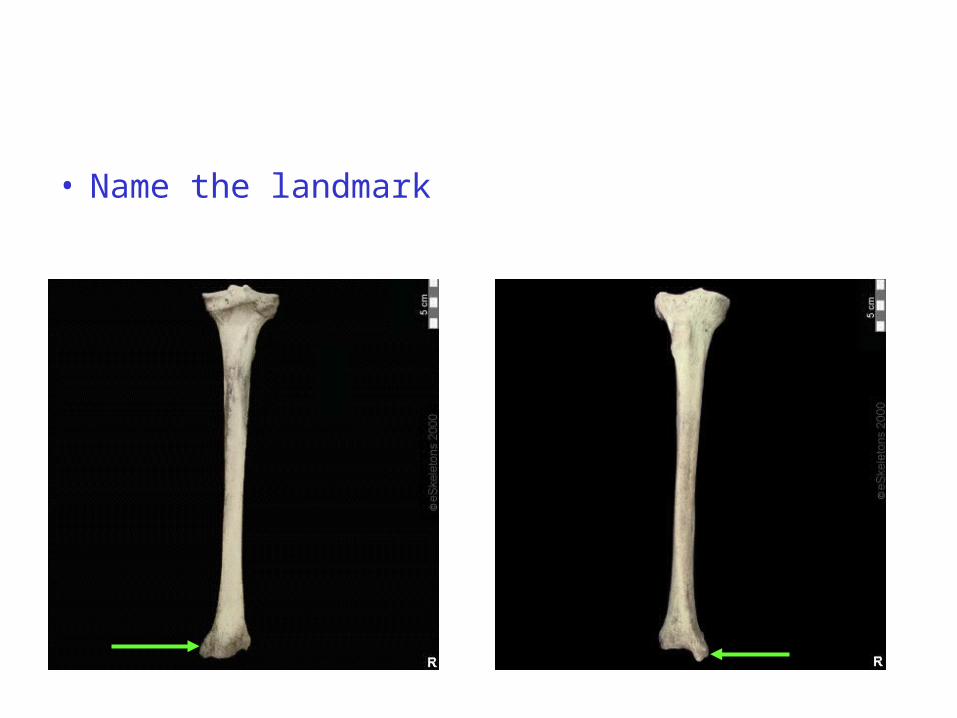
• Name the landmark

Name the actions of the ankle to the left and the ankle to the right.
Eversion Inversion
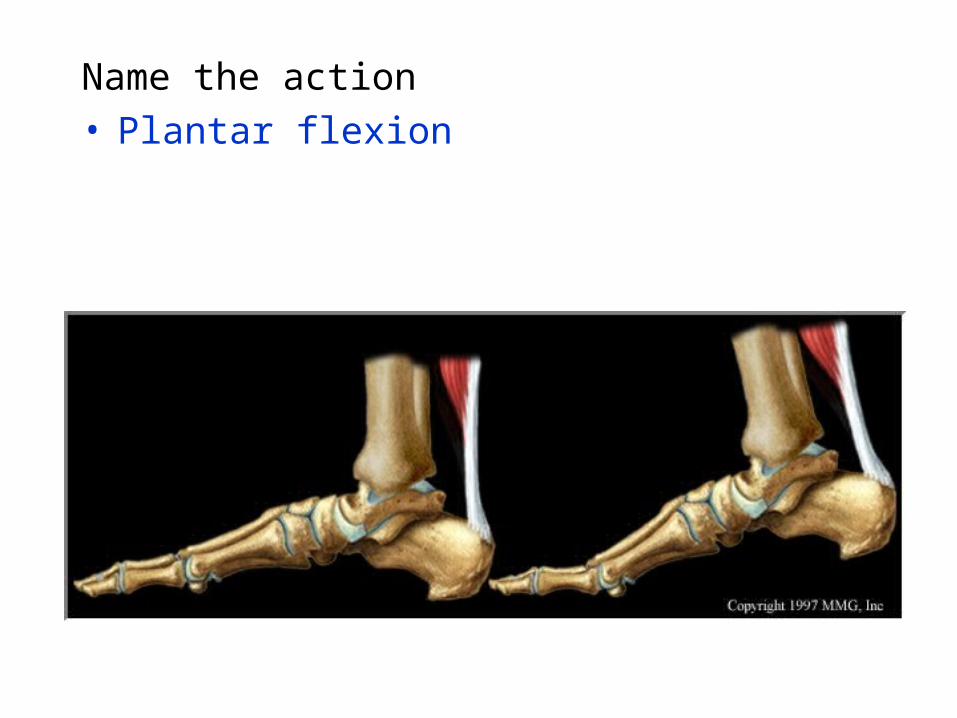
Name the action• Plantar flexion
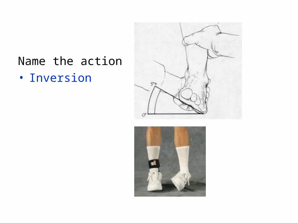
Name the action• Inversion
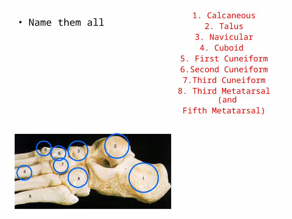
• Name them all1. Calcaneous
2. Talus3. Navicular4. Cuboid
5. First Cuneiform6.Second Cuneiform7.Third Cuneiform
8. Third Metatarsal (and Fifth Metatarsal)
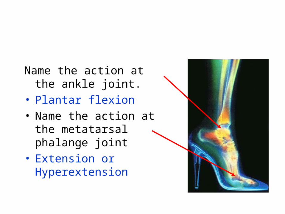
Name the action at the ankle joint.
• Plantar flexion• Name the action at the
metatarsal phalange joint• Extension or Hyperextension
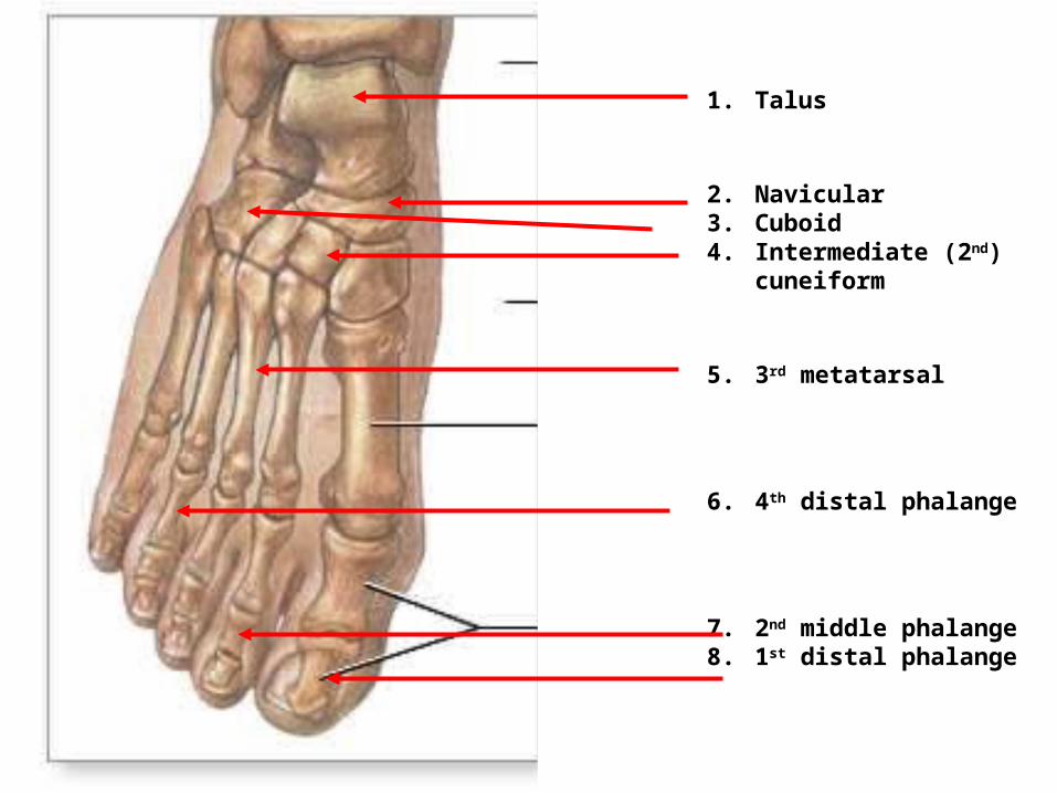
1. Talus
2. Navicular3. Cuboid4. Intermediate (2nd)
cuneiform
5. 3rd metatarsal
6. 4th distal phalange
7. 2nd middle phalange8. 1st distal phalange
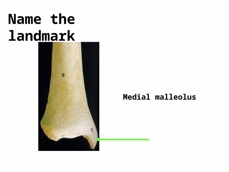
Name the landmark
Medial malleolus
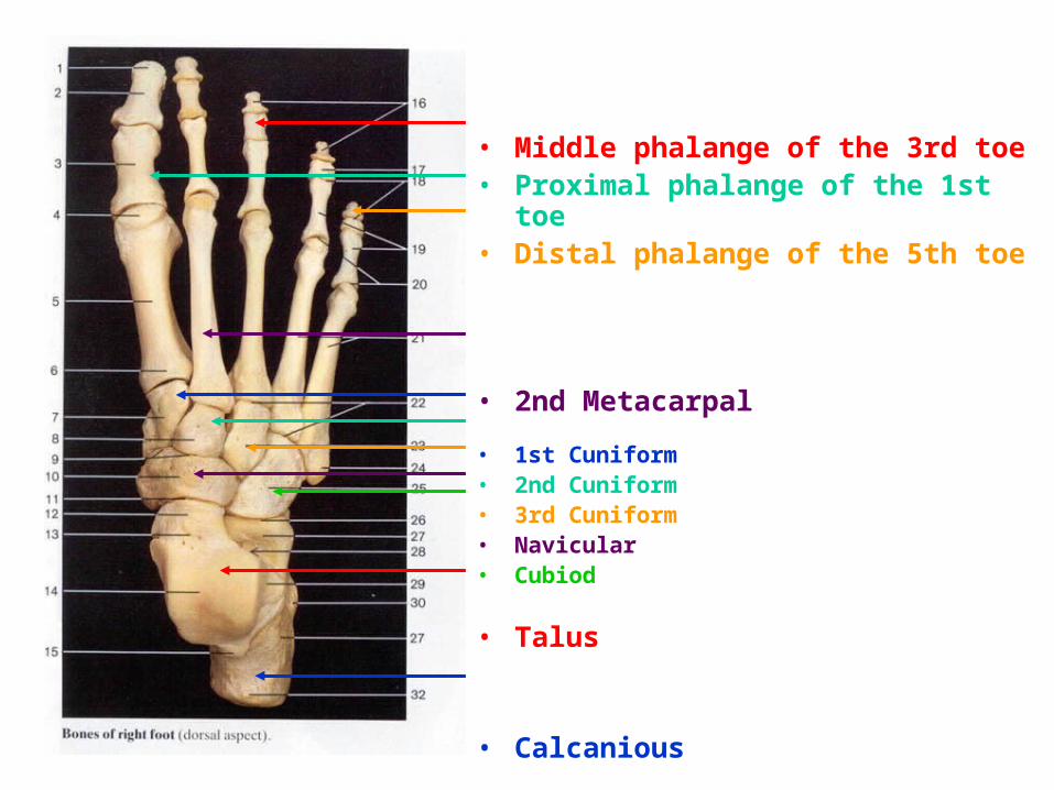
• Middle phalange of the 3rd toe• Proximal phalange of the 1st toe• Distal phalange of the 5th toe
• 2nd Metacarpal
• 1st Cuniform• 2nd Cuniform• 3rd Cuniform• Navicular• Cubiod
• Talus
• Calcanious