Embed Size (px)
Citation preview
MOL 24380
1
The Aryl Hydrocarbon Receptor Signaling Pathway is Modified through Interactions with a Kelch Protein Elizabeth E. Dunham, Emily A. Stevens, Edward Glover, and Christopher A. Bradfield From the McArdle Laboratory for Cancer Research, University of Wisconsin Medical School, Madison, WI 53706
Molecular Pharmacology Fast Forward. Published on March 31, 2006 as doi:10.1124/mol.106.024380
Copyright 2006 by the American Society for Pharmacology and Experimental Therapeutics.
This article has not been copyedited and formatted. The final version may differ from this version.Molecular Pharmacology Fast Forward. Published on March 31, 2006 as DOI: 10.1124/mol.106.024380
at ASPE
T Journals on A
ugust 25, 2019m
olpharm.aspetjournals.org
Dow
nloaded from
MOL 24380
2
Running Title: Novel AHR Signaling Modifier Correspondence should be sent to: Christopher A. Bradfield McArdle Laboratory for Cancer Research 1400 University Avenue Madison, WI 53706-1599 Email: [email protected]
Number of pages: 19
Number of tables: 0
Number of figures: 6
Number of references: 37
Number of words in abstract: 199
Number of words in introduction: 436
Number of words in discussion: 2279 (results and discussion)
Abbreviations: AHR, aryl hydrocarbon receptor; ARNT, aryl hydrocarbon nuclear translocator; TCDD, 2,3,7,8-tetrachlorodibenzo-p-dioxin; ARA3, aryl hydrocarbon receptor associated 3; ARA9, aryl hydrocarbon receptor associated 9; NS1BP, influenza virus nonstructural protein 1 binding protein
This article has not been copyedited and formatted. The final version may differ from this version.Molecular Pharmacology Fast Forward. Published on March 31, 2006 as DOI: 10.1124/mol.106.024380
at ASPE
T Journals on A
ugust 25, 2019m
olpharm.aspetjournals.org
Dow
nloaded from
MOL 24380
3
ABSTRACT:
The aryl hydrocarbon receptor (AHR) is a ligand activated transcription factor with important roles in
metabolic adaptation, dioxin toxicology and vascular development. In order to understand the details of this signal
transduction pathway, we have used the yeast two-hybrid system to identify proteins that physically interact with the
AHR in a ligand dependent manner. Using this strategy, we identified a novel modifier of the AHR signaling pathway,
which we named Ah-receptor associated protein 3 (ARA3). Coexpression of ARA3 with an AHR chimera in yeast
and mammalian cells enhances signaling in response to agonists. The human full-length cDNA previously was
described as influenza virus nonstructural protein-1 binding protein (NS1BP). This protein contains four apparent
domains—a “broad-complex, tramtrack and bric a` brac” (BTB) domain, a “kelch” domain, a “BTB and C-terminal
kelch” (BACK) domain, and an intervening region (IVR). The carboxy-terminus of AHR’s “Per-ARNT-Sim” (PAS)
domain and the BACK/IVR domains of ARA3 mediate the AHR-ARA3 interaction. The BACK/IVR domains of
ARA3 also are sufficient to modify AHR signaling in yeast and mammalian cells. In an effort to provide a preliminary
model of NS1BP activity in AHR signaling, we demonstrate that NS1BP regulates the concentration of functional
AHR in mammalian cells.
This article has not been copyedited and formatted. The final version may differ from this version.Molecular Pharmacology Fast Forward. Published on March 31, 2006 as DOI: 10.1124/mol.106.024380
at ASPE
T Journals on A
ugust 25, 2019m
olpharm.aspetjournals.org
Dow
nloaded from
MOL 24380
4
The aryl hydrocarbon receptor (AHR) is a member of the basic-helix-loop-helix-Per-Arnt-Sim (bHLH-PAS)
superfamily. Upon binding ligands, the AHR mediates an adaptive metabolic response by upregulating the
transcription of a battery of xenobiotic metabolizing enzymes, including the cytochromes P450, CYP1A1, CYP1A2
and CYP1B1 (Schmidt and Bradfield, 1996). When stimulated by high-potency agonists, such as 2,3,7,8-
tetrachlorodibenzo-p-dioxin (TCDD), the AHR mediates an additional toxic response that includes hepatocellular
damage, thymic involution, teratogenesis, chloracne and cancer (Fernandez-Salguero et al., 1996; Pohjanvirta and
Tuomisto, 1994). Recently, it has been shown that the AHR also plays an important role in vascular development. In
this regard, patent ductus venosus is observed in 100% of mice with a null allele at the Ahr locus (Lahvis et al., 2005).
The presentation of vascular aberrations in AHR-mutant mice is consistent with the idea that the AHR is activated by
an unknown endogenous ligand.
The AHR signaling pathway is understood at a basic level: In the absence of ligand, the AHR has a higher
affinity for the cytosol where it exists in a complex with Hsp90 and the cochaperones, ARA9 (also known as AIP1 or
XAP2) and p23 (Carver et al., 1998; Kazlauskas et al., 1999; Ma and Whitlock, 1997; Meyer et al., 1998;
Wilhelmsson et al., 1990). Upon ligand binding, the AHR attains a higher affinity for the nuclear compartment where
it dimerizes with another bHLH-PAS protein known as the Ah-receptor nuclear translocator (ARNT) (Reyes et al.,
1992). The transcriptional activity of the AHR-ARNT heterodimer is modulated through interactions with cofactors
such as SRC-1 and RIP140 (Beischlag et al., 2002; Kumar et al., 1999). The AHR pathway can be attenuated by two
mechanisms, proteasome-dependent degradation of the AHR and the AHR-mediated transcription of a dominant
negative bHLH-PAS protein known as the Ah receptor repressor (AHRR) (Davarinos and Pollenz, 1999; Mimura et
al., 1999).
In previous studies we have demonstrated that the yeast, Saccharomyces cerevisiae, is an excellent model of
AHR signal transduction (Carver et al., 1994; Yao et al., 2004). In an effort to identify unknown components of the
AHR signaling pathway, we have been employing a yeast-two-hybrid strategy to screen for proteins that interact with
the AHR in a ligand-dependent manner. Previously this approach has led to the identification of ARA9, now a proven
AHR cochaperone (Bell and Poland, 2000; LaPres et al., 2000; Ma and Whitlock, 1997; Petrulis et al., 2000). Due to
this earlier success, we began characterizing the second clone identified in our yeast-two-hybrid assay, i.e., Ah
receptor associated 3 (ARA3) (Carver and Bradfield, 1997). Here we characterize the AHR-ARA3 interaction and its
effect on AHR signaling in yeast and mammalian cells.
This article has not been copyedited and formatted. The final version may differ from this version.Molecular Pharmacology Fast Forward. Published on March 31, 2006 as DOI: 10.1124/mol.106.024380
at ASPE
T Journals on A
ugust 25, 2019m
olpharm.aspetjournals.org
Dow
nloaded from
MOL 24380
5
MATERIALS AND METHODS:
Oligonucleotide Sequences: OL163: CCCAAGCTTACGCGTGCAGTGGTCTCTGAGTGGCGATGATGTAATCT
GG; OL180: GCGTCGACTGATGAGCAGCGGCGCCAACATCACC; OL258: GCCGTCGACGCGGCCGCGAA
GTCTAGCTTGTGTTTGG; OL392: CCGCTCGAGTGATGAGCAGCGGCGCCAACATCACC; OL822: TAATA
CGACTCACTATAGGG; OL926: GCTTGTGTTGCTGTTAG; OL1191: CCTCAGCCTGTCCACGAACTACCCT
TGGATGATC; OL1378: GATCCCTCGAGCCACCATGAGCAGCGGCGC; OL1379: GATGCTCGAGGTGGCCA
ATGCTGCTC; OL1469: GGCGAATTCACCATGGATCGAGTAAAGC; OL1490: GGCCAATTCACCATGATTC
CCAATGG; OL1886: CCTTATCCTCGCCTAGCCACATTC; OL1887: CCTTAACAACGGTTAGTTCTCAAC;
OL1888: CCTTATCGTGCGTACTGCATAGGAG; OL2006: GGGAATTCATTATGGCTAGCATGACTGGTGGA
CAGCAAATGGGTATGGATGGAGTAAAGCAGGTTTGTGG; OL2372: GCTCGAGTTATCCGGTAGCAAACA
TGAAGGGCAG
Strains and Plasmids: Saccharomyces cerevisiae strain L40 (Mat a, his3∆200, trp1-901, leu2-3, 112, ade2,
LYS2::(lexAop)4-HIS3, URA3::(lexAop)8-lacZ, gal80) was used in both the yeast two-hybrid assays and the
pharmacology experiments (Vojtek et al., 1993). The plasmid pBTM116 is a 2µm TRP-marked yeast expression
vector for making LexA DNA binding domain (LexA) fusion proteins under regulation of the ADH1 promoter (Bartel
et al., 1993). The plasmid pYX242 (Novagen, Madison WI) is a 2µm LEU-marked yeast expression vector under the
regulation of the TP1 promoter. The plasmid pACT (Clontech, Mountain View CA) is a 2µm LEU-marked vector used
to make GAL4 transcription activation domain (TAD) fusion proteins. The plasmid pSPORT AHR (PL65) was used
as a template for any AHR PCR amplifications and has been described previously (Dolwick et al., 1993). The plasmid
pYX AHR (PL1033) was constructed by PCR using OL1378 and OL1379 using PL65 as the template and was
subcloned from pGEMT (Promega, Madison WI) using XhoI and SalI into the SalI site of pYX242. The plasmid pACT
ARA3 (PL792) was isolated as previously described from a two-hybrid screen with a human B-cell library using
pBTM LexA-AHR∆TAD (PL739) as the bait construct (Carver and Bradfield, 1997). The plasmids pBTM LexA-
ARA3 (PL1416) and pYX ARA3 (PL1468) were constructed by PCR amplification of PL792 using OL1469 and
OL926, ligated into pGEMT, then subcloned into EcoRI and SalI of pBTM116 or pYX242. The plasmids pSGBCU
ARNT (PL574), pBTM LexA-∆bHLHAHR (PL703) and pY2 N-LexA-C (PL740) have been described previously
(Carver et al., 1994; Hogenesch et al., 1997; LaPres et al., 2000).
This article has not been copyedited and formatted. The final version may differ from this version.Molecular Pharmacology Fast Forward. Published on March 31, 2006 as DOI: 10.1124/mol.106.024380
at ASPE
T Journals on A
ugust 25, 2019m
olpharm.aspetjournals.org
Dow
nloaded from
MOL 24380
6
Transformations and Library Screening: Transformations into S. cerevisiae strain L40 were performed by a
modified lithium acetate method as previously described (Carver et al., 1994). The plates were incubated for 2 days at
30°C and then replica-plated onto media containing 100 µl of the vehicle dimethylsulfoxide (DMSO) or 100 µl of
various concentrations of the ligand β-napthoflavone (βNF) or deoxycorticosterone (DOC). After 2 days of growth,
triplicate groups of 6 –10 colonies for each condition were resuspended in 500 µl Buffer Z (60 mM Na2HPO4, 40 mM
NaH2 PO4, 10 mM KCl, 1 mM MgSO4, 35 mM β-2-mercaptoethanol). The A600 was measured on a 1:10 dilution of
this cell suspension to determine cell density, and β-galactosidase (lacZ) expression was determined on 150 µl of this
cell suspension as described previously (Carver et al., 1998). Percent maximal response was plotted using the program
PRISM (GraphPad Software, San Diego CA). Statistical significance also was determined with ANOVA followed by
Tukey’s multiple comparison test in PRISM.
Cloning 5’-end of human ARA3: The 5’-end of human ARA3 cDNA was obtained by a 5’ RACE (rapid
amplification of cDNA ends) procedure utilizing Marathon-Ready human heart cDNA library kit per the
manufacturer’s protocol (Clontech, Palo Alto CA). The OL 1191 was used as the gene specific primer along with the
vector specific primer AP1 to generate a 5’-end cDNA product. The resulting product was cloned into pGEMT and
sequenced. In order to generate the full-length human ARA3 cDNA (FLARA3), the 5’RACE EcoRI/XbaI fragment
was cloned into PL1425 to generate pGEMT-FL ARA3 (PL1424).
ARA3 deletions: The T7-tagged ARA3 constructs were created by including the T7 epitope
(MASMTGGQQMG) on the 5’ end of the sense oligonucleotide 2006 used to PCR amplify these constructs. The
sense oligonucleotide 1469 was used to amplify products without a T7 tag to generate LexA fusions. All ARA3
deletions were created by PCR amplification of PL792 and subcloned from pGEMT into pYX242 and pBTM116 using
the restriction enzymes EcoRI and SalI. The following antisense oligonucleotides were used to delete the ARA3 kelch
repeats are as follows: OL1886 to delete the last 2 kelch repeats, OL1887 to delete last 4 kelch repeats and OL1888 to
delete all 6 kelch repeats. The deleted kelch repeat constructs are as follows: pYX T7ARA3∆KR5-6 (PL1443), pBTM
LexA-ARA3∆KR5-6 (PL1451), pYX T7ARA3∆KR3-6 (PL1444), pBTM LexA-ARA3∆KR3-6 (PL1452), pYX
T7ARA3∆KR1-6 (PL1445), pBTM LexA-ARA3∆KR1-6 (PL1453). The plasmid pYX T7ARA3 (PL1417) was created
by PCR amplification of the template PL792 with OL2006 and OL926. The T7FLARA3 construct was created by
PCR amplification using OL1490 and OL926 of the template PL1424 and ligated into pTarget (Promega, Madison
WI). The FLARA3 insert was first subcloned into pET17B (Novagen, Madison WI) using EcoRI and SalI to create the
This article has not been copyedited and formatted. The final version may differ from this version.Molecular Pharmacology Fast Forward. Published on March 31, 2006 as DOI: 10.1124/mol.106.024380
at ASPE
T Journals on A
ugust 25, 2019m
olpharm.aspetjournals.org
Dow
nloaded from
MOL 24380
7
in-frame fusion with the amino terminus T7 tag. This T7FLARA3 construct was then PCR-amplified with OL822 and
OL926 and cloned into pTarget (PL1430). The PL1430 was first digested with XhoI, and then the XhoI overhangs
were filled in with the Klenow fragment. This T7FLARA3 insert was then subcloned into the EcoRV and SmaI sites
of pYX242 to create pYX T7FLARA3 (PL1442). The LexA fusion of FLARA3 was constructed by subcloning the
insert from pTarget FLARA3 (PL1430) using EcoR I and SalI into the EcoRI/SalI sites of pBTM116 to create pBTM
LexA-FLARA3 (PL1456). Expression of ARA3 and the amino and carboxy-teriminal deletions of ARA3 were
confirmed by western blot analysis on yeast cell extracts as described previously (Hogenesch et al., 1997).
Hydroxylamine Mutagenesis: Hydroxylamine mutagenesis was performed as described (Garabedian, 1993).
A 1 M solution of hydroxylamine was prepared immediately prior to use in 50 mM EDTA. Equal volumes of 1 M
hydroxylamine solution and 1 M potassium phosphate buffer were mixed together with 2 µg of purified plasmid DNA
pYX T7ARA3 (PL1417) or the control plasmid pBluescript II +/- (Gibco/Invitrogen, Carlsbad CA). The
hydroxylamine/DNA mixture was incubated at 25oC for 48 hours. The DNA was purified over a G50 column (Roche,
Basel Switzerland) and transformed into JM109 bacteria. Plasmid DNA from the bacteria was purified on a maxiprep
column (Qiagen, Valencia CA). The mutagenized PL1417 was then transformed with PL703 into the L40 strain of S.
cerevisiae by modified lithium acetate method as described above. The wild-type PL1417 and the empty vector
pYX242 were also transformed with PL703 to serve as controls for the experiment. All of the yeast transformations
were replica plated onto media containing 10 µM βNF. Activation of the lacZ reporter was determined. Any mutant
white colonies after 1 hour were picked and rescued into HB101 bacteria. All rescued plasmids were retransformed
into L40 yeast along with PL703 to confirm the loss of function and a western analysis was performed on yeast
extracts to confirm the expression. Any mutants expressed at wild-type levels were then sequenced to identify the
mutation. The mutants, pYX T7ARA3V198M (PL1448) and pYX T7ARA3E288K (PL1449) were PCR amplified
using OL1469 and OL926, TA-cloned into pGEMT, and subcloned into pBTM116 to create pBTM ARA3 V198M
(PL1457) and pBTM ARA3 E288K (PL1458).
AHR deletions: The plasmid pSPORT AHR∆313 (PL145) has been described previously (Dolwick et al.,
1993). The plasmid pBTM LexA-∆bHLHAHR∆TAD (PL1297) was created by deleting the EcoRI fragment from
pBTM LexA-AHR∆TAD (PL739). The AHR deletions pBTM LexA-AHR∆425 (PL1301) and pBTM LexA-
AHR∆402 (PL1377) were constructed by PCR amplification of the template PL65 using OL392 as the sense primer
and OL163 and OL2372 respectively. All were cloned into pGEMT and subcloned into the SalI site of pBTM116
This article has not been copyedited and formatted. The final version may differ from this version.Molecular Pharmacology Fast Forward. Published on March 31, 2006 as DOI: 10.1124/mol.106.024380
at ASPE
T Journals on A
ugust 25, 2019m
olpharm.aspetjournals.org
Dow
nloaded from
MOL 24380
8
using XhoI and SalI. The plasmid pBTM LexA-∆bHLHAHR∆402 (PL1324) was created by deleting the EcoRI
fragment from pBTM LexA-AHR∆402. The plasmid pBTM LexA-AHR∆313 (PL1300) was constructed by PCR
amplification using OL1378 and OL822 of the template PL145 and the insert was subcloned from pGEMT into the
SalI site of pBTM116 using XhoI and SalI. The plasmid pBTM LexA-AHR∆516 (PL1322) was generated by PCR
amplification using OL180 and OL258 of the template PL65 and the insert was subcloned from pGEMT using SalI
into the SalI site of pGAD424 (Clontech, Palo Alto CA). The pGAD AHR∆516 (PL718) construct was digested with
XmaI and the PstI and the AHR insert was subcloned into the SmaI and PstI sites of pBTM116.
Mammalian cell-culture assays: The plasmid pSG Gal4-∆bHLHAHR (PL118) has been described previously
(Jain et al., 1994). The pTarget T7ARA3 (PL1797) and pTarget T7ARA3∆kelch (PL1429) constructs were generated
by PCR amplification of PL792 using OL2006/OL926 and OL2006/OL1888. PL1430 is described above. The plasmid
pG5 Luciferase (Promega, Madison WI) is a luciferase reporter construct containing five upstream GAL4 DNA
response elements. The PL186 is a β-galactosidase expression vector driven by a SV-40 promoter. Mammalian cell
culture experiments were performed in Cos-1 cells maintained at 37oC and 5% CO2 in DMEM supplemented with
10% fetal bovine serum, penicillin (0.1 unit/ml), and streptomycin (0.1 µg/ml), Hepes buffer (10 mM), minimal
essential amino acids (0.1 mM), and sodium pyruvate (1 mM) (Gibco/Invitrogen, Carlsbad CA). Transient
transfections were carried out in 6-well plates with Effectene (Qiagen, Valencia CA) per manufacturer’s instructions.
DNA used in transfections consisted of 800 ng of PL118, 800 ng of PL1797, PL1429, PL1430, or an empty vector. In
addition 800 ng pG5 Luciferase was used as a reporter and 600 ng PL186 to control for transfection efficiency. Media
was changed at 4 hours post-transfection, and at 24 hours post-transfection 10 nM of TCDD or DMSO (vehicle
control) was added. Cells were incubated for another 24 hours and harvested using Reporter Lysis Buffer (Promega,
Madison WI). Luciferase and β-galactosidase activity were measured with the appropriate assay system per
manufacturer’s instructions (Promega, Madison WI).
Transient transfection for receptor binding: Cos-1 cells were maintained and transfected as described above
with the following modifications. Cells were maintained in 10 cm2 dishes and transfected with 300 ng PL118 and 300
ng PL1429 or an empty vector. Forty hours post-transfection, the cells were harvested with trypsin and washed twice
with PBS. Cell pellets were resuspended in MDEENG Buffer (25 mM MOPS, pH7.4, 1 mM dithiothreitol, 1 mM
EDTA, 5 mM EGTA, 0.02% NaN3, 10% glycerol) supplemented with 10 mM Na2MoO4. Samples were disrupted
using a Dounce homogenizer in the presence of a protease inhibitor cocktail (Roche Basel Switzerland). The samples
This article has not been copyedited and formatted. The final version may differ from this version.Molecular Pharmacology Fast Forward. Published on March 31, 2006 as DOI: 10.1124/mol.106.024380
at ASPE
T Journals on A
ugust 25, 2019m
olpharm.aspetjournals.org
Dow
nloaded from
MOL 24380
9
were subjected to centrifugation at 14,000 x g (10 minutes, 4oC). An additional centrifugation (100,000 x g 60
minutes, 4oC) was performed on the supernatants to obtain the soluble or “cytosolic” fraction. Protein concentrations
of the cytosols were determined by Coomassie Reagent assay (Pierce, Rockford IL) and samples were diluted to a
final protein concentration of 1 mg/ml.
Photoaffinity labeling: The photoaffinity labeling of the AHR was carried out as described previously
(Bradfield et al., 1988). Briefly, 1 nM of the photoaffinity ligand, 2-azido-3-[125I]iodo-7,8-dibromodibenzo-p-dioxin,
was added to 150 µg/ml of cytosolic protein and incubated for 30 minutes at 20oC, then 5 minutes on ice. Unbound
ligand was removed with charcoal/dextran (10%/1%, w/v). The receptor/ligand complex was crosslinked by exposure
to ultraviolet light (310 nm. 80 W, 4 cm) for 30 seconds. Four milliliters of acetone was added and the mixture was
incubated for 16 hours at –20oC. The acetone precipitate was removed by centrifugation (2,000 x g, 10 minutes, 4oC)
and the pellet was washed with 90% acetone/water. The samples were resuspended in electrophoresis sample buffer
and run on a 7.5% SDS-PAGE gel. Gels were analyzed by autoradiography and appropriate bands were cut out and
counted on a Minaxi γ gamma counter (Packard, Meriden CT). Statistical Significance was determined with a
Student’s t-test.
This article has not been copyedited and formatted. The final version may differ from this version.Molecular Pharmacology Fast Forward. Published on March 31, 2006 as DOI: 10.1124/mol.106.024380
at ASPE
T Journals on A
ugust 25, 2019m
olpharm.aspetjournals.org
Dow
nloaded from
MOL 24380
10
RESULTS AND DISCUSSION
We previously reported a modified yeast two-hybrid assay to screen for proteins that interact with the AHR
in a ligand-dependent manner. This screen of over 700,000 human cDNAs yielded ten positive clones (Carver and
Bradfield, 1997). Sequence analysis revealed that five of these clones represented a unique cDNA that was designated
ARA9. The other five clones represented a second distinct cDNA that we designated ARA3. Given that the importance
of ARA9 in AHR signaling has been demonstrated repeatedly since this initial report, we returned to characterize the
function of ARA3 (Bell and Poland, 2000; LaPres et al., 2000; Ma and Whitlock, 1997; Petrulis et al., 2000).
Analysis of the ARA3 cDNA: Sequence analysis of the five ARA3 cDNAs indicated that they were identical,
and encoded a large open reading frame of 531 amino acids. Given that a consensus initiation ATG codon was not
identified in the original cDNA, the 5’ end was amplified by PCR of a human heart library (Clontech, Mountain View
California). As the result of this experiment, the full-length open reading frame of ARA3 (FLARA3) was found to be
642 amino acids in length and contained a consensus initiation ATG codon with an upstream, in-frame stop codon
(Kozak, 1987) (Figure 1)1. Comparison of FLARA3 with other proteins in the GenBank database revealed a
preliminary domain map. At the N-terminus of FLARA3 is a “broad-complex, tramtrack and bric a` brac” (BTB)
domain found in several transcriptional regulators and non-DNA binding proteins (Collins et al., 2001; Zollman et al.,
1994). In addition to mediating hetero/homo-dimerization, the BTB domain has been shown to recruit corepressors
and histone deacetylases (Melnick et al., 2002). At its C-terminus ARA3 harbors a “kelch” domain, consisting of six
imperfect repeats of approximately 50 amino acids. This domain has been shown to have the potential to form a β-
propeller structure (Adams et al., 2000). Like many other BTB-kelch proteins, FLARA3 also harbors a motif referred
to as a “BTB and C-terminal kelch” homology domain (BACK) (Stogios and Prive, 2004). In some BTB-kelch
proteins the BACK domain aids in the interaction of the BTB domain with ubiquitin-ligase complexes (Furukawa and
Xiong, 2005; Stogios and Prive, 2004). A fourth region with no significant amino acid sequence homology exists
between the BACK and kelch domains. We designate this domain as the “intervening region” (IVR).
Since the time of our initial report of ARA3, the human and mouse cDNAs have been reported by other
laboratories. The human cDNA was first reported as an influenza virus NS1 binding protein (NS1BP) (Wolff et al.,
1998). The murine NS1BP orthologue, designated Nd1, and a splice variant also were cloned in a screen for cDNAs
differentially expressed in neurons of Ncx-deficient mice that stabilize actin filaments (Sasagawa et al., 2002). Given
this provenance, we now refer to FLARA3 as NS1BP and use the ARA3 designation to refer to the clone obtained
This article has not been copyedited and formatted. The final version may differ from this version.Molecular Pharmacology Fast Forward. Published on March 31, 2006 as DOI: 10.1124/mol.106.024380
at ASPE
T Journals on A
ugust 25, 2019m
olpharm.aspetjournals.org
Dow
nloaded from
MOL 24380
11
from the original two-hybrid screen (Figure 1). Murine NS1BP transcripts were previously found in all tissues
examined with the highest expression in the heart, kidney, and intestines (Sasagawa et al., 2002). By northern blot
analysis on murine tissue, we also detected NS1BP mRNA in the heart and kidney, as well as muscle, lung, testis, and
brain (data not shown).
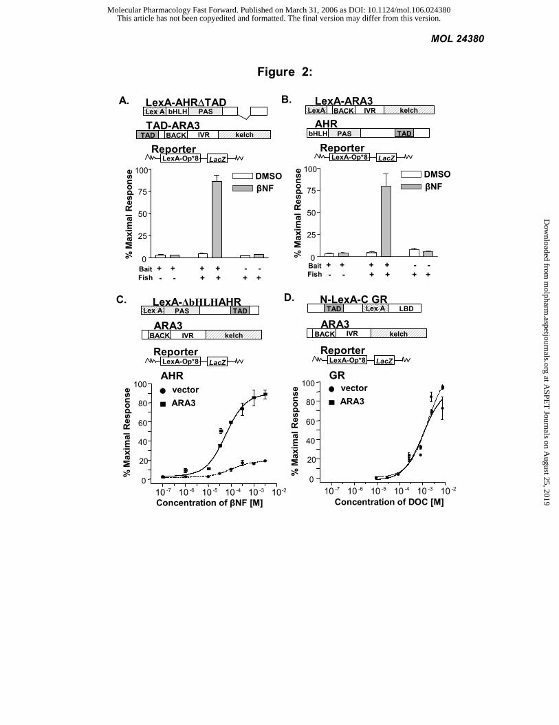
AHR and ARA3 proteins interact: To confirm the ligand-dependent nature of the AHR-ARA3 interaction,
plasmids harboring a LexA-AHR fusion (LexA-AHR∆TAD) and a TAD ARA3 fusion (TAD-ARA3) were
transformed into the L40 reporter strain. The separation of the LexA DNA-binding domain and the TAD on two
proteins requires that the two proteins interact for reporter activation. A 21-fold increase in β-galactosidase (LacZ)
reporter activity was observed when the cotransformed yeast were exposed to the AHR agonist βNF (Figure 2A). To
determine if this interaction was context dependent, the reverse-two hybrid experiment was performed. To this end, a
LexA-ARA3 fusion (LexA-ARA3) was screened against the full-length AHR containing its cognate TAD (AHR).
Again, an 18.5-fold increase in LacZ reporter activity was seen when these constructs were coexpressed in the
presence of βNF (Figure 2B).
AHR signaling is enhanced in the presence of ARA3: We have previously shown that an AHR chimera
harboring its cognate TAD, LexA-∆bHLHAHR, displays a similar pharmacological response to βNF in yeast as does
the AHR/ARNT heterodimer in mammalian cells (Carver et al., 1994). By removing the bHLH domain of AHR, the
dependence of AHR on ARNT is removed. To determine the effect of ARA3 on AHR signaling, L40 yeast were
transformed with LexA-∆bHLHAHR along with ARA3 cDNA or control vector. When grown in the presence of
increasing concentrations of βNF, we observed that coexpression of ARA3 influenced the shape of the dose response
curve significantly. In the presence of ARA3 the EC50 decreased from 1 x 10⁻⁴ to 6 x 10⁻⁵ and the maximal response
increased approximately 5-fold. This change in dose-response is consistent with the idea that ARA3 increases the
concentration of AHR available to bind ligand and activate transcription (Figure 2C) (Bourne and von Zastrow, 2001).
To determine whether the effect of ARA3 was specific to AHR signaling, a similar pharmacology experiment was
performed with a LexA fusion of the glucocorticoid receptor (GR)N-LexA-CGR) with its ligand deoxycorticosterone
(DOC). Coexpression of ARA3 did not influence GR’s signaling in this assay (Figure 2D).
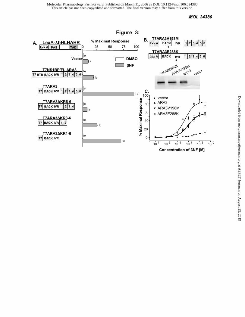
The BACK/IVR domains of NS1BP/FLARA3 modify AHR signaling: We first mapped the functional domains
of NS1BP/FLARA3 by examining specific deletions for their effect on AHR signaling in yeast. The deletions were
constructed with a T7-epitope tag to monitor protein expression levels. In this system, LexA-∆bHLHAHR was
This article has not been copyedited and formatted. The final version may differ from this version.Molecular Pharmacology Fast Forward. Published on March 31, 2006 as DOI: 10.1124/mol.106.024380
at ASPE
T Journals on A
ugust 25, 2019m
olpharm.aspetjournals.org
Dow
nloaded from
MOL 24380
12
cotransformed with each NS1BP/FLARA3 deletion. We observed that NS1BP/FLARA3 increased the signaling of the
AHR chimera by 2-fold (P<0.01), while the original ARA3 cDNA, with the BTB domain deleted increased AHR
signaling by approximately 10-fold (P<0.001). This suggests that the BTB domain inhibits the effect of ARA3 on
AHR signaling. Deletion of the entire kelch domain resulted in only a modest (25%) reduction in signaling. An “all or
nothing” effect was observed with deletions of individual kelch repeats disrupting ARA3 function. This observation
suggests that the kelch domain must be expressed in its entirety for normal folding and stability and that the kelch
domain of ARA3 does not play an essential role in modifying AHR signaling (Figure 3A).
To address potential effects on protein stability and folding caused by large deletions, we next used
hydroxylamine to induce random mutations in ARA3 (Garabedian, 1993). To this end, a library of point mutants was
cotransformed with LexA-∆bHLHAHR in L40 yeast and plated onto media containing 10 µM βNF. Expression levels
and size of the mutants were confirmed by western blot analysis using a T7 antibody. All full-length mutants were
sequenced and two single point mutations were identified. One mutation resulted in a valine to methionine substitution
at amino acid 198 (V198M) within the BACK domain, and the second resulted in a glutamic acid to lysine
substitution at amino acid 288 (E288K) within in the IVR (Figure 3B). A dose-response analysis revealed that both
mutations yielded partial loss-of-function phenotypes, decreasing the maximal response by 35% and decreasing the
effect on the EC50 by 50% (Figure 3C). The point mutants further confirmed that the BACK/IVR domains of ARA3
represent the minimal effector domain.
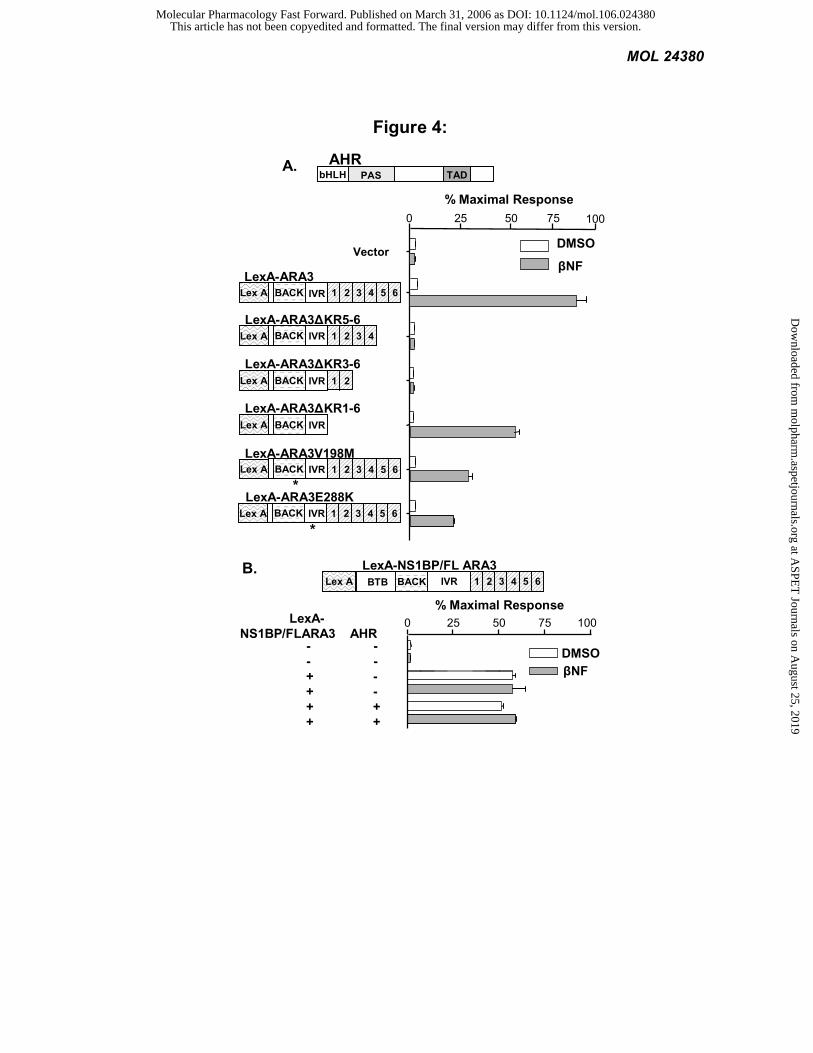
The BACK/IVR domains of NS1BP/FLARA3 also mediate the interaction with AHR: We also mapped the
NS1BP/FLARA3 domains that interact with AHR. In this series of experiments, we cloned the ARA3 deletions
described above into the vector pBTM116 to generate LexA fusions. These deletions constructs were then
cotransformed with the AHR in yeast. In this assay, reporter induction occurs when the LexA ARA3 fusions
physically interact with the AHR. As in Figure 2B, LexA-ARA3 interacts with AHR in a ligand-dependent manner. In
a manner similar to the pharmacology experiments in Figure 3C, subdeletions of the kelch domain significantly
disrupt the AHR-ARA3 interaction, whereas complete removal of the kelch domain reveals a mutant with near wild-
type interaction activity (comparing LexA-ARA3∆KR5-6 and 3-6 with LexA-ARA3∆KR1-6). Also similar to the
pharmacology experiments in Figure 3C, the two point mutations significantly inhibited the AHR-ARA3 interaction
by 70% (Figure 4A). We were unable to determine the role of the BTB domain of NS1BP on the interaction with
AHR, or whether NS1BP interacted with AHR at all, because LexA-NS1BP/FLARA3 activated the reporter
This article has not been copyedited and formatted. The final version may differ from this version.Molecular Pharmacology Fast Forward. Published on March 31, 2006 as DOI: 10.1124/mol.106.024380
at ASPE
T Journals on A
ugust 25, 2019m
olpharm.aspetjournals.org
Dow
nloaded from
MOL 24380
13
independent of its interaction with AHR or the presence of ligand (Figure 4B). The fact that expression of LexA-
NS1BP lead to reporter induction in the absence of ligand or TAD containing construct suggests that the BTB domain
might allow NS1BP to heterodimerize with other transcription factors containing TADs. In summary, Figures 3 and 4
demonstrate that the BACK/IVR domains of NS1BP/ARA3 are sufficient for the AHR-ARA3 interaction, as well as
the ability of ARA3 to modify AHR signaling.
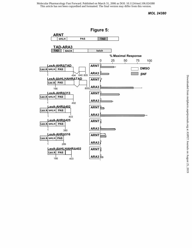
The C-terminus of the AHR PAS domain interacts with ARA3: We used a series of AHR deletions to
determine those regions important for its interaction with ARA3. We compared the AHR-ARA3 interaction to the
interaction of AHR with its known dimerization partner, ARNT. We observed that the C-terminus of the AHR PAS
domain, specifically AA 289-403, is essential for the AHR-ARA3 interaction. In contrast a distinct region, AA 380-
492, is required for the AHR-ARNT interaction in this system. The bHLH of AHR also is necessary for the AHR-
ARNT interaction, but its role in the AHR-ARA3 interaction is less clear. The bHLH inhibits ARA3’s interaction with
LexA-AHR∆TAD, but is necessary for ARA3 to interact with LexA-AHR∆402 (Figure 5). We previously have
demonstrated that AA 130-491 of AHR mediate the AHR-ARA9 interaction (Carver et al., 1998). Taken in sum, these
data suggest that the AHR uses overlapping, but distinct domains for interactions with ARA3, ARA9, and ARNT.
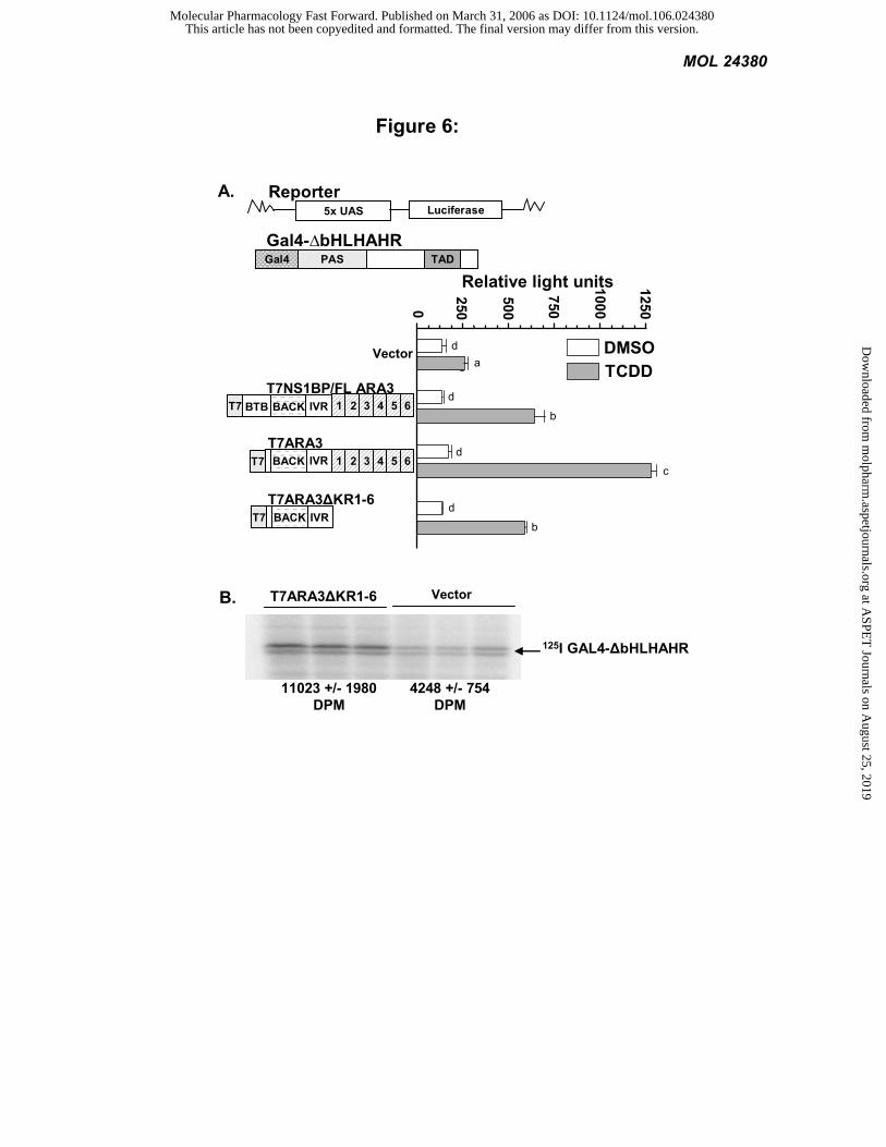
ARA3 increases signaling of an AHR chimera in mammalian cells: The influence of NS1BP/FLARA3 on
signaling by a Gal4-AHR fusion first was investigated in Cos-1 cells by transfections of a luciferase reporter gene
driven by the Gal4 promoter. In these experiments, the ∆bHLHAHR was fused to the DNA binding domain of Gal4
(Gal4-∆bHLHAHR) and cotransfected with T7ARA3, T7NS1BP/FLARA3, T7ARA3∆KR1-6 or vector control. The
addition of NS1BP/FLARA3 increased signaling of the AHR chimera in response to TCDD by more than 2-fold
(P<0.001). In agreement with the results of the LexA-AHR chimera in yeast, removal of the BTB domain increased
the modifier activity of ARA3 even further. That is, addition of the original ARA3 cDNA increased signaling of the
AHR chimera in mammalian cells by almost 5-fold (P<0.001) (Figure 6A). The fact that NS1BP was significantly less
effective on AHR chimera signaling than ARA3 provides further evidence that the BTB domain of ARA3 plays a
negative role in AHR signaling.
The yeast pharmacology experiments depicted in figure 2C are consistent with the idea that ARA3 increases
the concentration functional AHR in cells. An increase in AHR could account for the modifier activity of ARA3.
Given that the BACK/IVR domains of NS1BP are sufficient to mediate the AHR-ARA3 interaction and represent the
minimal effector domain of NS1BP, we tested the hypothesis that this region increases the concentration of functional
This article has not been copyedited and formatted. The final version may differ from this version.Molecular Pharmacology Fast Forward. Published on March 31, 2006 as DOI: 10.1124/mol.106.024380
at ASPE
T Journals on A
ugust 25, 2019m
olpharm.aspetjournals.org
Dow
nloaded from
MOL 24380
14
AHR in mammalian cells. To this end, we transfected Cos-1 cells with the Gal4-AHR chimera in the presence of the
NS1BP BACK/IVR domains or an empty vector and measured the amount of AHR with a radioactive ligand. That is,
with a saturating concentration of ligand, we observed a two-fold increase in the concentration of the AHR chimera in
the presence of the NS1BP BACK/IVR (P<0.05;Figure 6B). These results demonstrate that the BACK/IVR domains
of NS1BP do increase the functional concentration of AHR in the cytosol.
We have demonstrated that ARA3 binds AHR in a highly specific yeast-two-hybrid assay and enhances AHR
signaling in yeast, as well as mammalian cells. The dose response analysis suggested that ARA3 increases the
concentration of functional AHR. We have cloned full-length ARA3 and determined that it is the previously identified
NS1BP without the BTB domain. Through a series of deletions and mutants, we have shown that the BACK/IVR of
NS1BP is sufficient for its interaction with AHR and its effect on AHR signaling. In addition, we determined that the
C-terminus of AHR’s PAS domain is essential for the AHR-NS1BP interaction. Importantly, we have demonstrated
that deletion of the BTB domain significantly enhances the modifier activity NS1BP by increasing the cytosolic
concentration of AHR.
Model: Based upon the domain maps described above and what is known about other BTB-kelch proteins, a
model of NS1BP in AHR signaling can be proposed. The data presented here are consistent with the idea that
NS1BP/FLARA3 influences the cytosolic concentration of AHR by two mechanisms. First, NS1BP may tether AHR
to the actin cytoskeleton. In this regard, it has been shown that NS1BP colocalizes with actin and can bind actin
filaments through its kelch domain (Sasagawa et al., 2002). Second, the BTB domain of NS1BP may direct the
proteosomal degradation of AHR. The BTB domain in some kelch proteins interacts with Cullin3, a member of the E3
ubiquitin-ligase complex (Furukawa and Xiong, 2005; Stogios and Prive, 2004). In agreement with this model is the
observation that, in both yeast and mammalian cells, the NS1BP construct with a deletion of the BTB domain (i.e.,
ARA3) significantly increased AHR signaling compared to full length NS1BP. That is, ARA3, without the BTB
domain, may behave in a dominant-negative manner by competing with NS1BP. Likewise, the BACK/IVR domains
of NS1BP alone also may function in a dominant-negative manner, albeit less effectively than ARA3 with the kelch
domain that links it to the cytoplasm. In fact, we have shown that the BACK/IVR domains of NS1BP alone increase
the concentration of functional AHR. In summary, NS1BP modifies AHR signaling by both positively and negatively
influencing the concentration of AHR through the kelch and BTB domains, respectively.
This article has not been copyedited and formatted. The final version may differ from this version.Molecular Pharmacology Fast Forward. Published on March 31, 2006 as DOI: 10.1124/mol.106.024380
at ASPE
T Journals on A
ugust 25, 2019m
olpharm.aspetjournals.org
Dow
nloaded from
MOL 24380
15
REFERENCES
Adams J, Kelso R and Cooley L (2000) The kelch repeat superfamily of proteins: propellers of cell function. Trends
Cell Biol 10(1):17-24. Bartel PL, Chien C, Sternglanz R and Fields S (1993) Using the two-hybrid system to detect protein-protein
interactions., in Cellular interactions in development: A practical approach (Hartley DA ed) pp 153-179, IRL Press, Oxford.
Beischlag TV, Wang S, Rose DW, Torchia J, Reisz-Porszasz S, Muhammad K, Nelson WE, Probst MR, Rosenfeld MG and Hankinson O (2002) Recruitment of the NCoA/SRC-1/p160 family of transcriptional coactivators by the aryl hydrocarbon receptor/aryl hydrocarbon receptor nuclear translocator complex. Mol Cell Biol 22(12):4319-4333.
Bell DR and Poland A (2000) Binding of aryl hydrocarbon receptor (AhR) to AhR-interacting protein. The role of hsp90. Journal of Biological Chemistry 275(46):36407-36414.
Bourne H and von Zastrow M (2001) Drug Receptors and Pharmacodynamics, in Basic and Clinical Pharmacology (Katzung B ed) pp 9-34, Lange Medical Books/McGraw-Hill, New York.
Bradfield CA, Kende AS and Poland A (1988) Kinetic and equilibrium studies of Ah receptor-ligand binding: use of [125I]2-iodo-7,8-dibromodibenzo-p-dioxin. Mol Pharmacol 34(2):229-237.
Carver LA and Bradfield CA (1997) Ligand dependent interaction of the Ah receptor with a novel immunophilin homolog in vivo. J Biol Chem 272(17):11452-11456.
Carver LA, Jackiw V and Bradfield CA (1994) The 90-kDa heat shock protein is essential for Ah receptor signaling in a yeast expression system. J Biol Chem 269:30109-30112.
Carver LA, LaPres JJ, Jain S, Dunham EE and Bradfield CA (1998) Characterization of the Ah receptor-associated protein, ARA9. J Biol Chem 273(50):33580-33587.
Collins T, Stone JR and Williams AJ (2001) All in the family: the BTB/POZ, KRAB, and SCAN domains. Mol Cell Biol 21(11):3609-3615.
Davarinos NA and Pollenz RS (1999) Aryl hydrocarbon receptor imported into the nucleus following ligand binding is rapidly degraded via the cytosplasmic proteasome following nuclear export. Journal of Biological Chemistry 274(40):28708-28715.
Dolwick KM, Schmidt JV, Carver LA, Swanson HI and Bradfield CA (1993) Cloning and expression of a human Ah receptor cDNA. Mol Pharmacol 44(5):911-917.
Fernandez-Salguero PM, Hilbert DM, Rudikoff S, Ward JM and Gonzalez FJ (1996) Aryl-hydrocarbon receptor-deficient mice are resistant to 2,3,7,8-tetrachlorodibenzo-p-dioxin-induced toxicity. Toxicology & Applied Pharmacology 140(1):173-179.
Furukawa M and Xiong Y (2005) BTB protein Keap1 targets antioxidant transcription factor Nrf2 for ubiquitination by the Cullin 3-Roc1 ligase. Mol Cell Biol 25(1):162-171.
Garabedian MJ (1993) Genetic Approaches to Mammalian Nuclear Receptor Function in Yeast. Methods: A Companion to Methods in Enzymology 5:138-146.
Hogenesch JB, Chan WK, Jackiw VH, Brown RC, Gu YZ, Pray-Grant M, Perdew GH and Bradfield CA (1997) Characterization of a subset of the basic-helix-loop-helix-PAS superfamily that interacts with components of the dioxin signaling pathway. J Biol Chem 272(13):8581-8593.
Jain S, Dolwick KM, Schmidt JV and Bradfield CA (1994) Potent transactivation domains of the Ah receptor and the Ah receptor nuclear translocator map to their carboxyl termini. J Biol Chem 269(50):31518-31524.
Kazlauskas A, Poellinger L and Pongratz I (1999) Evidence that the co-chaperone p23 regulates ligand responsiveness of the dioxin (Aryl hydrocarbon) receptor. Journal of Biological Chemistry 274(19):13519-13524.
Kozak M (1987) An analysis of 5'-noncoding sequences from 699 vertebrate messenger RNAs. Nucleic Acids Res 15(20):8125-8132.
Kumar MB, Tarpey RW and Perdew GH (1999) Differential recruitment of coactivator RIP140 by Ah and estrogen receptors. Absence of a role for LXXLL motifs. J Biol Chem 274(32):22155-22164.
Lahvis GP, Pyzalski RW, Glover E, Pitot HC, McElwee MK and Bradfield CA (2005) The aryl hydrocarbon receptor is required for developmental closure of the ductus venosus in the neonatal mouse. Mol Pharmacol 67(3):714-720.
LaPres JJ, Glover E, Dunham EE, Bunger MK and Bradfield CA (2000) ARA9 modifies agonist signaling through an increase in cytosolic aryl hydrocarbon receptor. J Biol Chem 275(9):6153-6159.
This article has not been copyedited and formatted. The final version may differ from this version.Molecular Pharmacology Fast Forward. Published on March 31, 2006 as DOI: 10.1124/mol.106.024380
at ASPE
T Journals on A
ugust 25, 2019m
olpharm.aspetjournals.org
Dow
nloaded from
MOL 24380
16
Ma Q and Whitlock JP, Jr. (1997) A novel cytoplasmic protein that interacts with the Ah receptor, contains tetratricopeptide repeat motifs, and augments the transcriptional response to 2,3,7,8-tetrachlorodibenzo-p-dioxin. J Biol Chem 272(14):8878-8884.
Melnick A, Carlile G, Ahmad KF, Kiang CL, Corcoran C, Bardwell V, Prive GG and Licht JD (2002) Critical residues within the BTB domain of PLZF and Bcl-6 modulate interaction with corepressors. Mol Cell Biol 22(6):1804-1818.
Meyer BK, Pray-Grant MG, Vanden Heuvel JP and Perdew GH (1998) Hepatitis B virus X-associated protein 2 is a subunit of the unliganded aryl hydrocarbon receptor core complex and exhibits transcriptional enhancer activity. Molecular & Cellular Biology 18(2):978-988.
Mimura J, Ema M, Sogawa K and Fujii-Kuriyama Y (1999) Identification of a novel mechanism of regulation of Ah (dioxin) receptor function. Genes & Development 13(1):20-25.
Petrulis JR, Hord NG and Perdew GH (2000) Subcellular Localization of the Aryl Hydrocarbon Receptor Is Modulated by the Immunophilin Homolog Hepatitis B Virus X-associated Protein 2. J Biol Chem 275(48):37448-37453.
Pohjanvirta R and Tuomisto J (1994) Short-term toxicity of 2,3,7,8-tetrachlorodibenzo-p-dioxin in laboratory animals: Effects, mechanisms, and animal models. Pharmacol Rev 46(4):483-549.
Reyes H, Reisz-Porszasz S and Hankinson O (1992) Identification of the Ah receptor nuclear translocator protein (Arnt) as a component of the DNA binding form of the Ah receptor. Science 256(5060):1193-1195.
Sasagawa K, Matsudo Y, Kang M, Fujimura L, Iitsuka Y, Okada S, Ochiai T, Tokuhisa T and Hatano M (2002) Identification of Nd1, a novel murine kelch family protein, involved in stabilization of actin filaments. J Biol Chem 277(46):44140-44146.
Schmidt JV and Bradfield CA (1996) Ah receptor signaling pathways. Annu Rev Cell Dev Biol 12:55-89. Stogios PJ and Prive GG (2004) The BACK domain in BTB-kelch proteins. Trends Biochem Sci 29(12):634-637. Vojtek AB, Hollenberg SM and Cooper JA (1993) Mammalian Ras interacts directly with the serine/threonine kinase
Raf. Cell 74(1):205-214. Wilhelmsson A, Cuthill S, Denis M, Wikstrom AC, Gustafsson JA and Poellinger L (1990) The specific DNA binding
activity of the dioxin receptor is modulated by the 90 kd heat shock protein. EMBO J 9(1):69-76. Wolff T, O'Neill RE and Palese P (1998) NS1-Binding protein (NS1-BP): a novel human protein that interacts with
the influenza A virus nonstructural NS1 protein is relocalized in the nuclei of infected cells. J Virol 72(9):7170-7180.
Yao G, Craven M, Drinkwater N and Bradfield CA (2004) Interaction networks in yeast define and enumerate the signaling steps of the vertebrate aryl hydrocarbon receptor. PLoS Biol 2(3):E65.
Zollman S, Godt D, Prive GG, Couderc JL and Laski FA (1994) The BTB domain, found primarily in zinc finger proteins, defines an evolutionarily conserved family that includes several developmentally regulated genes in Drosophila. Proc Natl Acad Sci U S A 91(22):10717-10721.
This article has not been copyedited and formatted. The final version may differ from this version.Molecular Pharmacology Fast Forward. Published on March 31, 2006 as DOI: 10.1124/mol.106.024380
at ASPE
T Journals on A
ugust 25, 2019m
olpharm.aspetjournals.org
Dow
nloaded from
MOL 24380
17
FOOTNOTES
This work was supported by the National Institutes of Health grants R37-ES005703, T32-CA009135, and P30-
CA014520.
Please send all reprint requests to: Christopher A. Bradfield, McArdle Laboratory for Cancer Research, 1400
University Avenue, Madison, WI 53706, Email: [email protected]
1 The sequences of ARA3 and NS1BP/FLARA3 have been submitted to GenBank and have the respective accession
numbers: DQ443529and DQ443528.
This article has not been copyedited and formatted. The final version may differ from this version.Molecular Pharmacology Fast Forward. Published on March 31, 2006 as DOI: 10.1124/mol.106.024380
at ASPE
T Journals on A
ugust 25, 2019m
olpharm.aspetjournals.org
Dow
nloaded from
MOL 24380
18
FIGURE LEGENDS
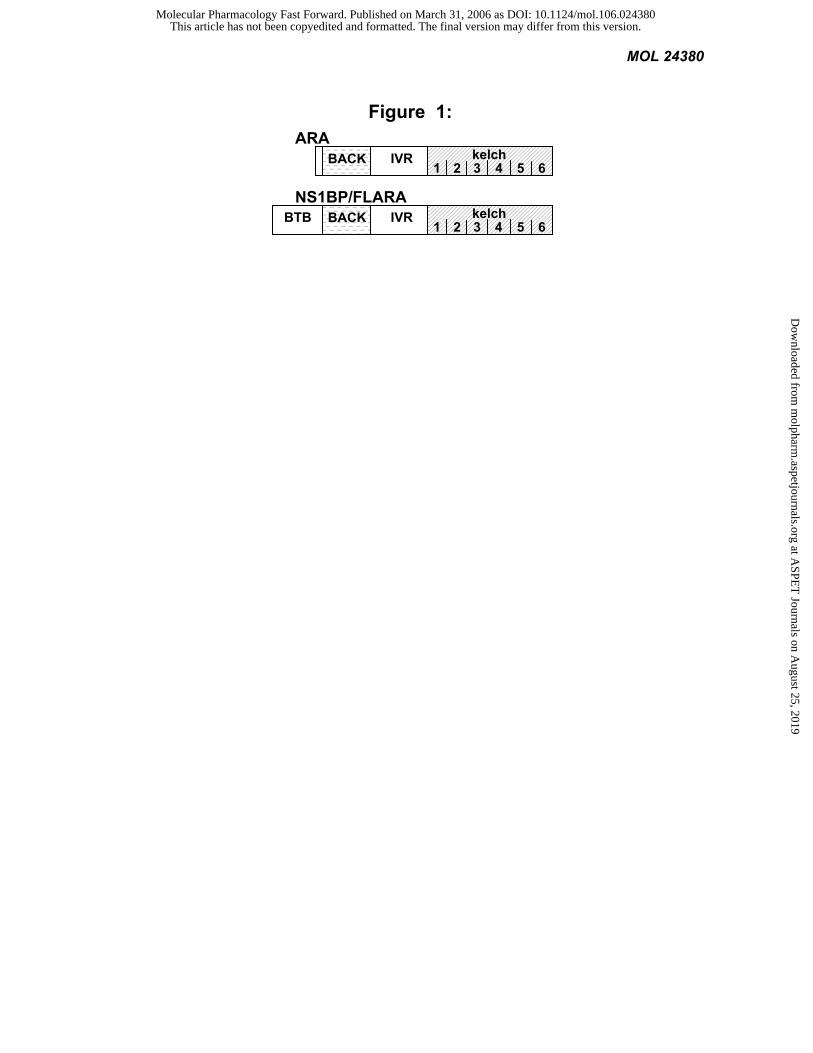
Figure 1: Analysis of the ARA3 cDNA revealed three characterized protein domains. The ARA3 yeast two
hybrid clones originally identified in our screen encoded a 531 amino acid (AA) protein. There was no Kozak
consensus initiation ATG present. Therefore the human full-length ARA3 (FLARA3) was cloned from a human heart
cDNA library and sequenced. FLARA3 is 642 AA in length, while the original ARA3 clone was missing the first 111
AA. Similarity searches identified FLARA3 as a protein originally called NS1BP.The NS1BP/FLARA3 protein
contains a BTB domain from AA 22-129, a BACK domain from AA 134-233, and a kelch domain consisting of six
imperfect 50 AA kelch repeats from AA 357-635 (http://www.sanger.ac.uk). A low homology region exists between
the BACK and kelch domains that we refer to as the intervening region (IVR).
Figure 2: ARA3 interacts with AHR ligand-dependently and modifies the AHR signaling pathway. The
L40 yeast, expressing an integrated β-galactosidase (lacZ) reporter, were transformed with constructs depicted above
each graph and treated with the appropriate ligand or vehicle. LacZ activity was determined and normalized to the
number of cells. Values are the average of three replicates and represent at least two independent experiments. A)
Yeast-two-hybrid assay. LexA-AHR∆TAD, TAD-ARA3, or control vectors were transformed in yeast and treated
with either 1 mM βNF or vehicle. B) A reverse-two-hybrid assay was performed as in A. C) Aryl hydrocarbon
receptor pharmacology. Yeast were transformed with the LexA-AHR chimera and ARA3 or control vector and treated
with increasing concentrations of βNF. D) Glucocorticoid receptor pharmacology. Yeast were transformed with the
LexA-GR chimera and ARA3 or control vector and treated with increasing concentrations of DOC.
Figure 3: The BACK/IVR domains of NS1BP/FLARA3 modify AHR signaling. The L40 yeast, expressing
an integrated β-galactosidase (lacZ) reporter, were transformed with the AHR chimera LexA-∆bHLHAHR and T7-
tagged ARA3 constructs or a vector control. LacZ activity was determined and normalized to the number of cells.
Values are the average of three replicates and represent at least two independent experiments. A) AHR pharmacology.
Yeast were transformed with the LexA-AHR chimera and ARA3 constructs or vector control and grown in the
presence of 1 mM βNF or vehicle. Statistical significance was determined with ANOVA followed by Tukey’s
multiple comparison test in PRISM. The same subscript depicts values that are statistically the same (P>0.05).
Different subscripts designate a statistical difference in the values (P<0.01). B) Expression of ARA3 mutants. ARA3
point mutations were generated with hydroxylamine and screened as described in the Methods. A schematic of the
mutants is shown. To confirm protein expression levels of the mutant ARA3, a western blot was performed on L40
This article has not been copyedited and formatted. The final version may differ from this version.Molecular Pharmacology Fast Forward. Published on March 31, 2006 as DOI: 10.1124/mol.106.024380
at ASPE
T Journals on A
ugust 25, 2019m
olpharm.aspetjournals.org
Dow
nloaded from
MOL 24380
19
yeast extracts. C) Aryl hydrocarbon receptor pharmacology. The two ARA3 point mutants, an empty vector or ARA3
were transformed with LexA-∆bHLHAHR exposed to increasing amounts of βNF.
Figure 4: The BACK/IVR domains of NS1BP/FLARA3 also mediate the interaction with AHR. The L40
yeast, expressing an integrated β-galactosidase (lacZ) reporter, were transformed with full-length AHR and LexA
fusions of the ARA3 constructs or a vector control. The yeast were exposed to 1 mM ßNF or vehicle. LacZ activity
was determined and normalized to the number of cells. Values are the average of three replicates and represent at least
two independent experiments. A) Yeast-two-hybrid assay. Location of point mutations is depicted by an asterisk. B)
Control yeast-two-hybrid assay.
Figure 5: The C-terminus of AHR’s PAS domain is necessary for the interaction with ARA3. The L40
yeast, expressing an integrated β-galactosidase (lacZ) reporter, were transformed with full-length ARNT or TAD-
ARA3 and LexA fusions of a AHR deletion series. The yeast were exposed to 1 mM ßNF or vehicle. LacZ activity
was determined and normalized to the number of cells. Values are the average of three replicates and represent at least
two independent experiments. The regions of AHR important for its interaction with ARA3 and ARNT were
compared.
Figure 6: ARA3 modifies signaling of an AHR chimera in mammalian cells. A) Cos-1 cells were transiently
transfected with ∆bHLHAHR fused to the GAL4 DNA binding domain (Gal4-∆bHLHAHR), a luciferase reporter,
and a ß-galactosidase transfection control vector. In addition NS1BP/FLARA3, ARA3, ARA3∆K1-6, or an empty
vector was transfected into the Cos-1 cells. The cells were treated with 10 nM TCDD or vehicle, and Luciferase and
LacZ activity was determined. Luciferase activity was normalized to LacZ activity. Values are the average of two
replicates and represent at least two independent experiments. Statistical significance was determined with ANOVA
followed by Tukey’s multiple comparison test in PRISM. The same subscript depicts values that are statistically the
same (P>0.05). Different subscripts designate a statistical difference in the values (P<0.001). B) Cos-1 cells were
transiently transfected with the Gal4-AHR chimera in the presence of ARA3∆K1-6 or an empty vector. Cytosolic
protein was labeled with the photoaffinity AHR ligand and the samples were run on a 7.5% SDS-PAGE gel. Gels
were analyzed by autoradiography and appropriate bands were cut out and counted on a Minaxi γ gamma counter.
Statistical Significance was determined with a Student’s t-test (P<0.05).
This article has not been copyedited and formatted. The final version may differ from this version.Molecular Pharmacology Fast Forward. Published on March 31, 2006 as DOI: 10.1124/mol.106.024380
at ASPE
T Journals on A
ugust 25, 2019m
olpharm.aspetjournals.org
Dow
nloaded from
MOL 24380
Figure 1: ARA
BACK IVR 1 2 53 4
kelch 6
BACK NS1BP/FLARA
IVR 1 2 53 4
kelch BTB 6
This article has not been copyedited and formatted. The final version may differ from this version.Molecular Pharmacology Fast Forward. Published on March 31, 2006 as DOI: 10.1124/mol.106.024380
at ASPE
T Journals on A
ugust 25, 2019m
olpharm.aspetjournals.org
Dow
nloaded from
MOL 24380
Figure 2:
B.
TADbHLH PASAHR
LexA-ARA3LexA
LacZLexA-Op*8Reporter
kelchBACK
D.TAD Lex A LBD
N-LexA-C GR
ARA3 kelchBACK
LacZLexA-Op*8Reporter
GR
Concentration of DOC [M]10 -7 10 -6 10 -5 10 -4 10 -3 10 -2
vectorARA3
0
25
50
75
100
% M
axim
al R
espo
nse
0
20
40
60
80
100
% M
axim
al R
espo
nse
BaitFish
+ + + + - -- - + + + +
DMSOβNF
IVRA.
0
25
50
75
100
% M
axim
al R
espo
nse
BaitFish
+ + + + - -- - + + + +
Lex A PASLexA-AHR∆TAD
TAD-ARA3 bHLH
TAD kelchBACK
LacZLexA-Op*8Reporter
C.TADLex A PAS
LexA-∆bHLHAHR
ARA3 kelchBACK
LacZLexA-Op*8Reporter
vectorARA3
AHR
10 -7 10 -6 10 -5 10 -4 10 -3 10-20
20
40
60
80
100
Concentration of βNF [M]
% M
axim
al R
espo
nse
DMSOβNF
IVR
IVR IVR
This article has not been copyedited and formatted. The final version may differ from this version.Molecular Pharmacology Fast Forward. Published on March 31, 2006 as DOI: 10.1124/mol.106.024380
at ASPE
T Journals on A
ugust 25, 2019m
olpharm.aspetjournals.org
Dow
nloaded from
MOL 24380
Figure 3:
C.
ARA3E288K
ARA3V198M
ARA3vector
10-7 10-6 10 -5 10-4 10 -3 10 -2
Concentration of βNF [M]
0
20
40
60
80
100
% M
axim
al R
espo
nse vector
ARA3ARA3V198MARA3E288K
B.Lex A BACK 1 2 63 4
*Lex A BACK 1 2 5 63 4
5
*
T7ARA3V198M
T7ARA3E288K
IVR
IVR
% Maximal Response
DMSO
βNF
DMSO
βNF
1 2
A.0 25 50 75 100TADLex A PAS
LexA-∆bHLHAHR
T7ARA3∆KR3-6
T7ARA3∆KR1-6
Vector
BTBT7 BACK 1 2 5 63 4T7NS1BP/FL ARA3
IVR
T7 BACK 1 2 5 63 4T7ARA3
IVR
1 2 3 4T7ARA3∆KR5-6
a
a
b
c
b
d
e
e
e
e
e
e
T7 BACK
T7 BACK
T7 BACK
IVR
IVR
IVR
This article has not been copyedited and formatted. The final version may differ from this version.Molecular Pharmacology Fast Forward. Published on March 31, 2006 as DOI: 10.1124/mol.106.024380
at ASPE
T Journals on A
ugust 25, 2019m
olpharm.aspetjournals.org
Dow
nloaded from
MOL 24380
Figure 4:
- -- -+ -+ -+ ++ +
NS1BP/FLARA3 AHR0 25 50 75 100
% Maximal ResponseLexA-
DMSO
βNF
TADbHLH PASAHR
DMSOβNF
0 25 50 75 100% Maximal Response
B.
A.
Vector
BTB BACK 1 2 5 63 4Lex ALexA-NS1BP/FL ARA3
IVR
DMSOβNF
1 2 5 63 4
1 2 3 4
1 2
1 2 63 4
*1 2 5 63 4
5
LexA-ARA3
LexA-ARA3∆KR5-6
LexA-ARA3∆KR3-6
LexA-ARA3∆KR1-6
*
LexA-ARA3V198M
LexA-ARA3E288K
Lex A BACK IVR
Lex A BACK IVR
Lex A BACK IVR
Lex A BACK IVR
Lex A BACK IVR
Lex A BACK IVR
This article has not been copyedited and formatted. The final version may differ from this version.Molecular Pharmacology Fast Forward. Published on March 31, 2006 as DOI: 10.1124/mol.106.024380
at ASPE
T Journals on A
ugust 25, 2019m
olpharm.aspetjournals.org
Dow
nloaded from
MOL 24380
Figure 5:
494 6401
492
TAD-ARA3
Lex A PASbHLH
PAS
166
403
380
289
403
ARNTTADbHLH PAS
0 25 50 75 100% Maximal Response
TAD kelchBACK
LexA-AHR∆TAD
LexA-∆bHLHAHR∆TAD
LexA-AHR∆313
LexA-AHR∆402
LexA-AHR∆425
LexA-AHR∆516
LexA-∆bHLHAHR∆402
DMSO
βNF805
805
ARNT
ARA3
ARNT
ARA3
ARNT
ARA3
ARNT
ARA3
ARNT
ARA3
ARNT
ARA3
ARNT
ARA3
Lex A
1
Lex A PASbHLH
1
Lex A PASbHLH
1
Lex A PASbHLH
1
Lex A PASbHLH
PAS
166
Lex A
This article has not been copyedited and formatted. The final version may differ from this version.Molecular Pharmacology Fast Forward. Published on March 31, 2006 as DOI: 10.1124/mol.106.024380
at ASPE
T Journals on A
ugust 25, 2019m
olpharm.aspetjournals.org
Dow
nloaded from
MOL 24380
Figure 6:
Luciferase5x UASReporter
Relative light unitsTADGal4 PAS
Gal4-∆bHLHAHR
0
250
500
750
1000
1250
Vector DMSOTCDDa
b
c
d
b
d
d
dT7ARA3∆KR1-6T7 BACK IVR
BTBT7 BACK 1 2 5 63 4T7NS1BP/FL ARA3
IVR
T7 BACK 1 2 5 63 4T7ARA3
IVR
125I GAL4-∆bHLHAHR
VectorT7ARA3∆KR1-6
A.
B.
11023 +/- 1980DPM
4248 +/- 754DPM
This article has not been copyedited and formatted. The final version may differ from this version.Molecular Pharmacology Fast Forward. Published on March 31, 2006 as DOI: 10.1124/mol.106.024380
at ASPE
T Journals on A
ugust 25, 2019m
olpharm.aspetjournals.org
Dow
nloaded from