Embed Size (px)
Citation preview
The body plan of the cnidarian medusa: distinct differences in positional
origins of polyp tentacles and medusa tentacles
Hiroshi Shimizu�,a and Hiroshi Namikawab
aDepartment of Developmental Genetics, National Institute of Genetics, 1111 Yata, Mishima, 411-8540 Shizuoka, JapanbShowa Memorial Institute, National Museum of Nature and Science, 4-1-1 Amakubo, Tsukuba, 305-0005, Ibaraki, Japan�Author for correspondence (email: [email protected])
Cnidarians have two typical body forms, the attached form
(polyp) and the swimming form (medusa) (Fig. 1). The polyp
has a mouth with tentacles closely surrounding it at the apical
end of a cylindrical body column (Fig. 1A). The morphology
of a medusa is characterized by a body like an inverted bowl,
termed the umbrella (Fig. 1B). The umbrella has tentacles
hanging down from its margin (marginal tentacles). The
mouth is located at the end of a columnar structure termed
the manubrium, which is located at the center of the umbrella.
Despite these significant differences in morphology, zoology
textbooks typically state that ‘‘the medusa is the upside down
form of a polyp,’’ implying that the two forms have basically
the same body plan (Ruppert and Barnes 1996). As a result, it
has generally been assumed that the tentacles of the polyp and
the marginal tentacles of the medusa are homologous. How-
ever, it remains unknown whether the textbooks’ description
is correct or not. Based upon classical observations and recent
results of molecular studies, we present a simple but novel
answer to this question by showing that animals that belong
to Cnidaria have two areas in the body where tentacles
are formed, one at the oral end, and the other in an aboral
location on the animal.
We focused our analysis on the differences in positional
origins of hydropolyp tentacles and hydromedusa marginal
tentacles in this study. Although the medusa form occurs
in three cnidarian classes (viz., Scyphozoa, Cubozoa, and
Hydrozoa), of these about 75% of the species that have the
medusa stage belong to Hydrozoa (Brusca and Brusca 1990;
Boero et al. 1992; Bouillon and Boero 2000). Furthermore,
Hydrozoa is currently the only class where molecular analysis
has been performed, although extensive gene expression stud-
ies have been carried out mostly with polyps.
The term tentacle is generally used to describe the flexible
appendages that are located near the mouth opening in cnid-
arians and are involved in capturing prey using nematocytes
(Ewer 1947). If a tentacle produced in the polyp showed
developmental continuity into the medusa, this would be
direct evidence that the polyp tentacles and the medusa
tentacles are essentially the same structure. This continuity,
however, is not seen. Instead, medusa tentacles are newly
formed during the process of medusa formation (namely,
asexual budding in Hydrozoa).
The morphology ofHydra, a hydrozoan research model, is
characterized by the solitary polyp form with the hypostome
surrounded by tentacles at the oral end, a large body column/
gastric region, and with the peduncle and foot region at the
aboral end (Fig. 1C). Hydra has been considered to represent
the typical solitary polyp form of this class. However, some
hydrozoans have polyps in which tentacles emerge in two
regions. A polyp of the hydrozoan family Corymorphidae
(Fig. 1D) has tentacles that are at a glance concentrated at
the oral end, suggesting that the morphology of the animal
is basically similar to Hydra. This view is, however, entirely
erroneous. The digestive region (denoted by DR) of a hydra
polyp, which is pink in color because of digested food content,
occupies about 3/4 of the body column length, whereas the rest
(about 1/4) corresponds to the peduncle region (Fig. 1C). In
contrast, the digestive region of a corymorphid polyp is the
region that lies between the two tentacle rings occupying only
about 1/10 of the body. The rest of the polyp (about 9/10) is
occupied by the peduncle region (Fig. 1D), demonstrating that
the two polyps have different proportions of the body parts in
apparently similar cylindrical morphology. Therefore, the po-
sition of the secondary tentacle ring of the corymorphid polyp
corresponds, in a hydra polyp, to the boundary between the
digestive region and the peduncle region, which is located on
the aboral side of the animal. Because the secondary tentacle
ring of the corymorphid polyp is located aboral to the digestive
region of the animal, the tentacles in the secondary ring should
be considered aboral tentacles. This implies that there are two
tentacle regions in the corymorphid polyp, one on the oral side
of the digestive region and the other in the aboral side of this
region. It should be noted that the tentacles in the two regions
are involved in capturing prey in a cooperative manner.
When three subclasses of Hydrozoa (Limnomedusae,
Anthmedusae [Athecata], and Leptomedusae [Thecata]) are
EVOLUTION & DEVELOPMENT 11:6, 619 –621 (2009)
DOI: 10.1111/j.1525-142X.2009.00368.x
& 2009 Wiley Periodicals, Inc. 619
surveyed based upon the results of Petersen (Petersen 1990)
according to the position of tentacles on the polyp form
termed hydropolyp, we find that in the Limnomedusae and
Leptomedusae, and order Filifera of Anthomedusae, tentacles
occur solely as one tentacle ring closely surrounding the
mouth of polyps, that is as oral tentacles. In contrast, we
find in the order Capitata of subclass Anthomedusae that
the majority of animals have tentacle rings in two regions of
hydropolyps, whereas some like Hydra have only one ring.
Despite this diversity, hydropolyps tend to be considered as
polyps having only one tentacle region at the oral end, as seen
with Hydra.
Formation of a hydromedusa occurs by an asexual repro-
ductive process termed budding (Boero et al. 1992). A min-
iature medusa-like structure is formed on the side of the body
column of a polyp. When this miniature medusa detaches
and becomes independent, it grows directly into the mature
medusa form. It is often the case in a hydromedusa that ten-
tacles are formed in two regions, first around the mouth as
‘‘oral tentacles’’ and second around the margin of the um-
brella as ‘‘marginal tentacles’’ (Fig. 1E). The oral tentacles of
hydromedusae are usually thinner and shorter than marginal
tentacles, and hence are sometimes unnoticed or ignored.
The formation of tentacles in two regions that occurs in
the hydropolyp and hydromedusa is, in fact, observed even in
the juvenile form of the animal termed the planula larva that
appears during development from the fertilized embryo to
the polyp form in some but not all species of hydrozoans. In
Tubularia mesembryanthemum for example, a larva having
tentacles on opposite sides of the animal appears (Fig. 1F)
and is termed an actinula (Hawes 1958). When the actinula
larva metamorphoses into a polyp, the short tentacles develop
into the oral tentacles of the polyp whereas the long tentacles
on the other side develop into the aboral tentacles, suggesting
that hydrozoans have two regions of tentacle formation in a
wide range of developmental stages.
We have presented several cases where there are two
tentacle regions in the polyp, the medusa, and the actinula
larva. As a natural outcome, the possibility arises that the oral
and aboral tentacles in the polyp are homologous to the oral
and marginal tentacles in the medusa. There is currently little
information on the molecular biology of medusae that is
relevant to this hypothesis. However, a study of one gene,
which encodes the homodomain-containing protein Otx, does
provide insight into the relationship between polyp and
medusa tentacles.
Otx plays a crucial role in specifying the forebrain and
midbrain of vertebrates (Millet et al. 1996). CnOtx, an
orthologue of Otx in Hydra, is expressed in the epithelial cells
in the digestive region of the body column (Smith et al. 1999).
If the pattern of CnOtx expression in hydropolyps that have
two tentacle regions is similar to the pattern in Hydra, the
predicted area of expression is the area between the two
tentacle regions. In Podocoryne carnea a marine hydrozoan,
the muscular tissue in the inner surface of the umbrella
expresses the Otx orthologue (Muller et al. 1999). This area of
tissue is located between the oral tentacles and the marginal
tentacles. Thus, the two tentacle regions, one in the polyp and
the other in the medusa, sandwich the CnOtx expressing tis-
sue. These results are consistent with the view that the tentacle
rings in the polyp and the medusa are homologous structures.
Based upon this view, we can simulate a virtual surgical
operation on a medusa (Fig. 2). By removing the thick ex-
tracellular matrix layer that resides in the extracellular space
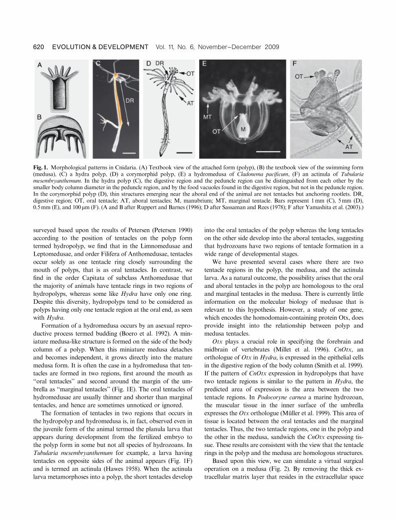
Fig. 1. Morphological patterns in Cnidaria. (A) Textbook view of the attached form (polyp), (B) the textbook view of the swimming form(medusa), (C) a hydra polyp, (D) a corymorphid polyp, (E) a hydromedusa of Cladonema pacificum, (F) an actinula of Tubulariamesembryanthemum. In the hydra polyp (C), the digestive region and the peduncle region can be distinguished from each other by thesmaller body column diameter in the peduncle region, and by the food vacuoles found in the digestive region, but not in the peduncle region.In the corymorphid polyp (D), thin structures emerging near the aboral end of the animal are not tentacles but anchoring rootlets. DR,digestive region; OT, oral tentacle; AT, aboral tentacles; M, manubrium; MT, marginal tentacle. Bars represent 1mm (C), 5mm (D),0.5mm (E), and 100mm (F). (A and B after Ruppert and Barnes (1996); D after Sassaman and Rees (1978); F after Yamashita et al. (2003).)
620 EVOLUTION & DEVELOPMENT Vol. 11, No. 6, November--December 2009
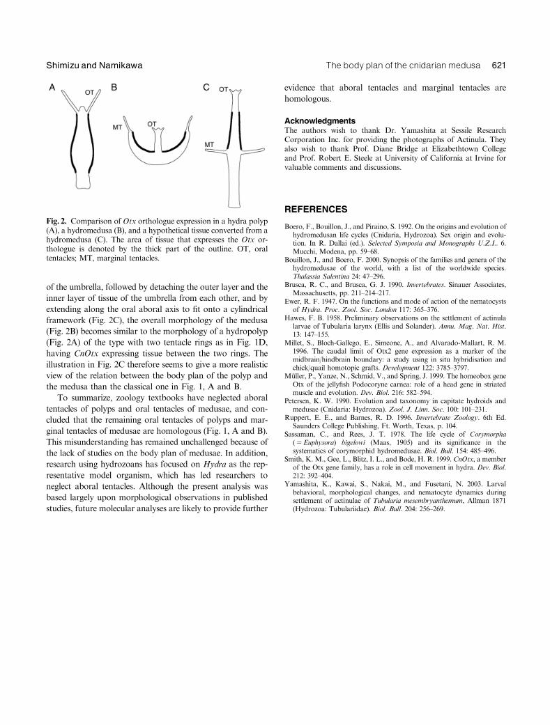
of the umbrella, followed by detaching the outer layer and the
inner layer of tissue of the umbrella from each other, and by
extending along the oral aboral axis to fit onto a cylindrical
framework (Fig. 2C), the overall morphology of the medusa
(Fig. 2B) becomes similar to the morphology of a hydropolyp
(Fig. 2A) of the type with two tentacle rings as in Fig. 1D,
having CnOtx expressing tissue between the two rings. The
illustration in Fig. 2C therefore seems to give a more realistic
view of the relation between the body plan of the polyp and
the medusa than the classical one in Fig. 1, A and B.
To summarize, zoology textbooks have neglected aboral
tentacles of polyps and oral tentacles of medusae, and con-
cluded that the remaining oral tentacles of polyps and mar-
ginal tentacles of medusae are homologous (Fig. 1, A and B).
This misunderstanding has remained unchallenged because of
the lack of studies on the body plan of medusae. In addition,
research using hydrozoans has focused on Hydra as the rep-
resentative model organism, which has led researchers to
neglect aboral tentacles. Although the present analysis was
based largely upon morphological observations in published
studies, future molecular analyses are likely to provide further
evidence that aboral tentacles and marginal tentacles are
homologous.
AcknowledgmentsThe authors wish to thank Dr. Yamashita at Sessile ResearchCorporation Inc. for providing the photographs of Actinula. Theyalso wish to thank Prof. Diane Bridge at Elizabethtown Collegeand Prof. Robert E. Steele at University of California at Irvine forvaluable comments and discussions.
REFERENCES
Boero, F., Bouillon, J., and Piraino, S. 1992. On the origins and evolution ofhydromedusan life cycles (Cnidaria, Hydrozoa). Sex origin and evolu-tion. In R. Dallai (ed.). Selected Symposia and Monographs U.Z.I.. 6.Mucchi, Modena, pp. 59–68.
Bouillon, J., and Boero, F. 2000. Synopsis of the families and genera of thehydromedusae of the world, with a list of the worldwide species.Thalassia Salentina 24: 47–296.
Brusca, R. C., and Brusca, G. J. 1990. Invertebrates. Sinauer Associates,Massachusetts, pp. 211–214–217.
Ewer, R. F. 1947. On the functions and mode of action of the nematocystsof Hydra. Proc. Zool. Soc. London 117: 365–376.
Hawes, F. B. 1958. Preliminary observations on the settlement of actinulalarvae of Tubularia larynx (Ellis and Solander). Annu. Mag. Nat. Hist.13: 147–155.
Millet, S., Bloch-Gallego, E., Simeone, A., and Alvarado-Mallart, R. M.1996. The caudal limit of Otx2 gene expression as a marker of themidbrain/hindbrain boundary: a study using in situ hybridisation andchick/quail homotopic grafts. Development 122: 3785–3797.
Muller, P., Yanze, N., Schmid, V., and Spring, J. 1999. The homeobox geneOtx of the jellyfish Podocoryne carnea: role of a head gene in striatedmuscle and evolution. Dev. Biol. 216: 582–594.
Petersen, K. W. 1990. Evolution and taxonomy in capitate hydroids andmedusae (Cnidaria: Hydrozoa). Zool. J. Linn. Soc. 100: 101–231.
Ruppert, E. E., and Barnes, R. D. 1996. Invertebrate Zoology. 6th Ed.Saunders College Publishing, Ft. Worth, Texas, p. 104.
Sassaman, C., and Rees, J. T. 1978. The life cycle of Corymorpha(5Euphysora) bigelowi (Maas, 1905) and its significance in thesystematics of corymorphid hydromedusae. Biol. Bull. 154: 485–496.
Smith, K. M., Gee, L., Blitz, I. L., and Bode, H. R. 1999. CnOtx, a memberof the Otx gene family, has a role in cell movement in hydra. Dev. Biol.212: 392–404.
Yamashita, K., Kawai, S., Nakai, M., and Fusetani, N. 2003. Larvalbehavioral, morphological changes, and nematocyte dynamics duringsettlement of actinulae of Tubularia mesembryanthemum, Allman 1871(Hydrozoa: Tubulariidae). Biol. Bull. 204: 256–269.
Fig. 2. Comparison of Otx orthologue expression in a hydra polyp(A), a hydromedusa (B), and a hypothetical tissue converted from ahydromedusa (C). The area of tissue that expresses the Otx or-thologue is denoted by the thick part of the outline. OT, oraltentacles; MT, marginal tentacles.
The body plan of the cnidarianmedusa 621Shimizu and Namikawa