Embed Size (px)
Citation preview
© 2012 Pearson Education, Inc.
PowerPoint® Lecture Presentations prepared by Jason LaPres Lone Star College—North Harris
3 The Cellular Level of Organization
NOTE: Presentations extensively modi6ied for use in MCB 244 & 246 at the University of Illinois by Drs. Kwast & Brown (2013-‐2014)
© 2012 Pearson Education, Inc.
An Introduction to Cells: Learning Outcomes
• Understand the basic anatomy & physiology (function) of the cell, including structure/function of the plasma membrane and cellular organelles (For a review of basic cell biology, please read pp. 63 – 85, sec1 – 4).
• Understand the difference between osmolarity and tonicity.
• Understand and be able to describe the different types of membrane transport (passive vs. active; uniport vs. symport vs. antiport; endocytosis vs. exocytosis).
• Describe the overall function of cellular transport systems.
2
© 2012 Pearson Education, Inc.
An Introduction to Cells • Cell Theory
• Developed from Robert Hooke’s research
• Cells are the building blocks of all plants and animals
• All cells come from the division of preexisting cells
• Cells are the smallest units that perform all vital physiological functions
• Each cell maintains homeostasis at the cellular level
• Two General Classes of Cells: 1. Germ (sex) cells
a. Sperm b. Oocyte
2. Somatic cells (soma = body) All body cells except sex cells
3
© 2012 Pearson Education, Inc.
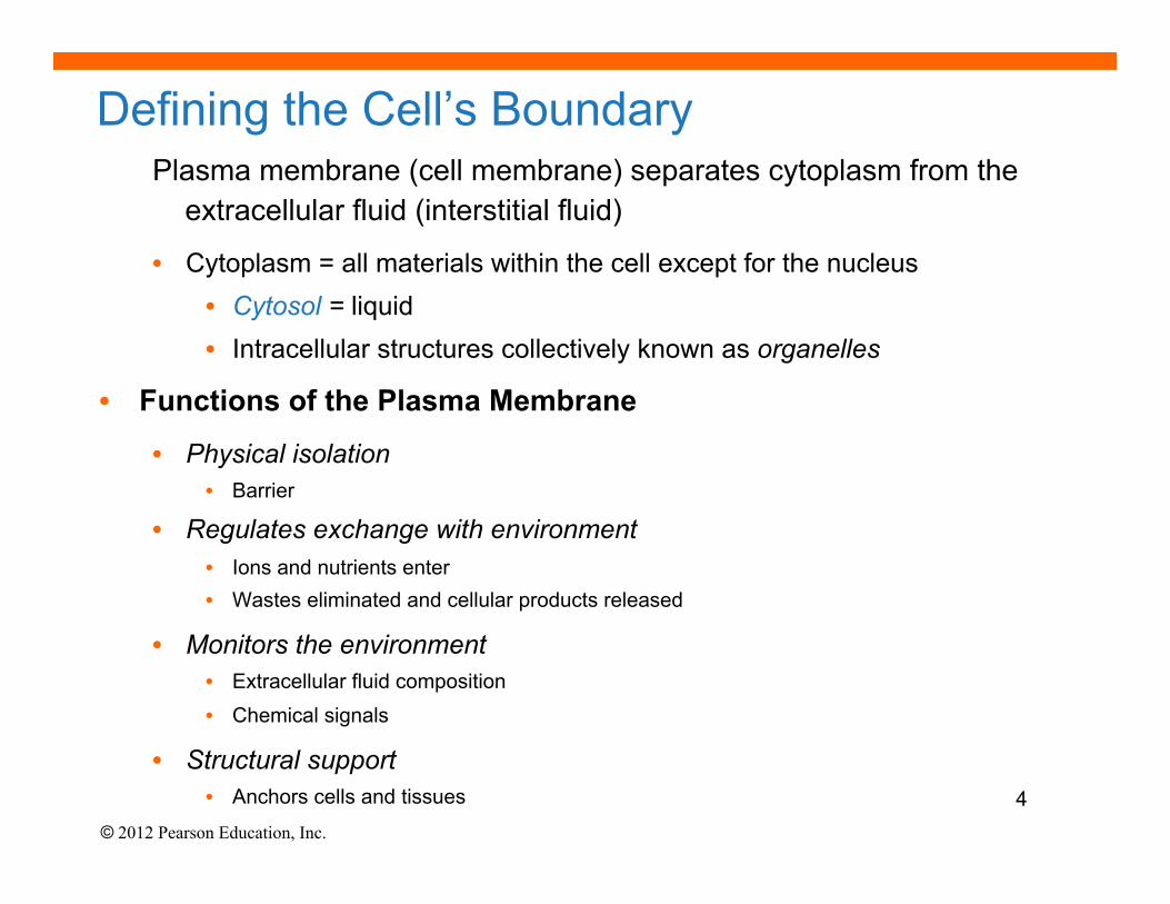
Defining the Cell’s Boundary Plasma membrane (cell membrane) separates cytoplasm from the
extracellular fluid (interstitial fluid)
• Cytoplasm = all materials within the cell except for the nucleus • Cytosol = liquid • Intracellular structures collectively known as organelles
• Functions of the Plasma Membrane
• Physical isolation • Barrier
• Regulates exchange with environment • Ions and nutrients enter • Wastes eliminated and cellular products released
• Monitors the environment • Extracellular fluid composition • Chemical signals
• Structural support • Anchors cells and tissues 4
© 2012 Pearson Education, Inc.
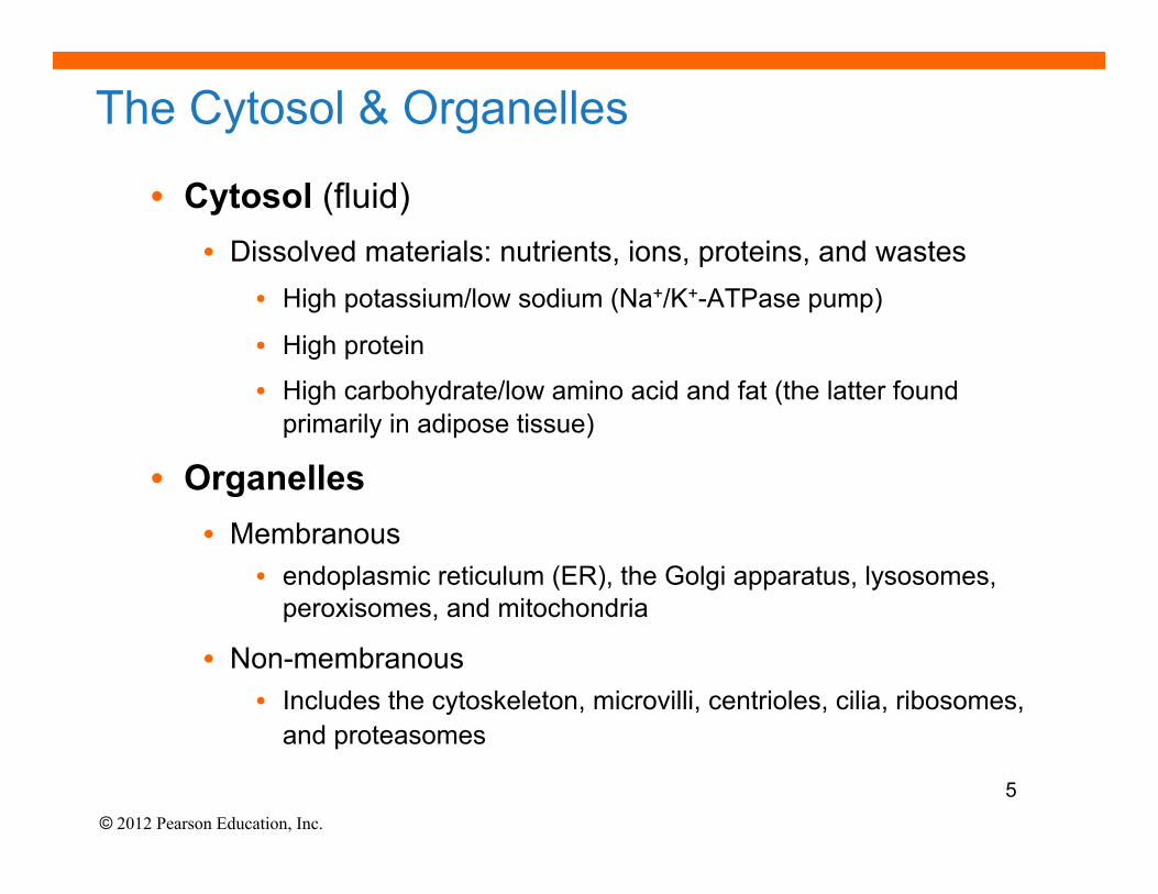
The Cytosol & Organelles
• Cytosol (fluid) • Dissolved materials: nutrients, ions, proteins, and wastes
• High potassium/low sodium (Na+/K+-ATPase pump)
• High protein
• High carbohydrate/low amino acid and fat (the latter found primarily in adipose tissue)
• Organelles • Membranous
• endoplasmic reticulum (ER), the Golgi apparatus, lysosomes, peroxisomes, and mitochondria
• Non-membranous • Includes the cytoskeleton, microvilli, centrioles, cilia, ribosomes,
and proteasomes
5
© 2012 Pearson Education, Inc.
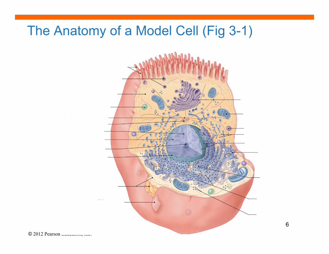
The Anatomy of a Model Cell (Fig 3-1)
6
© 2012 Pearson Education, Inc.
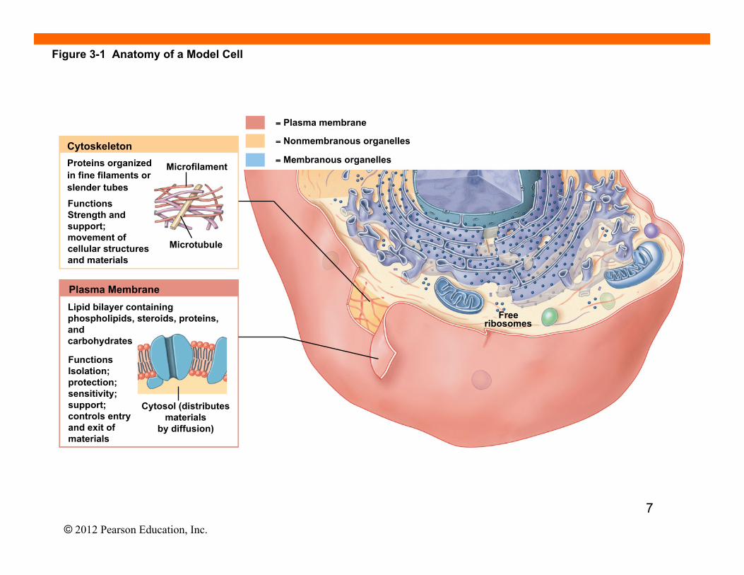
Figure 3-1 Anatomy of a Model Cell
Proteins organized in fine filaments or slender tubes Functions Strength and support; movement of cellular structures and materials
Plasma Membrane
Functions Isolation; protection; sensitivity; support; controls entry and exit of materials
Free ribosomes
= Plasma membrane
= Nonmembranous organelles
= Membranous organelles Microfilament
Microtubule
Lipid bilayer containing phospholipids, steroids, proteins, and carbohydrates
Cytosol (distributes materials
by diffusion)
Cytoskeleton
7
© 2012 Pearson Education, Inc.
Figure 3-1 Anatomy of a Model Cell
= Plasma membrane
= Nonmembranous organelles
= Membranous organelles
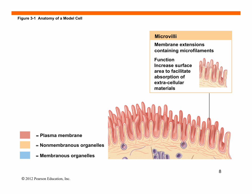
Microvilli Membrane extensions containing microfilaments
Function Increase surface area to facilitate absorption of extra-cellular materials
8
© 2012 Pearson Education, Inc.
Figure 3-1 Anatomy of a Model Cell
= Plasma membrane = Nonmembranous organelles
= Membranous organelles
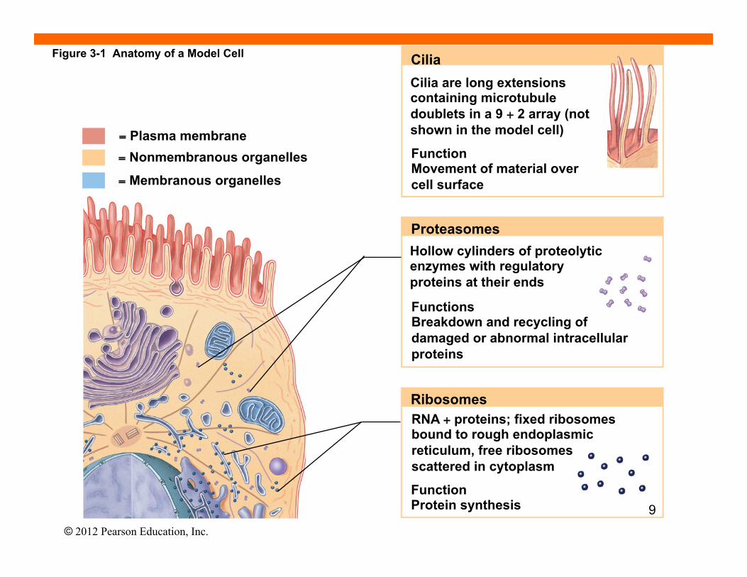
Proteasomes Hollow cylinders of proteolytic enzymes with regulatory proteins at their ends
Functions Breakdown and recycling of damaged or abnormal intracellular proteins
Cilia Cilia are long extensions containing microtubule doublets in a 9 + 2 array (not shown in the model cell)
Function Movement of material over cell surface
Ribosomes RNA + proteins; fixed ribosomes bound to rough endoplasmic reticulum, free ribosomes scattered in cytoplasm
Function Protein synthesis 9
© 2012 Pearson Education, Inc.
Figure 3-1 Anatomy of a Model Cell
= Plasma membrane
= Nonmembranous organelles
= Membranous organelles Secretory vesicles
CYTOSOL
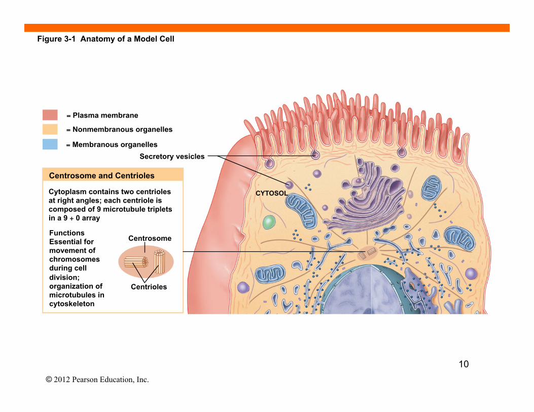
Centrosome and Centrioles
Centrosome
Centrioles
Cytoplasm contains two centrioles at right angles; each centriole is composed of 9 microtubule triplets in a 9 + 0 array Functions Essential for movement of chromosomes during cell division; organization of microtubules in cytoskeleton
10
© 2012 Pearson Education, Inc.
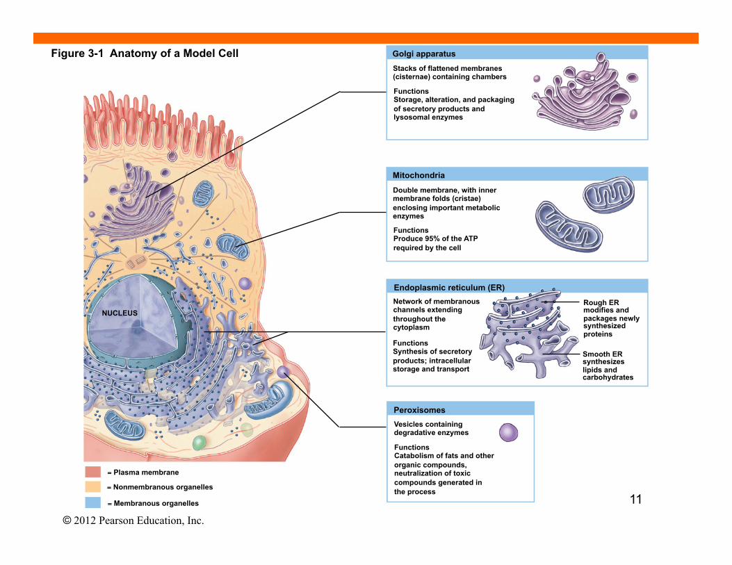
Golgi apparatus Stacks of flattened membranes (cisternae) containing chambers
Functions Storage, alteration, and packaging of secretory products and lysosomal enzymes
Mitochondria
Double membrane, with inner membrane folds (cristae) enclosing important metabolic enzymes
Functions Produce 95% of the ATP required by the cell
Endoplasmic reticulum (ER) Network of membranous channels extending throughout the cytoplasm
Functions Synthesis of secretory products; intracellular storage and transport
Rough ER modifies and packages newly synthesized proteins
Smooth ER synthesizes lipids and carbohydrates
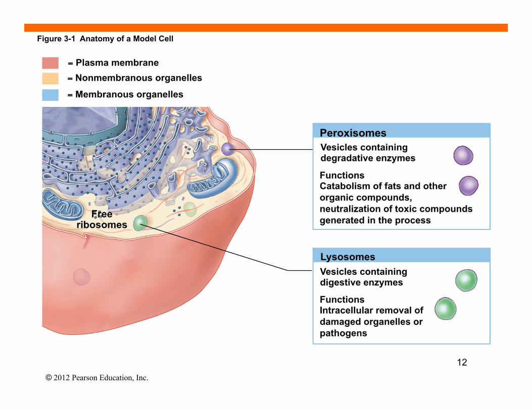
Peroxisomes Vesicles containing degradative enzymes
Functions Catabolism of fats and other organic compounds, neutralization of toxic compounds generated in the process
NUCLEUS
= Plasma membrane
= Nonmembranous organelles
= Membranous organelles
Figure 3-1 Anatomy of a Model Cell
11
© 2012 Pearson Education, Inc.
Peroxisomes Vesicles containing degradative enzymes
Functions Catabolism of fats and other organic compounds, neutralization of toxic compounds generated in the process
Lysosomes
Free ribosomes
Vesicles containing digestive enzymes
Functions Intracellular removal of damaged organelles or pathogens
= Plasma membrane = Nonmembranous organelles
= Membranous organelles
Figure 3-1 Anatomy of a Model Cell
12
© 2012 Pearson Education, Inc.
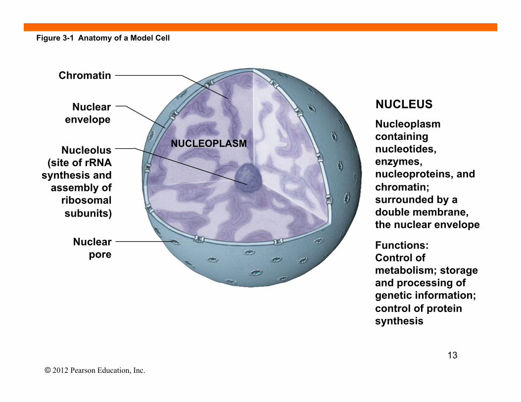
Chromatin
Nuclear envelope
Nucleolus (site of rRNA
synthesis and assembly of
ribosomal subunits)
Nuclear pore
NUCLEOPLASM
NUCLEUS Nucleoplasm containing nucleotides, enzymes, nucleoproteins, and chromatin; surrounded by a double membrane, the nuclear envelope
Functions: Control of metabolism; storage and processing of genetic information; control of protein synthesis
Figure 3-1 Anatomy of a Model Cell
13
© 2012 Pearson Education, Inc.
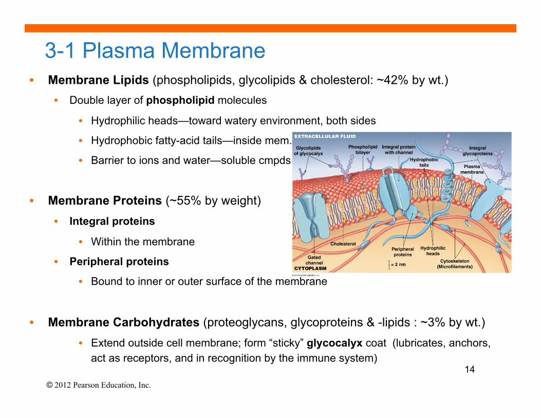
3-1 Plasma Membrane • Membrane Lipids (phospholipids, glycolipids & cholesterol: ~42% by wt.)
• Double layer of phospholipid molecules
• Hydrophilic heads—toward watery environment, both sides
• Hydrophobic fatty-acid tails—inside mem.
• Barrier to ions and water—soluble cmpds
• Membrane Proteins (~55% by weight) • Integral proteins
• Within the membrane
• Peripheral proteins
• Bound to inner or outer surface of the membrane
• Membrane Carbohydrates (proteoglycans, glycoproteins & -lipids : ~3% by wt.) • Extend outside cell membrane; form “sticky” glycocalyx coat (lubricates, anchors,
act as receptors, and in recognition by the immune system) 14
© 2012 Pearson Education, Inc.
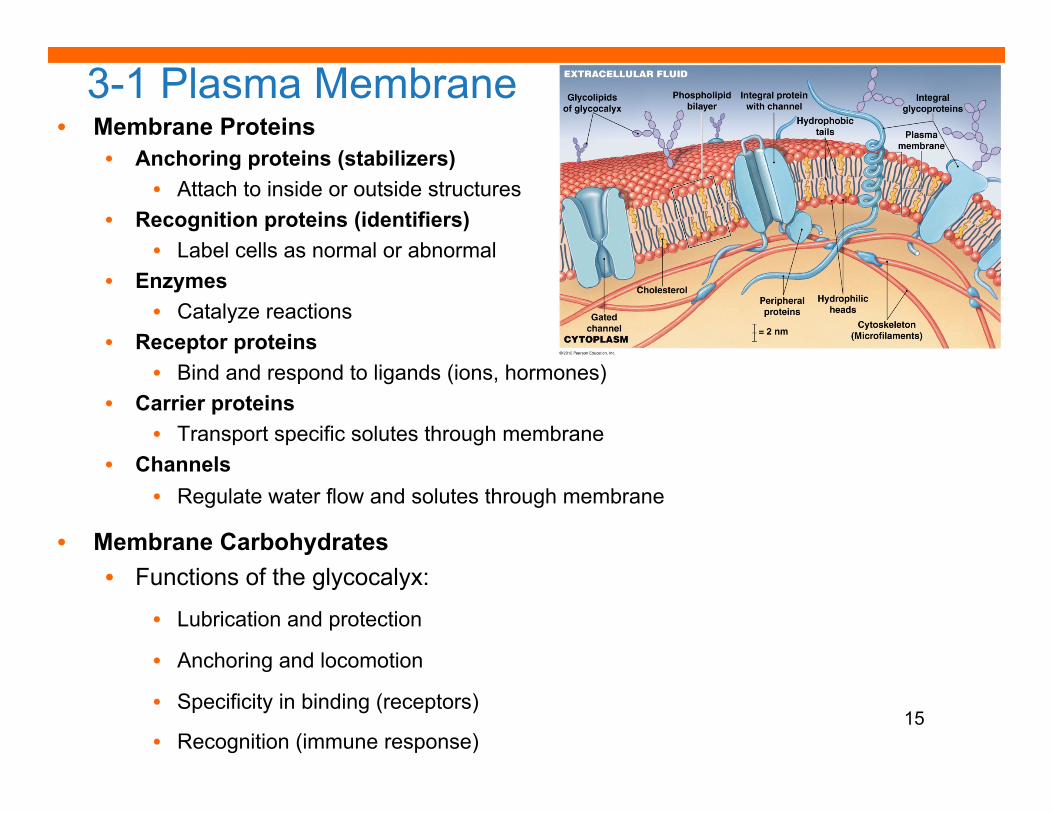
3-1 Plasma Membrane • Membrane Proteins
• Anchoring proteins (stabilizers) • Attach to inside or outside structures
• Recognition proteins (identifiers) • Label cells as normal or abnormal
• Enzymes • Catalyze reactions
• Receptor proteins • Bind and respond to ligands (ions, hormones)
• Carrier proteins • Transport specific solutes through membrane
• Channels • Regulate water flow and solutes through membrane
• Membrane Carbohydrates • Functions of the glycocalyx:
• Lubrication and protection
• Anchoring and locomotion
• Specificity in binding (receptors)
• Recognition (immune response) 15
© 2012 Pearson Education, Inc.
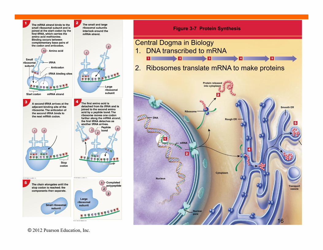
Figure 3-7 Protein Synthesis
DNA
mRNA
Ribosome
Nucleus
Cytoplasm
Rough ER
Protein released into cytoplasm
Smooth ER
Transport vesicle
Nuclear pore
Central Dogma in Biology 1. DNA transcribed to mRNA
2. Ribosomes translate mRNA to make proteins
16
© 2012 Pearson Education, Inc.
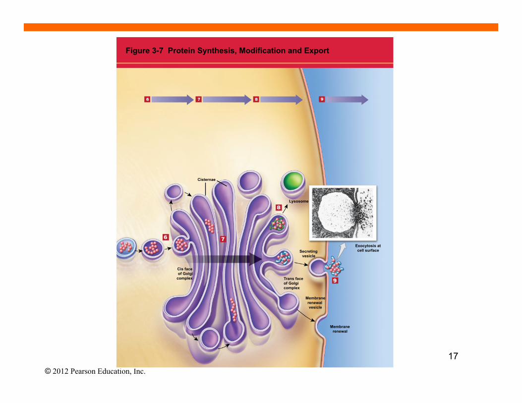
Cisternae
Lysosome
Secreting vesicle
Exocytosis at cell surface
Trans face of Golgi complex
Membrane renewal vesicle
Membrane renewal
Cis face of Golgi
complex
Figure 3-7 Protein Synthesis, Modification and Export
17
© 2012 Pearson Education, Inc.
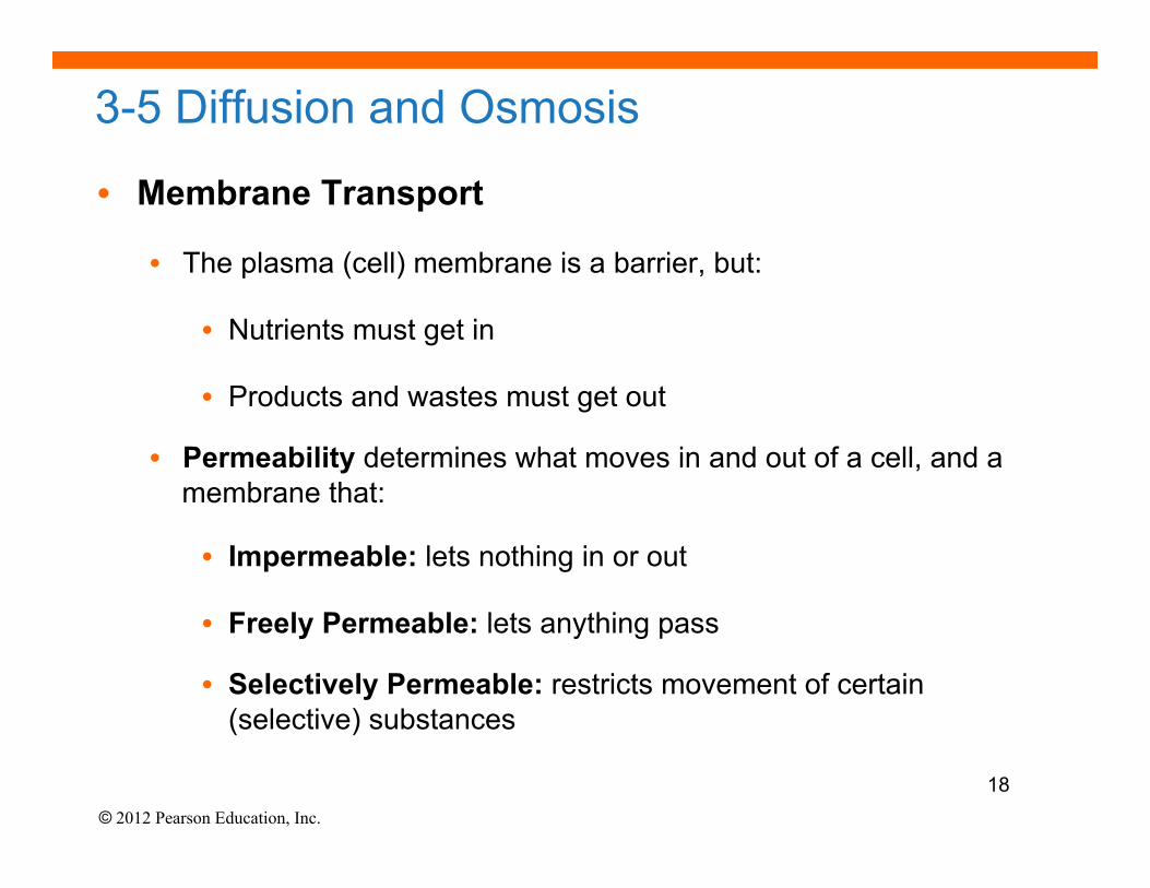
3-5 Diffusion and Osmosis
• Membrane Transport
• The plasma (cell) membrane is a barrier, but:
• Nutrients must get in
• Products and wastes must get out
• Permeability determines what moves in and out of a cell, and a membrane that:
• Impermeable: lets nothing in or out
• Freely Permeable: lets anything pass
• Selectively Permeable: restricts movement of certain (selective) substances
18
© 2012 Pearson Education, Inc.
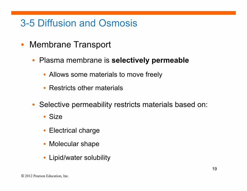
3-5 Diffusion and Osmosis
• Membrane Transport
• Plasma membrane is selectively permeable
• Allows some materials to move freely
• Restricts other materials
• Selective permeability restricts materials based on: • Size
• Electrical charge
• Molecular shape
• Lipid/water solubility 19
© 2012 Pearson Education, Inc.
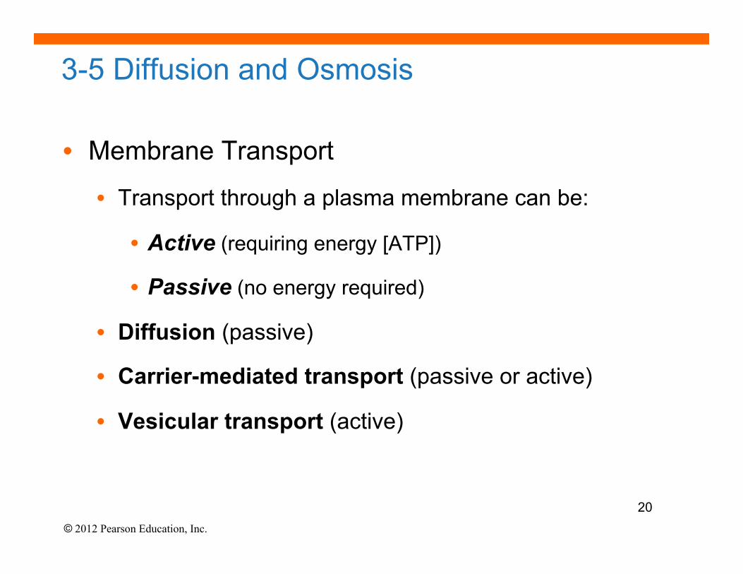
3-5 Diffusion and Osmosis
• Membrane Transport
• Transport through a plasma membrane can be:
• Active (requiring energy [ATP])
• Passive (no energy required)
• Diffusion (passive)
• Carrier-mediated transport (passive or active)
• Vesicular transport (active)
20
© 2012 Pearson Education, Inc.
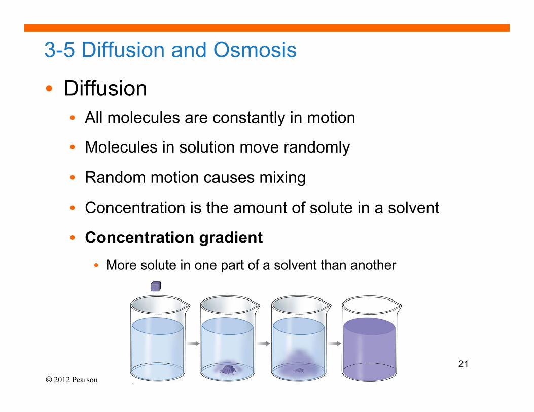
3-5 Diffusion and Osmosis
• Diffusion • All molecules are constantly in motion
• Molecules in solution move randomly
• Random motion causes mixing
• Concentration is the amount of solute in a solvent
• Concentration gradient • More solute in one part of a solvent than another
21
© 2012 Pearson Education, Inc.
3-5 Diffusion and Osmosis
• Factors Influencing Diffusion • Distance the particle has to move
• Molecule Size • Smaller is faster
• Temperature • More heat, faster motion
• Concentration Gradient • The difference between high and low concentrations
• Electrical Forces
• Opposites attract, like charges repel
22
© 2012 Pearson Education, Inc.
3-5 Diffusion and Osmosis
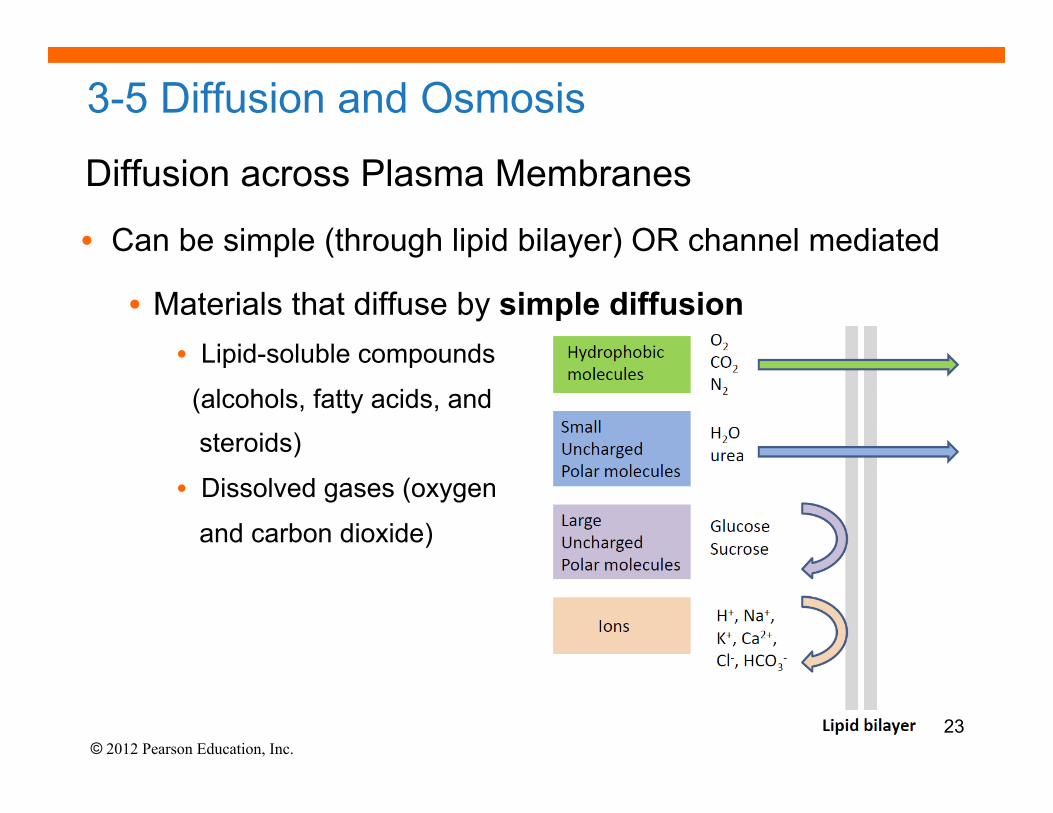
Diffusion across Plasma Membranes
• Can be simple (through lipid bilayer) OR channel mediated
• Materials that diffuse by simple diffusion • Lipid-soluble compounds
(alcohols, fatty acids, and steroids)
• Dissolved gases (oxygen
and carbon dioxide)
23
© 2012 Pearson Education, Inc.
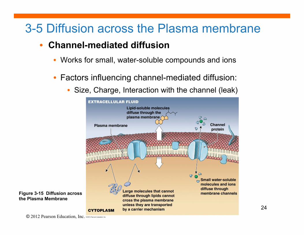
3-5 Diffusion across the Plasma membrane • Channel-mediated diffusion
• Works for small, water-soluble compounds and ions
• Factors influencing channel-mediated diffusion: • Size, Charge, Interaction with the channel (leak)
Figure 3-15 Diffusion across the Plasma Membrane
24
© 2012 Pearson Education, Inc.
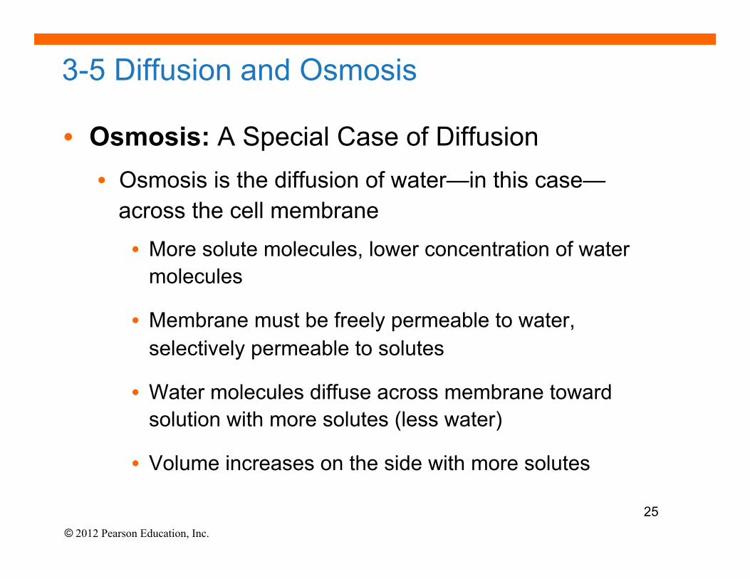
3-5 Diffusion and Osmosis
• Osmosis: A Special Case of Diffusion • Osmosis is the diffusion of water—in this case—
across the cell membrane
• More solute molecules, lower concentration of water molecules
• Membrane must be freely permeable to water, selectively permeable to solutes
• Water molecules diffuse across membrane toward solution with more solutes (less water)
• Volume increases on the side with more solutes
25
© 2012 Pearson Education, Inc.
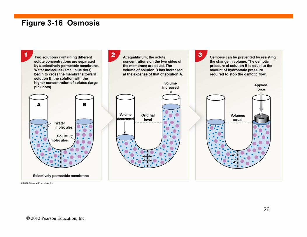
Figure 3-16 Osmosis
26
© 2012 Pearson Education, Inc.
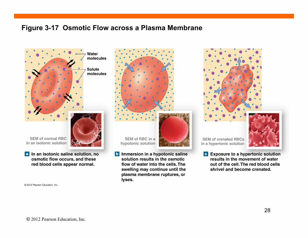
3-5 Osmolarity and Tonicity • Osmolarity is a measure of solute concentration (number of osmoles (Osm)
of solute per liter) • Osmolarity is distinct from molarity because it measures moles of solute particles
rather than moles of solute--distinction arises because some compounds can dissociate in solution whereas others cannot.
• Tonicity describes the osmotic effect of a solution on a cell (shrinks, swells, or no change) • two fluids may have equal osmolarity but different tonicity (e.g., because the
cell is selectively permeable to some but not all solutes)
• Isotonic (iso- = same, tonos = tension) • A solution that does not cause osmotic flow of water in or out of a cell
• Hypotonic (hypo- = below) • A solution that causes the cell to swell (for erythrocyte [RBC], if it burst = hemolysis)
• Hypertonic (hyper- = above)
• A solution that causes the cell to shrink (called crenation for erythrocyte) 27
© 2012 Pearson Education, Inc.
Figure 3-17 Osmotic Flow across a Plasma Membrane
28
© 2012 Pearson Education, Inc.
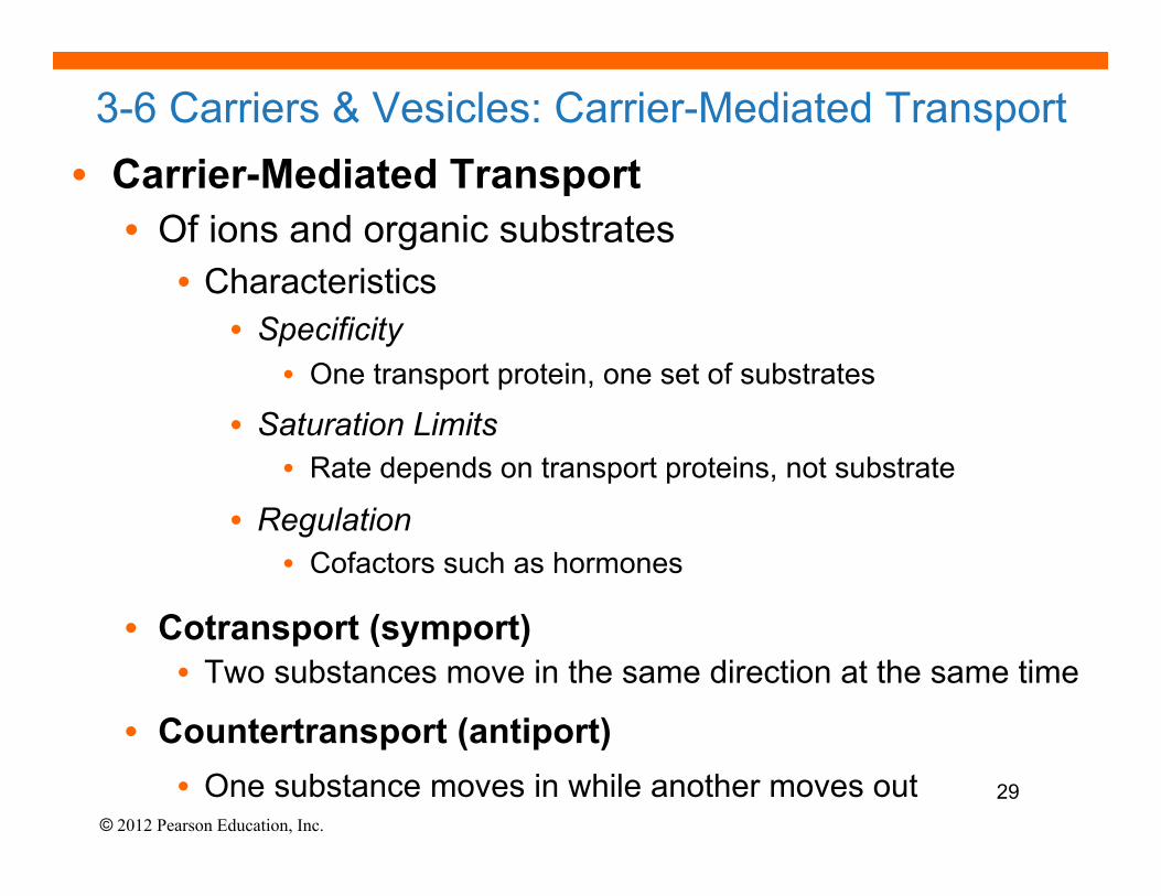
3-6 Carriers & Vesicles: Carrier-Mediated Transport • Carrier-Mediated Transport • Of ions and organic substrates
• Characteristics • Specificity
• One transport protein, one set of substrates
• Saturation Limits • Rate depends on transport proteins, not substrate
• Regulation • Cofactors such as hormones
• Cotransport (symport) • Two substances move in the same direction at the same time
• Countertransport (antiport) • One substance moves in while another moves out 29
© 2012 Pearson Education, Inc.
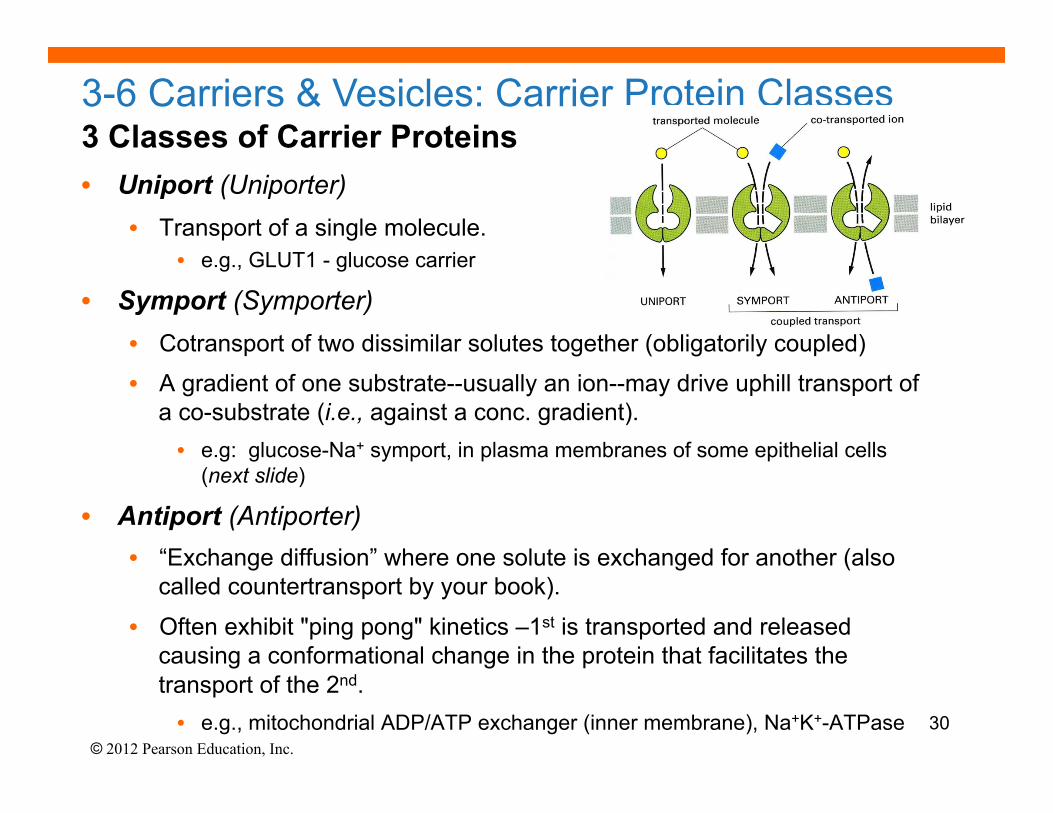
3 Classes of Carrier Proteins • Uniport (Uniporter)
• Transport of a single molecule. • e.g., GLUT1 - glucose carrier
• Symport (Symporter) • Cotransport of two dissimilar solutes together (obligatorily coupled)
• A gradient of one substrate--usually an ion--may drive uphill transport of a co-substrate (i.e., against a conc. gradient). • e.g: glucose-Na+ symport, in plasma membranes of some epithelial cells
(next slide)
• Antiport (Antiporter) • “Exchange diffusion” where one solute is exchanged for another (also
called countertransport by your book).
• Often exhibit "ping pong" kinetics –1st is transported and released causing a conformational change in the protein that facilitates the transport of the 2nd. • e.g., mitochondrial ADP/ATP exchanger (inner membrane), Na+K+-ATPase
3-6 Carriers & Vesicles: Carrier Protein Classes
30
© 2012 Pearson Education, Inc.
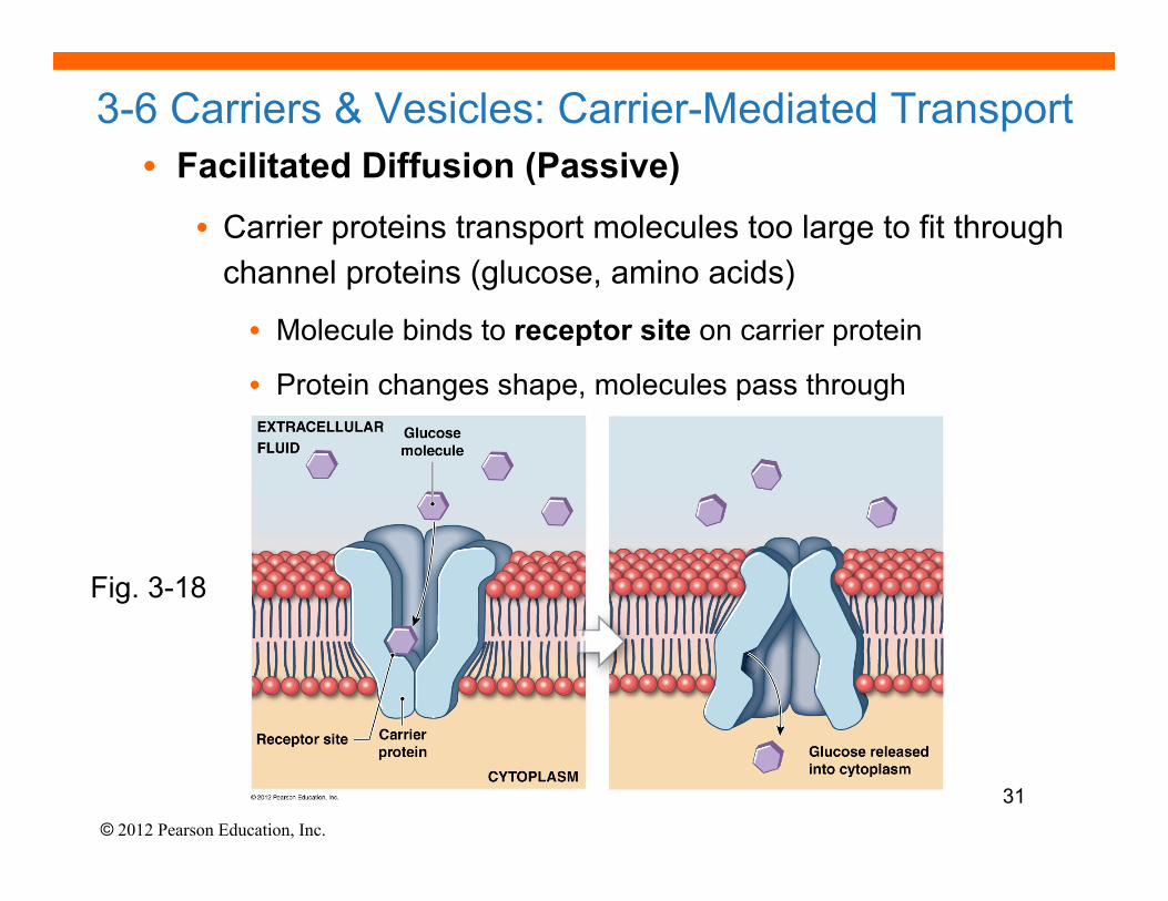
3-6 Carriers & Vesicles: Carrier-Mediated Transport • Facilitated Diffusion (Passive)
• Carrier proteins transport molecules too large to fit through channel proteins (glucose, amino acids)
• Molecule binds to receptor site on carrier protein
• Protein changes shape, molecules pass through
Fig. 3-18
31
© 2012 Pearson Education, Inc.
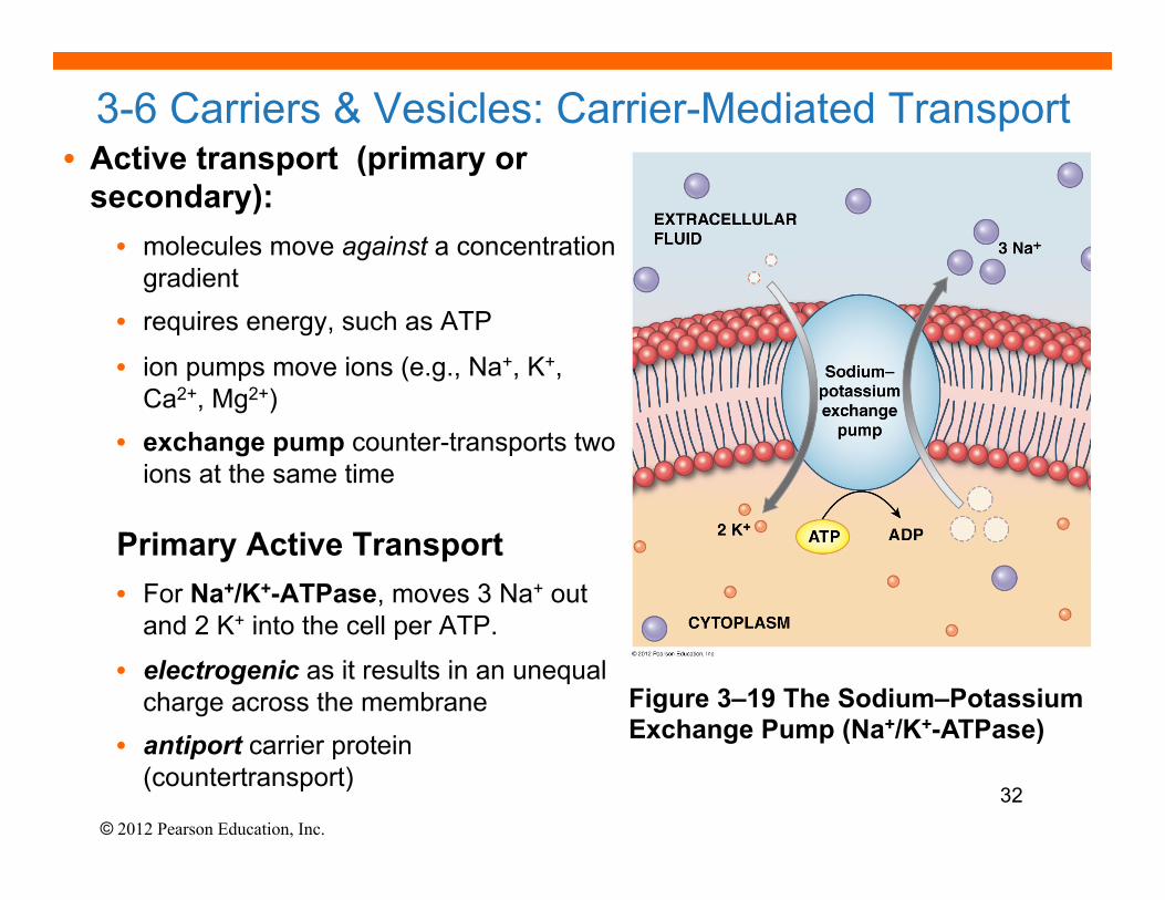
3-6 Carriers & Vesicles: Carrier-Mediated Transport • Active transport (primary or
secondary): • molecules move against a concentration
gradient • requires energy, such as ATP
• ion pumps move ions (e.g., Na+, K+, Ca2+, Mg2+)
• exchange pump counter-transports two ions at the same time
Primary Active Transport • For Na+/K+-ATPase, moves 3 Na+ out
and 2 K+ into the cell per ATP.
• electrogenic as it results in an unequal charge across the membrane
• antiport carrier protein (countertransport)
•
Figure 3–19 The Sodium–Potassium Exchange Pump (Na+/K+-ATPase)
32
© 2012 Pearson Education, Inc.
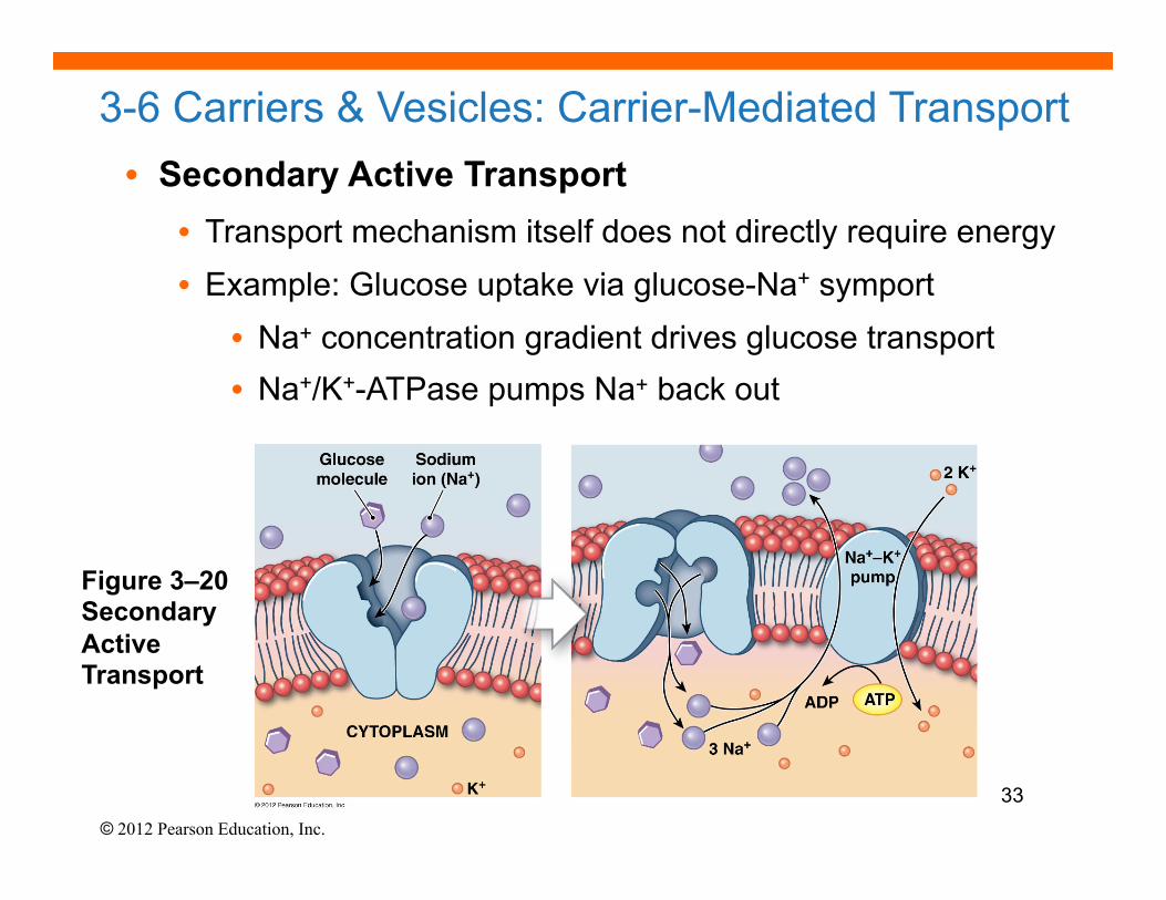
3-6 Carriers & Vesicles: Carrier-Mediated Transport • Secondary Active Transport
• Transport mechanism itself does not directly require energy • Example: Glucose uptake via glucose-Na+ symport
• Na+ concentration gradient drives glucose transport • Na+/K+-ATPase pumps Na+ back out
Figure 3–20 Secondary Active Transport
33
© 2012 Pearson Education, Inc.
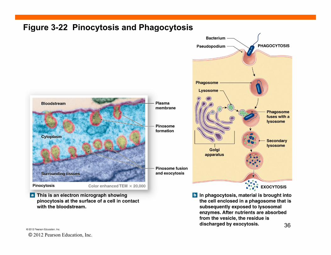
3-6 Carriers and Vesicles: Vesicular Transport
• Vesicular Transport (Bulk Transport)
• Materials move into or out of cell in vesicles
• Endocytosis – the cell takes in macromolecules by forming new
vesicles from the plasma membrane (3 types)
1. Phagocytosis – “cell eating” – for solid substances
2. Pinocytosis - “cell drinking” – for dissolved substances
3. Receptor-Mediated (receptors = glycoproteins)
• Exocytosis - transport vesicles migrate to plasma membrane,
fuse with it and release their contents
• Granules or droplets are released from the cell 34
© 2012 Pearson Education, Inc.
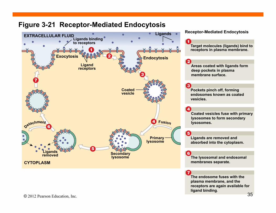
Figure 3-21 Receptor-Mediated Endocytosis EXTRACELLULAR FLUID
Exocytosis
Ligand receptors
Coated vesicle
Ligands binding to receptors
Endocytosis
Ligands
Ligands removed Secondary
lysosome
Primary lysosome
F u s i o c h m e n t
CYTOPLASM
Receptor-Mediated Endocytosis
Target molecules (ligands) bind to receptors in plasma membrane.
Areas coated with ligands form deep pockets in plasma membrane surface.
Pockets pinch off, forming endosomes known as coated vesicles.
Coated vesicles fuse with primary lysosomes to form secondary lysosomes.
Ligands are removed and absorbed into the cytoplasm.
The lysosomal and endosomal membranes separate.
The endosome fuses with the plasma membrane, and the receptors are again available for ligand binding.
35
© 2012 Pearson Education, Inc.
Figure 3-22 Pinocytosis and Phagocytosis
36
© 2012 Pearson Education, Inc.
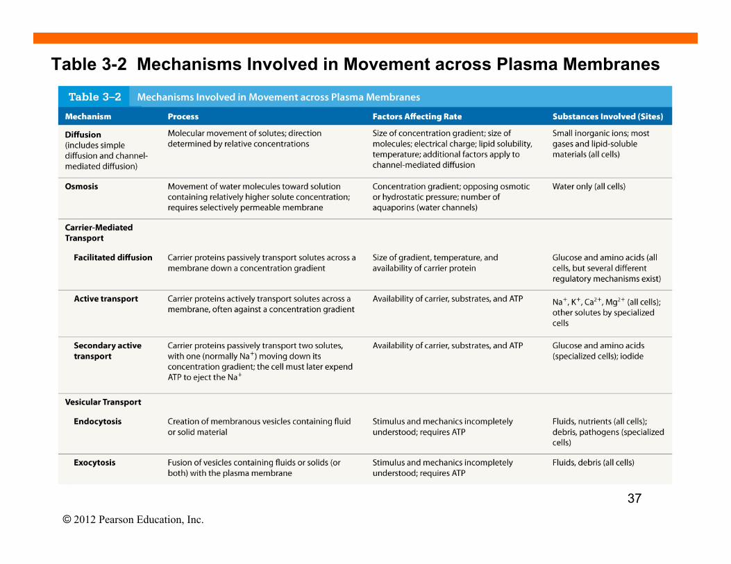
Table 3-2 Mechanisms Involved in Movement across Plasma Membranes
37
© 2012 Pearson Education, Inc.
Chapter 3 Summary Questions and Goals
• Be able to describe the overall function of the cell’s major organelles.
• Be able to describe the various mechanisms for transporting substances across the plasma membrane.
• Know the 3 types or classes of carrier proteins, the difference between passive and active transport (both primary and secondary).
• Know the difference between tonicity and osmolarity.
• Be able to describe the 3 types of mechanisms by which bulk substances enter the cell.
38