Embed Size (px)
Citation preview
Journal of
Clinical Medicine
Review
The Clinical and Neuropathological Features of Sporadic(Late-Onset) and Genetic Forms of Alzheimer’s Disease
Tanzil Rujeedawa 1, Eva Carrillo Félez 1, Isabel C. H. Clare 1,2 , Juan Fortea 3,4,5, Andre Strydom 6,7,Anne-Sophie Rebillat 8 , Antonia Coppus 9, Johannes Levin 10,11,12 and Shahid H. Zaman 1,2,*
�����������������
Citation: Rujeedawa, T.; Carrillo
Félez, E.; Clare, I.C.H.; Fortea, J.;
Strydom, A.; Rebillat, A.-S.; Coppus,
A.; Levin, J.; Zaman, S.H. The Clinical
and Neuropathological Features of
Sporadic (Late-Onset) and Genetic
Forms of Alzheimer’s Disease. J. Clin.
Med. 2021, 10, 4582. https://doi.org/
10.3390/jcm10194582
Academic Editors: Ann-Charlotte
Granholm, Melissa J. Alldred and
Alessandra C. Martini
Received: 31 July 2021
Accepted: 28 September 2021
Published: 3 October 2021
Publisher’s Note: MDPI stays neutral
with regard to jurisdictional claims in
published maps and institutional affil-
iations.
Copyright: © 2021 by the authors.
Licensee MDPI, Basel, Switzerland.
This article is an open access article
distributed under the terms and
conditions of the Creative Commons
Attribution (CC BY) license (https://
creativecommons.org/licenses/by/
4.0/).
1 Cambridge Intellectual & Developmental Disabilities Research Group, Department of Psychiatry,University of Cambridge, Cambridge CB2 8PQ, UK; [email protected] (T.R.); [email protected] (E.C.F.);[email protected] (I.C.H.C.)
2 Cambridgeshire and Peterborough Foundation NHS Trust, Fulbourn CB21 5EF, UK3 Sant Pau Memory Unit, Department of Neurology, Hospital de la Santa Creu i Sant Pau, Biomedical Research
Institute Sant Pau, Universitat Autònoma de Barcelona, 08193 Barcelona, Spain; [email protected] Center of Biomedical Investigation Network for Neurodegenerative Diseases (CIBERNED),
28031 Madrid, Spain5 Barcelona Down Medical Center, Fundació Catalana Síndrome de Down, 08029 Barcelona, Spain6 Department of Forensic and Neurodevelopmental Sciences, Institute of Psychiatry, Psychology and
Neuroscience, King’s College London, London SE5 8AF, UK; [email protected] South London and the Maudsley NHS Foundation Trust, The LonDowns Consortium, London SE5 8AZ, UK8 Geriatrics, Institut Jérôme Lejeune, 75015 Paris, France; [email protected] Department of Primary and Community Care (149 ELG), Radboud University Nijmegen Medical Center,
P.O. Box 9101, 6525 GA Nijmegen, The Netherlands; [email protected] Department of Neurology, Ludwig-Maximilians-Universität München, 80539 Munich, Germany;
[email protected] German Center for Neurodegenerative Diseases, Feodor-Lynen-Strasse 17, 81377 Munich, Germany12 Munich Cluster for Systems Neurology (SyNergy), Feodor-Lynen-Strasse 17, 81377 Munich, Germany* Correspondence: [email protected]
Abstract: The purpose of this review is to compare and highlight the clinical and pathological aspectsof genetic versus acquired Alzheimer’s disease: Down syndrome-associated Alzheimer’s disease in(DSAD) and Autosomal Dominant Alzheimer’s disease (ADAD) are compared with the late-onsetform of the disease (LOAD). DSAD and ADAD present in a younger population and are morelikely to manifest with non-amnestic (such as dysexecutive function features) in the prodromalphase or neurological features (such as seizures and paralysis) especially in ADAD. The very largevariety of mutations associated with ADAD explains the wider range of phenotypes. In the LOAD,age-associated comorbidities explain many of the phenotypic differences.
Keywords: late-onset Alzheimer’s disease; down syndrome; autosomal dominant Alzheimer ’sdisease; clinical features; neuropathology
1. Introduction and Background
This review aims to highlight the similarities and differences between the clinical andneuropathological manifestations of Alzheimer’s disease (AD) in sporadic (or late-onsetAD) and autosomal dominant AD (ADAD) and Down syndrome-associated AD (DSAD).
AD is a neurodegenerative disease that results in neuronal cell death, causing brainatrophy. The neurodegeneration is thought to be due to the immediate or downstreamconsequences of the abnormal accumulation of beta-amyloid (Aβ) and hyperphosphory-lated Tau proteins that manifest as Aβ neuritic plaques and neurofibrillary tangles (NFTs),respectively. Several other pathological processes also play a role, including the innateimmune system, the inflammatory response and mitochondrial dysfunction and the conse-quent oxidative damage. It is associated with reactive astroglia [1], immune-responsivemicroglia [2] and neurovascular issues [3]. AD is the most common form of dementia and
J. Clin. Med. 2021, 10, 4582. https://doi.org/10.3390/jcm10194582 https://www.mdpi.com/journal/jcm
J. Clin. Med. 2021, 10, 4582 2 of 28
is characterized by memory loss and cognitive decline in several modalities. In some cases,it can also present with atypical symptoms. AD affects 30–35 million people worldwideand as life expectancy increases, the prediction is that by 2030, it will be experienced by82 million people at any one time [4].
Late-onset AD (LOAD) is also referred to as sporadic AD and clinically presentsin those aged over 65 years. However, there are different subtypes of LOAD and theremay be heterogeneities in both the clinical features and neuropathology they demonstrate.AD presenting before 65 years of age is referred to as early onset AD (EOAD). Between5–7% of all AD presents as EOAD [5]. Though estimates of EOAD usually do not includepeople with DS, EOAD should comprise both Down syndrome associated AD (DSAD) andautosomal dominant AD (ADAD). However, not all EOAD are DSAD or ADAD.
Amongst all live births, Down syndrome (DS) is the most common aneuploidy. Inc.95% of cases the cause is a full trisomy of chromosome 21-the rest are due to partialtrisomies, translocations or mosaicisms. DS is associated with growth delays, characteristicfacial features, intellectual disability and multiple comorbidities such as congenital heartdefects, thyroid dysfunction, autism spectrum disorder, sleep apnoea, hearing loss andvisual impairment [6]. All these features, together with the “accelerated ageing” phenotype,used to result in a short life span: in the 1930s, most people with DS lived only up to 10years of age [7]. This improved lifespan gradually increased to 35 years in the 1980s [8],and nowadays people with DS live on average of 60 years [9]. The increase in longevity hasallowed the manifestation and study of AD in DS [10,11]. Virtually all adults with DS over40 years of age present the typical AD-like neuropathological features of fibrillary (senile)plaques and neurofibrillary tangles (NFTs) in their brains [12]. The prevalence of AD inDS increases with ageing in a more pronounced way, and at a much earlier age than inthe general population, or among other groups of people with intellectual disabilities [13].AD in people with DS is often diagnosed first in young adults and increases exponentiallyuntil the majority have a clinical diagnosis of AD around the age of 60 [14–16]. Accordingto a twenty year longitudinal study following people with DS [16], 97.4% developeddementia with the risk of dementia increasing from 23% at around 50 to 80% at age 65and above. However, some people with DS who may have died younger because of otherco-morbidities will influence these estimates and so result in a survival bias of the figures.
Autosomal dominant AD (ADAD) is caused by fully penetrant mutations in one of the,thus far recognised, three genes, amyloid precursor protein (APP) and presenilin (PSEN) 1and 2, which follow a Mendelian autosomal dominant inheritance pattern. ADAD repre-sents < 1% of all AD cases and the relative frequencies due to mutations in PSEN1, PSEN2and APP are 69%, 2% and 13%, respectively [17]. The number of disease gene mutationsdiscovered is huge: over sixty, seventy and three hundred specific gene mutations areassociated with APP, PSEN1 and PSEN2, respectively; knowing the specific mutation for agiven case can help inform precision medicine approaches for patients.
APP is encoded on chromosome 21 and is pathological with respect to dementia whenit is duplicated(dupAPP) [18]. The dupAPP results in an increase in APP gene dosage,similar to that of trisomy of chromosome 21 in DS.
APP is a transmembrane protein that is proteolytically cleaved at different specificresidues by secretases: the α-secretase, which is responsible for the products of the “non-amyloidogenic” pathway, and the β- and γ-secretases that result in the products of the“amyloidogenic” pathway. Cleavage via the β- and γ-secretases at different sites leads tothe formation of Aβ peptides of different sizes such as Aβ40 and Aβ42, which are 40 and42 residues long, respectively. AD causing mutations in the APP gene are clustered aroundthe three cleavage sites, with most of them affecting the γ-secretase site of cleavage. Thepositions of these mutation sites increases the generation of the Aβ42 which is more insolu-ble and more prevalent in cored Aβ-plaques compared to the Aβ40 peptide [19]. PSEN isone of the catalytic subunits of the γ-secretase complex [20]. Most PSEN mutations cause aloss of function of γ-secretase and increase the Aβ42/Aβ40 ratio [20], thereby promotingoligomer formation. Aβ peptides undergo a biophysical transformation from monomers
J. Clin. Med. 2021, 10, 4582 3 of 28
to oligomers before being deposited in Aβ plaques. Consistent with the amyloid cascadehypothesis [21], it is such monomers and oligomers that are thought to be particularly toxicto brain tissue. Hereditary forms of AD provides strong support to the amyloid cascadehypothesis as they show how alterations in the processing of APP with resulting aberrantlevels of Aβ42 represent a strong driver of synaptic and neuronal loss [20]. However, it isnoted that the amyloid cascade hypothesis hinges on neurodevelopmental processes and istherefore best suited to genetic or chromosomal causes of dementia.
When considering ADAD, the phenotype variants between carriers of the different ge-netic mutations, as well as between variants of the same gene need to be considered [20,22].ADAD phenotypes are influenced by mutation position and causative gene [22]. Thedifferent phenotypes are characterised not only by different clinical features and ages ofonset but also by aspects of underlying pathology. For example, APP Flemish and Dutchmutations present with recurrent cerebrovascular events, due to amyloid accumulationin the blood vessels rather than as parenchymal amyloid plaques [23]. In contrast, theAPP Icelandic mutation has a protective effect against AD and cognitive decline [24]. WithPSEN1, mutations before and after codon 200 are pathologically different [25] and havedifferent ages of onset [22]. PSEN2 mutation carriers show atypical presentations thatresemble dementia with Lewy bodies or frontotemporal dementia [26], when compared tothe other types of ADAD. The heterogeneity associated with different ADAD mutations istherefore very important to consider.
The different forms of AD have many similarities and differences, understanding ofwhich can allow a deeper insight into the mechanisms of AD. Notably, it can clarify the rolethat different genes, their mutations and proteins play in the development of the disease.In addition, given the difficulty in the general population of predicting the transition frompreclinical to clinical AD, studies of EOAD are valuable as they are expected to increasepredictability, are very valuable. Clinical studies in DS have some advantages as thereis both a high risk and predictability of developing DSAD, and the AD neuropathologyseems more homogeneous than in LOAD. Moreover, in ADAD and DS, the earlier age ofonset reduces the impact of confounding factors associated with ageing, thereby allowingthe pathological characteristics of AD to be better discriminated. However, the triplicatedgenes on chromosome 21 may limit the extrapolation of data from DS populations to otherpopulations, and neurodevelopmental factors need to be taken into account.
There are many factors associated with the development of AD. In the non-DS pop-ulation, these factors include not only ageing, but also cardiovascular risk factors (17),traumatic brain injury [27], number of years of education [28] and genetic risk factors [29].Genetic risk factors include a family history of AD, including the ADAD mutations inPSEN and APP and the possession of some SNPs or gene alleles that, through the geneticanalysis of large populations [30], have been linked with the disease. The possession ofthe E4 allele of the apolipoprotein E (APOE) gene is one of the major factors that influencethe development of AD [31]. The evidence for APOE’s effect on the clinical presentation ofADAD [22,32] and DSAD [33–35] is becoming clearer despite genetic mutations that causeAD masking the effect of APOE. For instance, it has been found that the APOE ε4 allelecorrelated with earlier clinical and biomarker changes of AD in DS [36]. It is also noted thatthe Christchurch mutation, R136S in APOE3 (homozygous) in a PSEN1-E280A mutationcarrier, reported relatively little decline in cognition despite ageing and little evidence oftau-deposition despite relatively greater amounts of amyloid being detected using PETimaging [37].
In DS, an extra copy of APP is sufficient to cause AD [38]. However, there are othergenes on chromosome 21 [39] whose overexpression may enhance or modify the risk forAD [40]. Some of these candidate pathogenesis gene modifiers include beta-secretase-2(BACE2) [27] and DYRK1A [41,42], both of which are related to the calcineurin-NFATsignaling pathway that is altered in AD [43]. Some studies however argue against theactivity of BACE2 in DS [44] as being pathological since it is able to cleave APP toward thenon-amyloidogenic pathway. Other studies have argued that BACE1 could instead play a

J. Clin. Med. 2021, 10, 4582 4 of 28
greater role [45]. It is also possible that DS cells respond to the increased gene dosage byenhancing DNA methylation [46] thereby accelerating epigenetic changes that are usuallyassociated with ageing [47]. This could account for the “accelerated ageing” phenotypeobserved in DS.
Comparisons of dementia in LOAD and ADAD [19,48] and in LOAD and DSAD [49]have been carried out. However, there is very limited literature comparing DSAD withADAD or all three conditions [50]. The aim of this review is to synthesise and present theliterature on all the three conditions.
2. Methods
A search through PubMed and the Cochrane library was performed to exclude theexistence of another review in this topic. Then, a structured review was undertakenfollowing the guidelines provided by PRISMA Protocols [51]. First, a search in PubMed wascarried out of the literature published about humans since 2000 and relating to Alzheimer’sdisease in Down syndrome, using the MeSH terms: (“Alzheimer Disease” [Mesh] AND“Down Syndrome” [Mesh]). From the 482 results, 219 were assessed for eligibility basedon their relevance from their title and abstract, and from those, 133 were selected after afull-text review. For the autosomal form, broader search methods were applied. As there isno MeSH term for this condition, three searches were undertaken with the same parametersas for Down syndrome: (“Alzheimer Disease” [Mesh] AND (clinical features [All Fields])),this produced 340 results from which 27 were screened and 18 selected; (“Neuropathology”[Mesh] AND “Alzheimer Disease” [Mesh]), with 19 results from which 2 were eligible butexcluded as duplicated; and (“Histology” [Mesh] AND “Alzheimer Disease” [Mesh]), with161 results from which 2 were screened and 1 included. We included 36 articles from the84 publications highlighted by the DIAN study in thie website [52] as their collection ofimportant publications was considered a valuable resource of information for ADAD. DIANstands for the Dominantly Inherited Alzheimer Network, a longitudinal observationalstudy to monitor individuals who carry one of the gene mutations known to cause ADADwith the main goal of identifying changes and establish reliable biomarkers. For LOAD, nospecial search was undertaken initially, as the information about this condition is implicitin the articles from the two other conditions. However, some specific searches were carriedout to cover omissions in the information, adding a total of 6 articles.
The exclusion criteria applied to select the articles from their abstracts were thefollowing: (a) n = 1, (b) only the title or abstract available, (c) opinion articles, clinicaltrials, novel test descriptions or protocol revisions, (d) no specific content about AD inpapers relating to Down syndrome (e) no specific reference to familial or genetic causesfor dementia in the search for ADAD. From the selected articles, we have finally includedthose that did not fulfil any of the previous exclusion criteria nor, following a full-textreview contain any new information. Finally, reference lists of selected papers were alsosearched for potentially relevant studies, adding 42 papers to the 75 included for Downsyndrome, 19 to the 30 included for the autosomal dominant form, and 21 papers to the 7for the sporadic form of the disease-references from these papers also were examined (anaddition of 94). The search process is summarized in Figure 1.
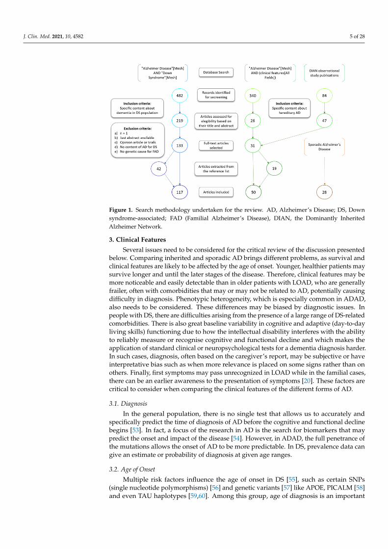
J. Clin. Med. 2021, 10, 4582 5 of 28J. Clin. Med. 2021, 10, x FOR PEER REVIEW 5 of 29
Figure 1. Search methodology undertaken for the review. AD, Alzheimer’s Disease; DS, Down syndrome-associated; FAD
(Familial Alzheimer’s Disease), DIAN, the Dominantly Inherited Alzheimer Network.
3. Clinical Features
Several issues need to be considered for the critical review of the discussion pre-
sented below. Comparing inherited and sporadic AD brings different problems, as sur-
vival and clinical features are likely to be affected by the age of onset. Younger, healthier
patients may survive longer and until the later stages of the disease. Therefore, clinical
features may be more noticeable and easily detectable than in older patients with LOAD,
who are generally frailer, often with comorbidities that may or may not be related to AD,
potentially causing difficulty in diagnosis. Phenotypic heterogeneity, which is especially
common in ADAD, also needs to be considered. These differences may be biased by diag-
nostic issues. In people with DS, there are difficulties arising from the presence of a large
range of DS-related comorbidities. There is also great baseline variability in cognitive and
adaptive (day-to-day living skills) functioning due to how the intellectual disability inter-
feres with the ability to reliably measure or recognise cognitive and functional decline and
which makes the application of standard clinical or neuropsychological tests for a demen-
tia diagnosis harder. In such cases, diagnosis, often based on the caregiver’s report, may
be subjective or have interpretative bias such as when more relevance is placed on some
signs rather than on others. Finally, first symptoms may pass unrecognized in LOAD
while in the familial cases, there can be an earlier awareness to the presentation of symp-
toms [20]. These factors are critical to consider when comparing the clinical features of the
different forms of AD.
3.1. Diagnosis
In the general population, there is no single test that allows us to accurately and spe-
cifically predict the time of diagnosis of AD before the cognitive and functional decline
begins [53]. In fact, a focus of the research in AD is the search for biomarkers that may
predict the onset and impact of the disease [54]. However, in ADAD, the full penetrance
of the mutations allows the onset of AD to be more predictable. In DS, prevalence data
can give an estimate or probability of diagnosis at given age ranges.
3.2. Age of Onset
Multiple risk factors influence the age of onset in DS [55], such as certain SNPs (single
nucleotide polymorphisms) [56]and genetic variants [57] like APOE, PICALM [58] and
Figure 1. Search methodology undertaken for the review. AD, Alzheimer’s Disease; DS, Downsyndrome-associated; FAD (Familial Alzheimer’s Disease), DIAN, the Dominantly InheritedAlzheimer Network.
3. Clinical Features
Several issues need to be considered for the critical review of the discussion presentedbelow. Comparing inherited and sporadic AD brings different problems, as survival andclinical features are likely to be affected by the age of onset. Younger, healthier patients maysurvive longer and until the later stages of the disease. Therefore, clinical features may bemore noticeable and easily detectable than in older patients with LOAD, who are generallyfrailer, often with comorbidities that may or may not be related to AD, potentially causingdifficulty in diagnosis. Phenotypic heterogeneity, which is especially common in ADAD,also needs to be considered. These differences may be biased by diagnostic issues. Inpeople with DS, there are difficulties arising from the presence of a large range of DS-relatedcomorbidities. There is also great baseline variability in cognitive and adaptive (day-to-dayliving skills) functioning due to how the intellectual disability interferes with the abilityto reliably measure or recognise cognitive and functional decline and which makes theapplication of standard clinical or neuropsychological tests for a dementia diagnosis harder.In such cases, diagnosis, often based on the caregiver’s report, may be subjective or haveinterpretative bias such as when more relevance is placed on some signs rather than onothers. Finally, first symptoms may pass unrecognized in LOAD while in the familial cases,there can be an earlier awareness to the presentation of symptoms [20]. These factors arecritical to consider when comparing the clinical features of the different forms of AD.
3.1. Diagnosis
In the general population, there is no single test that allows us to accurately andspecifically predict the time of diagnosis of AD before the cognitive and functional declinebegins [53]. In fact, a focus of the research in AD is the search for biomarkers that maypredict the onset and impact of the disease [54]. However, in ADAD, the full penetrance ofthe mutations allows the onset of AD to be more predictable. In DS, prevalence data cangive an estimate or probability of diagnosis at given age ranges.
3.2. Age of Onset
Multiple risk factors influence the age of onset in DS [55], such as certain SNPs(single nucleotide polymorphisms) [56] and genetic variants [57] like APOE, PICALM [58]and even TAU haplotypes [59,60]. Among this group, age of diagnosis is an important
J. Clin. Med. 2021, 10, 4582 6 of 28
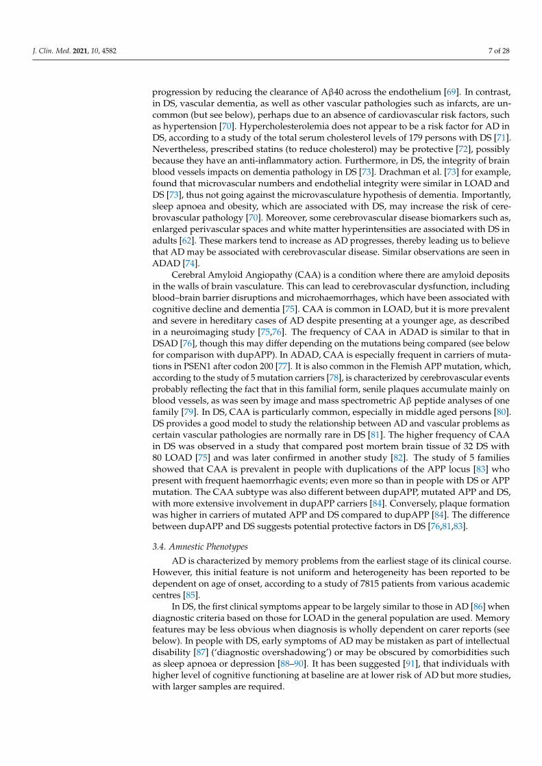
predictor of survival time along with severity of intellectual disability, socio-economicstatus, anti-dementia medication status and history of epilepsy [61].
Among those with DSAD and LOAD, there are sex differences in age of onset, withwomen at greater risk of developing the condition [62]. Longitudinal studies have sug-gested that variants in the oestrogen receptor genes-β [63] and -α [64]. Subsequently, it hasbeen suggested that age at menopause increases the risk of AD in DSAD and in LOAD,possibly menopausal oestrogen deficiency being a factor. The impact of oestrogen may beimportant in familial AD too, since the risk of the condition is associated with in oestrogensreceptor-1 gene variants [65].
Figure 2 shows the age of onset of diagnosis and survival time for the different formsof AD. Despite differences in age of onset, individuals with ADAD and LOAD have similarsurvival times. Among those with DSAD, the time between diagnosis and death in DS mayreflect a bias from late diagnosis, or the impact of comorbidities. Such an explanation isconsistent with the clinical course of DSAD appearing similar to that of individuals withan APP duplication (dupAPP).
Similarly, shorter survival times [32] are associated with ADAD with clinical symp-toms appearing at an earlier or later age, rather than in midlife (between 35 and 65 years)The duration of survival for younger people is likely to reflect the highly pathogenic natureof the mutations [32], while for their older counterparts, it is likely to be limited by expectedlifespan. PSEN2 mutation carriers typically have a later onset than the other hereditarycases as shown by a study of 1307 ADAD mutation carriers [32]. This may lead them to becategorised as LOAD [66]. It is important to note that Figure 2 does not account for thedifferences due to the different mutations that can occur a particular gene in ADAD. Forinstance, PSEN1 mutations before codon 200 are usually associated with an earlier onset[41.3± 7.2] than those with mutations at sites beyond [45.8± 6.4], as seen in a retrospectiveanalysis of 168 PSEN1 mutation carriers (p < 0.0001) [22].
J. Clin. Med. 2021, 10, x FOR PEER REVIEW 6 of 29
even TAU haplotypes [59,60]. Among this group, age of diagnosis is an important predic-
tor of survival time along with severity of intellectual disability, socio-economic status,
anti-dementia medication status and history of epilepsy [61].
Among those with DSAD and LOAD, there are sex differences in age of onset, with
women at greater risk of developing the condition [62]. Longitudinal studies have sug-
gested that variants in the oestrogen receptor genes-β [63] and -α [64]. Subsequently, it
has been suggested that age at menopause increases the risk of AD in DSAD and in LOAD,
possibly menopausal oestrogen deficiency being a factor. The impact of oestrogen may be
important in familial AD too, since the risk of the condition is associated with in oestro-
gens receptor-1 gene variants [65].
Figure 2 shows the age of onset of diagnosis and survival time for the different forms
of AD. Despite differences in age of onset, individuals with ADAD and LOAD have sim-
ilar survival times. Among those with DSAD, the time between diagnosis and death in DS
may reflect a bias from late diagnosis, or the impact of comorbidities. Such an explanation
is consistent with the clinical course of DSAD appearing similar to that of individuals with
an APP duplication (dupAPP).
Similarly, shorter survival times [32] are associated with ADAD with clinical symp-
toms appearing at an earlier or later age, rather than in midlife (between 35 and 65 years)
The duration of survival for younger people is likely to reflect the highly pathogenic na-
ture of the mutations [32], while for their older counterparts, it is likely to be limited by
expected lifespan. PSEN2 mutation carriers typically have a later onset than the other he-
reditary cases as shown by a study of 1307 ADAD mutation carriers [32]. This may lead
them to be categorised as LOAD [66]. It is important to note that Figure 2 does not account
for the differences due to the different mutations that can occur a particular gene in
ADAD. For instance, PSEN1 mutations before codon 200 are usually associated with an
earlier onset [41.3 ± 7.2] than those with mutations at sites beyond [45.8 ± 6.4], as seen in a
retrospective analysis of 168 PSEN1 mutation carriers (p < 0.0001) [22].
Figure 2. Schematic representation of the average duration of Alzheimer’s clinical course that has been reported for the
sporadic form of the disease (LOAD) [67], the familial one (PSEN1, PSEN2, APP and dAPP) [68]; and AD in the DS popu-
lation (DS) [61]. * The mean age of death for LOAD was calculated from the mean age at onset plus the average survival;
LOAD= Late-onset Alzheimer’s disease.
3.3. Vasculature
The vasculature plays a very important role in the pathology of dementia. In LOAD,
the association with vascular risk factors may be detrimental as it may accelerate dementia
80.2 ± 6
43.3 ± 8.6
58.1 ± 9.5
47.6 ± 7.151.5 ± 5.3
55.8 ± 6.29
86.1 ± 7.04*
50.5 ± 9.7
71.8 ± 10.6
58 ± 8.460.4 ± 6.2 59.98 ± 5.98
-
10.00
20.00
30.00
40.00
50.00
60.00
70.00
80.00
90.00
100.00
Age (years)
LOAD PSEN1 PSEN2 APP dAPP DS
Mean years of AD clinical course
Mean age at onset Mean age of death
Figure 2. Schematic representation of the average duration of Alzheimer’s clinical course that has been reported for thesporadic form of the disease (LOAD) [67], the familial one (PSEN1, PSEN2, APP and dAPP) [68]; and AD in the DSpopulation (DS) [61]. * The mean age of death for LOAD was calculated from the mean age at onset plus the averagesurvival; LOAD = Late-onset Alzheimer’s disease.
3.3. Vasculature
The vasculature plays a very important role in the pathology of dementia. In LOAD,the association with vascular risk factors may be detrimental as it may accelerate dementia
J. Clin. Med. 2021, 10, 4582 7 of 28
progression by reducing the clearance of Aβ40 across the endothelium [69]. In contrast,in DS, vascular dementia, as well as other vascular pathologies such as infarcts, are un-common (but see below), perhaps due to an absence of cardiovascular risk factors, suchas hypertension [70]. Hypercholesterolemia does not appear to be a risk factor for AD inDS, according to a study of the total serum cholesterol levels of 179 persons with DS [71].Nevertheless, prescribed statins (to reduce cholesterol) may be protective [72], possiblybecause they have an anti-inflammatory action. Furthermore, in DS, the integrity of brainblood vessels impacts on dementia pathology in DS [73]. Drachman et al. [73] for example,found that microvascular numbers and endothelial integrity were similar in LOAD andDS [73], thus not going against the microvasculature hypothesis of dementia. Importantly,sleep apnoea and obesity, which are associated with DS, may increase the risk of cere-brovascular pathology [70]. Moreover, some cerebrovascular disease biomarkers such as,enlarged perivascular spaces and white matter hyperintensities are associated with DS inadults [62]. These markers tend to increase as AD progresses, thereby leading us to believethat AD may be associated with cerebrovascular disease. Similar observations are seen inADAD [74].
Cerebral Amyloid Angiopathy (CAA) is a condition where there are amyloid depositsin the walls of brain vasculature. This can lead to cerebrovascular dysfunction, includingblood–brain barrier disruptions and microhaemorrhages, which have been associated withcognitive decline and dementia [75]. CAA is common in LOAD, but it is more prevalentand severe in hereditary cases of AD despite presenting at a younger age, as describedin a neuroimaging study [75,76]. The frequency of CAA in ADAD is similar to that inDSAD [76], though this may differ depending on the mutations being compared (see belowfor comparison with dupAPP). In ADAD, CAA is especially frequent in carriers of muta-tions in PSEN1 after codon 200 [77]. It is also common in the Flemish APP mutation, which,according to the study of 5 mutation carriers [78], is characterized by cerebrovascular eventsprobably reflecting the fact that in this familial form, senile plaques accumulate mainly onblood vessels, as was seen by image and mass spectrometric Aβ peptide analyses of onefamily [79]. In DS, CAA is particularly common, especially in middle aged persons [80].DS provides a good model to study the relationship between AD and vascular problems ascertain vascular pathologies are normally rare in DS [81]. The higher frequency of CAAin DS was observed in a study that compared post mortem brain tissue of 32 DS with80 LOAD [75] and was later confirmed in another study [82]. The study of 5 familiesshowed that CAA is prevalent in people with duplications of the APP locus [83] whopresent with frequent haemorrhagic events; even more so than in people with DS or APPmutation. The CAA subtype was also different between dupAPP, mutated APP and DS,with more extensive involvement in dupAPP carriers [84]. Conversely, plaque formationwas higher in carriers of mutated APP and DS compared to dupAPP [84]. The differencebetween dupAPP and DS suggests potential protective factors in DS [76,81,83].
3.4. Amnestic Phenotypes
AD is characterized by memory problems from the earliest stage of its clinical course.However, this initial feature is not uniform and heterogeneity has been reported to bedependent on age of onset, according to a study of 7815 patients from various academiccentres [85].
In DS, the first clinical symptoms appear to be largely similar to those in AD [86] whendiagnostic criteria based on those for LOAD in the general population are used. Memoryfeatures may be less obvious when diagnosis is wholly dependent on carer reports (seebelow). In people with DS, early symptoms of AD may be mistaken as part of intellectualdisability [87] (‘diagnostic overshadowing’) or may be obscured by comorbidities suchas sleep apnoea or depression [88–90]. It has been suggested [91], that individuals withhigher level of cognitive functioning at baseline are at lower risk of AD but more studies,with larger samples are required.
J. Clin. Med. 2021, 10, 4582 8 of 28
Starting with a cross-sectional study of 68 institutionalised people with DS, it has beenfound that one of the earliest signs of dementia is an impairment of recent memory in theabsence of loss of long-term memory [92]. The earliest stages of DSAD are characterisedby, episodic memory loss [93–97], similar to that of the mild cognitive impairment (MCI)phase described in people with LOAD [98] and ADAD [20]. The loss of episodic mem-ory precedes functional deterioration [99,100] by many years. Similarly, a retrospectivestudy of 449 PSEN E280A mutation carriers described an amnestic cognitive impairmentphase preceding the onset of dementia by up to two decades [101]. The loss of episodicmemory, measured by free recall correlates with pre-clinical accelerated hippocampalatrophy (see the DIAN cohort studies; [102], and has been associated with early navigationproblems [103]. In later stages, a deterioration in cued recall is found [99].
The timeline of deterioration of skills depends on the particular form of AD. In a10 year longitudinal prospective study of 19 people with ADAD, it was observed that inthe early stages, naming, spelling and visuo-perceptual skills were better preserved than inLOAD [104]. Visual deficits occurred in the late stages [20], particularly in PSEN1 mutationsafter codon 200 [68]. Similarly, for those with DS, loss of visuospatial skills occurs early [93–97]. However, visuo-perceptual deficits, similar to those reported in LOAD [105], have alsobeen reported in people with DS without dementia using psychophysical tests [106] andvisual evoked potentials [107]. The mean ages of these DS cohorts were c. 40 years and 26years, respectively. How these pre-dementia diagnossis changes in DS relate to age-relateddecline is unclear.
In DS, attention deficits have been described in the early stages of AD [87,97,108],while in ADAD, attention deficits are observed in the preclinical phases of the disease [109].With regard to language, in DS, there is already a major neurodevelopmental effect onexpressive language, and cases of LOAD can also present with language deficits [110]. Infact, assessment of language skills could potentially help in identifying earlier DSAD [111].In ADAD, language deterioration has been associated with a specific PSEN2 [112] andanother PSEN1 mutation, whereas memory has been relatively well-preserved [113].
With AD progression in DS, amnestic symptoms increase [114]. Carers report generalslowness, greater effects on cognitive domains, and the loss of adaptive skills [49,115] overtime. In contrast, among DS individuals without dementia, a decline in functional skills ismuch more common [116], perhaps reflecting better established skills in individuals whodo not have dementia and therefore not so demanding.
3.5. Non-Amnestic Phenotypes
In AD, non-amnestic phenotypes are rarer than amnestic phenotypes. Such atypicalpresentations may be associated with the altered proteolytic processing of other substratesrather than of APP. Both atypical and typical symptoms do not correlate well with Aβplaques but rather NFT pathology [117]. In the past, individuals with atypical pheno-types were often diagnosed later but developments in biomarker testing is making earlierdiagnosis possible [118].
Executive dysfunction is a common characteristic of LOAD, and may affect up to two-thirds of people with this diagnosis [119]; however, in a minority of case (possibly arounda quarter) atypical presentations occur [19,26]. As mentioned, non-amnestic phenotypesalso present in people with DS and they already perform more poorly pre-morbidly ontasks of executive functioning than other people with intellectual disabilities [120]; furtheramong those with DSAD, performance is markedly poorer [121].
Personality changes, observed by carers [24], and often referred to as ‘behaviouraland psychological symptoms of dementia (BPSD)’ [122] may be one of the first signs of thepre-clinical stages of DSAD [123]. BPSD comprises reduced empathy, emotional lability,apathy and social withdrawal [49,124], but not necessarily depression. The constellationof symptoms was first described in a longitudinal study of 108 individuals with DS, 68 ofwhom later developed dementia [125]. Later studies have supported these findings forexample, personality changes were reported during the first stages of AD in a sample of
J. Clin. Med. 2021, 10, 4582 9 of 28
participants (n = 331) from the DIAN cohort [24,126]. Changes in personality may, indeed,frequently occur before a decline in memory during the clinical stage of DSAD [124].Despite the appearance of BPSD, which affects daily functioning [86,127] and is thereforeeasier to identify compared to memory changes, depression remains the major mentalhealth issue affecting adults with DS [128]. As with other groups, the symptoms ofdepression may overlap with those of dementia [129].
The frequent presence of personality changes together with the executive dysfunctionin DS [121] have suggested an early impact of dementia on the frontal lobes and/or theirunderlying neuronal circuits [130,131]. The pre-frontal cortex is an area that is underde-veloped and may be more susceptible to neurodegeneration in people with intellectualdisability because of a reduction in cognitive reserve [132]. The findings of a neuroimagingstudy of fractional anisotropy, involving 20 adults with DS, half of whom had a diagnosisof DSAD, indicated a decrease in frontal white matter integrity that was associated witha decline in cognition [133]. A different perspective is that personality changes, at least,reflect the impact of negative life events, which are known to increase the risk of thedevelopment of AD [134]. Some of the available evidence does not support this view. Forexample, behavioural changes, such as disinhibition and problems in executive functioning(e.g., in planning, sequencing) are seen in the early stages of DSAD, while apathy appearsmore frequently only in the advanced stages [135]. In LOAD, in contrast, disinhibition isnot seen until dementia advanced [136]. Further studies that take into account the relativeunderdevelopment of the frontal lobes in individuals are needed. Similarly, it will beimportant to consider the heterogeneity of the DS population: in people with severe orprofound intellectual disabilities, early behavioural or personality changes are much morereadily identifiable than changes in memory [124].
Personality changes that are so marked that they present as psychosis have also beenreported among all groups with dementia. The symptoms affect about half of those withLOAD [137] but their prevalence remains uncertain in DSAD and the hereditary formsof dementia.
3.6. Neurological Symptoms
Neurological symptoms can present as the non-amnestic phenotype of AD. These tendto be more prominent and occur earlier in DSAD than in LOAD as shown by a 14 yearslongitudinal study of 77 persons with DS where it was found that neurological symptomswere more prevalent in DS with dementia than in non-demented DS (p = 0.0075) [15].Similarly, presenting neurological symptoms are more common in ADAD [19] than inLOAD, especially in kindreds with a very early onset age of dementia (<40 years) [20].
The prevalence of neurological symptoms in ADAD was further corroborated byVoglein et al., 2019 [138]: motor symptoms had a prevalence of about 30% in ADAD, withincreasing severity as the disease progresses [138]. In ADAD, there was a higher frequencyof motor findings than in LOAD (28.4% versus 12.8%; p < 0.001) and the severity was alsogreater (mean UPDRS-III scores 2.0 versus 0.4; p < 0.001). Bradykinesia was particularlycommon in ADAD [138]. In the same study it was also found that the frequency andseverity of the motor symptoms correlated positively with Aβ deposition within thebasal ganglia. Indeed, there was a greater amount of Aβ in ADAD compared to LOAD(Pittsburgh compound B-standardized uptake value ratio 2.472 versus 1.928; p = 0.002).The heterogeneity in ADAD is noted as the frequency and severity of the motor symptomswere higher in PSEN1 mutations after codon 200 compared to those before codon 200 [138].
Some APP mutations in the Aβ coding domain (positions 692–694) have a clinical pre-sentation at onset with epilepsy or resembling Dementia with Lewy Bodies [26]. Between16% to 25% of PSEN1 mutation carriers, mainly with mutations in exon 8, after codon200, show a non-amnestic phenotype [22,26] and present with behavioural and psychiatricsymptoms, aphasia, visual agnosia, neurological signs and dysexecutive syndrome [26]. Aretrospective study of 85 PSEN1 and 36 APP mutation carriers revealed the presence ofspastic paraparesis, extrapyramidal signs, cerebellar ataxia and myoclonus [22]; but this is
J. Clin. Med. 2021, 10, 4582 10 of 28
a rare presentation. Myoclonus was the most common symptom and with an increasedthe likelihood of developing seizures (p = 0.001 for PSEN1 and p = 0.036 for APP). Spasticparaparesis in some PSEN1 mutation carriers have been reported in association with cottonwool plaques [139,140], Lewy Body pathology (LBP) [26] or white matter and connectivitydefects [141].
Myoclonus and seizures have both been associated with an earlier age of onset andmore severe course of the disease in ADAD as reported in a systematic review [142]. Astudy of 5 families with APP duplications found that seizures were common (reported in12 out of 21 participants) for this genotype [143]. In DS, epilepsy occurs at a frequency ofup to 13% [144] and increases in those with AD diagnosis and up to 75% of adults withDSAD having epilepsy [145,146]. In contrast, the prevalence of epilepsy is lower in bothLOAD (1.5–12.7%) and ADAD (2.8–41.7%) [147]. In a retrospective cohort study comparing6430 individuals with DS to 19,176 controls, it was found that the incidence of epilepsyis elevated at all ages (p < 0.001), at least in Western countries [148]. Seizures occur as abimodal distribution: early-onset epilepsy is associated with an absence of dementia, whilemyoclonic epilepsy in the late fourth decade of life or later is associated with the onsetof AD [149]. In addition, seizures that had presented after the onset of dementia wereassociated with an accelerated functional decline. Such findings are supported by a studyof 11 patients with DS or AD with myoclonic epilepsy [150]. Nevertheless, a review of thesymptoms of people with DS in Japan and China, where epilepsy is less prevalent amongall the population, indicates [150], high rates of epilepsy are not found always found. Thedifference could potentially be explained by an environmental impact.
The cause of seizures remains uncertain. No association has been found with APOEε4 status in ADAD [22], but in mouse models, both TAU [151] and Aβ deposition [152]have been related to epileptiform activity. Furthermore, as reviewed in [153], DS neurode-velopmental abnormalities, including structural and biochemical alterations, may play arole. It is also possible that the increased prevalence is actually due to an over-reportingof the atypical symptoms for people with ADAD and DSAD or that their younger age atonset allows them to survive until the more severe stages of the disease than in LOAD [20].Another explanation is that this is related to increased plaque load due to Aβ42 depositionwhich would explain common observation of seizures in DS, dupAPP and certain otherAPP mutations [50]. Alternatively, seizures may be associated with a GABA/Glutamate (in-hibition/excitation) imbalance. In fact, GABAergic dysfunction has been seen in AD [154]and is altered in DS [155].
Another characteristic of DS are the musculoskeletal disorders which appear earlierthan in the general population, probably due to the accelerated ageing, and which cantrigger abnormal posture and unsteady gait. This decline has also been associated withLOAD in its pre-clinical stage [156].
4. Neuroimaging and Neuropathology
A cross-sectional functional MRI connectivity study of 83 mutation carriers fromthe DIAN cohort, harbouring either APP, PSEN1 or PSEN2 mutations, reported that theclinical diagnosis of AD can be preceded by many years by functional disruptions of thedefault mode network (DMN) in ADAD [157]. Similar changes in resting state network,though occurring later, are found in LOAD [157,158]. Altered default mode connectivitywas also observed in adults with DS with detectable fibrillar Aβ as measured by positronemission tomography (PET) using PiB as the tracer [159]. There are different explanationsfor the functional changes described. In ADAD, pre-clinical white matter degenerationmay contribute to DMN disruption [160]. A similar explanation may be applicable toDS [12]. In DSAD, the preclinical stage could be characterised by different compensatoryresponses [161]. For example, an increase in glucose uptake was described in a cross-sectional PET study that compared the regional cerebral glucose metabolic rates (GMR) of17 DS with both demented (n = 10) and non-demented controls (n = 24) during a cognitivetask [162].
J. Clin. Med. 2021, 10, 4582 11 of 28
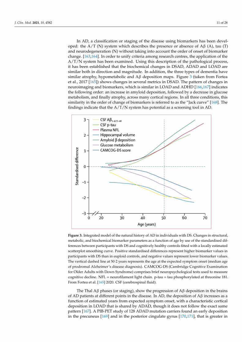
In AD, a classification or staging of the disease using biomarkers has been devel-oped: the A/T (N) system which describes the presence or absence of Aβ (A), tau (T)and neurodegeneration (N) without taking into account the order of onset of biomarkerchange. [163,164]. In order to unify criteria among research centres, the application of theA/T/N system has been examined. Using this description of the pathological process,it has been established that the biochemical changes in DSAD, ADAD and LOAD aresimilar both in direction and magnitude. In addition, the three types of dementia havesimilar atrophy, hypometabolic and Aβ deposition maps. Figure 3 (taken from Forteaet al., 2017 [165]) shows changes in several metrics in DSAD. The pattern of changes inneuroimaging and biomarkers, which is similar in LOAD and ADHD [166,167] indicatesthe following order: an increase in amyloid deposition, followed by a decrease in glucosemetabolism, and finally atrophy, across many cortical regions. In all three conditions, thissimilarity in the order of change of biomarkers is referred to as the “Jack curve” [168]. Thefindings indicate that the A/T/N system has potential as a screening tool in AD.
J. Clin. Med. 2021, 10, x FOR PEER REVIEW 11 of 29
sponses [161]. For example, an increase in glucose uptake was described in a cross-sec-
tional PET study that compared the regional cerebral glucose metabolic rates (GMR) of 17
DS with both demented (n = 10) and non-demented controls (n = 24) during a cognitive
task [162].
In AD, a classification or staging of the disease using biomarkers has been developed:
the A/T (N) system which describes the presence or absence of Aβ (A), tau (T) and neuro-
degeneration (N) without taking into account the order of onset of biomarker change.
[163,164]. In order to unify criteria among research centres, the application of the A/T/N
system has been examined. Using this description of the pathological process, it has been
established that the biochemical changes in DSAD, ADAD and LOAD are similar both in
direction and magnitude. In addition, the three types of dementia have similar atrophy,
hypometabolic and Aβ deposition maps. Figure 3 (taken from Fortea et al. 2017 [165])
shows changes in several metrics in DSAD. The pattern of changes in neuroimaging and
biomarkers, which is similar in LOAD and ADHD [166,167] indicates the following order:
an increase in amyloid deposition, followed by a decrease in glucose metabolism, and
finally atrophy, across many cortical regions. In all three conditions, this similarity in the
order of change of biomarkers is referred to as the “Jack curve” [168]. The findings indicate
that the A/T/N system has potential as a screening tool in AD.
Figure 3. Integrated model of the natural history of AD in individuals with DS. Changes in struc-
tural, metabolic, and biochemical biomarker parameters as a function of age by use of the standard-
ised differences between participants with DS and cognitively healthy controls fitted with a locally
estimated scatterplot smoothing curve. Positive standardised differences represent higher bi-
omarker values in participants with DS than in euploid controls, and negative values represent
lower biomarker values. The vertical dashed line at 50·2 years represents the age at the expected
symptom onset (median age of prodromal Alzheimer’s disease diagnosis). CAMCOG-DS (Cam-
bridge Cognitive Examination for Older Adults with Down Syndrome) comprises brief neuropsy-
chological tests used to measure cognitive decline. NFL = neurofilament light chain. p-tau = tau
phosphorylated at threonine 181. From Fortea et al.,[165] 2020 . CSF (cerebrospinal fluid).
Figure 3. Integrated model of the natural history of AD in individuals with DS. Changes in structural,metabolic, and biochemical biomarker parameters as a function of age by use of the standardised dif-ferences between participants with DS and cognitively healthy controls fitted with a locally estimatedscatterplot smoothing curve. Positive standardised differences represent higher biomarker values inparticipants with DS than in euploid controls, and negative values represent lower biomarker values.The vertical dashed line at 50·2 years represents the age at the expected symptom onset (median ageof prodromal Alzheimer’s disease diagnosis). CAMCOG-DS (Cambridge Cognitive Examinationfor Older Adults with Down Syndrome) comprises brief neuropsychological tests used to measurecognitive decline. NFL = neurofilament light chain. p-tau = tau phosphorylated at threonine 181.From Fortea et al. [165] 2020. CSF (cerebrospinal fluid).
The Thal Aβ phases (or staging), show the progression of Aβ deposition in the brainsof AD patients at different points in the disease. In AD, the deposition of Aβ increases as afunction of estimated years from expected symptom onset, with a characteristic corticaldeposition in LOAD that is shared by ADAD, though it does not follow the exact samepattern [167]. A PIB-PET study of 128 ADAD mutation carriers found an early depositionin the precuneus [169] and in the posterior cingulate gyrus [170,171], that is greater in
J. Clin. Med. 2021, 10, 4582 12 of 28
those with PSEN1 mutations after codon 200, as well as for those carrying an ε4 allele [20].Similar brain amyloid deposition patterns are seen in DSAD, whereby there are neuriticplaques in the parieto-temporal, precuneus, posterior cingulate, and frontal regions similarto LOAD [172]. A (18F) FDDNP PET amyloid study showed that plaque density was higherin the brains of 19 adults with DS without dementia when compared with 10 patients withLOAD, and its deposition was greater in the frontal and parietal cortex [173].
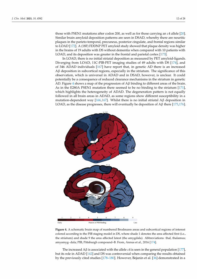
In LOAD, there is no initial striatal deposition as measured by PET amyloid-ligands.Diverging from LOAD, 11C-PIB-PET imaging studies of 49 adults with DS [174], andof 346 ADAD individuals [167] have report that, in genetic AD there is an increasedAβ deposition in subcortical regions, especially in the striatum. The significance of thisobservation, which is universal in ADAD and in DSAD, however, is unclear. It couldpotentially be a consequence of reduced clearance mechanisms in the striatum in geneticAD. Figure 4 shows a map of the progression of Aβ binding to different areas of the brain.As in the E280A PSEN1 mutation there seemed to be no binding to the striatum [171],which highlights the heterogeneity of ADAD. The degeneration pattern is not equallyfollowed in all brain areas in ADAD, as some regions show different susceptibility in amutation-dependent way [166,167]. Whilst there is no initial striatal Aβ deposition inLOAD, as the disease progresses, there will eventually be deposition of Aβ there [175,176].
J. Clin. Med. 2021, 10, x FOR PEER REVIEW 13 of 29
Figure 4. A schematic brain map of numbered Brodmann areas and subcortical regions of interest colored according to
the PIB staging model in DS, where shade 1 denotes the area affected first (i.e., the striatum) and shade 9 the area affected
latest (the amygdala). Abbreviations: thal, thalamus; amyamyg- dala; PIB, Pittsburgh compound–B. From, Annus et al.,
2016 [174].
Aβ42 plaques are more prevalent than Aβ40 plaques at all ages in ADAD and DS
[12,182]. In DS, concentrations of Aβ40 and Aβ42 are higher compared to LOAD [183].
However, the Aβ42/Aβ40 ratio shows no difference between the two conditions, demon-
strating similar processing and deposition of Aβ40 and Aβ42 [183] during the “transition”
phase to greater plaque numbers. Novel mass spectrometry techniques that accurately
detect Aβ in LOAD have not yet been reported in DS [184]. In ADAD, a study comparing
18 children with the PSEN1 E280A mutations with 19 noncarriers showed that ADAD
mutation carriers had increased plasma Aβ42 levels when compared to controls (p < 0.001)
[171]. Exceptions, however, are notable. For instance, in the APP mutations—Dutch mu-
tation E693Q or the Italian mutation E693K—the Aβ42/40 ratio is reduced [50]. These ex-
ceptions may, at first sight, seem to contradict the amyloid hypothesis. However, a reduc-
tion in the ratio does not necessarily mean a reduction in the absolute level of Aβ. A re-
duction in the ratio can be due to increases in Aβ40 and/or reduction in A42, but with an
overall increase in total Aβ.
Different studies have reported a range of associations between changes in Aβ levels
and dementia status. A prospective study of 530 individuals found a positive association
between AD and alterations in the concentration of Aβ42 but not Aβ40 [185], a 10-year
follow-up case–cohort study of 6713 participants found that both concentrations were af-
fected [186] and a longitudinal study of 237 ADAD [187] reported no changes in Aβ con-
centration. These different results are also seen in DSAD [12]. A prospective study of 204
adults with DS found alterations just in Aβ42 concentration related to AD [179]. Another
Figure 4. A schematic brain map of numbered Brodmann areas and subcortical regions of interestcolored according to the PIB staging model in DS, where shade 1 denotes the area affected first (i.e.,the striatum) and shade 9 the area affected latest (the amygdala). Abbreviations: thal, thalamus;amyamyg- dala; PIB, Pittsburgh compound–B. From, Annus et al., 2016 [174].
The increased Aβ is associated with the allele ε4 is seen in the general population [177],but its role in ADAD [142] and DS was controversial when comparing the results obtainedby the previously cited studies [178–180]. However, Bejanin et al. [36] demonstrated in a
J. Clin. Med. 2021, 10, 4582 13 of 28
DS cohort of 464 that the ε4 allele does indeed influence the earlier onset of the presence ofamyloid biomarkers.
Aβ42 plaques are more prevalent than Aβ40 plaques at all ages in ADAD andDS [12,181]. In DS, concentrations of Aβ40 and Aβ42 are higher compared to LOAD [182].However, the Aβ42/Aβ40 ratio shows no difference between the two conditions, demon-strating similar processing and deposition of Aβ40 and Aβ42 [182] during the “transition”phase to greater plaque numbers. Novel mass spectrometry techniques that accuratelydetect Aβ in LOAD have not yet been reported in DS [183]. In ADAD, a study comparing18 children with the PSEN1 E280A mutations with 19 noncarriers showed that ADAD muta-tion carriers had increased plasma Aβ42 levels when compared to controls (p < 0.001) [171].Exceptions, however, are notable. For instance, in the APP mutations—Dutch mutationE693Q or the Italian mutation E693K—the Aβ42/40 ratio is reduced [50]. These exceptionsmay, at first sight, seem to contradict the amyloid hypothesis. However, a reduction in theratio does not necessarily mean a reduction in the absolute level of Aβ. A reduction inthe ratio can be due to increases in Aβ40 and/or reduction in Aβ42, but with an overallincrease in total Aβ.
Different studies have reported a range of associations between changes in Aβ levelsand dementia status. A prospective study of 530 individuals found a positive associationbetween AD and alterations in the concentration of Aβ42 but not Aβ40 [184], a 10-yearfollow-up case–cohort study of 6713 participants found that both concentrations wereaffected [185] and a longitudinal study of 237 ADAD [186] reported no changes in Aβconcentration. These different results are also seen in DSAD [12]. A prospective study of 204adults with DS found alterations just in Aβ42 concentration related to AD [179]. Anotherproject with 225 adults with DS found an increased Aβ42 and decreased Aβ40 [187].Similarly, lower levels of Aβ40 were found in 44 participants with DS and dementiacompared with 83 individuals with DS and no dementia [188]. A study of 506 DS foundincreases in both peptides [189]. In a meta-analysis Alhajraf et al., 2019 [190] showed thatAβ40 levels in the plasma increased and that plasma Aβ40 levels could potentially act as amarker for predicting AD in DS. Some studies reported no changes in Aβ concentration aswas the case reported in a longitudinal study with 78 DS participants [178], and anotherstudy with 60 [180]. It is critical to point out, however, that is no agreement about the bestkind of plasma biomarker assay. The contradictory findings may reflect the use of differentassays in different studies.
CSF soluble Aβ42 levels in ADAD [186] and DS [191] are high early in life and thendecline rapidly as they presumably start depositing into plaques [192]. ADAD mutations,an ApoE ε4 allele and the chromosome 21 trisomy have been for a long time associated witha higher density of plaques, as described by in a report of the distribution of senile plaquesin AD [193]. In a post-mortem study of 60 patients with ADAD, neuritic plaques werefound at a higher level compared to 120 participants with LOAD [77]. In PSEN1 mutations,particularly those after codon 200, cotton-wool plaques seem to be more frequent [194].In DS, a histological study described that, not only the density of plaques is altered, butalso its form: for 12 DS cases, amyloid plaques were larger and had a more amorphousmorphology than those present in 10 LOAD cases [195]. However, certain studies haveshown that Aβ plaques and neurofibrillary tangles in DSAD are similar in appearance toLOAD [196].
Following Aβ deposition metabolic deficits start; in a retrospective study of 146 withDS metabolism was associated with a tendency from aerobic (lower lactic acid levels)towards more glycolysis and subsequent lactic acid fermentation metabolism [197]. Hy-pometabolism drives widespread cell stress that leads to neuronal loss, especially in theprecuneus of ADAD mutation carriers, as described by a FDG-PET study of 20 ADADversus 20 LOAD subjects and in the posterior cingulated cortices of ADAD [198] andDS [162]. The hypometabolism pattern in DS is similar to that in LOAD involving theparietal, precuneus and posterior cingulate [199,200]. Some studies have reported hyper-
J. Clin. Med. 2021, 10, 4582 14 of 28
metabolism in DS, especially in young adults as a consequence of less efficient glycolysisor potential compensatory mechanisms [162,201,202].
Atrophy tends to occur in AD. Serial MRI scans of 66 ADAD participants compared to28 controls [102] showed that approaching the age of onset of dementia, there is increasedventricular volume and hippocampal atrophy. In comparison with LOAD, a prospectiveMRI study of 12 patients showed that atrophy has an accelerated course in ADAD [203].Atrophy was also seen in the Down Syndrome Biomarker Initiative, in which 12 DS adultstook part for the volumetric study [204]. The atrophy pattern in DS is similar to thatin LOAD, involving posterior dominant cortical thinning with atrophy of hippocampus,thalamus, and striatum [205,206]. At this stage, the brain reserve is not sufficient tocompensate for the deficits, and the symptoms that characterise and are associated witha clinical diagnosis of dementia become evident. In DS, atrophy seems to be less intenseperhaps suggesting that chromosome 21 may encode a gene that is neuroprotective whentriplicated [40]. Another possibility, however, is that the more limited atrophy may simplyreflect smaller whole brain volume in DS, as described in a study from 1991 of 7 adultswith DS [207].
Moreover, the regional distribution of tau (both from PET studies and histopathology)is broadly similar in DS and LOAD [208]. The pattern accords with the tau Braak stagingsystem [209,210]. Braak staging shows the distribution of tau within the brain at differentpoints in AD. Amyloid deposition precedes tau pathology in the cerebral cortex andsubcortical nuclei of the forebrain, akin to LOAD [208]. In addition, and as in LOAD, taupathology is first seen in the entorhinal cortex of the temporal lobe [208]. Importantly,the early involvement of brainstem monoamine producing neuron systems has also beendescribed in DS [208]. The distribution of tau in ADAD is also similar to that in LOADwith the areas of high tau PET binding overlapping with those in LOAD [211]. Similar toLOAD, tau is present in the temporal and parietal regions [211]. However, despite similarcognitive impairment in the two conditions, there seems to be greater cortical involvementand higher levels of binding in ADAD [211].
Later in life, there is an increase in CSF tau concentration levels in LOAD [212] andDS [192], while in ADAD, there is a decrease in its levels, according to a longitudinal studyof 411 individuals [213]. In fact, CSF and plasma biomarkers, notably neurofilament lightand tau181, have been shown to have good potential for predicting AD in DS [165,214,215].As plaques continue to develop, tangles form, supporting the hypothesis of Aβ as thedriving force in AD [216]. The time lag between Aβ pathology and NFT pathology issimilar in ADAD and DS [217]. The distribution of plaques and NFT is similar between thethree groups [40] and in a 15 year prospective study of 92 hospitalised adults with DS, it wasshown that NFTs also correlate better with dementia than with amyloid deposition [218].
The different subtypes of LOAD and the neuroimaging findings and biomarkerchanges for these subtypes reveal respective differences [219]. For instance, four dis-tinct trajectories of tau deposition have been demonstrated in LOAD [220], and in termsof the temporal complexity, three subtypes can be classified [221]. In discussing the neu-roimaging and neuropathology of LOAD, the focus throughout this review has been onthe LOAD that presents with amnestic symptoms.
5. Co-Pathologies
AD is often associated with co-pathologies such as Lewy body pathology (LBP) andTDP-43 pathology [219]. The number of co-pathologies increases with age [222]. Thesecan affect both diagnosis and disease progression. Although they are not the focus of thisreview, they are briefly considered here.
5.1. Lewy Body Pathology
Lewy bodies (LB) are pathological aggregates of proteins in the brain. In an autopsystudy, it was found that the number of LB deposits in DS increases with age [223]. Clinically,this can manifest as LB dementia but it is rare in DS, but the first case was reported in
J. Clin. Med. 2021, 10, 4582 15 of 28
2010 [224]. In both LOAD and ADAD, LB dementia is a frequent comorbidity as hasbeen reported in both the ADNI and DIAN cohorts [225]. In ADAD, LB dementia is morecommon than might be expected to occur in a young population; we speculate this mayalso be true in DS. The explanation may lie in APP mismetabolism [77], following thehypothesis that LB could be induced by the accumulation of Aβ [20]. LB are less extensivein ADAD compared to LOAD [77], with the prevalence in LOAD being between 6 and39% [226].
5.2. TDP-43 Pathology
TDP-43 pathology co-occurs in all three forms of AD and seems to be associated withthe development of amnestic phenotypes [227]. The distribution of TDP-43 affects theamygdala and hippocampus more than the neocortical regions, where there is an absenceof TDP-43 [228]. However, the frequency at which TDP-43 pathology cooccurance in thethree forms differs. TDP-43 pathology seems to be more prevalent in LOAD compared toADAD and DS [227]. In LOAD, limbic predominant age-related TDP-43 encephalopathyneuropathological change (LATE-NC) occurred in 57% of cases and correlated with fasterdisease progression and cognitive impairment [229]. This, and similar, findings have ledto the hypothesis that TDP-43 pathology may be a side effect of ageing rather than ofAD [227].
6. Other Similarities and Differences
There are other similarities and differences observed in neuropathology between theLOAD and in DS that have not yet been thoroughly examined. These include impairmentsof the noradrenergic [230] and immune systems [231], NGF metabolism [232,233], enhancedinflammation, and increased oxidative stress [231]. Such features warrant further inves-tigation since although APP plays an important role, there are many oxidative [234,235]and inflammatory [236] genes on chromosome 21 that overexpressed in DS. These genesmay cause a neuroinflammatory states that as in LOAD, may result in a self-amplifyingcycle that leads to the development and/or maintenance of AD [237]. In common withLOAD [238], DS shows upregulation of inflammatory response, as seen by elevated lev-els of cytokines [239] or the association of proteins of the complement cascade to Aβplaques [240]. Moreover, in DS, there is also increased microglial activation with increasedage compared with healthy controls [241]. Nevertheless, there appear to be differences: forexample, reflecting the distinct profile of microglial states in LOAD, the neuroinflammatoryphenotypes of the two conditions are not the same [242].
There are other areas to investigate. First, there is neopterin, a marker for cell-mediatedimmune activation and inflammation. Higher plasma concentrations have been foundin individuals with DSAD compared with those with DS without dementia [243]. At thesame time, neopterin levels in urine predict cognitive decline in people with DS overtime [244]. A different focus is IL1β. Levels of IL1β levels were higher in DS comparedto LOAD [182]. Moreover, IL1β was correlated with t-tau, suggesting that it may beassociated with neurodegeneration [182]. It should be noted, however, that high levelsof IL1β in DS may simply reflect increased prevalence of autoimmune conditions and/orheightened vulnerability to infections in people with the condition [182], presenting asautoimmune diseases [245] such as hypothyroidism [246]. Secondly, the development ofDSAD may also be facilitated by increased vulnerability to certain infections. For example,the severe immunodeficiency in the salivary IgA for DS [247] along with the increasedsusceptibility for impairments of DS’s gingival fibroblasts [248] may be related to the higherprevalence of severe early-onset periodontal diseases in DS [249]. Periodontitis caused byPorphyromonas gingivalis has been linked to an increased risk of developing AD [250,251].
In DS, mitochondrial dysfunction and its consequent higher levels of reactive oxygenspecies (ROS) have been reported [12]. Oxidative stress is enhanced in DS compared to thegeneral population and may contribute to increased lipid and protein peroxidation thatpromotes increase in DSAD [252]. Moreover, the level of superoxide dismutase enzymes,
J. Clin. Med. 2021, 10, 4582 16 of 28
which are antioxidant enzymes, could predict memory decline over time in DS [253]. Fur-ther corroborating the relationship between AD and oxidative stress is the suggestion thatchanges in iPF2α, a marker of oxidative stress, are correlated with cognitive decline [254].Increased oxidation has also been linked with increased Aβ production [255,256]. Moreover,as in LOAD, the mitochondrial DNA (mtDNA) mutation rate is particularly high in thebrains of people with DSAD [257]. All these processes could contribute to accelerating theonset of dementia in DS [235]. It has been suggested however that other researchers suggestthat the systemic accumulation of Aβ or the lipid peroxidation may alter the mitochondriaintegrity with the resulting dysfunction leading to a self-amplifying loop [234].
7. Conclusions
In the genetic cases of AD (DSAD and ADAD), there are life-long neuropathologicalchanges that are not present in the general population until the early stages of AD. In themajority of cases in all the three groups compared, the clinical appearance of dementiastarts with memory deficits. However, in ADAD and DS, there are more non-amnesticphenotypes. Moreover, in those with DS and most forms of ADAD, there is also moresevere CAA and increased neurological symptoms compared to LOAD. Both people withDS and carriers of ADAD mutations show a higher and earlier brain Aβ load, as well as anincreased accumulation of Aβ plaques and NFTs. The increased Aβ deposition probablyaccounts for the early age of onset described in these hereditary AD cases. Though themagnitude and direction of changes in the three conditions are generally similar, thereare some differences. In contrast to LOAD, ADAD and DSAD have an increased initialaccumulation in the subcortical regions, particularly in the striatum. Table 1 recapitulatesthe similarities and differences between the three forms of AD, highlighting the hetero-geneities, particularly in ADAD and age associated comorbidities There remain gaps inour understanding the reasons for the differences in clinical presentation and the genotype-phenotype relations of these conditions. With on-going large longitudinal clinical studiesof DS, ADAD and LOAD (all funded by the National Institutes of Health), namely ABC-DS (Alzheimer’s Biomarkers Consortium-DS), DIAN and ADNI, respectively, data fromthese studies are beginning to provide a fine-grained characterisation and understandingof AD. Concurrently, biochemical and cellular understanding is being made possible bystudying known mutations of the genetic forms of the disease aided by the use of inducedpluripotent stem cells, organoids and gene-editing techniques. Importantly, a multi-scaleunderstanding (for example, molecular changes and their impact on tissue pathology orcognition) and the generation of new hypotheses is likely when individual case studies arecomprehensively investigated.
There were multiple difficulties encountered throughout this review. The main lim-itation was the lack of head-to-head comparisons. It is also important to highlight thatmany features of DSAD, especially those defined decades ago have not been replicated. Itis difficult to extrapolate which features belong to ADAD in general, rather than to specificmutations. For the mutations in a particular gene or from the same kindred, we cannotdiscard that the characteristics studied belong to the specific mutation of those patients,as would happen with a case-report, rather than being a shared feature of mutations inthat gene. This is especially the case in PSEN2, which is rare and so cohorts studied tendto be small. Taking all these considerations into account, we consider that much is leftto be explored and more research is a must. Understanding the pathology behind thedifferent forms of AD and the differences and similarities with LOAD will hopefully allowus a deeper insight into the causes of AD and potentially lead to the development of newtargeted and personalised therapies.
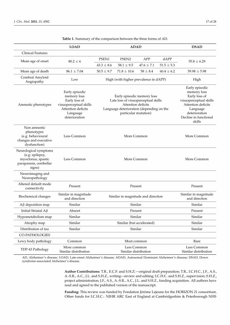
J. Clin. Med. 2021, 10, 4582 17 of 28
Table 1. Summary of the comparison between the three forms of AD.
LOAD ADAD DSAD
Clinical Features
Mean age of onset 80.2 ± 6PSEN1 PSEN2 APP dAPP
55.8 ± 6.2943.3 ± 8.6 58.1 ± 9.5 47.6 ± 7.1 51.5 ± 5.3
Mean age of death 86.1 ± 7.04 50.5 ± 9.7 71.8 ± 10.6 58 ± 8.4 60.4 ± 6.2 59.98 ± 5.98
Cerebral AmyloidAngiopathy Low High (with higher prevalence in dAPP) High
Amnestic phenotypes
Early episodicmemory lossEarly loss of
visuoperceptual skillsAttention deficits
Languagedeterioration
Early episodic memory lossLate loss of visuoperceptual skills
Attention deficitsLanguage deterioration (depending on the
particular mutation)
Early episodicmemory lossEarly loss of
visuoperceptual skillsAttention deficits
Languagedeterioration
Decline in functionalskills
Non amnesticphenotypes
(e.g: behaviouralchanges and executive
dysfunction)
Less Common More Common More Common
Neurological symptoms(e.g: epilepsy,
myoclonus, spasticparaparesis, cerebellar
signs)
Less Common More Common More Common
Neuroimaging andNeuropathology
Altered default modeconnectivity Present Present Present
Biochemical changes Similar in magnitudeand direction Similar in magnitude and direction Similar in magnitude
and direction
Aβ deposition map Similar Similar Similar
Initial Striatal Aβ Absent Present Present
Hypometabolism map Similar Similar Similar
Atrophy map Similar Similar (but accelerated) Similar
Distribution of tau Similar Similar Similar
CO-PATHOLOGIES
Lewy body pathology Common Most common Rare
TDP-43 Pathology More commonSimilar distribution
Less CommonSimilar distribution
Less CommonSimilar distribution
AD, Alzheimer’s disease; LOAD, Late-onset Alzheimer’s disease; ADAD, Autosomal Dominant Alzheimer’s disease; DSAD, Downsyndrome-associated Alzheimer’s disease.
Author Contributions: T.R., E.C.F. and S.H.Z.—original draft preparation; T.R., I.C.H.C., J.F., A.S.,A.-S.R., A.C., J.L. and S.H.Z., writing—review and editing; I.C.H.C. and S.H.Z., supervision; S.H.Z.,project administration; J.F., A.S., A.-S.R., A.C., J.L. and S.H.Z., funding acquisition. All authors haveread and agreed to the published version of the manuscript.
Funding: This review was funded by Fondation Jérôme Lejeune for the HORIZON 21 consortium.Other funds for I.C.H.C.: NIHR ARC East of England at Cambridgeshire & Peterborough NHS
J. Clin. Med. 2021, 10, 4582 18 of 28
Foundation Trust; J.F.: the Fondo de Investigaciones Sanitario (FIS), Instituto de Salud Carlos III(PI14/01126, PI17/01019 and PI20/01473) and the CIBERNED program (Program 1, AlzheimerDisease and SIGNAL study, www.signalstudy.es, accessed on 1 July 2021), partly jointly fundedby Fondo Europeo de Desarrollo Regional, Unión Europea, Una manera de hacer Europa. Thework was also supported by the National Institutes of Health (NIA grants 1R01AG056850-01A1;R21AG056974 and R01AG061566), Fundació La Marató de TV3 (20141210), grants from FundacióVíctor Grífols i Lucas, and by the Generalitat de Catalunya (SLT006/17/00119); A.S.:the MRC(MR/S011277/1; MR/S005145/1; MR/R024901/1), Lumind IDSC, The LeJeune Foundation and theEuropean Commission (H2020 SC1 Gene overdosage and comorbidities during the early lifetime inDown Syndrome GO-DS21- 848077). A.-S.R.: Fondation Jérôme Lejeune for the TRIAL 21 study andthe HORIZON 21 consortium; S.H.Z.: Fondation Jérôme Lejeune for the HORIZON 21 consortium.
Conflicts of Interest: All the authors declare no conflict of interest, except: J.L.: declares speakerfees from Bayer Vital, Biogen and Roche, consulting fees from Axon Neuroscience, author fees fromThieme medical publishers and W. Kohlhammer GmbH medical publishers, non-financial supportfrom Abbvie and compensation for duty as part-time CMO from MODAG, outside the submittedwork. The funders had no role in the design of the study; in the collection, analyses, or interpretationof data; in the writing of the manuscript, or in the decision to publish the results.
References1. Carter, S.F.; Herholz, K.; Rosa-Neto, P.; Pellerin, L.; Nordberg, A.; Zimmer, E.R. Astrocyte biomarkers in Alzheimer’s disease.
Trends Mol. Med. 2019, 25, 77–95. [CrossRef]2. Keren-Shaul, H.; Spinrad, A.; Weiner, A.; Matcovitch-Natan, O.; Dvir-Szternfeld, R.; Ulland, T.K.; David, E.; Baruch, K.; Lara-
Astaiso, D.; Toth, B.; et al. A unique microglia type associated with restricting development of Alzheimer’s disease. Cell 2017, 169,1276–1290.e17. [CrossRef]
3. Kisler, K.; Nelson, A.R.; Montagne, A.; Zlokovic, B.V. Cerebral blood flow regulation and neurovascular dysfunction in Alzheimerdisease. Nat. Rev. Neurosci. 2017, 18, 419–434. [CrossRef]
4. WHO. World Health Organization. Available online: https://www.alzint.org/resource/numbers-of-people-with-dementia-worldwide/ (accessed on 30 September 2021).
5. Zhu, X.-C.; Tan, L.; Wang, H.-F.; Jiang, T.; Cao, L.; Wang, C.; Wang, J.; Tan, C.-C.; Meng, X.-F.; Yu, J.-T. Rate of early onsetAlzheimer’s disease: A systematic review and meta-analysis. Ann. Transl. Med. 2015, 3, 38. [CrossRef] [PubMed]
6. Malt, E.A.; Dahl, R.C.; Haugsand, T.M.; Ulvestad, I.H.; Emilsen, N.M.; Hansen, B.; Cardenas, Y.E.G.; Skøld, R.O.; Thorsen, A.T.B.;Davidsen, E.M.M. Health and disease in adults with Down syndrome. Tidsskr Den Nor Legeforening 2013, 133, 290–294. [CrossRef][PubMed]
7. Yang, Q.; Rasmussen, S.; Friedman, J. Mortality associated with Down′s syndrome in the USA from 1983 to 1997: A population-based study. Lancet 2002, 359, 1019–1025. [CrossRef]
8. Thase, M.E. Longevity and mortality in Down′s syndrome. J. Intellect. Disabil. Res. 2008, 26, 177–192. [CrossRef]9. Englund, A.; Jonsson, B.; Zander, C.S.; Gustafsson, J.; Annerén, G. Changes in mortality and causes of death in the Swedish Down
syndrome population. Am. J. Med. Genet. Part A 2013, 161, 642–649. [CrossRef] [PubMed]10. Oliver, C.; Holland, A.J. Down′s Syndrome and Alzheimer’s disease: A review. Psychol. Med. 1986, 16, 307–322. [CrossRef]
[PubMed]11. Handen, B.L.; Lott, I.T.; Christian, B.T.; Schupf, N.; Obryant, S.; Mapstone, M.; Fagan, A.M.; Lee, J.H.; Tudorascu, D.; Wang, M.;
et al. The Alzheimer’s Biomarker Consortium-Down Syndrome: Rationale and methodology. Alzheimer Dement. Diagn. Assess.Dis. Monit. 2020, 12, e12065. [CrossRef]
12. Head, E.; Lott, I.T.; Wilcock, D.M.; Lemere, C.A. Aging in Down syndrome and the development of Alzheimer’s diseaseneuropathology. Curr. Alzheimer Res. 2015, 13, 18–29. [CrossRef]
13. Evans, E. Dementia in People with Intellectual Disability: Guidelines for Australian GPs. Ph.D. Thesis, University of New SouthWales, Sydney, Australia, 2018.
14. Scheepers, M. Faculty for People with Intellectual Disabilities Dementia and People with Intellectual Disabilities Guidance on the Assessment,Diagnosis, Interventions and Support of People with Intellectual Disabilities Who Develop Dementia; Jessica Kingsley Publishers: London,UK, 2014.
15. McCarron, M.; McCallion, P.; Reilly, E.; Mulryan, N. A prospective 14-year longitudinal follow-up of dementia in persons withDown syndrome. J. Intellect. Disabil. Res. 2013, 58, 61–70. [CrossRef]
16. McCarron, M.; McCallion, P.; Reilly, E.; Dunne, P.; Carroll, R.; Mulryan, N. A prospective 20-year longitudinal follow-up ofdementia in persons with Down syndrome. J. Intellect. Disabil. Res. 2017, 61, 843–852. [CrossRef] [PubMed]
17. Orphanet. Available online: https://www.orpha.net/consor/cgi-bin/index.php (accessed on 1 July 2021).18. Buss, L.; Fisher, E.; Hardy, J.; Nizetic, D.; Groet, J.; Pulford, L.; Strydom, A. Intracerebral haemorrhage in Down syndrome:
Protected or predisposed? F1000Research 2016, 5, 876. [CrossRef] [PubMed]
J. Clin. Med. 2021, 10, 4582 19 of 28
19. Bateman, R.J.; Aisen, P.S.; De Strooper, B.; Fox, N.C.; Lemere, C.A.; Ringman, J.M.; Salloway, S.; Sperling, R.A.; Windisch, M.;Xiong, C. Autosomal-dominant Alzheimer’s disease: A review and proposal for the prevention of Alzheimer’s disease. AlzheimerRes. Ther. 2011, 3, 1. [CrossRef] [PubMed]
20. Ryan, N.S.; Rossor, M.N. Correlating familial Alzheimer’s disease gene mutations with clinical phenotype. Biomark. Med. 2010, 4,99–112. [CrossRef] [PubMed]
21. Castellani, R.J.; Plascencia-Villa, G.; Perry, G. The amyloid cascade and Alzheimer’s disease therapeutics: Theory versusobservation. Lab. Investig. 2019, 99, 958–970. [CrossRef] [PubMed]
22. Ryan, N.S.; Nicholas, J.M.; Weston, P.; Liang, Y.; Lashley, T.; Guerreiro, R.; Adamson, G.; Kenny, J.; Beck, J.; Chavez-Gutierrez, L.;et al. Clinical phenotype and genetic associations in autosomal dominant familial Alzheimer’s disease: A case series. LancetNeurol. 2016, 15, 1326–1335. [CrossRef]
23. Zou, Z.; Liu, C.; Che, C.; Huang, H. Clinical genetics of Alzheimer’s disease. BioMed Res. Int. 2014, 2014, 291862. [CrossRef]24. Storandt, M.; Balota, D.A.; Aschenbrenner, A.J.; Morris, J.C. Clinical and psychological characteristics of the initial cohort of the
Dominantly Inherited Alzheimer Network (DIAN). Neuropsychology 2014, 28, 19–29. [CrossRef]25. Mann, D.M.; Pickering-Brown, S.M.; Takeuchi, A.; Iwatsubo, T. Members of the familial Alzheimer’s disease pathology study
group. Amyloid angiopathy and variability in amyloid beta deposition is determined by mutation position in presenilin-1-linkedAlzheimer’s disease. Am. J. Pathol. 2001, 158, 2165–2175. [CrossRef]
26. Alberici, A.; Benussi, A.; Premi, E.; Borroni, B.; Padovani, A. Clinical, genetic, and neuroimaging features of early onset Alzheimerdisease: The challenges of diagnosis and treatment. Curr. Alzheimer Res. 2014, 11, 909–917. [CrossRef] [PubMed]
27. Dams-O′Connor, K.; Guetta, G.; Hahn-Ketter, A.E.; Fedor, A. Traumatic brain injury as a risk factor for Alzheimer’s disease:Current knowledge and future directions. Neurodegener. Dis. Manag. 2016, 6, 417–429. [CrossRef] [PubMed]
28. Franzmeier, N.; Düzel, E.; Jessen, F.; Buerger, K.; Levin, J.; Duering, M.; Dichgans, M.; Haass, C.; Suárez-Calvet, M.; Fagan, A.M.;et al. Left frontal hub connectivity delays cognitive impairment in autosomal-dominant and sporadic Alzheimer’s disease. Brain2018, 141, 1186–1200. [CrossRef] [PubMed]
29. Karch, C.M.; Goate, A.M. Alzheimer’s disease risk genes and mechanisms of disease pathogenesis. Biol. Psychiatry 2015, 77, 43–51.[CrossRef]
30. Kunkle, B.W.; Grenier-Boley, B.; Sims, R.; Bis, J.C.; Damotte, V.; Naj, A.C.; Boland, A.; Vronskaya, M.; Van Der Lee, S.J.; Amlie-Wolf,A.; et al. Genetic meta-analysis of diagnosed Alzheimer’s disease identifies new risk loci and implicates Aβ, tau, immunity andlipid processing. Nat. Genet. 2019, 51, 414–430. [CrossRef]
31. Safieh, M.; Korczyn, A.D.; Michaelson, D.M. ApoE4: An emerging therapeutic target for Alzheimer’s disease. BMC Med. 2019, 17,64. [CrossRef]
32. Ryman, D.C.; Acosta-Baena, N.; Aisen, P.S.; Bird, T.; Danek, A.; Fox, N.; Goate, A.; Frommelt, P.; Ghetti, B.; Langbaum, J.B.; et al.Symptom onset in autosomal dominant Alzheimer disease: A systematic review and meta-analysis. Neurology 2014, 83, 253–260.[CrossRef]
33. Lai, F.; Kammann, E.; Rebeck, G.W.; Anderson, A.; Chen, Y.; Nixon, R.A. APOE genotype and gender effects on Alzheimer diseasein 100 adults with Down syndrome. Neurology 1999, 53, 331. [CrossRef]
34. Prasher, V.P.; Sajith, S.G.; Rees, S.D.; Patel, A.; Tewari, S.; Schupf, N.; Zigman, W.B. Significant effect of APOE epsilon 4 genotypeon the risk of dementia in Alzheimer’s disease and mortality in persons with Down syndrome. Int. J. Geriatr. Psychiatry 2008, 23,1134–1140. [CrossRef]
35. Coppus, A.; Evenhuis, H.; Verberne, G.-J.; Visser, F.; Arias-Vasquez, A.; Sayed-Tabatabaei, F.; Vergeer-Drop, J.; Eikelenboom, P.;van Gool, W.; van Duijn, C. The impact of apolipoprotein E on dementia in persons with Down′s syndrome. Neurobiol. Aging2008, 29, 828–835. [CrossRef]
36. Benejam, B.; Videla, L.; Barroeta, I.; Fernandez, S. Association of apolipoprotein E ε 4 allele with clinical and multimodalbiomarker changes of Alzheimer disease in adults with down syndrome. JAMA Neurol. 2021, 78, 937–947.
37. Arboleda-Velasquez, J.F.; Lopera, F.; O’Hare, M.; Delgado-Tirado, S.; Marino, C.; Chmielewska, N.; Saez-Torres, K.L.; Amarnani,D.; Schultz, A.P.; Sperling, R.A.; et al. Resistance to autosomal dominant Alzheimer’s disease in an APOE3 christchurchhomozygote: A case report. Nat. Med. 2019, 25, 1680–1683. [CrossRef]
38. McNaughton, D.; Knight, W.; Guerreiro, R.; Ryan, N.; Lowe, J.; Poulter, M.; Nicholl, D.J.; Hardy, J.; Revesz, T.; Lowe, J.; et al.Duplication of Amyloid Precursor Protein (APP), but not Prion Protein (PRNP) gene is a significant cause of early onset dementiain a large UK series. Neurobiol. Aging 2012, 33, 426.e13–426.e21. [CrossRef]
39. Mok, K.Y.; Jones, E.L.; Hanney, M.; Harold, D.; Sims, R.; Williams, J.; Ballard, C.; Hardy, J. Polymorphisms in BACE2 may affectthe age of onset Alzheimer’s dementia in Down syndrome. Neurobiol. Aging 2013, 35, 1513.e1–1513.e5. [CrossRef]
40. Wiseman, F.K.; Al-Janabi, T.; Hardy, J.; Karmiloff-Smith, A.; Nizetic, D.; Tybulewicz, V.L.J.; Fisher, E.M.C.; Strydom, A. A geneticcause of Alzheimer disease: Mechanistic insights from Down syndrome. Nat. Rev. Neurosci. 2015, 16, 564–574. [CrossRef][PubMed]
41. Ryoo, S.R.; Cho, H.J.; Lee, H.W.; Jeong, H.K.; Radnaabazar, C.; Kim, Y.S.; Kim, M.J.; Son, M.Y.; Seo, H.; Chung, S.H.; et al.Dual-specificity tyrosine(Y)-phosphorylation regulated kinase 1A-mediated phos-phorylation of amyloid precursor protein:Evidence for a functional link between Down syndrome and Alzheimer’s disease. J. Neurochem. 2008, 104, 1333–1344. [CrossRef]
J. Clin. Med. 2021, 10, 4582 20 of 28
42. Wegiel, J.; Dowjat, K.; Kaczmarski, W.; Kuchna, I.; Nowicki, K.; Frackowiak, J.; Kolecka, B.M.; Wegiel, J.; Silverman, W.P.; Reisberg,B.; et al. The role of overexpressed DYRK1A protein in the early onset of neurofibrillary degeneration in Down syndrome. ActaNeuropathol. 2008, 116, 391–407. [CrossRef] [PubMed]
43. Asai, M.; Kinjo, A.; Kimura, S.; Mori, R.; Kawakubo, T.; Shirotani, K.; Yagishita, S.; Maruyama, K.; Iwata, N. Perturbed cal-cineurin-NFAT signaling is associated with the development of Alzheimer’s disease. Biol. Pharm. Bull. 2016, 39, 1646–1652.[CrossRef] [PubMed]
44. Sun, X.; He, G.; Song, W. BACE2, as a novel APP θ-secretase, is not responsible for the pathogenesis of Alzheimer’s disease inDown syndrome. FASEB J. 2006, 20, 1369–1376. [CrossRef]
45. Sun, X.; Tong, Y.; Qing, H.; Chen, C.-H.; Song, W. Increased BACE1 maturation contributes to the pathogenesis of Alzheimer’sdisease in Down syndrome. FASEB J. 2006, 20, 1361–1368. [CrossRef] [PubMed]
46. Kerkel, K.; Schupf, N.; Hatta, K.; Pang, D.; Salas, M.; Kratz, A.; Minden, M.; Murty, V.; Zigman, W.; Mayeux, R.P.; et al. AlteredDNA methylation in leukocytes with trisomy 21. PLoS Genet. 2010, 6, e1001212. [CrossRef] [PubMed]
47. Horvath, S.; Garagnani, P.; Bacalini, M.G.; Pirazzini, C.; Salvioli, S.; Gentilini, D.; Di Blasio, A.M.; Giulina, C.; Tung, S.; Vinters,H.V.; et al. Accelerated epigenetic aging in Down syndrome. Aging Cell 2015, 14, 491–495. [CrossRef] [PubMed]
48. Panegyres, P.K.; Chen, H.-Y. Differences between early and late onset Alzheimer’s disease. Am. J. Neurodegener. Dis. 2013, 2,300–306. [PubMed]
49. Deb, S.; Hare, M.; Prior, L. Symptoms of dementia among adults with Down′s syndrome: A qualitative study. J. Intellect. Disabil.Res. 2007, 51, 726–739. [CrossRef]
50. Zis, P.; Strydom, A. Clinical aspects and biomarkers of Alzheimer’s disease in Down syndrome. Free Radic. Biol. Med. 2018, 114,3–9. [CrossRef]
51. Shamseer, L.; Moher, D.; Clarke, M.; Ghersi, D.; Liberati, A.; Petticrew, M.; Shekelle, P.; Stewart, L.A.; The PRISMA-P Group.Preferred reporting items for systematic review and meta-analysis protocols (PRISMA-P) 2015: Elaboration and explanation. BMJ2015, 349, g7647. [CrossRef]
52. DIAN Observational Study. The Dominantly Inherited Alzheimer Network. Available online: https://dian.wustl.edu/ (accessedon 1 July 2021).
53. Weller, J.; Budson, A. Current understanding of Alzheimer’s disease diagnosis and treatment. F1000Research 2018, 7, 1161.[CrossRef]
54. McKhann, G.M.; Knopman, D.S.; Chertkow, H.; Hyman, B.T.; Jack, C.R., Jr.; Kawas, C.H.; Klunk, W.E.; Koroshetz, W.J.; Manly,J.J.; Mayeux, R.; et al. The diagnosis of dementia due to Alzheimer’s disease: Recommendations from the National Instituteon Aging—Alzheimer’s Association workgroups on diagnostic guidelines for Alzheimer’s disease. Alzheimer Dement. 2011, 7,263–269. [CrossRef]
55. Schupf, N. Genetic and host factors for dementia in Down′s syndrome. Br. J. Psychiatry 2002, 180, 405–410. [CrossRef]56. Lee, J.H.; Lee, A.J.; Dang, L.-H.; Pang, D.; Kisselev, S.; Krinsky-McHale, S.J.; Zigman, W.B.; Luchsinger, J.; Silverman, W.; Tycko,
B.; et al. Candidate gene analysis for Alzheimer’s disease in adults with Down syndrome. Neurobiol. Aging 2017, 56, 150–158.[CrossRef]
57. Patel, A.; Rees, S.D.; Kelly, M.A.; Bain, S.C.; Barnett, A.H.; Thalitaya, D.; Prasher, V.P. Association of variants within APOE,SORL1, RUNX1, BACE1 and ALDH18A1 with dementia in Alzheimer’s disease in subjects with Down syndrome. Neurosci. Lett.2011, 487, 144–148. [CrossRef]
58. Jones, E.L.; Mok, K.; Hanney, M.; Harold, D.; Sims, R.; Williams, J.; Ballard, C. Evidence that PICALM affects age at onset ofAlzheimer’s dementia in Down syndrome. Neurobiol. Aging 2013, 34, 2441.e1–2441.e5. [CrossRef]
59. Jones, E.L.; Margallo-Lana, M.; Prasher, V.P.; Ballard, C.G. The extended tau haplotype and the age of onset of dementia in Downsyndrome. Dement. Geriatr. Cogn. Disord. 2008, 26, 199–202. [CrossRef]
60. Raha-Chowdhury, R.; Henderson, J.W.; Raha, A.A.; Vuono, R.; Bickerton, A.; Jones, E.; Fincham, R.; Allinson, K.; Holland, A.;Zaman, S.H. Choroid plexus acts as gatekeeper for TREM2, abnormal accumulation of ApoE, and fibrillary tau in Alzheimer’sdisease and in Down syndrome dementia. J. Alzheimer Dis. 2019, 69, 91–109. [CrossRef] [PubMed]
61. Sinai, A.; Mokrysz, C.; Bernal, J.; Bohnen, I.; Bonell, S.; Courtenay, K.; Dodd, K.; Gazizova, D.; Hassiotis, A.; Hillier, R.; et al.Predictors of age of diagnosis and survival of Alzheimer’s disease in Down syndrome. J. Alzheimer Dis. 2017, 61, 717–728.[CrossRef] [PubMed]
62. Lao, P.J.; Gutierrez, J.; Keator, D.; Rizvi, B.; Banerjee, A.; Igwe, K.C.; Laing, K.K.; Sathishkumar, M.; Moni, F.; Andrews, H.; et al.Alzheimer-related cerebrovascular disease in Down syndrome. Ann. Neurol. 2020, 88, 1165–1177. [CrossRef] [PubMed]
63. Zhao, Q.; Lee, J.H.; Pang, D.; Temkin, A.; Park, N.; Janicki, S.C.; Zigman, W.B.; Silverman, W.; Tycko, B.; Schupf, N. Estrogenreceptor-beta variants are associated with increased risk of Alzheimer’s disease in women with down syndrome. Dement. Geriatr.Cogn. Disord. 2011, 32, 241–249. [CrossRef]
64. Schupf, N.; Lee, J.H.; Wei, M.; Pang, D.; Chace, C.; Cheng, R.; Zigman, W.B.; Tycko, B.; Silverman, W. Estrogen receptor-α variantsincrease risk of Alzheimer’s disease in women with Down syndrome. Dement. Geriatr. Cogn. Disord. 2008, 25, 476–482. [CrossRef][PubMed]
65. Mattila, K.M.; Axelman, K.; Rinne, J.O.; Blomberg, M.; Lehtimäki, T.; Laippala, P.; Röyttä, M.; Viitanen, M.; Wahlund, L.-O.;Winblad, B.; et al. Interaction between estrogen receptor 1 and the ε4 allele of apolipoprotein E increases the risk of familialAlzheimer’s disease in women. Neurosci. Lett. 2000, 282, 45–48. [CrossRef]
J. Clin. Med. 2021, 10, 4582 21 of 28
66. Jayadev, S.; Leverenz, J.; Steinbart, E.; Stahl, J.; Klunk, W.; Yu, C.-E.; Bird, T.D. Alzheimer’s disease phenotypes and genotypesassociated with mutations in presenilin 2. Brain 2010, 133, 1143–1154. [CrossRef]
67. Ganguli, M.; Dodge, H.H.; Shen, C.; Pandav, R.S.; DeKosky, S. Alzheimer disease and mortality. Arch. Neurol. 2005, 62, 779–784.[CrossRef] [PubMed]
68. Shea, Y.-F.; Chu, L.-W.; Chan, A.O.-K.; Ha, J.; Li, Y.; Song, Y.-Q. A systematic review of familial Alzheimer’s disease: Differencesin presentation of clinical features among three mutated genes and potential ethnic differences. J. Formos. Med. Assoc. 2016, 115,67–75. [CrossRef] [PubMed]
69. Wanleenuwat, P.; Iwanowski, P.; Kozubski, W. Alzheimer’s dementia: Pathogenesis and impact of cardiovascular risk factors oncognitive decline. Postgrad. Med. 2019, 131, 415–422. [CrossRef]
70. Wilcock, D.M.; Schmitt, F.A.; Head, E. Cerebrovascular contributions to aging and Alzheimer’s disease in Down syndrome.Biochim. Biophys. Acta BBA Mol. Basis Dis. 2016, 1862, 909–914. [CrossRef] [PubMed]
71. Prasher, V.P.; Airuehia, E.; Patel, A.; Haque, M.S. Total serum cholesterol levels and Alzheimer’s dementia in patients with downsyndrome. Int. J. Geriatr. Psychiatry 2008, 23, 937–942. [CrossRef] [PubMed]
72. Zigman, W.B.; Schupf, N.; Jenkins, E.C.; Urv, T.K.; Tycko, B.; Silverman, W. Cholesterol level, statin use and Alzheimer’s diseasein adults with Down syndrome. Neurosci. Lett. 2007, 416, 279–284. [CrossRef] [PubMed]
73. Drachman, D.A.; Smith, T.W.; Alkamachi, B.; Kane, K. Microvascular changes in Down syndrome with Alzheimer’s-typepathology: Insights into a potential vascular mechanism for Down syndrome and Alzheimer’s disease. Alzheimer Dement. 2017,13, 1389–1396. [CrossRef] [PubMed]
74. Lee, S.; Viqar, F.; Zimmerman, M.E.; Narkhede, A.; Tosto, G.; Benzinger, T.L.; Marcus, D.S.; Fagan, A.M.; Goate, A.; Fox, N.C.;et al. White matter hyperintensities are a core feature of Alzheimer’s disease: Evidence from the dominantly inherited Alzheimernetwork. Ann. Neurol. 2016, 79, 929–939. [CrossRef] [PubMed]
75. Head, E.; Phelan, M.J.; Doran, E.; Kim, R.C.; Poon, W.; Schmitt, F.A.; Lott, I.T. Cerebrovascular pathology in Down syndrome andAlzheimer disease. Acta Neuropathol. Commun. 2017, 5, 93. [CrossRef]
76. Carmona-Iragui, M.; Balasa, M.; Benejam, B.; Alcolea, D.; Fernández, S.; Videla, L.; Sala, I.; Sánchez-Saudinós, M.B.; Morenas-Rodriguez, E.; Ribosa-Nogué, R.; et al. Cerebral amyloid angiopathy in Down syndrome and sporadic and autosomal-dominantAlzheimer’s disease. Alzheimer Dement. 2017, 13, 1251–1260. [CrossRef] [PubMed]
77. Ringman, J.M.; Monsell, S.; Ng, D.W.; Zhou, Y.; Nguyen, A.; Coppola, G.; Van Berlo, V.; Mendez, M.F.; Tung, S.; Weintraub, S.;et al. Neuropathology of autosomal dominant Alzheimer disease in the National Alzheimer Coordinating Center Database.J. Neuropathol. Exp. Neurol. 2016, 75, 284–290. [CrossRef] [PubMed]
78. Basun, H.; Bogdanovic, N.; Ingelsson, M.; Almkvist, O.; Näslund, J.; Axelman, K.; Bird, T.D.; Nochlin, D.; Schellenberg, G.D.;Wahlund, L.-O.; et al. Clinical and neuropathological features of the arctic APP gene mutation causing early-onset Alzheimerdisease. Arch. Neurol. 2008, 65, 499–505. [CrossRef] [PubMed]
79. Kumar-Singh, S.; Cras, P.; Wang, R.; Kros, J.M.; van Swieten, J.; Lübke, U.; Ceuterick, C.; Serneels, S.; Vennekens, K.L.;Timmermans, J.P.; et al. Dense-core senile plaques in the Flemish variant of Alzheimer’s disease are vas-ocentric. Am. J. Pathol.2002, 161, 507–520. [CrossRef]
80. Yanai, K.; Ishida, Y.; Nishido, H.; Miyamoto, S.; Yamazaki, K.; Hoya, K. Multiple cerebral hemorrhagic lesions depicted bysusceptibility-weighted imaging in a patient with Down syndrome: Case report. J. Stroke Cerebrovasc. Dis. 2019, 28, e37–e38.[CrossRef] [PubMed]
81. Carmona-Iragui, M.; Videla, L.; Lleó, A.; Fortea, J. Down syndrome, Alzheimer disease, and cerebral amyloid angiopathy: Thecomplex triangle of brain amyloidosis. Dev. Neurobiol. 2019, 79, 716–737. [CrossRef] [PubMed]
82. Helman, A.M.; Siever, M.; McCarty, K.L.; Lott, I.T.; Doran, E.; Abner, E.L.; Schmitt, F.A.; Head, E. Microbleeds and cerebralamyloid angiopathy in the brains of people with Down syndrome with Alzheimer’s disease. J. Alzheimer Dis. 2019, 67, 103–112.[CrossRef]
83. Rovelet-Lecrux, A.; Hannequin, D.; Raux, G.; Le Meur, N.; Laquerrière, A.; Vital, A.; Dumanchin, C.; Feuillette, S.; Brice, A.;Vercelletto, M.; et al. APP locus duplication causes autosomal dominant early-onset Alzheimer disease with cerebral amyloidangiopathy. Nat. Genet. 2006, 38, 24–26. [CrossRef] [PubMed]
84. Mann, D.M.; Davidson, Y.S.; Robinson, A.C.; Allen, N.; Hashimoto, T.; Richardson, A.; Jones, M.; Snowden, J.S.; Pendleton, N.;Potier, M.C.; et al. Patterns and severity of vascular amyloid in Alzheimer’s disease associated with duplications and missensemutations in APP gene, Down syndrome and sporadic Alzheimer’s disease. Acta Neuropathol 2018, 136, 569–587. [CrossRef]
85. Barnes, J.; Dickerson, B.C.; Frost, C.; Jiskoot, L.C.; Wolk, D.; van der Flier, W.M. Alzheimer’s disease first symptoms are agedependent: Evidence from the NACC dataset. Alzheimer Dement. 2015, 11, 1349–1357. [CrossRef]
86. Lautarescu, B.A.; Holland, A.J.; Zaman, S.H. The early presentation of dementia in people with Down syndrome: A systematicreview of longitudinal studies. Neuropsychol. Rev. 2017, 27, 31–45. [CrossRef]
87. Benejam, B.; Videla, L.; Vilaplana, E.; Barroeta, I.; Carmona-Iragui, M.; Altuna-Azkargorta, M.; Valldeneu, S.; Fernandez, S.;Giménez, S.; Iulita, F.; et al. Diagnosis of prodromal and Alzheimer’s disease dementia in adults with Down syndrome usingneuropsychological tests. Alzheimer Dement. Diagn. Assess. Dis. Monit. 2020, 12, e12047. [CrossRef]
88. Dekker, A.D.; Sacco, S.; Carfi, A.; Benejam, B.; Vermeiren, Y.; Beugelsdijk, G.; Schippers, M.; Hassefras, L.; Eleveld, J.; Grefelman, S.;et al. The behavioral and psychological symptoms of dementia in Down syndrome (BPSD-DS) scale: Comprehensive assessmentof psychopathology in Down syndrome. J. Alzheimer Dis. 2018, 63, 797–819. [CrossRef]
J. Clin. Med. 2021, 10, 4582 22 of 28
89. Giménez, S.; Videla, L.; Romero, S.; Benejam, B.; Clos, S.; Fernández, S.; Martínez, M.; Carmona-Iragui, M.; Antonijoan, R.M.;Mayos, M.; et al. Prevalence of sleep disorders in adults with Down syndrome: A comparative study of self-reported, actigraphic,and polysomnographic findings. J. Clin. Sleep Med. 2018, 14, 1725–1733. [CrossRef]
90. Cody, K.A.; Piro-Gambetti, B.; Zammit, M.D.; Christian, B.T.; Handen, B.L.; Klunk, W.E.; Zaman, S.; Johnson, S.C.; Plante, D.T.;Hartley, S.L. Association of sleep with cognition and beta amyloid accumulation in adults with Down syndrome. Neurobiol. Aging2020, 93, 44–51. [CrossRef]
91. Temple, V.; Jozsvai, E.; Konstantareas, M.M.; Hewitt, T.-A. Alzheimer dementia in Down′s syndrome: The relevance of cognitiveability. J. Intellect. Disabil. Res. 2001, 45, 47–55. [CrossRef]
92. Blok, J.B.; Scheirs, J.G.M.; Thijm, N.S. Personality and behavioural changes do not precede memory problems as possible signs ofdementia in ageing people with Down syndrome. Int. J. Geriatr. Psychiatry 2017, 32, 1257–1263. [CrossRef] [PubMed]
93. Hartley, S.L.; Handen, B.L.; Devenny, D.; Tudorascu, D.; Piro-Gambetti, B.; Zammit, M.D.; Laymon, C.M.; Klunk, W.E.; Zaman, S.;Cohen, A.; et al. Cognitive indicators of transition to preclinical and prodromal stages of Alzheimer’s disease in Down syndrome.Alzheimer Dement. Diagn. Assess. Dis. Monit. 2020, 12, e12096. [CrossRef]
94. Hithersay, R.; Baksh, R.A.; Startin, C.M.; Wijeratne, P.; Hamburg, S.; Carter, B.; Strydom, A.; The LonDownS Consortium. Optimalage and outcome measures for Alzheimer’s disease prevention trials in people with Down syndrome. Alzheimer Dement. 2021, 17,595–604. [CrossRef]
95. Firth, N.C.; Startin, C.M.; Hithersay, R.; Hamburg, S.; Wijeratne, P.A.; Mok, K.Y.; Hardy, J.; Alexander, D.C.; Strydom, A.; TheLonDownS Consortium; et al. Aging related cognitive changes associated with Alzheimer’s disease in Down syndrome. Ann.Clin. Transl. Neurol. 2018, 5, 741–751. [CrossRef] [PubMed]
96. Hartley, S.L.; Handen, B.L.; Devenny, D.; Mihaila, I.; Hardison, R.; Lao, P.J.; Klunk, W.E.; Bulova, P.; Johnson, S.C.; Christian, B.T.Cognitive decline and brain amyloid-β accumulation across 3 years in adults with Down syndrome. Neurobiol. Aging 2017, 58,68–76. [CrossRef] [PubMed]
97. Startin, C.M.; Hamburg, S.; Hithersay, R.; Al-Janabi, T.; Mok, K.Y.; Hardy, J.; Strydom, A.; LonDownS Consortium. Cognitivemarkers of preclinical and prodromal Alzheimer’s disease in Down syndrome. Alzheimer Dement. 2019, 15, 245–257. [CrossRef][PubMed]
98. Gauthier, S.; Reisberg, B.; Zaudig, M.; Petersen, R.C.; Ritchie, K.; Broich, K.; Belleville, S.; Brodaty, H.; Bennett, D.; Chertkow, H.;et al. Mild cognitive impairment. Lancet 2006, 367, 1262–1270. [CrossRef]
99. Papp, K.V.; Amariglio, R.E.; Mormino, E.C.; Hedden, T.; Dekhytar, M.; Johnson, K.A.; Sperling, R.A.; Rentz, D.M. Free andcued memory in relation to biomarker-defined abnormalities in clinically normal older adults and those at risk for Alzheimer’sdis-ease. Neuropsychologia 2015, 73, 169–175. [CrossRef] [PubMed]
100. Almkvist, O.; Rodriguez-Vieitez, E.; Thordardottir, S.; Amberla, K.; Axelman, K.; Basun, H.; Kinhult-Ståhlbom, A.; Lilius, L.;Remes, A.; Wahlund, L.-O.; et al. Predicting cognitive decline across four decades in mutation carriers and non-carriers inautosomal-dominant Alzheimer’s disease. J. Int. Neuropsychol. Soc. 2017, 23, 195–203. [CrossRef]
101. Acosta-Baena, N.; Sepulveda-Falla, D.; Lopera-Gómez, C.M.; Elorza, M.C.J.; Moreno, S.; Aguirre-Acevedo, D.C.; Saldarriaga, A.;Lopera, F. Pre-dementia clinical stages in presenilin 1 E280A familial early-onset Alzheimer’s disease: A retrospective cohortstudy. Lancet Neurol. 2011, 10, 213–220. [CrossRef]
102. Kinnunen, K.M.; Cash, D.M.; Poole, T.; Frost, C.; Benzinger, T.L.; Ahsan, R.L.; Leung, K.K.; Cardoso, M.J.; Modat, M.; Malone,I.B.; et al. Presymptomatic atrophy in autosomal dominant Alzheimer’s disease: A serial magnetic resonance imaging study.Alzheimer Dement. 2018, 14, 43–53. [CrossRef]
103. Parizkova, M.; Lerch, O.; Moffat, S.D.; Andel, R.; Mazancova, A.F.; Nedelska, Z.; Vyhnalek, M.; Hort, J.; Laczó, J. The effect ofAlzheimer’s disease on spatial navigation strategies. Neurobiol. Aging 2018, 64, 107–115. [CrossRef]
104. Godbolt, A.K.; Cipolotti, L.; Watt, H.; Fox, N.C.; Janssen, J.C.; Rossor, M.N. The natural history of Alzheimer disease. Arch. Neurol.2004, 61, 1743–1748. [CrossRef]
105. Lenoir, H.; Siéroff, É. Visual perceptual disorders in Alzheimer’s disease. Geriatr. Psychol. Neuropsychiatry Vieil. 2019, 17, 307–316.106. Krinsky-McHale, S.J.; Silverman, W.; Gordon, J.; Devenny, D.A.; Oley, N.; Abramov, I. Vision deficits in adults with Down
syndrome. J. Appl. Res. Intellect. Disabil. 2013, 27, 247–263. [CrossRef]107. Del Viva, M.M.; Tozzi, A.; Bargagna, S.; Cioni, G. Motion perception deficit in Down Syndrome. Neuropsychologia 2015, 75,
214–220. [CrossRef] [PubMed]108. Krinsky-McHale, S.J.; Devenny, D.A.; Kittler, P.; Silverman, W. Selective attention deficits associated with mild cognitive
impairment and early stage Alzheimer’s disease in adults with Down syndrome. Am. J. Ment. Retard. 2008, 113, 369–386.[CrossRef]
109. Medina, L.D.; Woo, E.; Rodriguez-Agudelo, Y.; Maldonado, H.C.; Yi, D.; Coppola, G.; Zhou, Y.; Chui, H.C.; Ringman, J.M.Reaction time and response inhibition in autosomal dominant Alzheimer’s disease. Brain Cogn. 2021, 147, 105656. [CrossRef]
110. Carvalho, C.L.; Belan, A.F.R.; De Castro, L.R.; Radanovic, M. Analysis of the linguistic profile in down syndrome using thearizona battery for communication disorders of dementia—A pilot study. CoDAS 2018, 30. [CrossRef]
111. Pulsifer, M.B.; Evans, C.L.; Hom, C.; Krinsky-McHale, S.J.; Silverman, W.; Lai, F.; Lott, I.; Schupf, N.; Wen, J.; Rosas, H.D.Language skills as a predictor of cognitive decline in adults with Down syndrome. Alzheimer Dement. Diagn. Assess. Dis. Monit.2020, 12, e12080. [CrossRef]
J. Clin. Med. 2021, 10, 4582 23 of 28
112. Lindquist, S.G.; Hasholt, L.; Bahl, J.M.C.; Heegaard, N.H.H.; Andersen, B.B.; Nørremølle, A.; Stokholm, J.; Schwartz, M.; Batbayli,M.; Laursen, H.; et al. A novel presenilin 2 mutation (V393M) in early-onset dementia with profound language impairment. Eur.J. Neurol. 2008, 15, 1135–1139. [CrossRef]
113. Godbolt, A.K.; Beck, J.A.; Collinge, J.; Garrard, P.; Warren, J.D.; Fox, N.C.; Rossor, M.N. A presenilin 1 R278I mutation presentingwith language impairment. Neurology 2004, 63, 1702–1704. [CrossRef]
114. Fonseca, L.M.; Padilla, C.; Jones, E.; Neale, N.; Haddad, G.G.; Mattar, G.P.; Barros, E.; Clare, I.C.H.; Busatto, G.F.; Bottino,C.M.D.C.; et al. Amnestic and non-amnestic symptoms of dementia: An international study of Alzheimer’s disease in peoplewith Down′s syndrome. Int. J. Geriatr. Psychiatry 2020, 35, 650–661. [CrossRef] [PubMed]
115. Startin, C.M.; Lowe, B.; Hamburg, S.; Hithersay, R.; Strydom, A.; Fisher, E.; Nizetic, D.; Hardy, J.; Tybulewicz, V.; Karmiloff-Smith,A. Validating the cognitive scale for down syndrome (CS-DS) to detect longitudinal cognitive decline in adults with downsyndrome. Front. Psychiatry 2019, 10, 158. [CrossRef] [PubMed]
116. Krinsky-McHale, S.J.; Zigman, W.B.; Lee, J.H.; Schupf, N.; Pang, D.; Listwan, T.; Kovacs, C.; Silverman, W. Promising outcomemeasures of early Alzheimer’s dementia in adults with Down syndrome. Alzheimer Dement. Diagn. Assess. Dis. Monit. 2020, 12,e12044. [CrossRef]
117. Murray, M.E.; Graff-Radford, N.R.; Ross, O.A.; Petersen, R.C.; Duara, R.; Dickson, D.W. Neuropathologically defined subtypes ofAlzheimer’s disease with distinct clinical characteristics: A retrospective study. Lancet Neurol. 2011, 10, 785–796. [CrossRef]
118. Graff-Radford, J.; Yong, K.X.X.; Apostolova, L.G.; Bouwman, F.H.; Carrillo, M.; Dickerson, B.C.; Rabinovici, G.D.; Schott, J.M.;Jones, D.T.; Murray, M.E. New insights into atypical Alzheimer’s disease in the era of biomarkers. Lancet Neurol. 2021, 20, 222–234.[CrossRef]
119. Swanberg, M.M.; Tractenberg, R.E.; Mohs, R.; Thal, L.J.; Cummings, J.L. Executive dysfunction in Alzheimer disease. Arch. Neurol.2004, 61, 556–560. [CrossRef]
120. Rowe, J.; Lavender, A.; Turk, V. Cognitive executive function in Down′s syndrome. Br. J. Clin. Psychol. 2006, 45, 5–17. [CrossRef][PubMed]
121. Ball, S.L.; Holland, A.J.; Treppner, P.; Watson, P.; Huppert, F. Executive dysfunction and its association with personality andbehaviour changes in the development of Alzheimer’s disease in adults with Down syndrome and mild to moderate learningdisabilities. Br. J. Clin. Psychol. 2008, 47, 1–29. [CrossRef]
122. Finkel, I.S. Behavioral and psychological symptoms of dementia: A current focus for clinicians, researchers, and caregivers. J. Clin.Psychiatry 2001, 62, 3–6.
123. Dekker, A.D.; Strydom, A.; Coppus, A.M.; Nizetic, D.; Vermeiren, Y.; Naudé, P.J.; Van Dam, D.; Potier, M.-C.; Fortea, J.; De Deyn,P.P. Behavioural and psychological symptoms of dementia in Down syndrome: Early indicators of clinical Alzheimer’s disease?Cortex 2015, 73, 36–61. [CrossRef]
124. Ball, S.L.; Holland, A.J.; Hon, J.; Huppert, F.A.; Treppner, P.; Watson, P.C. Personality and behaviour changes mark the earlystages of Alzheimer’s disease in adults with Down′s syndrome: Findings from a prospective population-based study. Int. J.Geriatr. Psychiatry 2006, 21, 661–673. [CrossRef]
125. Balsis, S.; Carpenter, B.D.; Storandt, M. Personality change precedes clinical diagnosis of dementia of the Alzheimer type.J. Gerontol. Ser. B 2005, 60, P98–P101. [CrossRef]
126. Ringman, J.M.; Liang, L.-J.; Zhou, Y.; Vangala, S.; Teng, E.; Kremen, S.; Wharton, D.; Goate, A.; Marcus, D.S.; Farlow, M.; et al.Early behavioural changes in familial Alzheimer’s disease in the Dominantly Inherited Alzheimer Network. Brain 2015, 138,1036–1045. [CrossRef]
127. Adams, D.; Oliver, C. The relationship between acquired impairments of executive function and behaviour change in adults withDown syndrome. J. Intellect. Disabil. Res. 2010, 54, 393–405. [CrossRef]
128. Dykens, E.M. Psychiatric and behavioral disorders in persons with Down syndrome. Ment. Retard. Dev. Disabil. Res. Rev. 2007, 13,272–278. [CrossRef]
129. Määttä, T.; Tervo-Määttä, T.; Taanila, A.; Kaski, M.; Iivanainen, M. Adaptive behaviour change and health in adults with Downsyndrome: A prospective clinical follow-up study. In Pharmacology and Nutritional Intervention in the Treatment of Disease; InTechOpen: Rijeka, Croatia, 2014.
130. Nelson, L.D.; Orme, D.; Osann, K.; Lott, I.T. Neurological changes and emotional functioning in adults with Down syndrome.J. Intellect. Disabil. Res. 2001, 45, 450–456. [CrossRef] [PubMed]
131. Fonseca, L.M.; Yokomizo, J.E.; Bottino, C.M.; Fuentes, D. Frontal lobe degeneration in adults with Down syndrome andAlzheimer’s disease: A review. Dement. Geriatr. Cogn. Disord. 2016, 41, 123–136. [CrossRef]
132. Beacher, F.; Daly, E.; Simmons, A.; Prasher, V.; Morris, R.; Robinson, C.; Lovestone, S.; Murphy, K.; Murphy, D. Brain anatomy andageing in non-demented adults with Down′s syndrome: An in vivo MRI study. Psychol. Med. 2009, 40, 611–619. [CrossRef]
133. Powell, D.; Caban-Holt, A.; Jicha, G.; Robertson, W.; Davis, R.; Gold, B.T.; Schmitt, F.A.; Head, E. Frontal white matter integrity inadults with Down syndrome with and without dementia. Neurobiol. Aging 2014, 35, 1562–1569. [CrossRef]
134. Fonseca, L.M.; de Oliveira, M.C.; de Figueiredo Ferreira Guilhoto, L.M.; Cavalheiro, E.A.; Bottino, C.M. Bereavement andbe-havioral changes as risk factors for cognitive decline in adults with Down syndrome. Neuropsychiatr. Dis. Treat. 2014, 10,2209–2219. [CrossRef] [PubMed]
J. Clin. Med. 2021, 10, 4582 24 of 28
135. Fonseca, L.M.; Mattar, G.P.; Haddad, G.G.; Gonçalves, A.S.; Miguel, A.D.Q.C.; Guilhoto, L.M.; Zaman, S.; Holland, A.J.; Bottino,C.M.D.C.; Hoexter, M.Q. Frontal-subcortical behaviors during Alzheimer’s disease in individuals with Down syndrome. Neurobiol.Aging 2019, 78, 186–194. [CrossRef] [PubMed]
136. Stout, J.C.; Wyman, M.F.; Johnson, S.A.; Peavy, G.M.; Salmon, D.P. Frontal behavioral syndromes and functional status in probableAlzheimer disease. Am. J. Geriatr. Psychiatry 2003, 11, 683–686. [CrossRef] [PubMed]
137. Murray, P.S.; Kumar, S.; Demichele-Sweet, M.A.A.; Sweet, R.A. Psychosis in Alzheimer’s disease. Biol. Psychiatry 2014, 75, 542–552.[CrossRef]
138. Vöglein, J.; Paumier, K.; Jucker, M.; Preische, O.; McDade, E.; Hassenstab, J.; Benzinger, T.L.; Noble, J.; Berman, S.B.; Graff-Radford,N.R.; et al. Clinical, pathophysiological and genetic features of motor symptoms in autosomal dominant Alzheimer’s disease.Brain 2019, 142, 1429–1440. [CrossRef] [PubMed]
139. Karlstrom, H.; Brooks, W.S.; Kwok, J.B.J.; Broe, G.A.; Kril, J.J.; McCann, H.; Halliday, G.M.; Schofield, P.R. Variable phenotype ofAlzheimer’s disease with spastic paraparesis. J. Neurochem. 2007, 104, 573–583. [CrossRef] [PubMed]
140. Houlden, H.; Baker, M.; McGowan, E.; Lewis, P.; Hutton, M.; Crook, R.; Wood, N.W.; Kumar-Singh, S.; Geddes, J.; Swash, M.;et al. Variant Alzheimer’s disease with spastic paraparesis and cotton wool plaques is caused by PS-1 mutations that lead toexceptionally high amyloid-beta concentrations. Ann. Neurol. 2000, 48, 806–808. [CrossRef]
141. Soosman, S.K.; Joseph-Mathurin, N.; Braskie, M.N.; Bordelon, Y.M.; Wharton, D.; Casado, M.; Coppola, G.; McCallum, H.; Nuwer,M.; Coutin-Churchman, P.; et al. Widespread white matter and conduction defects in PSEN1-related spastic paraparesis. Neurobiol.Aging 2016, 47, 201–209. [CrossRef] [PubMed]
142. Tang, M.; Ryman, D.C.; McDade, E.; Jasielec, M.S.; Buckles, V.D.; Cairns, N.J.; Fagan, A.M.; Goate, A.; Marcus, D.S.; Xiong, C.; et al.Neurological manifestations of autosomal dominant familial Alzheimer’s disease: A comparison of the published literature withthe Dominantly Inherited Alzheimer Network observational study (DIAN-OBS). Lancet Neurol. 2016, 15, 1317–1325. [CrossRef]
143. Cabrejo, L.; Guyant-Maréchal, L.; Laquerrière, A.; Vercelletto, M.; De La Fournière, F.; Thomas-Antérion, C.; Verny, C.; Letournel,F.; Pasquier, F.; Vital, A.; et al. Phenotype associated with APP duplication in five families. Brain 2006, 129, 2966–2976. [CrossRef]
144. Meeus, M.; Kenis, S.; Wojciechowski, M.; Ceulemans, B. Epilepsy in children with Down syndrome: Not so benign as generallyaccepted. Acta Neurol. Belg. 2015, 115, 569–573. [CrossRef]
145. Gholipour, T.; Mitchell, S.; Sarkis, R.A.; Chemali, Z. The clinical and neurobehavioral course of Down syndrome and dementiawith or without new-onset epilepsy. Epilepsy Behav. 2017, 68, 11–16. [CrossRef]
146. Altuna, M.; Giménez, S.; Fortea, J. Epilepsy in Down syndrome: A highly prevalent comorbidity. J. Clin. Med. 2021, 10, 2776.[CrossRef]
147. Xu, Y.; Lavrencic, L.; Radford, K.; Booth, A.; Yoshimura, S.; Anstey, K.J.; Anderson, C.S.; Peters, R. Systematic review of coexistentepileptic seizures and Alzheimer’s disease: Incidence and prevalence. J. Am. Geriatr. Soc. 2021. [CrossRef]
148. Alexander, M.; Petri, H.; Ding, Y.; Wandel, C.; Khwaja, O.; Foskett, N. Morbidity and medication in a large population ofindividuals with Down syndrome compared to the general population. Dev. Med. Child Neurol. 2015, 58, 246–254. [CrossRef]
149. Menéndez, M. Down syndrome, Alzheimer’s disease and seizures. Brain Dev. 2005, 27, 246–252. [CrossRef]150. Aller-Alvarez, J.S.; Menéndez-González, M.; Ribacoba-Montero, R.; Salvado, M.; Vega, V.; Suárez-Moro, R.; Sueiras, M.; Toledo,
M.; Salas-Puig, J.; Álvarez-Sabin, J. Epilepsia mioclónica en el síndrome de Down y en la enfermedad de Alzheimer. Neurologia2017, 32, 69–73. [CrossRef] [PubMed]
151. Roberson, E.D.; Halabisky, B.; Yoo, J.W.; Yao, J.; Chin, J.; Yan, F.; Wu, T.; Hamto, P.; Devidze, N.; Yu, G.Q.; et al. Amyloid-β/Fyn-induced synaptic, network, and cognitive impairments depend on tau levels in multiple mouse models of Alzheimer’s disease.J. Neurosci. 2011, 31, 700–711. [CrossRef] [PubMed]
152. Palop, J.J.; Mucke, L. Epilepsy and cognitive impairments in Alzheimer disease. Arch. Neurol. 2009, 66, 435–440. [CrossRef]153. De Simone, R.; Puig, X.S.; Gélisse, P.; Crespel, A.; Genton, P. Senile myoclonic epilepsy: Delineation of a common condition
associated with Alzheimer’s disease in Down syndrome. Seizure 2010, 19, 383–389. [CrossRef]154. Rissman, R.A.; De Blas, A.L.; Armstrong, D.M. GABAA receptors in aging and Alzheimer’s disease. J. Neurochem. 2007, 103,
1285–1292. [CrossRef] [PubMed]155. Contestabile, A.; Magara, S.; Cancedda, L. The GABAergic hypothesis for cognitive disabilities in Down syndrome. Front. Cell.
Neurosci. 2017, 11, 54. [CrossRef]156. Anderson-Mooney, A.J.; Schmitt, F.A.; Head, E.; Lott, I.T.; Heilman, K.M. Gait dyspraxia as a clinical marker of cognitive decline
in Down syndrome: A review of theory and proposed mechanisms. Brain Cogn. 2016, 104, 48–57. [CrossRef]157. Chhatwal, J.P.; Schultz, A.P.; Johnson, K.; Benzinger, T.L.; Jack, C.; Ances, B.M.; Thompson, P.; Saykin, A.J.; Correia, S.; Marcus,
D.S.; et al. Impaired default network functional connectivity in autosomal dominant Alzheimer disease. Neurology 2013, 81,736–744. [CrossRef]
158. Thomas, J.B.; Brier, M.R.; Bateman, R.J.; Snyder, A.Z.; Benzinger, T.L.; Xiong, C.; Raichle, M.; Holtzman, D.M.; Sperling, R.A.;Mayeux, R.; et al. Functional connectivity in autosomal dominant and late-onset Alzheimer disease. JAMA Neurol. 2014, 71,1111–1122. [CrossRef] [PubMed]
159. Wilson, L.R.; Vatansever, D.; Annus, T.; Williams, G.B.; Hong, Y.T.; Fryer, T.D.; Nestor, P.J.; Holland, A.J.; Zaman, S.H. Differentialeffects of Down′s syndrome and Alzheimer’s neuropathology on default mode connectivity. Hum. Brain. Mapp. 2019, 40,4551–4563. [CrossRef] [PubMed]
J. Clin. Med. 2021, 10, 4582 25 of 28
160. Araque Caballero, M.Á.; Suárez-Calvet, M.; Duering, M.; Franzmeier, N.; Benzinger, T.; Fagan, A.M.; Bateman, R.J.; Jack, C.R.;Levin, J.; Dichgans, M.; et al. White matter diffusion alterations precede symptom onset in autosomal dominant Alzheimer’sdisease. Brain 2018, 141, 3065–3080. [CrossRef] [PubMed]
161. Haier, R.; Head, K.; Head, E.; Lott, I. Neuroimaging of individuals with Down′s syndrome at-risk for dementia: Evidence forpossible compensatory events. NeuroImage 2008, 39, 1324–1332. [CrossRef]
162. Haier, R.J.; Alkire, M.T.; White, N.S.; Uncapher, M.R.; Head, E.; Lott, I.T.; Cotman, C.W. Temporal cortex hypermetabolism inDown syndrome prior to the onset of dementia. Neurology 2003, 61, 1673–1679. [CrossRef]
163. Clifford, R.J., Jr.; Bennett, D.A.; Blennow, K.; Carrillo, M.C.; Feldman, H.H.; Frisoni, G.B.; Hampel, H.; Jagust, W.J.; Johnson, K.A.;Knopman, D.S.; et al. A/T/N: An unbiased descriptive classification scheme for Alzheimer disease biomarkers. Neurology 2016,87, 539–547. [CrossRef]
164. Alcolea, D.; Delaby, C.; Muñoz, L.; Torres, S.; Estellés, T.; Zhu, N.; Barroeta, I.; Carmona-Iragui, M.; Illán-Gala, I.; Santos-Santos,M.Á.; et al. Use of plasma biomarkers for AT(N) classification of neurodegenerative dementias. J. Neurol. Neurosurg. Psychiatry2021. [CrossRef]
165. Fortea, J.; Vilaplana, E.; Carmona-Iragui, M.; Benejam, B.; Videla, L.; Barroeta, I.; Fernández, S.; Altuna-Azkargorta, M.; Pegueroles,J.; Montal, V.; et al. Clinical and biomarker changes of Alzheimer’s disease in adults with Down syndrome: A cross-sectionalstudy. Lancet 2020, 395, 1988–1997. [CrossRef]
166. Benzinger, T.L.S.; Blazey, T.; Jack, C.R.; Koeppe, R.A.; Su, Y.; Xiong, C.; Raichle, M.E.; Snyder, A.Z.; Ances, B.; Bateman, R.; et al.Regional variability of imaging biomarkers in autosomal dominant Alzheimer’s disease. Proc. Natl. Acad. Sci. USA 2013, 110,E4502–E4509. [CrossRef]
167. Gordon, B.A.; Blazey, T.M.; Su, Y.; Hari-Raj, A.; Dincer, A.; Flores, S.; Christensen, J.; McDade, E.; Wang, G.; Xiong, C.; et al.Spatial patterns of neuroimaging biomarker change in individuals from families with autosomal dominant Alzheimer’s disease:A longitudinal study. Lancet Neurol. 2018, 17, 241–250. [CrossRef]
168. Jack, C.R., Jr.; Knopman, D.S.; Jagust, W.J.; Petersen, R.C.; Weiner, M.W.; Aisen, P.S.; Shaw, L.M.; Vemuri, P.; Wiste, H.J.; Weigand,S.D.; et al. Tracking pathophysiological processes in Alzheimer’s disease: An updated hypothetical model of dynamic biomarkers.Lancet Neurol. 2013, 12, 207–216. [CrossRef]
169. Bateman, R.J.; Xiong, C.; Benzinger, T.L.; Fagan, A.M.; Goate, A.; Fox, N.C.; Marcus, D.S.; Cairns, N.J.; Xie, X.; Blazey, T.M.; et al.Clinical and biomarker changes in dominantly inherited Alzheimer’s disease. N. Engl. J. Med. 2012, 367, 795–804. [CrossRef]
170. Villemagne, V.L.; Ataka, S.; Mizuno, T.; Brooks, W.S.; Wada, Y.; Kondo, M.; Jones, G.; Watanabe, Y.; Mulligan, R.; Nakagawa, M.;et al. High striatal amyloid β-peptide deposition across different autosomal Alzheimer disease mutation types. Arch. Neurol.2009, 66, 1537–1544. [CrossRef]
171. Fleisher, A.S.; Chen, K.; Quiroz, Y.T.; Jakimovich, L.J.; Gomez, M.G.; Langois, C.M.; Langbaum, J.B.; Ayutyanont, N.; Roontiva,A.; Thiyyagura, P.; et al. Florbetapir PET analysis of amyloid-β deposition in the presenilin 1 E280A au-tosomal dominantAlzheimer’s disease kindred: A cross-sectional study. Lancet Neurol. 2012, 11, 1057–1065. [CrossRef]
172. Cohen, A.D.; McDade, E.; Christian, B.; Price, J.; Mathis, C.; Klunk, W.; Handen, B.L. Early striatal amyloid deposition distin-guishes Down syndrome and autosomal dominant Alzheimer’s disease from late-onset amyloid deposition. Alzheimer Dement.2018, 14, 743–750. [CrossRef]
173. Nelson, L.D.; Siddarth, P.; Kepe, V.; Scheibel, K.E.; Huang, S.C.; Barrio, J.R.; Small, G.W. Positron emission tomography of brainβ-amyloid and tau levels in adults with Down syndrome. Arch. Neurol. 2011, 68, 768–774. [CrossRef]
174. Annus, T.; Wilson, L.R.; Hong, Y.T.; Acosta–Cabronero, J.; Fryer, T.D.; Cardenas–Blanco, A.; Smith, R.; Boros, I.; Coles, J.P.;Aigbirhio, F.I.; et al. The pattern of amyloid accumulation in the brains of adults with Down syndrome. Alzheimer Dement. 2016,12, 538–545. [CrossRef] [PubMed]
175. Grothe, M.J.; Barthel, H.; Sepulcre, J.; Dyrba, M.; Sabri, O.; Teipel, S.J.; Alzheimer’s Disease Neuroimaging Initiative. In Vivostaging of regional amyloid deposition. Neurology 2017, 89, 2031–2038. [CrossRef] [PubMed]
176. Thal, D.R.; Rüb, U.; Orantes, M.; Braak, H. Phases of Aß-deposition in the human brain and its relevance for the development ofAD. Neurology 2020, 58, 1791–1800. [CrossRef] [PubMed]
177. Liu, C.-C.; Kanekiyo, T.; Xu, H.; Bu, G. Apolipoprotein E and Alzheimer disease: Risk, mechanisms and therapy. Nat. Rev. Neurol.2013, 9, 106–118. [CrossRef]
178. Head, E.; Doran, E.; Nistor, M.; Hill, M.; Schmitt, F.A.; Haier, R.J.; Lott, I.T. Plasma amyloid-β as a function of age, level ofintellectual disability, and presence of dementia in Down syndrome. J. Alzheimer Dis. 2011, 23, 399–409. [CrossRef] [PubMed]
179. Schupf, N.; Patel, B.; Pang, D.; Zigman, W.; Silverman, W.; Mehta, P.D.; Mayeux, R. Elevated plasma β-amyloid peptide Aβ42levels, incident dementia, and mortality in Down syndrome. Arch. Neurol. 2007, 64, 1007–1013. [CrossRef]
180. Jones, E.L.; Hanney, M.; Francis, P.T.; Ballard, C.G. Amyloid β concentrations in older people with Down syndrome and dementia.Neurosci. Lett. 2009, 451, 162–164. [CrossRef]
181. Quiroz, Y.T.; Schultz, A.P.; Chen, K.; Protas, H.D.; Brickhouse, M.; Fleisher, A.S.; Langbaum, J.B.; Thiyyagura, P.; Fagan, A.M.;Shah, A.R.; et al. Brain imaging and blood biomarker abnormalities in children with autosomal dominant Alzheimer disease across-sectional study. JAMA Neurol. 2015, 72, 912–919. [CrossRef]
182. Startin, C.M.; LonDownS Consortium; Ashton, N.J.; Hamburg, S.; Hithersay, R.; Wiseman, F.K.; Mok, K.Y.; Hardy, J.; Lleó, A.;Lovestone, S.; et al. Plasma biomarkers for amyloid, tau, and cytokines in Down syndrome and sporadic Alzheimer’s disease.Alzheimer Res. Ther. 2019, 11, 26. [CrossRef] [PubMed]
J. Clin. Med. 2021, 10, 4582 26 of 28
183. Scheltens, P.; De Strooper, B.; Kivipelto, M.; Holstege, H.; Chételat, G.; Teunissen, C.E.; Cummings, J.; van der Flier, W.M.Alzheimer’s disease. Lancet 2021, 397, 1577–1590. [CrossRef]
184. Mayeux, R.; Honig, L.S.; Tang, M.X.; Manly, J.; Stern, Y.; Schupf, N.; Mehta, P.D. Plasma Aβ40 and Aβ42 and Alzheimer’s disease:Relation to age, mortality, and risk. Neurology 2003, 61, 1185–1190. [CrossRef]
185. Van Oijen, M.; Hofman, A.; Soares, H.D.; Koudstaal, P.J.; Breteler, M.M. Plasma Aβ1-40 and Aβ1-42 and the risk of dementia: Aprospective case-cohort study. Lancet Neurol 2006, 5, 655–660. [CrossRef]
186. Fagan, A.M.; Xiong, C.; Jasielec, M.S.; Bateman, R.; Goate, A.; Benzinger, T.; Ghetti, B.; Martins, R.; Masters, C.L.; Mayeux, R.;et al. Longitudinal change in CSF biomarkers in autosomal-dominant Alzheimer’s disease. Sci. Transl. Med. 2014, 6, 226ra30.[CrossRef]
187. Schupf, N.; Zigman, W.B.; Tang, M.X.; Pang, D.; Mayeux, R.; Mehta, P.; Silverman, W. Change in plasma Aβ peptides and onset ofdementia in adults with Down syndrome. Neurology 2010, 75, 1639–1644. [CrossRef]
188. Prasher, V.P.; Sajith, S.G.; Mehta, P.; Zigman, W.B.; Schupf, N. Plasma β-amyloid and duration of Alzheimer’s disease in adultswith Down syndrome. Int. J. Geriatr. Psychiatry 2010, 25, 202–207. [CrossRef]
189. Coppus, A.M.; Schuur, M.; Vergeer, J.; Janssens, A.C.J.; Oostra, B.A.; Verbeek, M.M.; van Duijn, C.M. Plasma β amyloid and therisk of Alzheimer’s disease in Down syndrome. Neurobiol. Aging 2012, 33, 1988–1994. [CrossRef] [PubMed]
190. Alhajraf, F.; Ness, D.; Hye, A.; Strydom, A. Plasma amyloid and tau as dementia biomarkers in Down syndrome: Systematicreview and meta-analyses. Dev. Neurobiol. 2019, 79, 684–698. [CrossRef]
191. Englund, H.; Annerén, G.; Gustafsson, J.; Wester, U.; Wiltfang, J.; Lannfelt, L.; Blennow, K.; Höglund, K. Increase in β-amyloidlevels in cerebrospinal fluid of children with Down syndrome. Dement. Geriatr. Cogn. Disord. 2007, 24, 369–374. [CrossRef][PubMed]
192. Dekker, A.; Fortea, J.; Blesa, R.; De Deyn, P.P. Cerebrospinal fluid biomarkers for Alzheimer’s disease in Down syndrome.Alzheimer Dementia Diagn. Assess. Dis. Monit. 2017, 8, 1–10. [CrossRef]
193. Hyman, B.T.; West, H.L.; Rebeck, G.W.; Buldyrev, S.V.; Mantegna, R.N.; Ukleja, M.; Havlin, S.; Stanley, H.E. Quantitative analysisof senile plaques in Alzheimer disease: Observation of log-normal size distribution and molecular epidemiology of differencesassociated with apolipoprotein E genotype and trisomy 21 (Down syndrome). Proc. Natl. Acad. Sci. USA 1995, 92, 3586–3590.[CrossRef]
194. Shepherd, C.; McCann, H.; Halliday, G.M. Variations in the neuropathology of familial Alzheimer’s disease. Acta Neuropathol.2009, 118, 37–52. [CrossRef]
195. Armstrong, A.R. Size frequency distributions of β-amyloid (Aβ) deposits: A comparative study of four neurodegenerativedisorders. Folia Neuropathol. 2012, 3, 240–249. [CrossRef]
196. Ohara, P.T. Electron microscopical study of the brain in Down′s syndrome. Brain 1972, 95, 681–684. [CrossRef]197. Gross, T.J.; Doran, E.; Cheema, A.K.; Head, E.; Lott, I.T.; Mapstone, M. Plasma metabolites related to cellular energy metabolism
are altered in adults with Down syndrome and Alzheimer’s disease. Dev. Neurobiol. 2019, 79, 622–638. [CrossRef] [PubMed]198. Sakamoto, S.; Ishii, K.; Sasaki, M.; Hosaka, K.; Mori, T.; Matsui, M.; Hirono, N.; Mori, E. Differences in cerebral metabolic
im-pairment between early and late onset types of Alzheimer’s disease. J. Neurol. Sci. 2002, 200, 27–32. [CrossRef]199. Lao, P.J.; Brickman, A.M. Multimodal neuroimaging study of cerebrovascular disease, amyloid deposition, and neuro-
degeneration in Alzheimer’s disease progression. Alzheimer Dement. Diagn. Assess. Dis. Monit. 2018, 10, 638–646.200. Zammit, M.D.; Laymon, C.M.; Betthauser, T.J.; Cody, K.A.; Tudorascu, D.L.; Minhas, D.S.; Sabbagh, M.N.; Johnson, S.C.; Zaman,
S.H.; Mathis, C.A.; et al. Amyloid accumulation in Down syndrome measured with amyloid load. Alzheimer Dement. Diagn.Assess. Dis. Monit. 2020, 12, e12020. [CrossRef] [PubMed]
201. Matthews, D.C.; Lukic, A.S.; Andrews, R.D.; Marendic, B.; Brewer, J.; Rissman, R.A.; Mosconi, L.; Strother, S.C.; Wernick, M.N.;Mobley, W.C.; et al. Dissociation of Down syndrome and Alzheimer’s disease effects with imaging. Alzheimer Dement. Transl. Res.Clin. Interv. 2016, 2, 69–81. [CrossRef] [PubMed]
202. Lengyel, Z.; Balogh, E.; Emri, M.; Szikszai, E.; Kollár, J.; Sikula, J.; Ésik, O.; Trón, L.; Oláh, É. Pattern of increased cerebral FDGuptake in Down syndrome patients. Pediatr. Neurol. 2006, 34, 270–275. [CrossRef]
203. Chan, D.; Janssen, J.C.; Whitwell, J.L.; Watt, H.C.; Jenkins, R.; Frost, C.; Rossor, M.N.; Fox, N.C. Change in rates of cerebralatrophy over time in early-onset Alzheimer’s disease: Longitudinal MRI study. Lancet 2003, 362, 1121–1122. [CrossRef]
204. Rafii, M.S.; Wishnek, H.; Brewer, J.B.; Donohue, M.C.; Ness, S.; Mobley, W.C.; Aisen, P.S.; Rissman, R.A. The Down SyndromeBiomarker Initiative (DSBI) pilot: Proof of concept for deep phenotyping of Alzheimer’s disease biomarkers in Down syndrome.Front. Behav. Neurosci. 2015, 9, 239. [CrossRef]
205. Neale, N.; Padilla, C.; Fonseca, L.; Holland, T.; Zaman, S. Neuroimaging and other modalities to assess Alzheimer’s disease inDown syndrome. NeuroImage Clin. 2018, 17, 263–271. [CrossRef]
206. Annus, T.; Wilson, L.R.; Acosta-Cabronero, J.; Cardenas-Blanco, A.; Hong, Y.T.; Fryer, T.D.; Coles, J.P.; Menon, D.K.; Zaman, S.H.;Holland, A.J.; et al. The Down syndrome brain in the presence and absence of fibrillar β-amyloidosis. Neurobiol. Aging 2017, 53,11–19. [CrossRef]
207. Weis, S.; Weber, G.; Neuhold, A.; Rett, A. Down syndrome: MR quantification of brain structures and comparison with normalcontrol subjects. AJNR Am. J. Neuroradiol. 1991, 12, 1207–1211. [PubMed]
208. Abrahamson, E.E.; Head, E.; Lott, I.T.; Handen, B.L.; Mufson, E.J.; Christian, B.T.; Klunk, W.E.; Ikonomovic, M.D. Neuropatholog-ical correlates of amyloid PET imaging in Down syndrome. Dev. Neurobiol. 2019, 79, 750–766. [CrossRef] [PubMed]
J. Clin. Med. 2021, 10, 4582 27 of 28
209. Tudorascu, D.L.; Laymon, C.M.; Zammit, M.; Minhas, D.S.; Anderson, S.J.; Ellison, P.A.; Zaman, S.; Ances, B.M.; Sabbagh, M.;Johnson, S.C.; et al. Relationship of amyloid beta and neurofibrillary tau deposition in Neu-rodegeneration in Aging DownSyndrome (NiAD) study at baseline. Alzheimer Dement. Transl. Res. Clin. Interv. 2020, 6, 1–8.
210. Rafii, M.S.; Lukic, A.S.; Andrews, R.D.; Brewer, J.; Rissman, R.A.; Strother, S.C.; Wernick, M.N.; Pennington, C.; Mobley, W.C.;Ness, S.; et al. PET imaging of tau pathology and relationship to amyloid, longitudinal MRI, and cognitive change in Downsyndrome: Results from the Down Syndrome Biomarker Initiative (DSBI). J. Alzheimer Dis. 2017, 60, 439–450. [CrossRef]
211. Gordon, B.; Blazey, T.M.; Christensen, J.; Dincer, A.; Flores, S.; Keefe, S.; Chen, C.; Su, Y.; McDade, E.M.; Wang, G.; et al. TauPET in autosomal dominant Alzheimer’s disease: Relationship with cognition, dementia and other biomarkers. Brain 2019, 142,1063–1076. [CrossRef]
212. Molina, L.; Touchon, J.; Herpé, M.; Lefranc, D.; Duplan, L.; Cristol, J.P.; Sabatier, R.; Vermersch, P.; Pau, B.; Mourton-Gilles, C. Tauand apo E in CSF: Potential aid for discriminating Alzheimer’s disease from other dementias. Neuroreport 1999, 10, 3491–3495.[CrossRef] [PubMed]
213. McDade, E.; Wang, G.; Gordon, B.A.; Hassenstab, J.; Benzinger, T.L.; Buckles, V.; Fagan, A.M.; Holtzman, D.M.; Cairns, N.J.;Goate, A.M.; et al. Longitudinal cognitive and biomarker changes in dominantly inherited Alzheimer disease. Neurology 2018, 91,e1295–e1306. [CrossRef]
214. Fortea, J.; Carmona-Iragui, M.; Benejam, B.; Fernández, S.; Videla, L.; Barroeta, I.; Alcolea, D.; Pegueroles, J.; Muñoz, L.; Belbin, O.;et al. Plasma and CSF biomarkers for the diagnosis of Alzheimer’s disease in adults with Down syndrome: A cross-sectionalstudy. Lancet Neurol. 2018, 17, 860–869. [CrossRef]
215. Lleó, A.; Zetterberg, H.; Pegueroles, J.; Karikari, T.K.; Carmona-Iragui, M.; Ashton, N.J.; Montal, V.; Barroeta, I.; Lantero-Rodríguez, J.; Videla, L.; et al. Phosphorylated tau181 in plasma as a potential biomarker for Alzheimer’s disease in adults withDown syndrome. Nat. Commun. 2021, 12, 1–8. [CrossRef]
216. Oddo, S.; Caccamo, A.; Kitazawa, M.; Tseng, B.P.; LaFerla, F.M. Amyloid deposition precedes tangle formation in a tripletransgenic model of Alzheimer’s disease. Neurobiol. Aging 2003, 24, 1063–1070. [CrossRef]
217. McDade, E.; Bateman, R.J. Tau positron emission tomography in autosomal dominant Alzheimer disease small windows, bigpicture. JAMA Neurol. 2018, 75, 536–538. [CrossRef] [PubMed]
218. Margallo-Lana, M.L.; Moore, P.B.; Kay, D.W.K.; Perry, R.H.; Reid, B.E.; Berney, T.P.; Tyrer, S.P. Fifteen-year follow-up of 92hospi-talized adults with Down′s syndrome: Incidence of cognitive decline, its relationship to age and neuropathology. J. Intellect.Disabil. Res. 2007, 51, 463–477. [CrossRef]
219. Jellinger, K.A. Pathobiological subtypes of Alzheimer disease. Dement. Geriatr. Cogn. Disord. 2020, 49, 321–333. [CrossRef]220. Vogel, J.W.; Initiative, T.A.D.N.; Young, A.L.; Oxtoby, N.P.; Smith, R.; Ossenkoppele, R.; Strandberg, O.T.; La Joie, R.; Aksman, L.M.;
Grothe, M.J.; et al. Four distinct trajectories of tau deposition identified in Alzheimer’s disease. Nat. Med. 2021, 27, 871–881. [CrossRef]221. Young, A.L.; Marinescu, R.V.; Oxtoby, N.P.; Bocchetta, M.; Yong, K.; Firth, N.C.; Cash, D.M.; Thomas, D.L.; Dick, K.M.; Cardoso, J.;
et al. Uncovering the heterogeneity and temporal complexity of neurodegen-erative diseases with subtype and stage inference.Nat. Commun. 2018, 9, 4273. [CrossRef]
222. Jellinger, K.A.; Attems, J. Challenges of multimorbidity of the aging brain: A critical update. J. Neural Transm. 2015, 122, 505–521.[CrossRef]
223. Davidson, Y.S.; Robinson, A.; Prasher, V.P.; Mann, D.M.A. The age of onset and evolution of Braak tangle stage and Thal amyloidpathology of Alzheimer’s disease in individuals with Down syndrome. Acta Neuropathol. Commun. 2018, 6, 56. [CrossRef]
224. Prasher, V.P.; Airuehia, E.; Carey, M. The first confirmed case of Down syndrome with dementia with lewy bodies. J. Appl. Res.Intellect. Disabil. 2010, 23, 296–300. [CrossRef]
225. Cairns, N.J.; Perrin, R.J.; Franklin, E.E.; Carter, D.; Vincent, B.; Xie, M.; Bateman, R.; Benzinger, T.; Friedrichsen, K.; Brooks, W.S.;et al. Neuropathologic assessment of participants in two multi-center longitudinal observational studies: The Alzheimer DiseaseNeuroimaging Initiative (ADNI) and the Dominantly Inherited Alzheimer Network (DIAN). Neuropathology 2015, 35, 390–400.[CrossRef] [PubMed]
226. Rahimi, J.; Kovacs, G.G. Prevalence of mixed pathologies in the aging brain. Alzheimer Res. Ther. 2014, 6, 82. [CrossRef]227. Davidson, Y.S.; Raby, S.; Foulds, P.G.; Robinson, A.; Thompson, J.C.; Sikkink, S.; Yusuf, I.; Amin, H.; DuPlessis, D.; Troakes, C.;
et al. TDP-43 pathological changes in early onset familial and sporadic Alzheimer’s disease, late onset Alzheimer’s disease andDown′s syndrome: Association with age, hippocampal sclerosis and clinical phenotype. Acta Neuropathol. 2011, 122, 703–713.[CrossRef] [PubMed]
228. Lippa, C.F.; Rosso, A.; Stutzbach, L.D.; Neumann, M.; Lee, V.M.-Y.; Trojanowski, J.Q. Transactive response DNA-binding protein43 burden in familial Alzheimer disease and Down syndrome. Arch. Neurol. 2009, 66, 1483–1488. [CrossRef] [PubMed]
229. Nelson, P.T.; Dickson, D.W.; Trojanowski, J.Q.; Jack, C.R.; Boyle, P.A.; Arfanakis, K.; Rademakers, R.; Alafuzoff, I.; Attems, J.;Brayne, C.; et al. Limbic-predominant Age-related TDP-43 Encephalopathy (LATE): Consensus working group report. Brain 2019,142, 1503–1527. [CrossRef] [PubMed]
230. Phillips, C.; Fahimi, A.; Das, D.; SMojabi, F.; Ponnusamy, R.; Salehi, A. Noradrenergic system in Down syndrome and Alz-heimer′sdisease a target for therapy. Curr. Alzheimer Res. 2015, 13, 68–83. [CrossRef]
231. Hartley, D.; Blumenthal, T.; Carrillo, M.; DiPaolo, G.; Esralew, L.; Gardiner, K.; Granholm, A.-C.; Iqbal, K.; Krams, M.; Lemere,C.; et al. Down syndrome and Alzheimer’s disease: Common pathways, common goals. Alzheimer Dement. 2015, 11, 700–709.[CrossRef]
J. Clin. Med. 2021, 10, 4582 28 of 28
232. Iulita, M.F.; Cuello, A.C. Nerve growth factor metabolic dysfunction in Alzheimer’s disease and Down syndrome. TrendsPharmacol. Sci. 2014, 35, 338–348. [CrossRef]
233. Florencia Iulita, M.; Claudio Cuello, A. The NGF metabolic pathway in the CNS and its dysregulation in Down syndrome andAlzheimer’s disease. Curr. Alzheimer Res. 2015, 13, 53–67. [CrossRef]
234. Barone, E.; Head, E.; Butterfield, D.A.; Perluigi, M. HNE-modified proteins in Down syndrome: Involvement in development ofAlzheimer disease neuropathology. Free Radic. Biol. Med. 2017, 111, 262–269. [CrossRef]
235. Zana, M.; Janka, Z.; Kálmán, J. Oxidative stress: A bridge between Down′s syndrome and Alzheimer’s disease. Neurobiol. Aging2007, 28, 648–676. [CrossRef]
236. Wilcock, D.M.; Griffin, W.S.T. Down′s syndrome, neuroinflammation, and Alzheimer neuropathogenesis. J. Neuroinflamm. 2013,10, 864. [CrossRef]
237. Griffin, W.S.T. Inflammation and neurodegenerative diseases. Am. J. Clin. Nutr. 2006, 83, 470S–474S. [CrossRef] [PubMed]238. Swanson, A.; Wolf, T.; Sitzmann, A.; Willette, A.A. Neuroinflammation in Alzheimer’s disease: Pleiotropic roles for cytokines and
neuronal pentraxins. Behav. Brain Res. 2018, 347, 49–56. [CrossRef] [PubMed]239. Carta, M.G.; Serra, P.; Ghiani, A.; Manca, E.; Hardoy, M.C.; Del Giacco, G.S.; Diaz, G.; Carpiniello, B.; Manconi, P.E. Chemokines
and pro-inflammatory cytokines in Down′s syndrome: An early marker for Alzheimer-type dementia? Psychother. Psychosom.2002, 71, 233–236. [CrossRef] [PubMed]
240. Head, E.; Azizeh, B.Y.; Lott, I.T.; Tenner, A.J.; Cotman, C.W.; Cribbs, D.H. Complement association with neurons and β-amyloiddeposition in the brains of aged individuals with Down syndrome. Neurobiol. Dis. 2001, 8, 252–265. [CrossRef] [PubMed]
241. Portelius, E.; Soininen, H.; Andreasson, U.; Zetterberg, H.; Persson, R.; Karlsson, G.; Blennow, K.; Herukka, S.-K.; Mattsson,N. Exploring Alzheimer molecular pathology in Down′s syndrome cerebrospinal fluid. Neurodegener. Dis. 2014, 14, 98–106.[CrossRef] [PubMed]
242. Wilcock, D.M.; Hurban, J.; Helman, A.M.; Sudduth, T.L.; McCarty, K.L.; Beckett, T.L.; Ferrell, J.C.; Murphy, M.P.; Abner, E.L.;Schmitt, F.A.; et al. Down syndrome individuals with Alzheimer’s disease have a distinct neuroinflammatory phenotypecompared to sporadic Alzheimer’s disease. Neurobiol. Aging 2015, 36, 2468–2474. [CrossRef] [PubMed]
243. Coppus, A.M.; Fekkes, D.; Verhoeven, W.M.; Evenhuis, H.M.; Van Duijn, C.M. Neopterin and the risk of dementia in personswith Down syndrome. Neurosci. Lett. 2009, 458, 60–64. [CrossRef]
244. Zis, P.; Strydom, A.; Buckley, D.; Adekitan, D.; McHugh, P. Cognitive ability in Down syndrome and its relationship to urinaryneopterin, a marker of activated cellular immunity. Neurosci. Lett. 2016, 636, 254–257. [CrossRef]
245. Verstegen, R.H.J.; Chang, K.J.J.; Kusters, M.A.A. Clinical implications of immune-mediated diseases in children with Downsyndrome. Pediatr. Allergy Immunol. 2019, 31, 117–123. [CrossRef]
246. Barnhart, R.C.; Connolly, B. Aging and Down syndrome: Implications for physical therapy. Phys. Ther. 2007, 87, 1399–1406.[CrossRef]
247. Chaushu, S.; Zigmond, M.; Yefenof, E.; Stabholz, A.; Shapira, J.; Merrick, J.; Bachrach, G. Age-dependent deficiency in saliva andsalivary antibodies secretion in Down′s syndrome. Arch. Oral Biol. 2007, 52, 1088–1096. [CrossRef]
248. Murakami, J.; Kato, T.; Kawai, S.; Akiyama, S.; Amano, A.; Morisaki, I. Cellular motility of Down syndrome gingival fibro-blastsis susceptible to impairment by Porphyromonas gingivalis invasion. J. Periodontol. 2008, 79, 721–727. [CrossRef]
249. Carrada, C.F.; Scalioni, F.A.R.; Cesar, D.E.; Devito, K.; Ribeiro, L.C.; Ribeiro, R.A. Salivary Periodontopathic bacteria in children andadolescents with Down syndrome. PLoS ONE 2016, 11, e0162988. [CrossRef]
250. Dominy, S.S.; Lynch, C.; Ermini, F.; Benedyk, M.; Marczyk, A.; Konradi, A.; Nguyen, M.; Haditsch, U.; Raha, D.; Griffin, C.;et al. Porphyromonas gingivalis in Alzheimer’s disease brains: Evidence for disease causation and treatment with small-moleculeinhibitors. Sci. Adv. 2019, 5, eaau3333. [CrossRef]
251. Kamer, A.R.; Fortea, J.O.; Videla, S.; Mayoral, A.; Janal, M.; Iragui, M.C.; Benejam, B.; Craig, R.G.; Saxena, D.; Corby, P.; et al.Periodontal disease′s contribution to Alzheimer’s disease progression in Down syndrome. Alzheimer Dement. Diagn. Assess. Dis.Monit. 2016, 2, 49–57. [CrossRef]
252. Di Domenico, F.; Pupo, G.; Tramutola, A.; Giorgi, A.; Schininà, M.E.; Coccia, R.; Head, E.; Butterfield, D.A.; Perluigi, M. Redoxproteomics analysis of HNE-modified proteins in Down syndrome brain: Clues for understanding the development of Alz-heimerdisease. Free Radic. Biol. Med. 2014, 71, 270–280. [CrossRef] [PubMed]
253. Zis, P.; Dickinson, M.; Shende, S.; Walker, Z.; Strydom, A. Oxidative stress and memory decline in adults with Down syndrome:Longitudinal study. J. Alzheimer Dis. 2012, 31, 277–283. [CrossRef] [PubMed]
254. Zis, P.; McHugh, P.; McQuillin, A.; Praticò, D.; Dickinson, M.; Shende, S.; Walker, Z.; Strydom, A. Memory decline in Downsyndrome and its relationship to iPF2alpha, a urinary marker of oxidative stress. PLoS ONE 2014, 9, e97709. [CrossRef] [PubMed]
255. Head, E.; Lott, I.T. Down syndrome and beta-amyloid deposition. Curr. Opin. Neurol. 2004, 17, 95–100. [CrossRef]256. Di Domenico, F.; Coccia, R.; Cocciolo, A.; Murphy, M.P.; Cenini, G.; Head, E.; Butterfield, D.A.; Giorgi, A.; Schinina, M.E.;
Mancuso, C.; et al. Impairment of proteostasis network in Down syndrome prior to the devel-opment of Alzheimer’s diseaseneuropathology: Redox proteomics analysis of human brain. Biochim. Biophys. Acta Mol. Basis Dis. 2013, 1832, 1249–1259.[CrossRef] [PubMed]
257. Coskun, P.E.; Wyrembak, J.; Derbereva, O.; Melkonian, G.; Doran, E.; Lott, I.T.; Head, E.; Cotman, C.W.; Wallace, D.C. Systemicmitochondrial dysfunction and the etiology of Alzheimer’s disease and Down syndrome dementia. J. Alzheimer Dis. 2010, 20,S293–S310. [CrossRef] [PubMed]