Embed Size (px)
Citation preview
The EEG in focal epilepsy
Bassel Abou-Khalil, M.D. Vanderbilt University Medical Center

I have no financial relationships to disclose that are relative to the content of my presentation
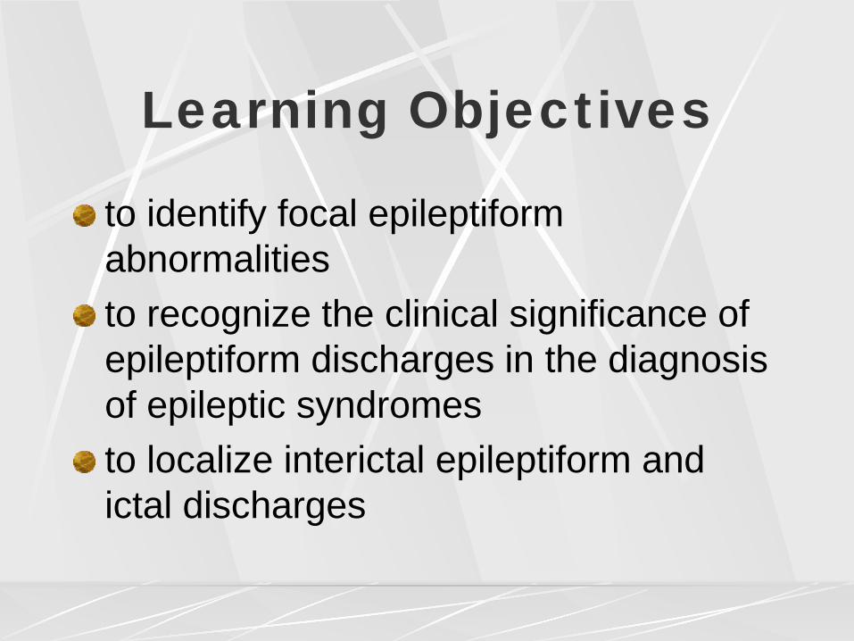
Learning Objectives
to identify focal epileptiform abnormalities to recognize the clinical significance of epileptiform discharges in the diagnosis of epileptic syndromes to localize interictal epileptiform and ictal discharges

Self assessment questions
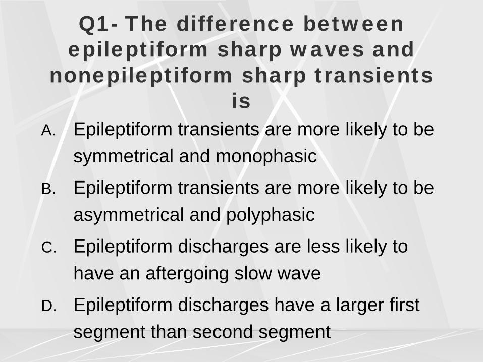
Q1- The difference between epileptiform sharp waves and
nonepileptiform sharp transients is
A. Epileptiform transients are more likely to be symmetrical and monophasic
B. Epileptiform transients are more likely to be asymmetrical and polyphasic
C. Epileptiform discharges are less likely to have an aftergoing slow wave
D. Epileptiform discharges have a larger first segment than second segment
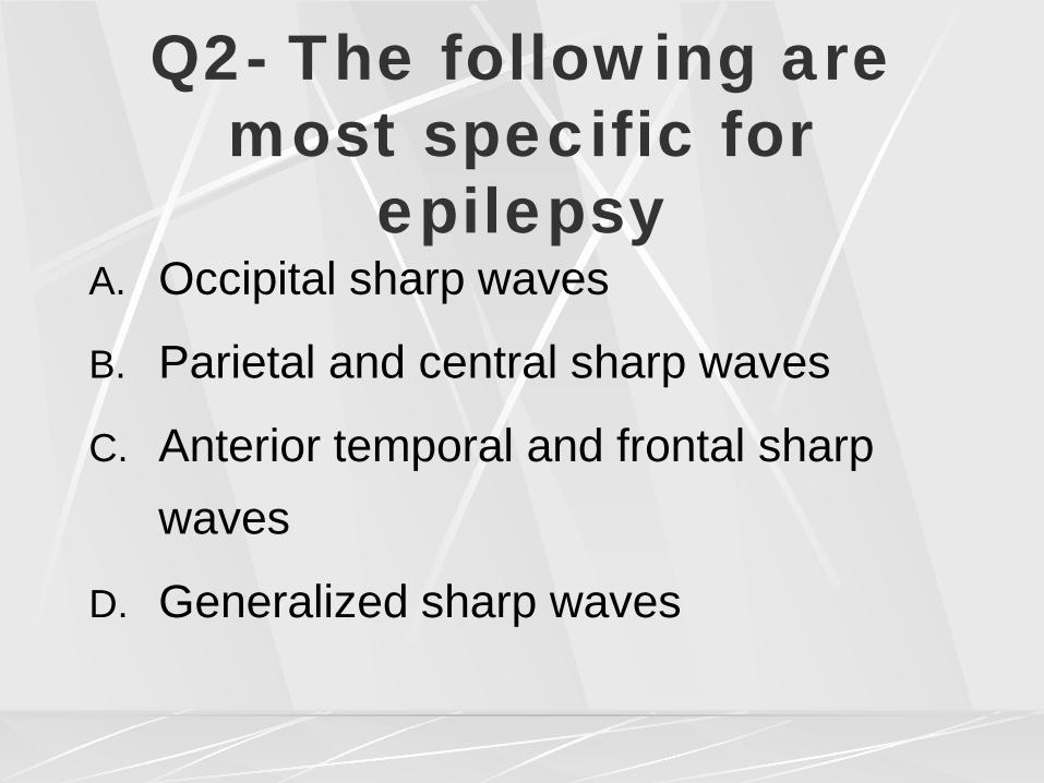
Q2- The following are most specific for
epilepsy A. Occipital sharp waves
B. Parietal and central sharp waves
C. Anterior temporal and frontal sharp waves
D. Generalized sharp waves
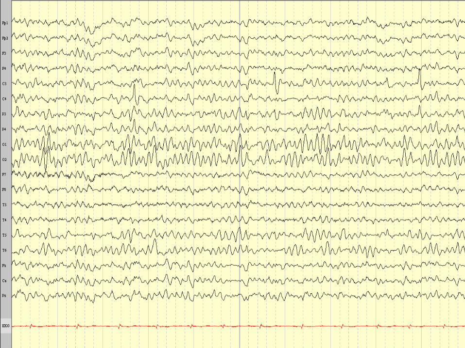
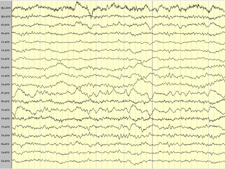
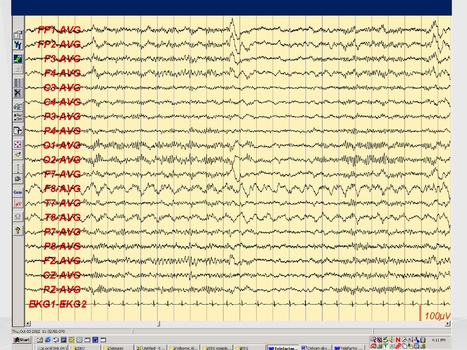
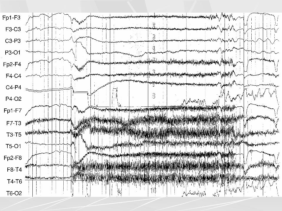
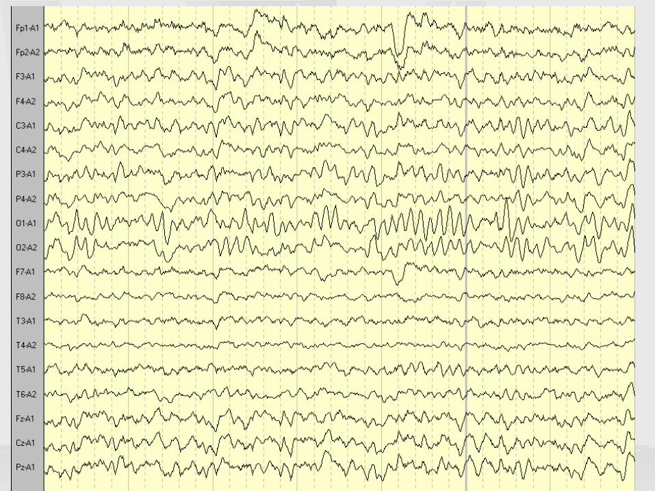
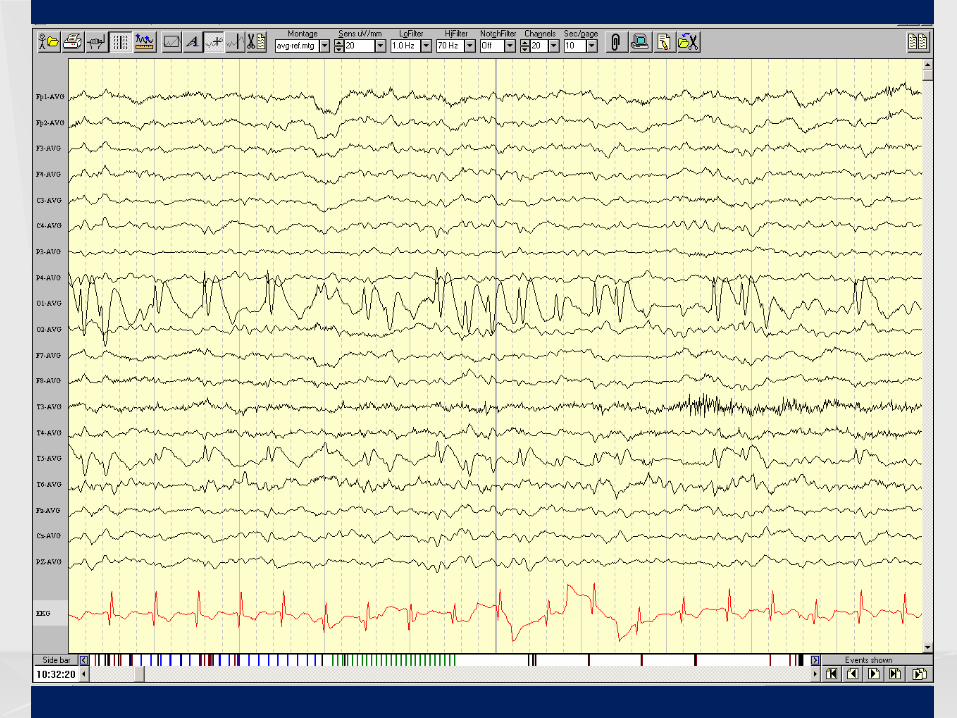
Q3- What is not true for the EEG above
A. Findings may be seen in the absence of epilepsy
B. Is expected to remit at puberty C. Seizures are likely to be hemifacial
sensorimotor D. Seizures likely manifest with
oroalimentary automatisms
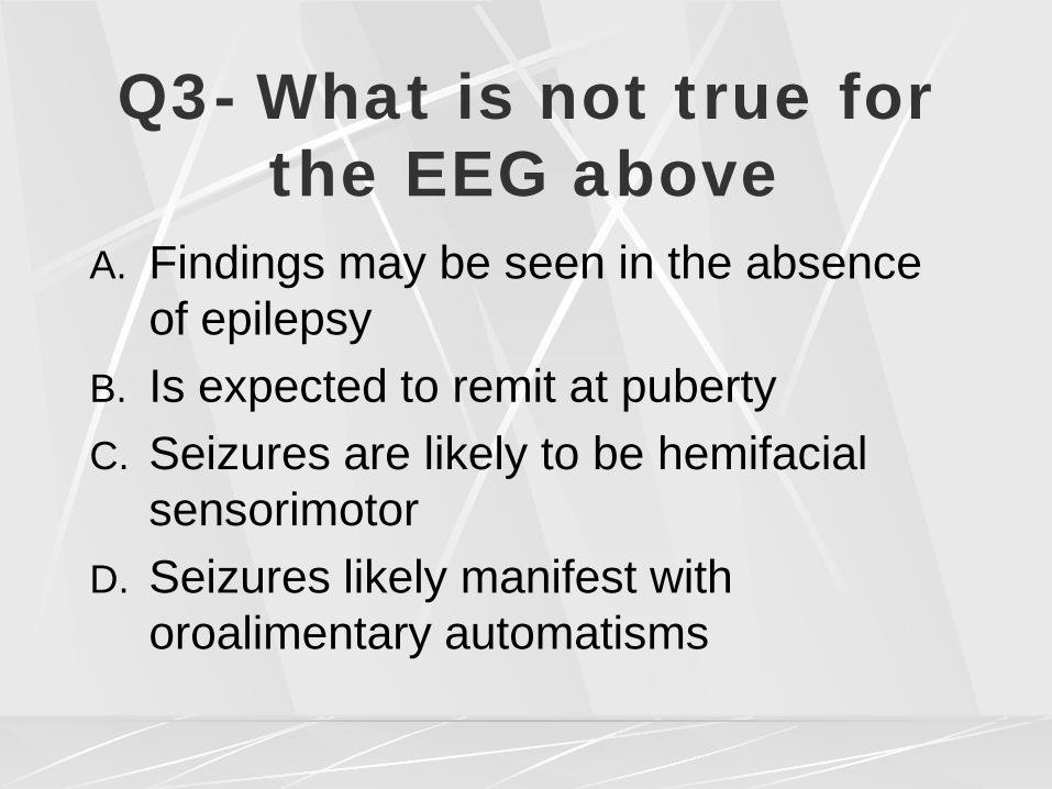
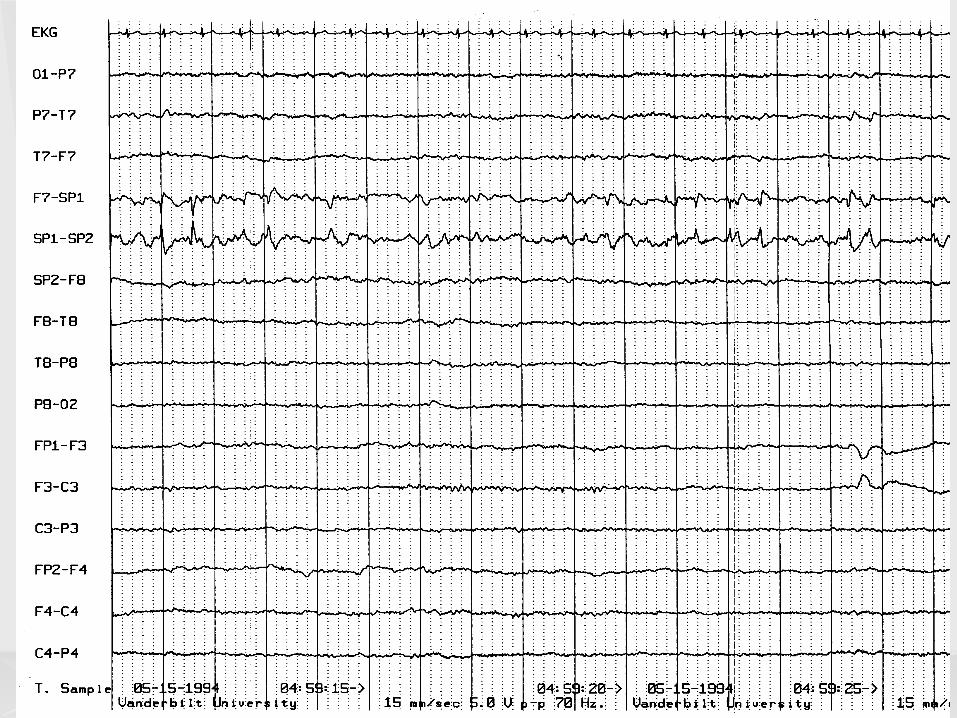
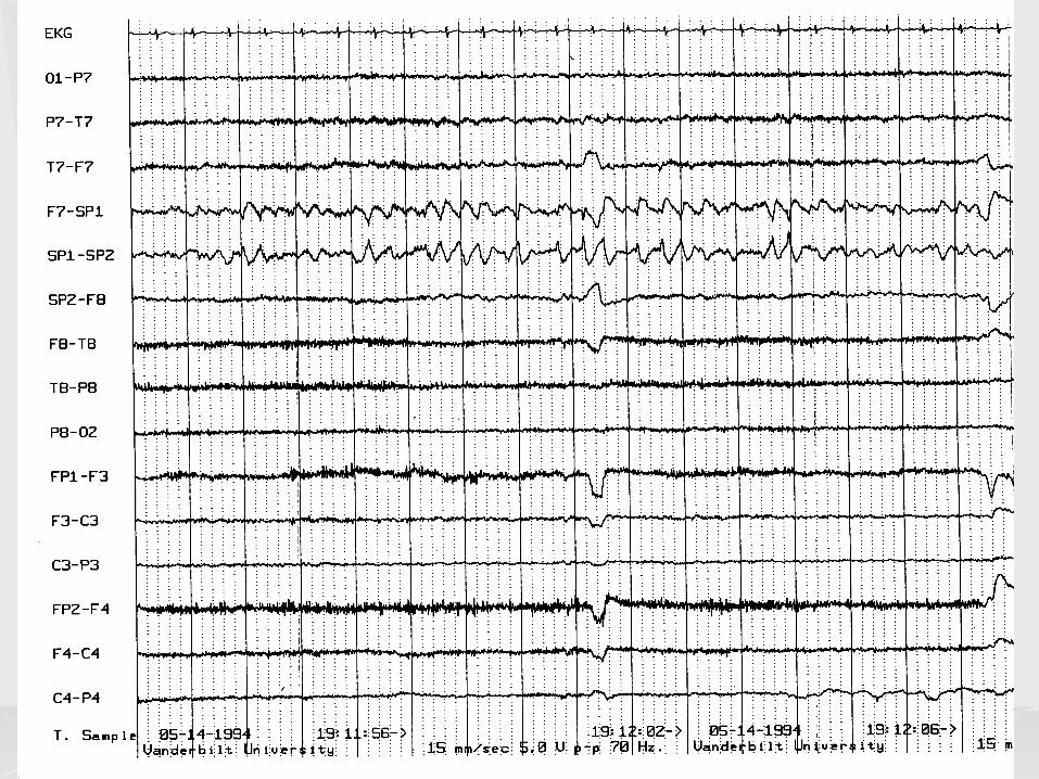
Fp1-Av Fp2-Av F3-Av F4-Av C3-Av C4-Av P3-Av P4-Av O1-Av O2-Av F7-Av F8-Av T1-Av T2-Av T7-Av T8-Av P7-Av P8-Av Fz-Av Cz-Av Pz-Av
ECG
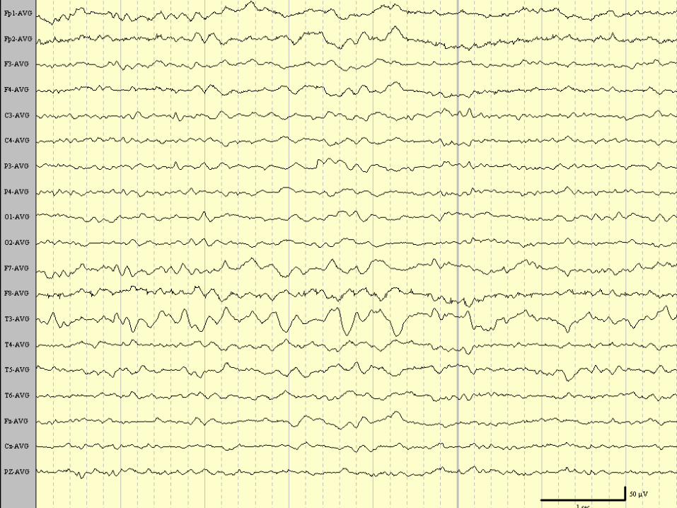
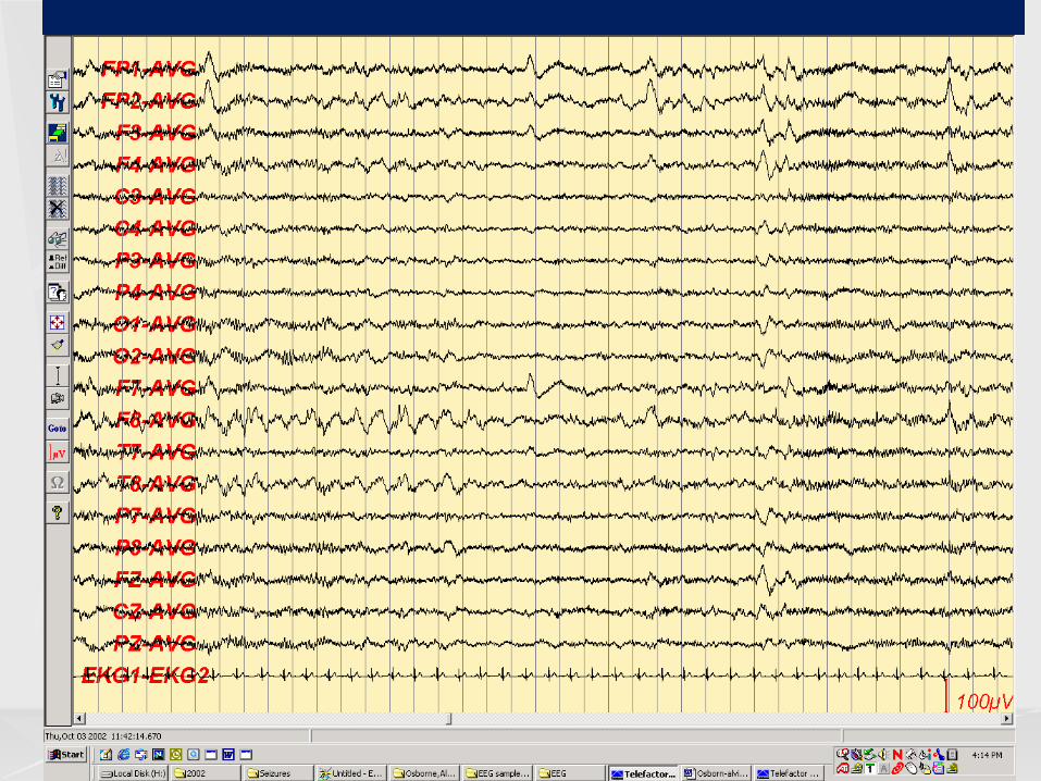
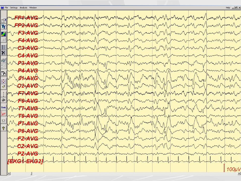
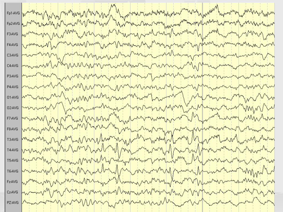
Q4- What is not true for the EEG above
A. Seizures originate from both temporal regions
B. Findings could be seen in parietal and insular lobe epilepsy
C. Seizures are more difficult to lateralize than for patients with unilateral discharges
D. Waking and REM recordings may help resolve lateralization
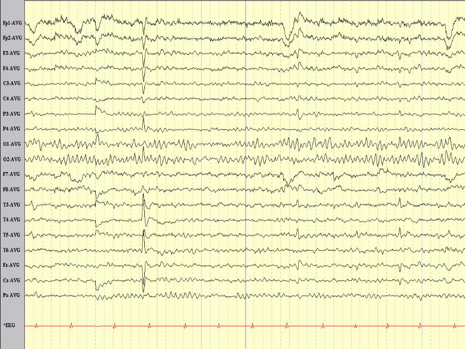
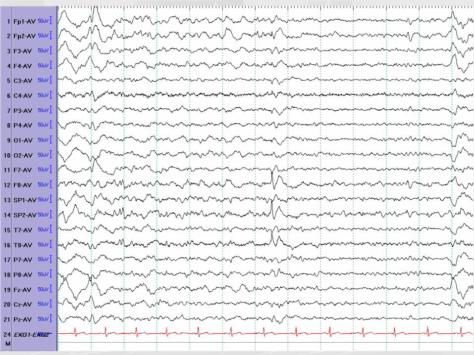
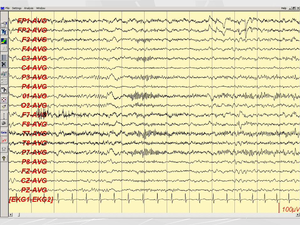
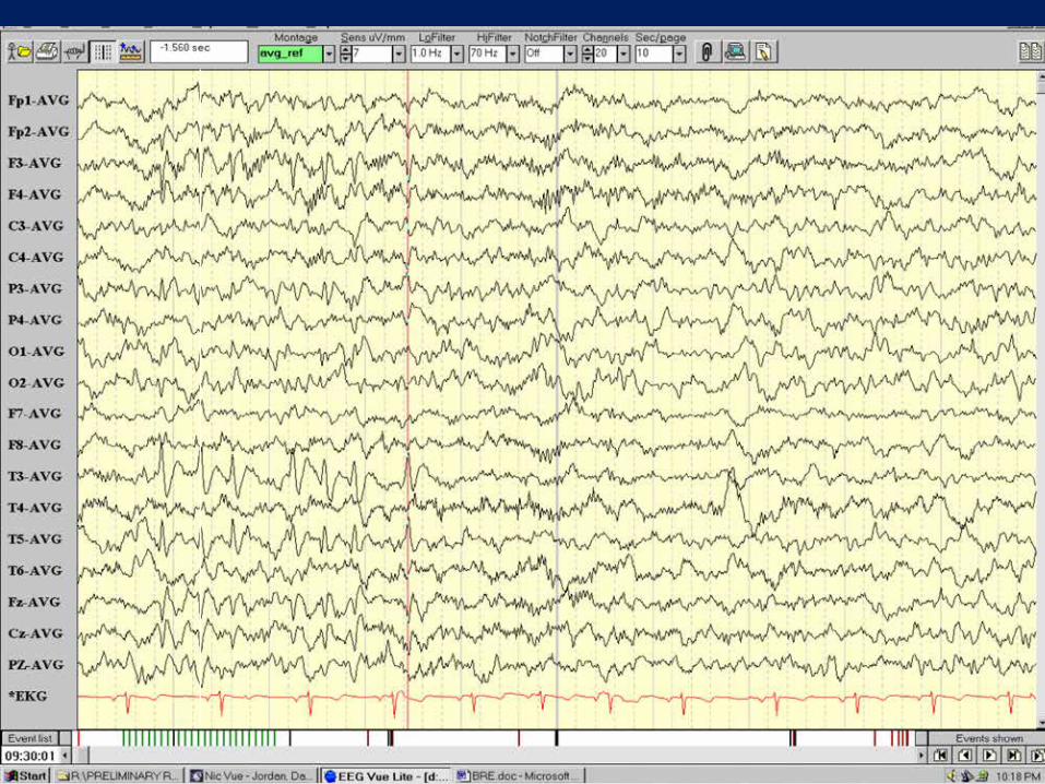
Q5- What is most likely true for the EEG above
A. The pattern is indicative of a contaminated reference
B. Seizures involve jerking of the right face
C. Seizures are most likely nocturnal D. Seizures originate in the midline frontal
region
The EEG in epilepsy
Role in diagnosis and management Focal interictal nonepileptiform and epileptiform abnormalities Focal ictal patterns Specificity, sensitivity, pitfalls
Role in diagnosis and management of epilepsy
Provide support for the clinical diagnosis (including status epilepticus) Help classify the epilepsy/ epileptic syndrome and localize the epileptogenic zone Unlikely to provide evidence for the etiology of epilepsy Occasionally help follow response to therapy (ex: absence) Help predict seizure recurrence after first unprovoked seizure or after AED withdrawal
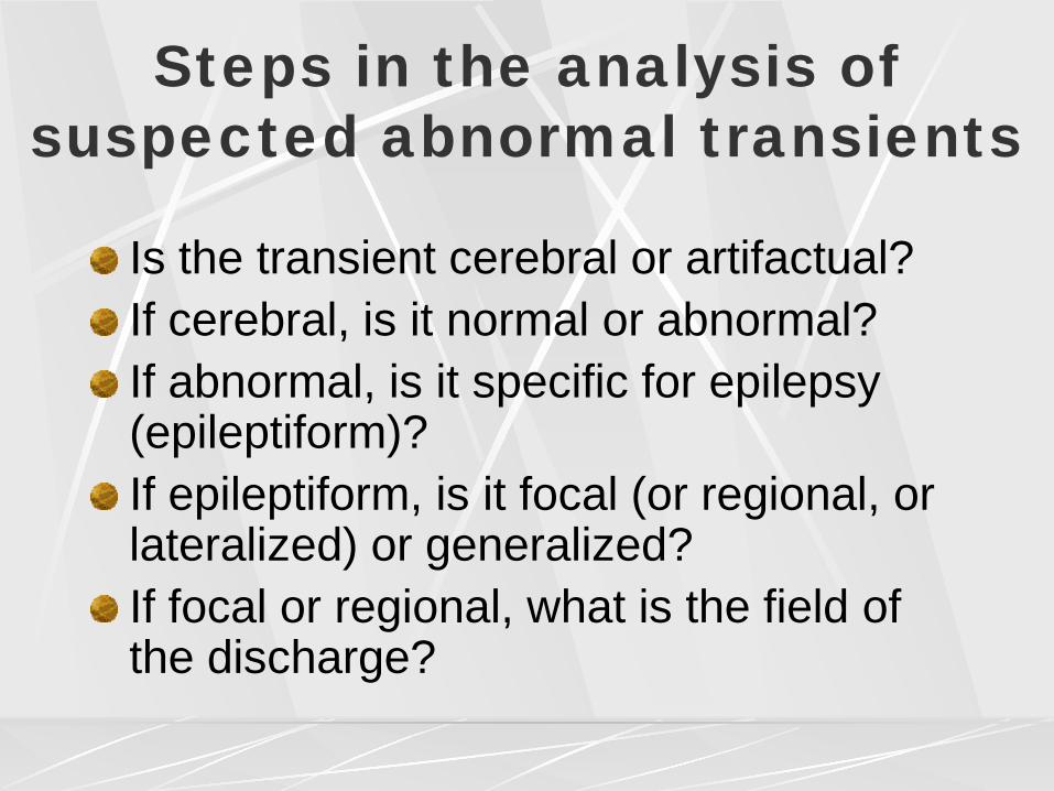
Steps in the analysis of suspected abnormal transients
Is the transient cerebral or artifactual? If cerebral, is it normal or abnormal? If abnormal, is it specific for epilepsy (epileptiform)? If epileptiform, is it focal (or regional, or lateralized) or generalized? If focal or regional, what is the field of the discharge?
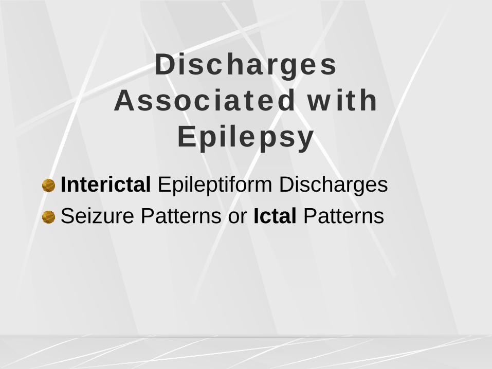
Discharges Associated with
Epilepsy Interictal Epileptiform Discharges Seizure Patterns or Ictal Patterns
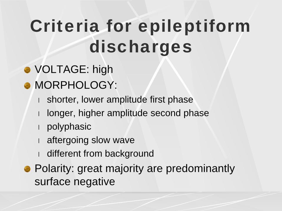
Criteria for epileptiform discharges
VOLTAGE: high MORPHOLOGY: l shorter, lower amplitude first phase l longer, higher amplitude second phase l polyphasic l aftergoing slow wave l different from background
Polarity: great majority are predominantly surface negative
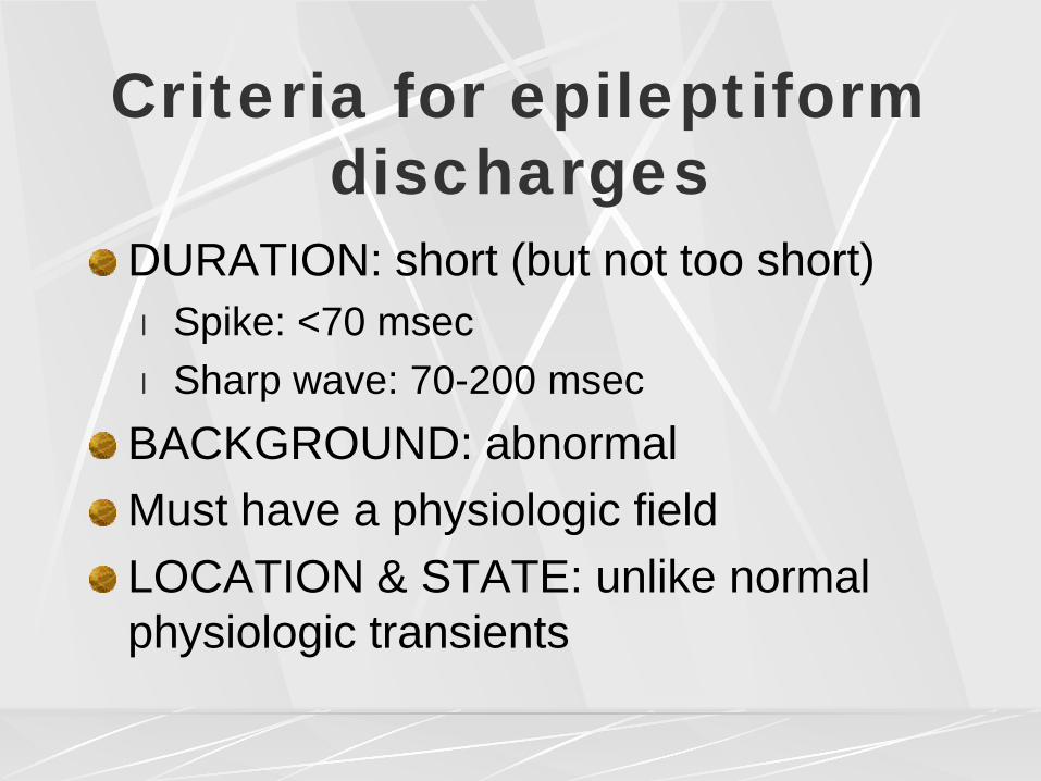
Criteria for epileptiform discharges
DURATION: short (but not too short) l Spike: <70 msec l Sharp wave: 70-200 msec
BACKGROUND: abnormal Must have a physiologic field LOCATION & STATE: unlike normal physiologic transients
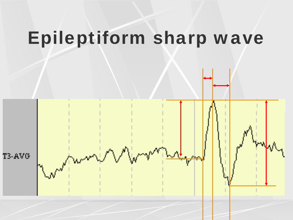
Epileptiform sharp wave
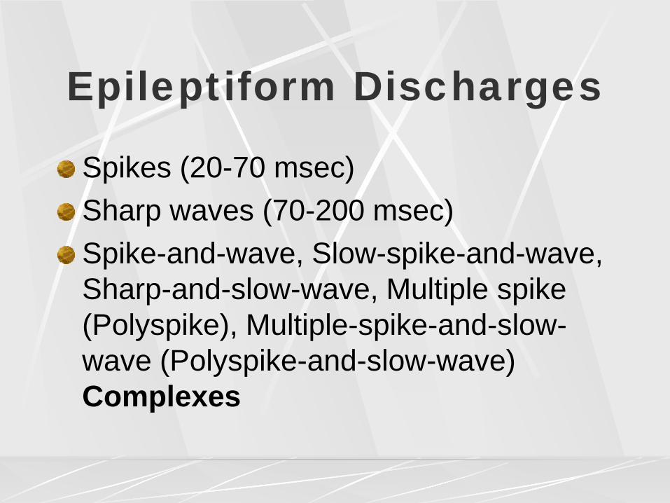
Epileptiform Discharges
Spikes (20-70 msec) Sharp waves (70-200 msec) Spike-and-wave, Slow-spike-and-wave, Sharp-and-slow-wave, Multiple spike (Polyspike), Multiple-spike-and-slow-wave (Polyspike-and-slow-wave) Complexes
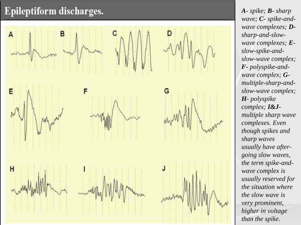
A- spike; B- sharp wave; C- spike-and-wave complexes; D- sharp-and-slow-wave complexes; E- slow-spike-and-slow-wave complex; F- polyspike-and-wave complex; G- multiple-sharp-and- slow-wave complex; H- polyspike complex; I&J- multiple sharp wave complexes. Even though spikes and sharp waves usually have after-going slow waves, the term spike-and-wave complex is usually reserved for the situation where the slow wave is very prominent, higher in voltage than the spike.
Ictal vs interictal discharges
Ictal discharge usually not a repetition of interictal discharges, except in generalized absence seizures Occasional patterns can be either ictal or interictal (spike-and-wave discharges, paroxysmal fast activity)
EEG findings in selected epileptic syndromes
TLE Other cryptogenic/symptomatic partial epilepsies Benign Partial Epilepsies
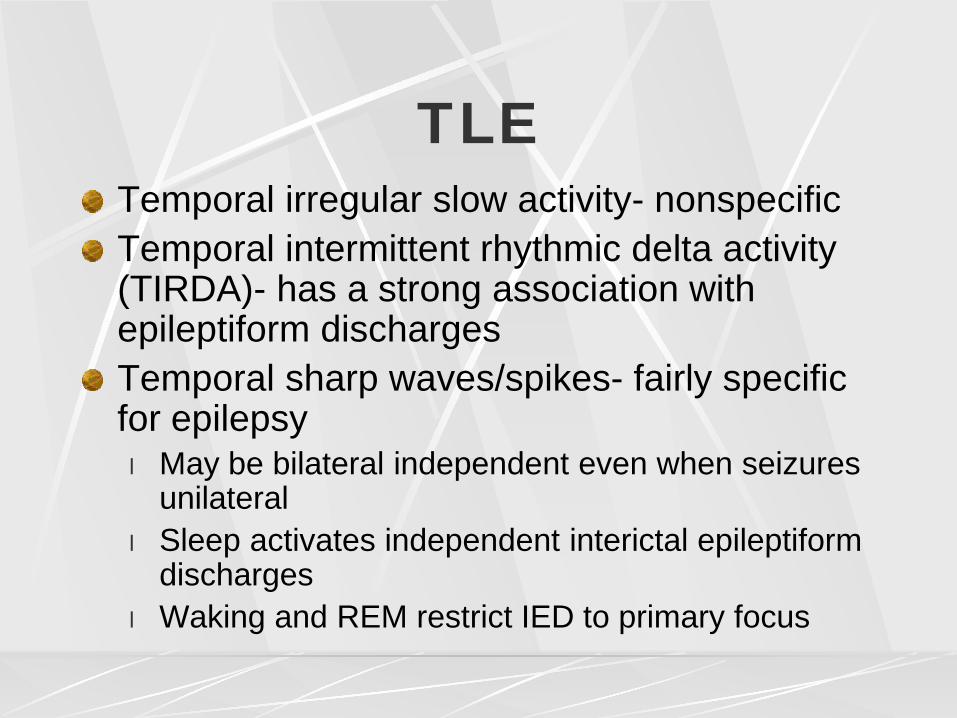
TLE Temporal irregular slow activity- nonspecific Temporal intermittent rhythmic delta activity (TIRDA)- has a strong association with epileptiform discharges Temporal sharp waves/spikes- fairly specific for epilepsy l May be bilateral independent even when seizures
unilateral l Sleep activates independent interictal epileptiform
discharges l Waking and REM restrict IED to primary focus
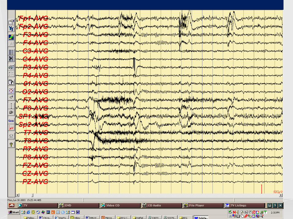
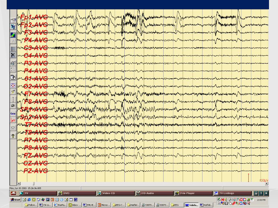
HOW CAN WE DISTINGUISH MESIAL FROM LATERAL TEMPORAL FOCI?

Interictal EEG features distinguishing mesial and
lateral temporal lobe epilepsy Mesial
IEDs predominantly over the ipsilateral mesial temporal regions (SP1/2 or FT9/10) TIRDA more likely
Lateral IEDs with lateral neocortical predominance (T7/T8 ie T3/T4; P7/P8 ie T5/T6)
l Patient groups do not differ in the presence of mesial versus lateral discharges; only in the predominance
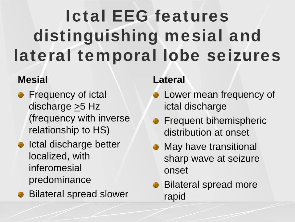
Ictal EEG features distinguishing mesial and
lateral temporal lobe seizures Mesial
Frequency of ictal discharge >5 Hz (frequency with inverse relationship to HS) Ictal discharge better localized, with inferomesial predominance Bilateral spread slower
Lateral Lower mean frequency of ictal discharge Frequent bihemispheric distribution at onset May have transitional sharp wave at seizure onset Bilateral spread more rapid
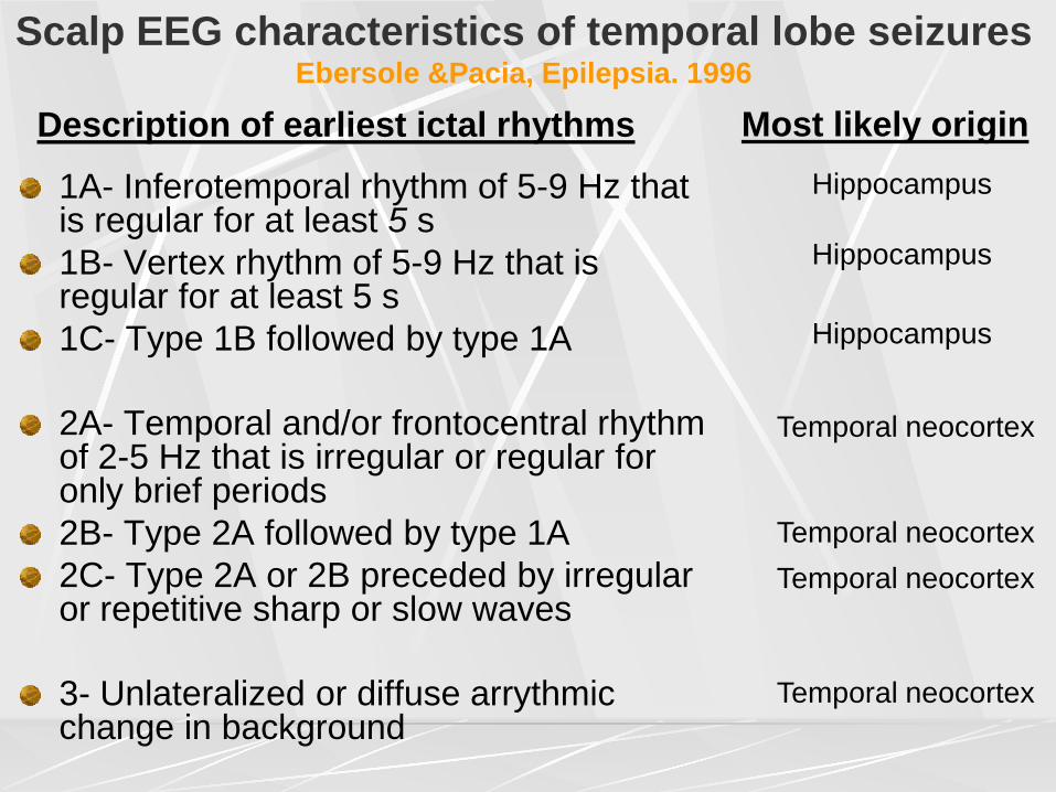
Scalp EEG characteristics of temporal lobe seizures Ebersole &Pacia, Epilepsia. 1996
1A- Inferotemporal rhythm of 5-9 Hz that is regular for at least 5 s 1B- Vertex rhythm of 5-9 Hz that is regular for at least 5 s 1C- Type 1B followed by type 1A 2A- Temporal and/or frontocentral rhythm of 2-5 Hz that is irregular or regular for only brief periods 2B- Type 2A followed by type 1A 2C- Type 2A or 2B preceded by irregular or repetitive sharp or slow waves 3- Unlateralized or diffuse arrythmic change in background
Hippocampus
Most likely origin
Temporal neocortex
Hippocampus
Hippocampus
Temporal neocortex Temporal neocortex
Temporal neocortex
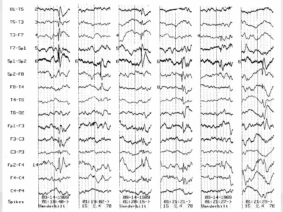
Description of earliest ictal rhythms
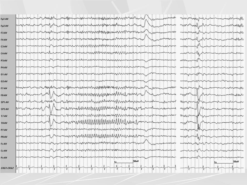
Transitional Sharp Waves at Ictal Onset- a Neocortical Ictal Pattern
Azar et al, JCN 2009
52 ictal discharges in 13 patients started with a TShW The center of TShW field was always concordant with the presumed final localization/lateralization, while that of the subsequent ictal discharge was concordant in only six patients. None of 61 ictal discharges in 15 patients with mesial temporal lobe epilepsy studied in the same time period started with a TShW. TShW are a marker of neocortical seizure onset. The TShW field provided more accurate localization or lateralization of the ictal focus than the following rhythmic ictal discharge. TShW at seizure onset should suggest a neocortical rather than hippocampal seizure onset.
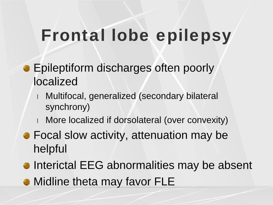
Frontal lobe epilepsy
Epileptiform discharges often poorly localized l Multifocal, generalized (secondary bilateral
synchrony) l More localized if dorsolateral (over convexity)
Focal slow activity, attenuation may be helpful Interictal EEG abnormalities may be absent Midline theta may favor FLE
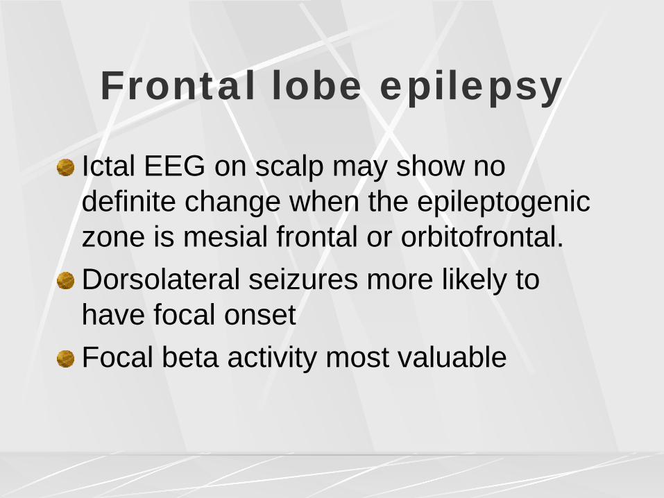
Frontal lobe epilepsy
Ictal EEG on scalp may show no definite change when the epileptogenic zone is mesial frontal or orbitofrontal. Dorsolateral seizures more likely to have focal onset Focal beta activity most valuable
Perry interictal
Perry ictal
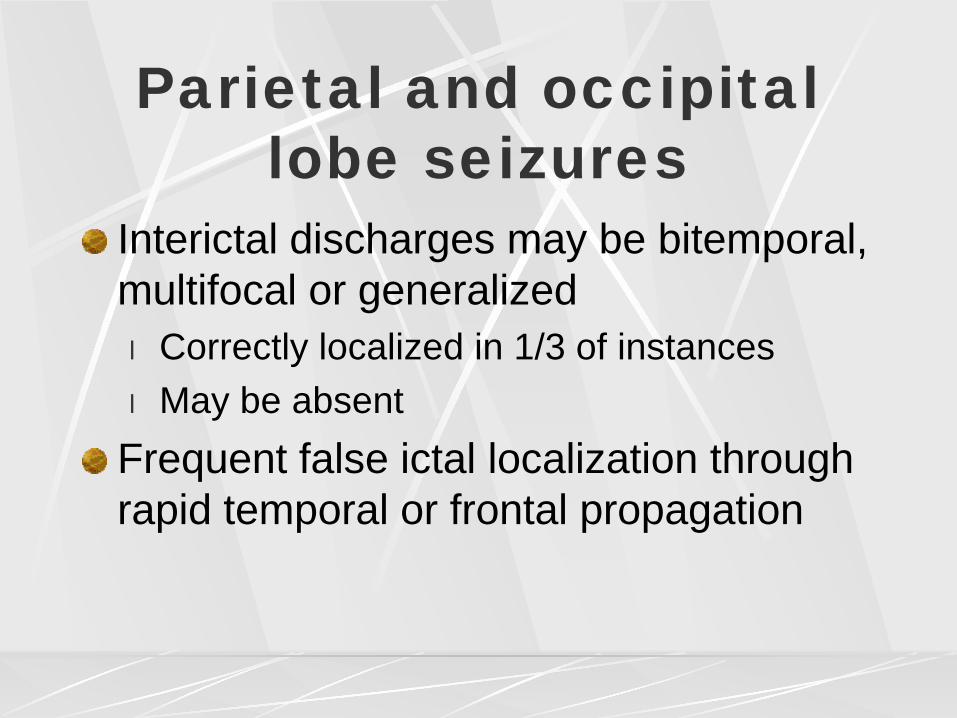
Parietal and occipital lobe seizures
Interictal discharges may be bitemporal, multifocal or generalized l Correctly localized in 1/3 of instances l May be absent
Frequent false ictal localization through rapid temporal or frontal propagation
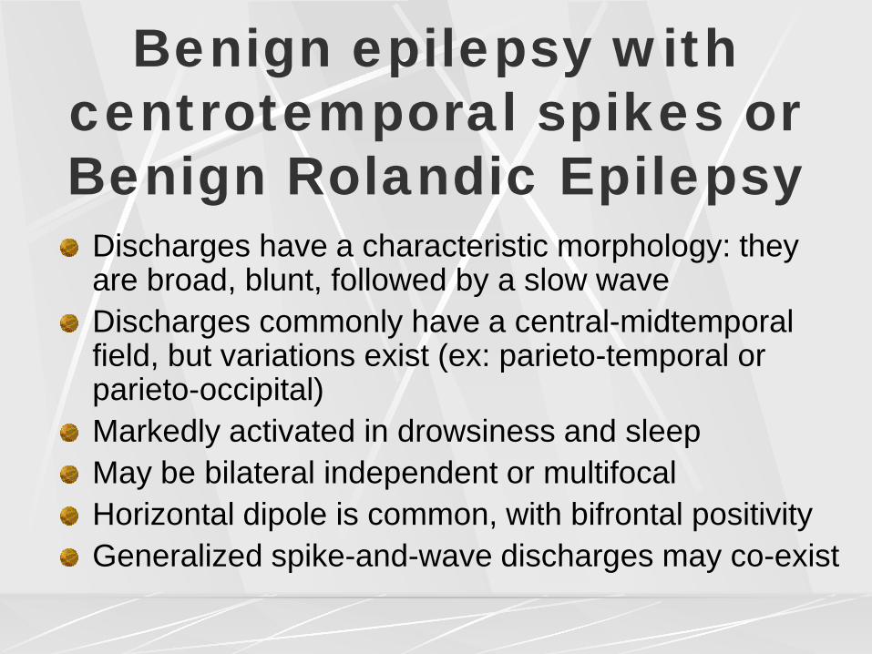
Benign epilepsy with centrotemporal spikes or Benign Rolandic Epilepsy
Discharges have a characteristic morphology: they are broad, blunt, followed by a slow wave Discharges commonly have a central-midtemporal field, but variations exist (ex: parieto-temporal or parieto-occipital) Markedly activated in drowsiness and sleep May be bilateral independent or multifocal Horizontal dipole is common, with bifrontal positivity Generalized spike-and-wave discharges may co-exist
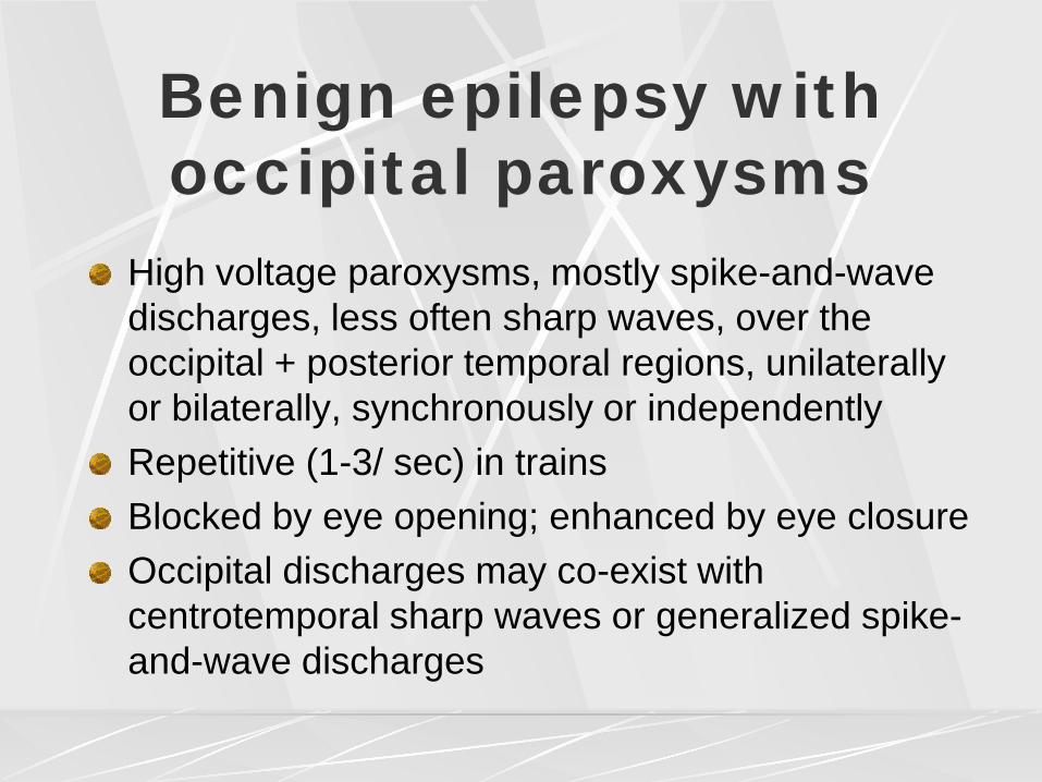
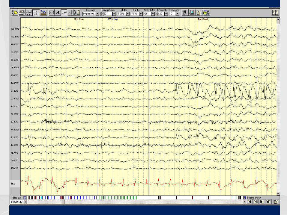
Benign epilepsy with occipital paroxysms
High voltage paroxysms, mostly spike-and-wave discharges, less often sharp waves, over the occipital + posterior temporal regions, unilaterally or bilaterally, synchronously or independently Repetitive (1-3/ sec) in trains Blocked by eye opening; enhanced by eye closure Occipital discharges may co-exist with centrotemporal sharp waves or generalized spike-and-wave discharges
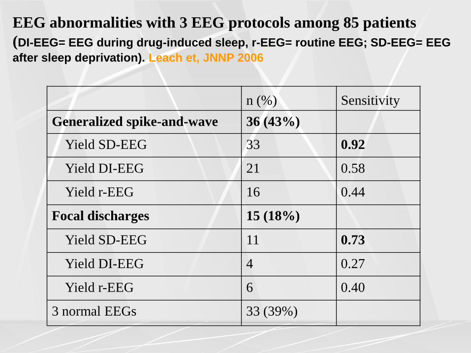
n (%) Sensitivity Generalized spike-and-wave 36 (43%)
Yield SD-EEG 33 0.92
Yield DI-EEG 21 0.58
Yield r-EEG 16 0.44
Focal discharges 15 (18%)
Yield SD-EEG 11 0.73
Yield DI-EEG 4 0.27
Yield r-EEG 6 0.40
3 normal EEGs 33 (39%)
EEG abnormalities with 3 EEG protocols among 85 patients (DI-EEG= EEG during drug-induced sleep, r-EEG= routine EEG; SD-EEG= EEG after sleep deprivation). Leach et, JNNP 2006
Effectiveness of Multiple EEGs in Supporting the Diagnosis of Epilepsy
Interictal epileptiform activity (IIEA) detected l on first EEG in 50% l by 3rd EEG in 84% l by 4th EEG in 92%
There is relatively little yield beyond the 4th EEG
Salinsky et al, Epilepsia 1987
Limitations of routine EEG Indirect assessment (seizures being an intermittent phenomenon) l Relies on interictal discharges; rare opportunity to
record seizures l 50% of patients with epilepsy will have a normal
first EEG; 10% will always have a normal interictal EEG
l Interictal EEG abnormalities may be unreliable for diagnosis and classification l Non-epileptic seizures coexistent with epilepsy l Generalized SW discharges in patients with partial
epilepsy, focal D/Cs in patients with generalized seizures
Methods of Activation
Sleep Sleep deprivation Hyperventilation Photic stimulation Specific modes of precipitation in patients with reflex seizures Prolonged recordings
Additional Electrodes
10/10 system T1/T2, zygomatic, cheek electrodes Nasopharyngeal electrodes Sphenoidal electrodes Minisphenoidal electrodes Supraorbital
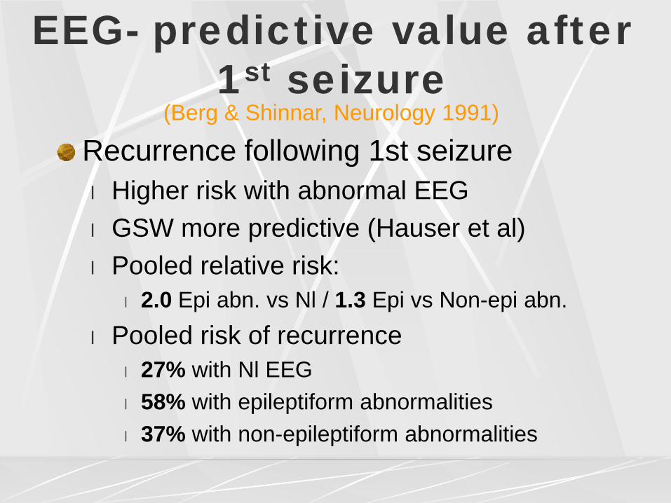
EEG- predictive value after 1st seizure
Recurrence following 1st seizure l Higher risk with abnormal EEG l GSW more predictive (Hauser et al) l Pooled relative risk:
l 2.0 Epi abn. vs Nl / 1.3 Epi vs Non-epi abn. l Pooled risk of recurrence
l 27% with Nl EEG l 58% with epileptiform abnormalities l 37% with non-epileptiform abnormalities
(Berg & Shinnar, Neurology 1991)
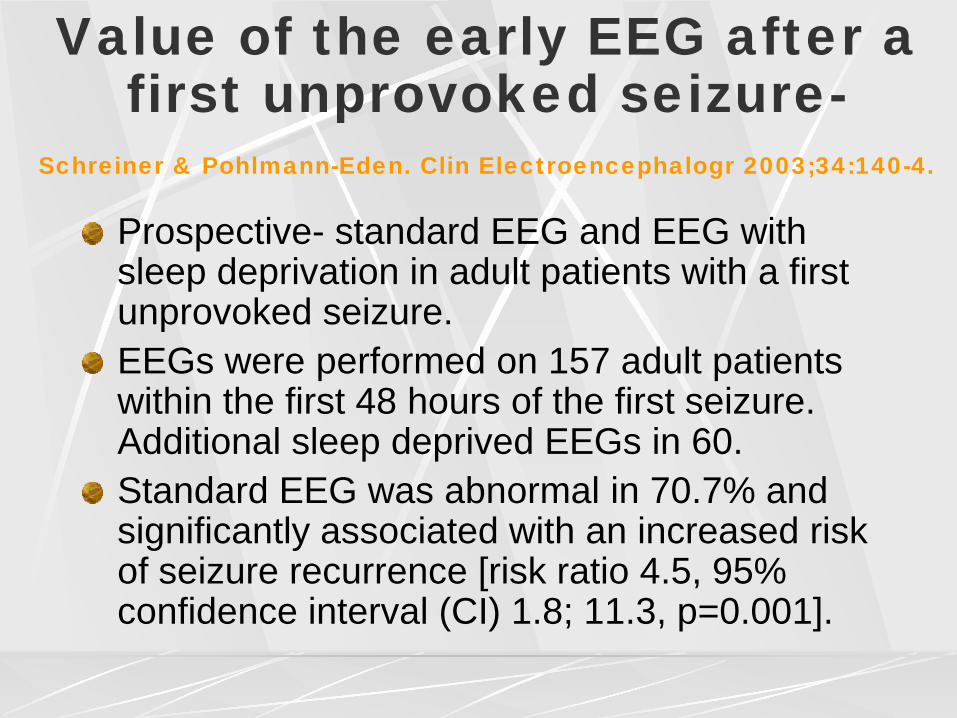
Value of the early EEG after a first unprovoked seizure-
Schreiner & Pohlmann-Eden. Clin Electroencephalogr 2003;34:140-4. Prospective- standard EEG and EEG with sleep deprivation in adult patients with a first unprovoked seizure. EEGs were performed on 157 adult patients within the first 48 hours of the first seizure. Additional sleep deprived EEGs in 60. Standard EEG was abnormal in 70.7% and significantly associated with an increased risk of seizure recurrence [risk ratio 4.5, 95% confidence interval (CI) 1.8; 11.3, p=0.001].
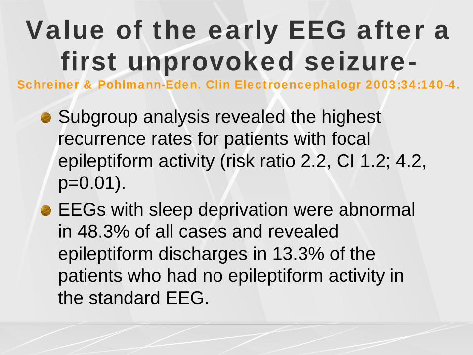
Value of the early EEG after a first unprovoked seizure-
Schreiner & Pohlmann-Eden. Clin Electroencephalogr 2003;34:140-4.
Subgroup analysis revealed the highest recurrence rates for patients with focal epileptiform activity (risk ratio 2.2, CI 1.2; 4.2, p=0.01). EEGs with sleep deprivation were abnormal in 48.3% of all cases and revealed epileptiform discharges in 13.3% of the patients who had no epileptiform activity in the standard EEG.
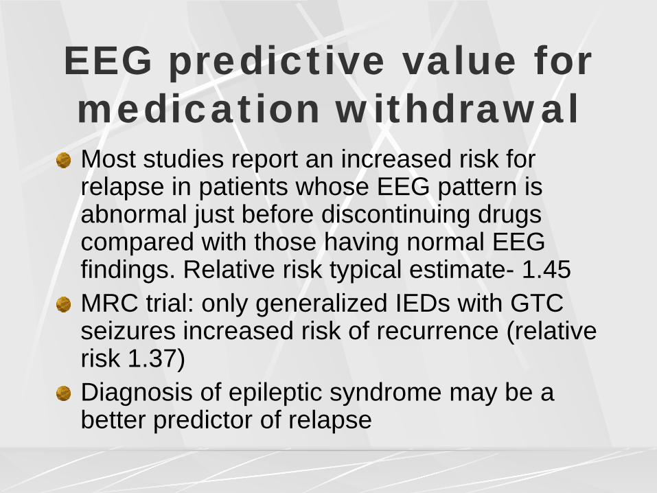
EEG predictive value for medication withdrawal Most studies report an increased risk for relapse in patients whose EEG pattern is abnormal just before discontinuing drugs compared with those having normal EEG findings. Relative risk typical estimate- 1.45 MRC trial: only generalized IEDs with GTC seizures increased risk of recurrence (relative risk 1.37) Diagnosis of epileptic syndrome may be a better predictor of relapse
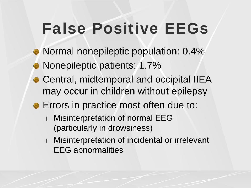
False Positive EEGs Normal nonepileptic population: 0.4% Nonepileptic patients: 1.7% Central, midtemporal and occipital IIEA may occur in children without epilepsy Errors in practice most often due to: l Misinterpretation of normal EEG
(particularly in drowsiness) l Misinterpretation of incidental or irrelevant
EEG abnormalities
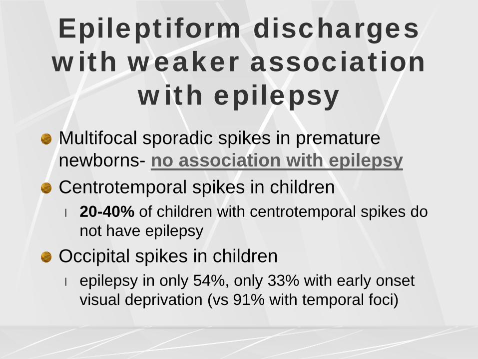
Epileptiform discharges with weaker association
with epilepsy Multifocal sporadic spikes in premature newborns- no association with epilepsy Centrotemporal spikes in children l 20-40% of children with centrotemporal spikes do
not have epilepsy Occipital spikes in children l epilepsy in only 54%, only 33% with early onset
visual deprivation (vs 91% with temporal foci)
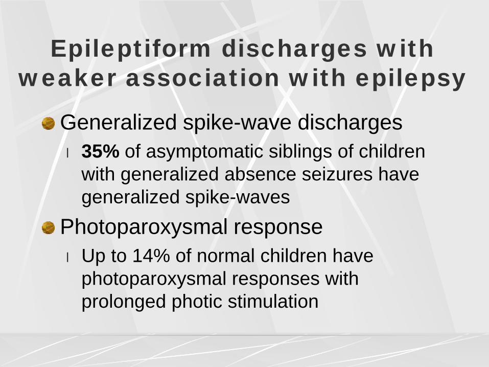
Epileptiform discharges with weaker association with epilepsy
Generalized spike-wave discharges l 35% of asymptomatic siblings of children
with generalized absence seizures have generalized spike-waves
Photoparoxysmal response l Up to 14% of normal children have
photoparoxysmal responses with prolonged photic stimulation
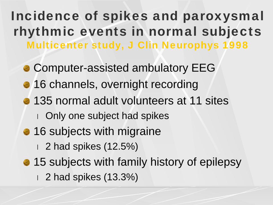
Incidence of spikes and paroxysmal rhythmic events in normal subjects
Multicenter study, J Clin Neurophys 1998
Computer-assisted ambulatory EEG 16 channels, overnight recording 135 normal adult volunteers at 11 sites l Only one subject had spikes
16 subjects with migraine l 2 had spikes (12.5%)
15 subjects with family history of epilepsy l 2 had spikes (13.3%)
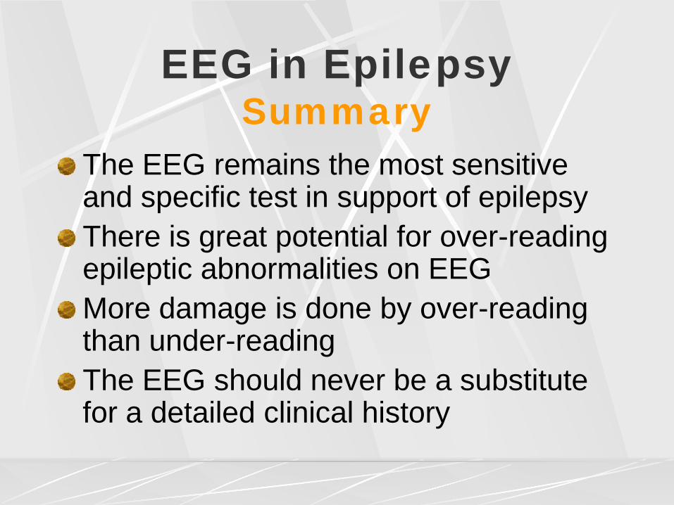
EEG in Epilepsy Summary
The EEG remains the most sensitive and specific test in support of epilepsy There is great potential for over-reading epileptic abnormalities on EEG More damage is done by over-reading than under-reading The EEG should never be a substitute for a detailed clinical history


![Animal in Animal Imaging'12 - University of Arizona · irritant effect on CNS (epileptiform activity seen on EEG) [Goble, E. and Ruhnke, A. Adverse Drug Reaction Bull 2009] – Use](https://img.pdfslide.net/doc/110x75/5eb7b5f8022e29278f78be7f/animal-in-animal-imaging12-university-of-arizona-irritant-effect-on-cns-epileptiform.jpg)