Embed Size (px)
Citation preview
Acta Biomaterialia xxx (2014) xxx–xxx
Contents lists available at ScienceDirect
Acta Biomaterialia
journal homepage: www.elsevier .com/locate /actabiomat
The effects of varying poly(ethylene glycol) hydrogel crosslinkingdensity and the crosslinking mechanism on protein accumulationin three-dimensional hydrogels
http://dx.doi.org/10.1016/j.actbio.2014.05.0231742-7061/� 2014 Acta Materialia Inc. Published by Elsevier Ltd. All rights reserved.
⇑ Corresponding author at: Departments of Orthopaedic Surgery and Bioengi-neering, Stanford University School of Medicine, Stanford, CA 94305, USA.
E-mail address: [email protected] (F. Yang).
Please cite this article in press as: Lee S et al. The effects of varying poly(ethylene glycol) hydrogel crosslinking density and the crosslinking mechanprotein accumulation in three-dimensional hydrogels. Acta Biomater (2014), http://dx.doi.org/10.1016/j.actbio.2014.05.023
Soah Lee a, Xinming Tong b, Fan Yang b,c,⇑a Department of Materials Science and Engineering, Stanford University, Stanford, CA, USAb Department of Orthopedic Surgery, Stanford University, Stanford, CA, USAc Department of Bioengineering, Stanford University, Stanford, CA, USA
a r t i c l e i n f o
Article history:Received 18 February 2014Received in revised form 28 April 2014Accepted 22 May 2014Available online xxxx
Keywords:Protein diffusionThree-dimensional hydrogelsPoly(ethylene glycol)Crosslinking densityCrosslinking mechanism
a b s t r a c t
Matrix stiffness has been shown to play an important role in modulating various cell fate processes suchas differentiation and cell cycle. Given that the stiffness can be easily tuned by varying the crosslinkingdensity, poly(ethylene glycol) (PEG) hydrogels have been widely used as an artificial cell niche. However,little is known about how changes in the hydrogel crosslinking density may affect the accumulation ofexogenous growth factors within 3-D hydrogel scaffolds formed by different crosslinking mechanisms.To address such shortcomings, we measured protein diffusivity and accumulation within PEG hydrogelswith varying PEG molecular weight, concentration and crosslinking mechanism. We found that proteinaccumulation increased substantially above a critical mesh size, which was distinct from the protein dif-fusivity trend, highlighting the importance of using protein accumulation as a parameter to better predictthe cell fates in addition to protein diffusivity, a parameter commonly reported by researchers studyingprotein diffusion in hydrogels. Furthermore, we found that chain-growth-polymerized gels allowed moreprotein accumulation than step-growth-polymerized gels, which may be the result of network heteroge-neity. The strategy used here can help quantify the effects of varying the hydrogel crosslinking densityand crosslinking mechanism on protein diffusion in different types of hydrogel. Such tools could bebroadly useful for interpreting cellular responses in hydrogels of varying stiffness for various tissueengineering applications.
� 2014 Acta Materialia Inc. Published by Elsevier Ltd. All rights reserved.
1. Introduction
Matrix stiffness has recently been recognized as playing animportant role in regulating cell fate and tissue development. Ithas been shown that stem cells specify their lineage and committo their fate by matrix stiffness, which mimics specific tissue-levelelasticity [1–3]. Matrix stiffness has also been shown to directlyinfluence other cellular fate processes, such as cell cycle, in avariety of cell types, including myofibroblasts [4], epithelial cells,vascular smooth muscle cells and osteoblasts [5]. Cancer cells arealso known to be mechanosensitive, and increasing matrix stiffnessresulted in an increase in cell growth, spreading and migration [6].
To study the effects of varying matrix stiffness on cell behavior,poly(ethylene glycol) (PEG) hydrogels have been widely used tocreate a biomimetic artificial niche with tunable biochemical and
biomechanical cues [7]. The biomechanical cue, specificallystiffness of the PEG hydrogel, can be easily tuned by varying themolecular weight or concentration of the PEG [8]. Increasing themolecular weight or decreasing the concentration of PEG reducesthe crosslinking density of the hydrogel, which results in a softergel. However, varying the crosslinking density simultaneouslychanges the mesh size of the hydrogel network, which, in turn,can influence protein diffusion in 3-D hydrogels [9,10]. Previousstudies have shown that increasing the gel crosslinking densityby increasing the molecular weight or decreasing the concentra-tion of PEG can lead to decreasing diffusivity of different solutes(vitamin B12, insulin, myoglobin, trypsin inhibitor, carbonicanhydrase, ovalbumin, bovine serum albumin (BSA), IgG) [9–11].
However, varying the gel crosslinking density influences notonly the protein diffusivity but also the protein accumulation withinhydrogels. To date, most studies have only looked at proteindiffusivity (how fast proteins can diffuse within hydrogels), ignoringprotein accumulation (how much proteins can actually go into 3-Dhydrogels), which can directly influence the encapsulated cells. A
ism on
2 S. Lee et al. / Acta Biomaterialia xxx (2014) xxx–xxx
recent study has shown that varying the hydrogel crosslinkingdensity can alter the stem cell differentiation, which is unlikely tobe related to mechanotransduction [12]. Since cells are known tobe sensitive to available soluble factors, it is important to character-ize the effects of varying the hydrogel crosslinking density onexogenous protein accumulation within 3-D hydrogels, therebyallowing the correct interpretation of the mechanisms that regulatecellular responses in hydrogels with varying stiffness.
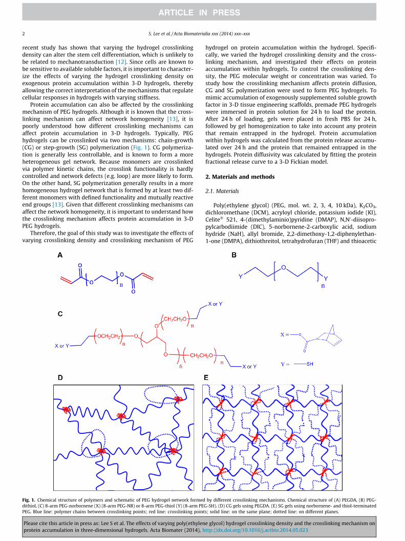
Protein accumulation can also be affected by the crosslinkingmechanism of PEG hydrogels. Although it is known that the cross-linking mechanism can affect network homogeneity [13], it ispoorly understood how different crosslinking mechanisms canaffect protein accumulation in 3-D hydrogels. Typically, PEGhydrogels can be crosslinked via two mechanisms: chain-growth(CG) or step-growth (SG) polymerization (Fig. 1). CG polymeriza-tion is generally less controllable, and is known to form a moreheterogeneous gel network. Because monomers are crosslinkedvia polymer kinetic chains, the crosslink functionality is hardlycontrolled and network defects (e.g. loop) are more likely to form.On the other hand, SG polymerization generally results in a morehomogeneous hydrogel network that is formed by at least two dif-ferent monomers with defined functionality and mutually reactiveend groups [13]. Given that different crosslinking mechanisms canaffect the network homogeneity, it is important to understand howthe crosslinking mechanism affects protein accumulation in 3-DPEG hydrogels.
Therefore, the goal of this study was to investigate the effects ofvarying crosslinking density and crosslinking mechanism of PEG
Fig. 1. Chemical structure of polymers and schematic of PEG hydrogel network formeddithiol, (C) 8-arm PEG-norbornene (X) (8-arm PEG-NB) or 8-arm PEG-thiol (Y) (8-arm PEPEG. Blue line: polymer chains between crosslinking points; red line: crosslinking point
Please cite this article in press as: Lee S et al. The effects of varying poly(ethylenprotein accumulation in three-dimensional hydrogels. Acta Biomater (2014), h
hydrogel on protein accumulation within the hydrogel. Specifi-cally, we varied the hydrogel crosslinking density and the cross-linking mechanism, and investigated their effects on proteinaccumulation within hydrogels. To control the crosslinking den-sity, the PEG molecular weight or concentration was varied. Tostudy how the crosslinking mechanism affects protein diffusion,CG and SG polymerization were used to form PEG hydrogels. Tomimic accumulation of exogenously supplemented soluble growthfactor in 3-D tissue engineering scaffolds, premade PEG hydrogelswere immersed in protein solution for 24 h to load the protein.After 24 h of loading, gels were placed in fresh PBS for 24 h,followed by gel homogenization to take into account any proteinthat remain entrapped in the hydrogel. Protein accumulationwithin hydrogels was calculated from the protein release accumu-lated over 24 h and the protein that remained entrapped in thehydrogels. Protein diffusivity was calculated by fitting the proteinfractional release curve to a 3-D Fickian model.
2. Materials and methods
2.1. Materials
Poly(ethylene glycol) (PEG, mol. wt. 2, 3, 4, 10 kDa), K2CO3,dichloromethane (DCM), acryloyl chloride, potassium iodide (KI),Celite� 521, 4-(dimethylaminio)pyridine (DMAP), N,N0-diisopro-pylcarbodiimide (DIC), 5-norbornene-2-carboxylic acid, sodiumhydride (NaH), allyl bromide, 2,2-dimethoxy-1,2-diphenylethan-1-one (DMPA), dithiothreitol, tetrahydrofuran (THF) and thioacetic
by different crosslinking mechanisms. Chemical structure of (A) PEGDA, (B) PEG-G-SH). (D) CG gels using PEGDA. (E) SG gels using norbornene- and thiol-terminateds; solid line: on the same plane; dotted line: on different planes.
e glycol) hydrogel crosslinking density and the crosslinking mechanism onttp://dx.doi.org/10.1016/j.actbio.2014.05.023
S. Lee et al. / Acta Biomaterialia xxx (2014) xxx–xxx 3
acid were purchased from Sigma–Aldrich (St Louis, MO).4-(2-Hydroxyethoxy)phenyl-(2-hydroxy-2-propyl)ketone (Irgacure2959) was purchased from BASF (Florham Park, NJ). PEG diacrylate(PEGDA, mol. wt. 5 kDa) was purchased from Laysan Bio (Arab, AL).8-arm PEG (mol. wt. 10 kDa) and 8-arm PEG-SH (mol. wt. 10 kDa),were purchased from JenKem Technology (Allen, TX). PEGDA (mol.wt. 2, 3, 4, 10 kDa), 8-arm PEG-norbornene (8-arm PEG-NB, mol.wt. 10 kDa) and PEG-diSH (mol. wt. 1.5, 10 kDa) were synthesizedin house. Regular BioRad Protein Assay was purchased from BioRad(Hercules, CA).
2.2. Synthesis
To synthesize PEGDA (mol. wt. 2, 3, 4, 10 kDa), linear PEG (2, 3,4, 10 kDa) was dissolved in DCM with the addition of 3 eq (moleequivalent with respect to hydroxyls) K2CO3. Next, 3 eq acryloylchloride and 0.1 eq KI were added to the solution. The solutionwas then stirred at 4 �C overnight and filtered using Celite� 521to remove any undissolved particles. The product was collectedby evaporation of the solvent and precipitation in cold ether. Itwas purified by dialysis against deionized water (MCO 1 kDa) for2 days, followed by lyophilization to collect the purified product.The structure of the products was confirmed by 1H nuclear mag-netic resonance (NMR) and the conversion ratio was over 95%.
8-arm PEG-norbornene (10 kDa) and PEG-dithiol (1.5, 10 kDa)were synthesized as previously reported [14,15]. To synthesizenorbornene-terminated 8-arm PEG, PEG (10 kDa) was dissolvedin DCM and 0.2 eq DMAP and 3 eq 5-norbornene-2-carboxylic acidwere added. After cooling the solution in an ice bath, 3 eq DIC wasadded. After stirring overnight, the reaction mixture was filteredand concentrated by evaporating away most of the solvent. Theconcentrated solution was then poured into ice-cold diethyl etherto precipitate the product. For the synthesis of PEG-dithiol (1.5,10 kDa), linear PEG-diol (1.5, 10 kDa) was dissolved in THF, and5 eq NaH and 2 eq allyl bromide were added. After stirring over-night, the solution was filtered and concentrated, then precipitatedin ice-cold diethyl ether. The resulting PEG allyl was then dissolvedin DCM containing 0.5% (w/v) DMPA and 2 eq thioacetic acid. Thesolution was then exposed to 365 nm UV (4 mW cm�2; UVPXX-15S lamp) for 1 h and the PEG thioester was precipitated inice-cold ether. The product was then dissolved in ammoniummethanol and 0.5 eq dithiothreitol was added to avoid disulfideformation. After stirring for 3 h, the product was precipitated inice-cold ether. The conversion ratio for both products was over95%, as confirmed by 1H NMR.
2.3. Hydrogel formation
Two different hydrogel structures were formed via CG polymer-ization and SG polymerization (Fig. 1). A total of 24 groups wereexamined with different crosslinking densities and crosslinkingmechanisms (Table S1). To form CG hydrogels, different concentra-tions (10, 15, 20% (w/v)) of PEGDA (mol. wt. 2 k, 3 k, 4 k, 5 k,10 kDa) were dissolved in phosphate-buffered saline (PBS)containing Irgacure 2959 (0.05% (w/v)). For SG hydrogels, norborn-ene-terminated PEG (8-arm PEG10 kDa-NB) and thiol-terminatedPEG (8-arm PEG10 kDa-SH, linear PEG1.5 k-dithiol, linearPEG10 k-dithiol) were mixed in a stoichiometrically balanced ratioin PBS solution containing Irgacure 2959 (0.05% (w/v)). The molec-ular weight of the SG gel (2.5 k, 4 k, 12.5 kDa) was defined as themolecular weight between two adjacent crosslinks. To form hydro-gels, 50 ll of the precursor solution was loaded into a cylindricalgel mold (5 mm diameter � 3 mm thickness) and exposed to UVlight (365 nm, 4 mW cm�2, 5 min). During UV exposure, CG-polymerized gels using PEGDA were formed by polyacrylate kinetic
Please cite this article in press as: Lee S et al. The effects of varying poly(ethylenprotein accumulation in three-dimensional hydrogels. Acta Biomater (2014), h
chains, while SG-polymerized gels were formed by mutuallyreactive groups, norbornene groups and thiol groups.
2.4. Mechanical test
The PEG hydrogel stiffness was measured by unconfined com-pression tests using an Instron 5944 materials testing system(Instron Corporation, Norwood, MA), as previously reported [16].Briefly, the specimen diameter and thickness were measured usingdigital calipers and the material testing system’s position read-out,respectively. Before each test, a preload of approximately 2 mNwas applied to the hydrogels. All tests were conducted in PBS solu-tion at room temperature. The upper platen was then lowered at arate of 1% strain s�1 to a maximum strain of 30%. Load and dis-placement data were recorded at 100 Hz. The compressive modu-lus was determined for strain ranges of 20–30% from the linearcurve fit of the stress vs. strain curve in each strain range.
2.5. Protein release test
To quantify the protein diffusivity and protein accumulationwithin 3-D hydrogels, BSA was chosen as the model solute for dif-fusion. To mimic exogeneously supplemented growth factor accu-mulation in 3-D tissue engineering scaffolds, premade hydrogelswere immersed in BSA solution (4 mg ml�1) at room temperaturefor 24 h to achieve uniform protein loading. For protein diffusivityon the order of 10�7 cm2 s�1, the diffusion time is 15 h for a diffu-sion length of 1.5 mm (t � L2/4D). Therefore, a 24 h loading timeshould be sufficient to achieve equilibrium protein concentrationswithin the gels. The loading time observed in similar studies ofvarious model proteins through even denser polymer networksfurther support the rationale for a 24 h loading time [9].
For protein release, BSA-loaded hydrogels were immersed infresh PBS. To maximize the driving force for diffusion, all of thesupernatant was collected and replaced by fresh PBS at differenttime points (t = 5, 10, 15, 20, 25, 30, 45, 60, 90 and 120 min and24 h). To release any proteins that remained entrapped in thehydrogels after release for 24 h, the hydrogels were mechanicallybroken down in fresh PBS using a homogenizer. To quantify theprotein in the supernatant, a Micro-BCA™ protein assay wasperformed following the manufacturer’s protocol.
Protein accumulation was calculated from the protein releasedinto supernatant at different time points and the protein thatremained trapped in the gel after the 24 h release. The protein con-centration within hydrogels was calculated by dividing the proteinaccumulation (mass) by the free water content (vol.) within thegels. The free water content (vol.) was calculated by using the freewater content (mass) from a swelling ratio test, assuming the PBSdensity to be 1 mg ml�1.
To determine the protein diffusivity (D) within differenthydrogels, fractional release profiles were fitted to the follow-ing equation, which represents a short-time approximation of a3-D Fickian diffusion model: a disk-shaped gel with uniform initialconcentration and equal surface concentration [17] (the short-timesolution of the 3-D Fickian diffusion model is valid for the first65–70% of total release):
Mt
M1¼ 4
Dtpa2
� �12
� p Dtpa2
� �� p
3Dtpa2
� �32
þ 4Dtpa2
� �12
� 2al
8Dtpa2
� �� 2p � Dt
pa2
� �32
� 2p3� Dt
pa2
� �2 !
Here Mt is the accumulated protein release up to the time point t;M1 is the accumulated protein release at infinite time, which wasdetermined by the protein accumulation; and D is the diffusivity
e glycol) hydrogel crosslinking density and the crosslinking mechanism onttp://dx.doi.org/10.1016/j.actbio.2014.05.023
4 S. Lee et al. / Acta Biomaterialia xxx (2014) xxx–xxx
of the BSA within the gel. Given that gels have different swellingratios, the final swollen gel geometry was taken into account whencalculating the protein diffusivity (a is the gel diameter and l is thegel thickness).
2.6. Swelling test for network mesh size calculation
Hydrogel samples were swollen in PBS at room temperature for24 h and weighed to obtain the equilibrium swollen mass (Ms). Toobtain the dry polymer mass (Md), the samples were placed indeionized water to remove the PBS salts, frozen and lyophilizedovernight. The volumetric swelling ratio (Qv) was calculated fromthe mass swelling ratio (Ms/Md) using the densities of PBS(1.01 g ml�1) and PEG (1.18 g ml�1) [18]:
Qv ¼Ms=qgel
Md=qPEG;qgel ¼ qPBS � 1�Md
Ms
� �þ qPEG �
Md
Msð1Þ
Qv was used to calculate the hydrogel network mesh size (n) accord-ing to the Flory–Rehner theory [19].
2.7. Statistical analysis
Data are presented as mean ± standard deviation. The standarddeviation was calculated based on three replicates (n = 3). AStudent’s t-test was used to compare data sets and a p value lessthan 0.05 was considered statistically significant.
3. Results
3.1. Stiffness of PEG hydrogels
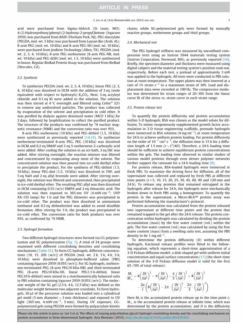
To evaluate how changing gel stiffness by varying gel crosslink-ing density can simultaneously alter protein accumulation within3-D hydrogels, we first evaluated the effect of varying the PEGcrosslinking density on gel stiffness. Regardless of the PEG cross-linking mechanism, increasing the PEG concentration or decreasingthe PEG molecular weight increased the hydrogel stiffness (Fig. 2).Decreasing the PEG molecular weight (CG 20% (w/v) gel: from 10 kto 2 k) led to a 15-fold increase in gel stiffness (Fig. 2A). Similarly,increasing the PEG concentration (CG 2 k gel: from 10 to 20% (w/v))resulted in a 10-fold increase in stiffness.
To study the effect of different crosslinking mechanisms on gelstiffness, we compared the stiffness of CG and SG gels with thesame molecular weight (Mc = 4 kDa) and concentration (10, 15and 20% (w/v)). The CG gels generally demonstrated higher stiff-ness than the corresponding SG gels (Fig. 2). For the CG gels,increasing the PEG concentration (from 10 to 20% (w/v)) led to
Fig. 2. The effects of varying PEG molecular weight (2, 2.5, 3, 4, 5, 10 and 12.5 kDa) orhydrogels.
Please cite this article in press as: Lee S et al. The effects of varying poly(ethylenprotein accumulation in three-dimensional hydrogels. Acta Biomater (2014), h
5-fold increase in stiffness, while it led to 3-fold increase for theSG gels.
3.2. Protein diffusion profile and diffusivity
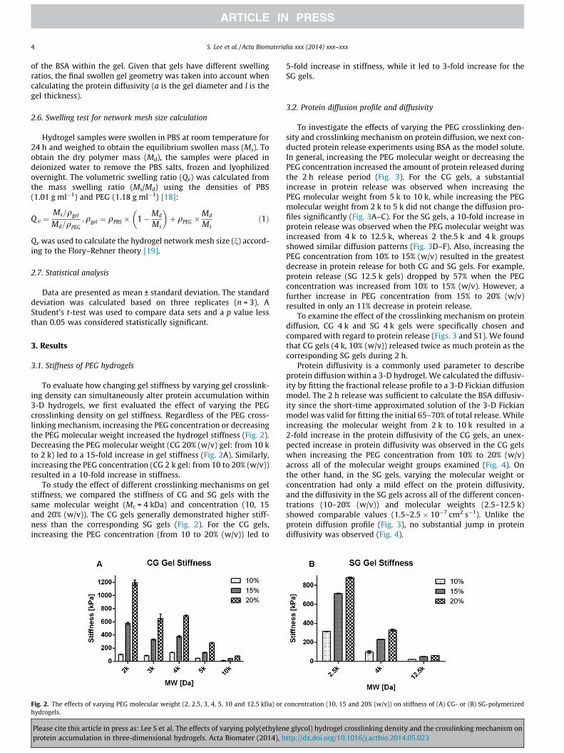
To investigate the effects of varying the PEG crosslinking den-sity and crosslinking mechanism on protein diffusion, we next con-ducted protein release experiments using BSA as the model solute.In general, increasing the PEG molecular weight or decreasing thePEG concentration increased the amount of protein released duringthe 2 h release period (Fig. 3). For the CG gels, a substantialincrease in protein release was observed when increasing thePEG molecular weight from 5 k to 10 k, while increasing the PEGmolecular weight from 2 k to 5 k did not change the diffusion pro-files significantly (Fig. 3A–C). For the SG gels, a 10-fold increase inprotein release was observed when the PEG molecular weight wasincreased from 4 k to 12.5 k, whereas 2 the.5 k and 4 k groupsshowed similar diffusion patterns (Fig. 3D–F). Also, increasing thePEG concentration from 10% to 15% (w/v) resulted in the greatestdecrease in protein release for both CG and SG gels. For example,protein release (SG 12.5 k gels) dropped by 57% when the PEGconcentration was increased from 10% to 15% (w/v). However, afurther increase in PEG concentration from 15% to 20% (w/v)resulted in only an 11% decrease in protein release.
To examine the effect of the crosslinking mechanism on proteindiffusion, CG 4 k and SG 4 k gels were specifically chosen andcompared with regard to protein release (Figs. 3 and S1). We foundthat CG gels (4 k, 10% (w/v)) released twice as much protein as thecorresponding SG gels during 2 h.
Protein diffusivity is a commonly used parameter to describeprotein diffusion within a 3-D hydrogel. We calculated the diffusiv-ity by fitting the fractional release profile to a 3-D Fickian diffusionmodel. The 2 h release was sufficient to calculate the BSA diffusiv-ity since the short-time approximated solution of the 3-D Fickianmodel was valid for fitting the initial 65–70% of total release. Whileincreasing the molecular weight from 2 k to 10 k resulted in a2-fold increase in the protein diffusivity of the CG gels, an unex-pected increase in protein diffusivity was observed in the CG gelswhen increasing the PEG concentration from 10% to 20% (w/v)across all of the molecular weight groups examined (Fig. 4). Onthe other hand, in the SG gels, varying the molecular weight orconcentration had only a mild effect on the protein diffusivity,and the diffusivity in the SG gels across all of the different concen-trations (10–20% (w/v)) and molecular weights (2.5–12.5 k)showed comparable values (1.5–2.5 � 10�7 cm2 s�1). Unlike theprotein diffusion profile (Fig. 3), no substantial jump in proteindiffusivity was observed (Fig. 4).
concentration (10, 15 and 20% (w/v)) on stiffness of (A) CG- or (B) SG-polymerized
e glycol) hydrogel crosslinking density and the crosslinking mechanism onttp://dx.doi.org/10.1016/j.actbio.2014.05.023
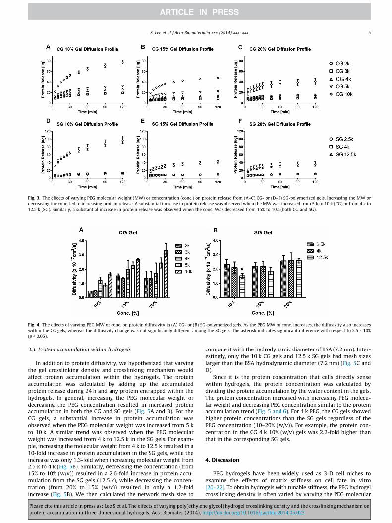
Fig. 3. The effects of varying PEG molecular weight (MW) or concentration (conc.) on protein release from (A–C) CG- or (D–F) SG-polymerized gels. Increasing the MW ordecreasing the conc. led to increasing protein release. A substantial increase in protein release was observed when the MW was increased from 5 k to 10 k (CG) or from 4 k to12.5 k (SG). Similarly, a substantial increase in protein release was observed when the conc. Was decreased from 15% to 10% (both CG and SG).
Fig. 4. The effects of varying PEG MW or conc. on protein diffusivity in (A) CG- or (B) SG-polymerized gels. As the PEG MW or conc. increases, the diffusivity also increaseswithin the CG gels, whereas the diffusivity change was not significantly different among the SG gels. The asterisk indicates significant difference with respect to 2.5 k 10%(p < 0.05).
S. Lee et al. / Acta Biomaterialia xxx (2014) xxx–xxx 5
3.3. Protein accumulation within hydrogels
In addition to protein diffusivity, we hypothesized that varyingthe gel crosslinking density and crosslinking mechanism wouldaffect protein accumulation within the hydrogels. The proteinaccumulation was calculated by adding up the accumulatedprotein release during 24 h and any protein entrapped within thehydrogels. In general, increasing the PEG molecular weight ordecreasing the PEG concentration resulted in increased proteinaccumulation in both the CG and SG gels (Fig. 5A and B). For theCG gels, a substantial increase in protein accumulation wasobserved when the PEG molecular weight was increased from 5 kto 10 k. A similar trend was observed when the PEG molecularweight was increased from 4 k to 12.5 k in the SG gels. For exam-ple, increasing the molecular weight from 4 k to 12.5 k resulted in a10-fold increase in protein accumulation in the SG gels, while theincrease was only 1.3-fold when increasing molecular weight from2.5 k to 4 k (Fig. 5B). Similarly, decreasing the concentration (from15% to 10% (w/v)) resulted in a 2.6-fold increase in protein accu-mulation from the SG gels (12.5 k), while decreasing the concen-tration (from 20% to 15% (w/v)) resulted in only a 1.2-foldincrease (Fig. 5B). We then calculated the network mesh size to
Please cite this article in press as: Lee S et al. The effects of varying poly(ethylenprotein accumulation in three-dimensional hydrogels. Acta Biomater (2014), h
compare it with the hydrodynamic diameter of BSA (7.2 nm). Inter-estingly, only the 10 k CG gels and 12.5 k SG gels had mesh sizeslarger than the BSA hydrodynamic diameter (7.2 nm) (Fig. 5C andD).
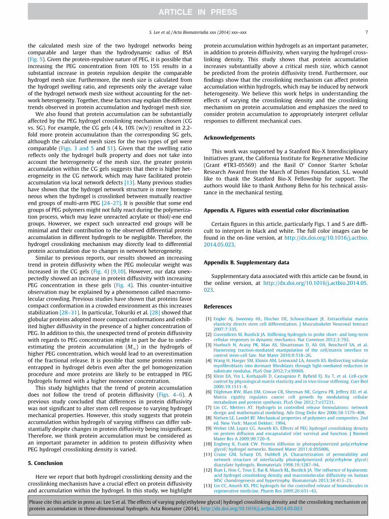
Since it is the protein concentration that cells directly sensewithin hydrogels, the protein concentration was calculated bydividing the protein accumulation by the water content in the gels.The protein concentration increased with increasing PEG molecu-lar weight and decreasing PEG concentration similar to the proteinaccumulation trend (Fig. 5 and 6). For 4 k PEG, the CG gels showedhigher protein concentrations than the SG gels regardless of thePEG concentration (10–20% (w/v)). For example, the protein con-centration in the CG 4 k 10% (w/v) gels was 2.2-fold higher thanthat in the corresponding SG gels.
4. Discussion
PEG hydrogels have been widely used as 3-D cell niches toexamine the effects of matrix stiffness on cell fate in vitro[20–22]. To obtain hydrogels with tunable stiffness, the PEG hydrogelcrosslinking density is often varied by varying the PEG molecular
e glycol) hydrogel crosslinking density and the crosslinking mechanism onttp://dx.doi.org/10.1016/j.actbio.2014.05.023
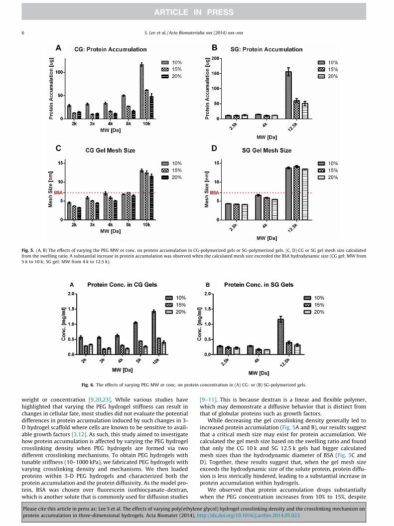
Fig. 5. (A, B) The effects of varying the PEG MW or conc. on protein accumulation in CG-polymerized gels or SG-polymerized gels. (C, D) CG or SG gel mesh size calculatedfrom the swelling ratio. A substantial increase in protein accumulation was observed when the calculated mesh size exceeded the BSA hydrodynamic size (CG gel: MW from5 k to 10 k; SG gel: MW from 4 k to 12.5 k).
Fig. 6. The effects of varying PEG MW or conc. on protein concentration in (A) CG- or (B) SG-polymerized gels.
6 S. Lee et al. / Acta Biomaterialia xxx (2014) xxx–xxx
weight or concentration [9,20,23]. While various studies havehighlighted that varying the PEG hydrogel stiffness can result inchanges in cellular fate, most studies did not evaluate the potentialdifferences in protein accumulation induced by such changes in 3-D hydrogel scaffold where cells are known to be sensitive to avail-able growth factors [3,12]. As such, this study aimed to investigatehow protein accumulation is affected by varying the PEG hydrogelcrosslinking density when PEG hydrogels are formed via twodifferent crosslinking mechanisms. To obtain PEG hydrogels withtunable stiffness (10–1000 kPa), we fabricated PEG hydrogels withvarying crosslinking density and mechanisms. We then loadedproteins within 3-D PEG hydrogels and characterized both theprotein accumulation and the protein diffusivity. As the model pro-tein, BSA was chosen over fluorescein isothiocyanate–dextran,which is another solute that is commonly used for diffusion studies
Please cite this article in press as: Lee S et al. The effects of varying poly(ethylenprotein accumulation in three-dimensional hydrogels. Acta Biomater (2014), h
[9–11]. This is because dextran is a linear and flexible polymer,which may demonstrate a diffusive behavior that is distinct fromthat of globular proteins such as growth factors.
While decreasing the gel crosslinking density generally led toincreased protein accumulation (Fig. 5A and B), our results suggestthat a critical mesh size may exist for protein accumulation. Wecalculated the gel mesh size based on the swelling ratio and foundthat only the CG 10 k and SG 12.5 k gels had bigger calculatedmesh sizes than the hydrodynamic diameter of BSA (Fig. 5C andD). Together, these results suggest that, when the gel mesh sizeexceeds the hydrodynamic size of the solute protein, protein diffu-sion is less sterically hindered, leading to a substantial increase inprotein accumulation within hydrogels.
We observed that protein accumulation drops substantiallywhen the PEG concentration increases from 10% to 15%, despite
e glycol) hydrogel crosslinking density and the crosslinking mechanism onttp://dx.doi.org/10.1016/j.actbio.2014.05.023
S. Lee et al. / Acta Biomaterialia xxx (2014) xxx–xxx 7
the calculated mesh size of the two hydrogel networks beingcomparable and larger than the hydrodynamic radius of BSA(Fig. 5). Given the protein-repulsive nature of PEG, it is possible thatincreasing the PEG concentration from 10% to 15% results in asubstantial increase in protein repulsion despite the comparablehydrogel mesh size. Furthermore, the mesh size is calculated fromthe hydrogel swelling ratio, and represents only the average valueof the hydrogel network mesh size without accounting for the net-work heterogeneity. Together, these factors may explain the differenttrends observed in protein accumulation and hydrogel mesh size.
We also found that protein accumulation can be substantiallyaffected by the PEG hydrogel crosslinking mechanism chosen (CGvs. SG). For example, the CG gels (4 k, 10% (w/v)) resulted in 2.2-fold more protein accumulation than the corresponding SG gels,although the calculated mesh sizes for the two types of gel werecomparable (Figs. 3 and 5 and S1). Given that the swelling ratioreflects only the hydrogel bulk property and does not take intoaccount the heterogeneity of the mesh size, the greater proteinaccumulation within the CG gels suggests that there is higher het-erogeneity in the CG network, which may have facilitated proteinaccumulation via local network defects [13]. Many previous studieshave shown that the hydrogel network structure is more homoge-neous when the hydrogel is crosslinked between mutually reactiveend groups of multi-arm PEG [24–27]. It is possible that some endgroups of PEG polymers might not fully react during the polymeriza-tion process, which may leave unreacted acrylate or thiol/-ene endgroups. However, we expect such unreacted end groups will beminimal and their contribution to the observed differential proteinaccumulation in different hydrogels to be negligible. Therefore, thehydrogel crosslinking mechanism may directly lead to differentialprotein accumulation due to changes in network heterogeneity.
Similar to previous reports, our results showed an increasingtrend in protein diffusivity when the PEG molecular weight wasincreased in the CG gels (Fig. 4) [9,10]. However, our data unex-pectedly showed an increase in protein diffusivity with increasingPEG concentration in these gels (Fig. 4). This counter-intuitiveobservation may be explained by a phenomenon called macromo-lecular crowding. Previous studies have shown that proteins favorcompact conformation in a crowded environment as this increasesstabilization [28–31]. In particular, Tokuriki et al. [28] showed thatglobular proteins adopted more compact conformations and exhib-ited higher diffusivity in the presence of a higher concentration ofPEG. In addition to this, the unexpected trend of protein diffusivitywith regards to PEG concentration might in part be due to under-estimating the protein accumulation (M1) in the hydrogels ofhigher PEG concentration, which would lead to an overestimationof the fractional release. It is possible that some proteins remainentrapped in hydrogel debris even after the gel homogenizationprocedure and more proteins are likely to be entrapped in PEGhydrogels formed with a higher monomer concentration.
This study highlights that the trend of protein accumulationdoes not follow the trend of protein diffusivity (Figs. 4–6). Aprevious study concluded that differences in protein diffusivitywas not significant to alter stem cell response to varying hydrogelmechanical properties. However, this study suggests that proteinaccumulation within hydrogels of varying stiffness can differ sub-stantially despite changes in protein diffusivity being insignificant.Therefore, we think protein accumulation must be considered asan important parameter in addition to protein diffusivity whenPEG hydrogel crosslinking density is varied.
5. Conclusion
Here we report that both hydrogel crosslinking density and thecrosslinking mechanism have a crucial effect on protein diffusivityand accumulation within the hydrogel. In this study, we highlight
Please cite this article in press as: Lee S et al. The effects of varying poly(ethylenprotein accumulation in three-dimensional hydrogels. Acta Biomater (2014), h
protein accumulation within hydrogels as an important parameter,in addition to protein diffusivity, when varying the hydrogel cross-linking density. This study shows that protein accumulationincreases substantially above a critical mesh size, which cannotbe predicted from the protein diffusivity trend. Furthermore, ourfindings show that the crosslinking mechanism can affect proteinaccumulation within hydrogels, which may be induced by networkheterogeneity. We believe this work helps in understanding theeffects of varying the crosslinking density and the crosslinkingmechanism on protein accumulation and emphasizes the need toconsider protein accumulation to appropriately interpret cellularresponses to different mechanical cues.
Acknowledgements
This work was supported by a Stanford Bio-X InterdisciplinaryInitiatives grant, the California Institute for Regenerative Medicine(Grant #TR3-05569) and the Basil O’ Connor Starter ScholarResearch Award from the March of Dimes Foundation. S.L. wouldlike to thank the Stanford Bio-X Fellowship for support. Theauthors would like to thank Anthony Behn for his technical assis-tance in the mechanical testing.
Appendix A. Figures with essential color discrimination
Certain figures in this article, particularly Figs. 1 and 5 are diffi-cult to interpret in black and white. The full color images can befound in the on-line version, at http://dx.doi.org/10.1016/j.actbio.2014.05.023.
Appendix B. Supplementary data
Supplementary data associated with this article can be found, inthe online version, at http://dx.doi.org/10.1016/j.actbio.2014.05.023.
References
[1] Engler AJ, Sweeney HL, Discher DE, Schwarzbauer JE. Extracellular matrixelasticity directs stem cell differentiation. J Musculoskelet Neuronal Interact2007;7:335.
[2] Guvendiren M, Burdick JA. Stiffening hydrogels to probe short- and long-termcellular responses to dynamic mechanics. Nat Commun 2012;3:792.
[3] Huebsch N, Arany PR, Mao AS, Shvartsman D, Ali OA, Bencherif SA, et al.Harnessing traction-mediated manipulation of the cell/matrix interface tocontrol stem-cell fate. Nat Mater 2010;9:518–26.
[4] Wang H, Haeger SM, Kloxin AM, Leinwand LA, Anseth KS. Redirecting valvularmyofibroblasts into dormant fibroblasts through light-mediated reduction insubstrate modulus. PLoS One 2012;7:e39969.
[5] Klein EA, Yin L, Kothapalli D, Castagnino P, Byfield FJ, Xu T, et al. Cell-cyclecontrol by physiological matrix elasticity and in vivo tissue stiffening. Curr Biol2009;19:1511–8.
[6] Tilghman RW, Blais EM, Cowan CR, Sherman NE, Grigera PR, Jeffery ED, et al.Matrix rigidity regulates cancer cell growth by modulating cellularmetabolism and protein synthesis. PLoS One 2012;7:e37231.
[7] Lin CC, Metters AT. Hydrogels in controlled release formulations: networkdesign and mathematical modeling. Adv Drug Deliv Rev 2006;58:1379–408.
[8] Nielsen LE, Landel RF. Mechanical properties of polymers and composites. 2nded. New York: Marcel Dekker; 1994.
[9] Weber LM, Lopez CG, Anseth KS. Effects of PEG hydrogel crosslinking densityon protein diffusion and encapsulated islet survival and function. J BiomedMater Res A 2009;90:720–9.
[10] Engberg K, Frank CW. Protein diffusion in photopolymerized poly(ethyleneglycol) hydrogel networks. Biomed Mater 2011;6:055006.
[11] Cruise GM, Scharp DS, Hubbell JA. Characterization of permeability andnetwork structure of interfacially photopolymerized poly(ethylene glycol)diacrylate hydrogels. Biomaterials 1998;19:1287–94.
[12] Bian L, Hou C, Tous E, Rai R, Mauck RL, Burdick JA. The influence of hyaluronicacid hydrogel crosslinking density and macromolecular diffusivity on humanMSC chondrogenesis and hypertrophy. Biomaterials 2013;34:413–21.
[13] Lin CC, Anseth KS. PEG hydrogels for the controlled release of biomolecules inregenerative medicine. Pharm Res 2009;26:631–43.
e glycol) hydrogel crosslinking density and the crosslinking mechanism onttp://dx.doi.org/10.1016/j.actbio.2014.05.023
8 S. Lee et al. / Acta Biomaterialia xxx (2014) xxx–xxx
[14] Fairbanks BD, Schwartz MP, Halevi AE, Nuttelman CR, Bowman CN, Anseth KS.A Versatile synthetic extracellular matrix mimic via thiol-norbornenephotopolymerization. Adv Mater 2009;21:5005.
[15] Anderson SB, Lin CC, Kuntzler DV, Anseth KS. The performance of humanmesenchymal stem cells encapsulated in cell-degradable polymer–peptidehydrogels. Biomaterials 2011;32:3564–74.
[16] Han L-H, Yu S, Wang T, Behn AW, Yang F. Microribbon-like elastomers forfabricating macroporous and highly flexible scaffolds that support cellproliferation in 3D. Adv Funct Mater 2013;23:346–58.
[17] Ritger PL, Peppas NA. A simple equation for description of solute release I.Fickian and non-Fickian release from non-swellable devices in the form ofslabs, spheres, cylinders or discs. J Control Release 1987;5:23–36.
[18] Beamish JA, Zhu J, Kottke-Marchant K, Marchant RE. The effects ofmonoacrylated poly(ethylene glycol) on the properties of poly(ethyleneglycol) diacrylate hydrogels used for tissue engineering. J Biomed Mater ResA 2010;92:441–50.
[19] Canal T, Peppas NA. Correlation between mesh size and equilibrium degree ofswelling of polymeric networks. J Biomed Mater Res 1989;23:1183–93.
[20] Wang C, Tong X, Yang F. Bioengineered 3D brain tumor model to elucidate theeffects of matrix stiffness on glioblastoma cell behavior using PEG-basedhydrogels. Mol Pharm 2014. http://dx.doi.org/10.1021/mp5000828.
[21] Ehrbar M, Sala A, Lienemann P, Ranga A, Mosiewicz K, Bittermann A, et al.Elucidating the role of matrix stiffness in 3D cell migration and remodeling.Biophys J 2011;100:284–93.
[22] Kraehenbuehl TP, Zammaretti P, Van der Vlies AJ, Schoenmakers RG, Lutolf MP,Jaconi ME, et al. Three-dimensional extracellular matrix-directed
Please cite this article in press as: Lee S et al. The effects of varying poly(ethylenprotein accumulation in three-dimensional hydrogels. Acta Biomater (2014), h
cardioprogenitor differentiation: systematic modulation of a synthetic cell-responsive PEG-hydrogel. Biomaterials 2008;29:2757–66.
[23] Parekh SH, Chatterjee K, Lin-Gibson S, Moore NM, Cicerone MT, Young MF,et al. Modulus-driven differentiation of marrow stromal cells in 3D scaffoldsthat is independent of myosin-based cytoskeletal tension. Biomaterials2011;32:2256–64.
[24] Kurakazu M, Katashima T, Chijiishi M, Nishi K, Akagi Y, Matsunaga T, et al.Evaluation of gelation kinetics of tetra-PEG gel. Macromolecules 2010;43:3935–40.
[25] Sakai T. Gelation mechanism and mechanical properties of tetra-PEG gel. ReactFunct Polym 2013;73:898–903.
[26] Sakai T, Matsunaga T, Yamamoto Y, Ito C, Yoshida R, Suzuki S, et al. Design andfabrication of a high-strength hydrogel with ideally homogeneous network structurefrom tetrahedron-like macromonomers. Macromolecules 2008;41:5379–84.
[27] Malkoch M, Vestberg R, Gupta N, Mespouille L, Dubois P, Mason AF, et al.Synthesis of well-defined hydrogel networks using click chemistry. ChemCommun (Camb) 2006:2774–6.
[28] Tokuriki N, Kinjo M, Negi S, Hoshino M, Goto Y, Urabe I, et al. Protein folding bythe effects of macromolecular crowding. Protein Sci 2004;13:125–33.
[29] Minton AP. Excluded volume as a determinant of macromolecular structureand reactivity. Biopolymers 1981;20:2093–120.
[30] Kozer N, Kuttner YY, Haran G, Schreiber G. Protein–protein association inpolymer solutions: from dilute to semidilute to concentrated. Biophys J2007;92:2139–49.
[31] Bhat R, Timasheff SN. Steric exclusion is the principal source of the preferentialhydration of proteins in the presence of polyethylene glycols. Protein Sci1992;1:1133–43.
e glycol) hydrogel crosslinking density and the crosslinking mechanism onttp://dx.doi.org/10.1016/j.actbio.2014.05.023