Embed Size (px)
Citation preview
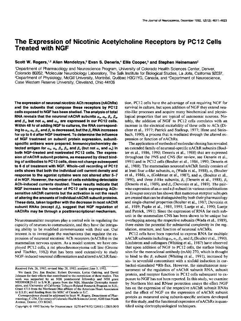
The Journal of Neuroscience, December 1992, 72(12): 461 l-4623
The Expression of Nicotinic Acetylcholine Receptors by PC1 2 Cells Treated with NGF
Scott W. Rogers, L* Allan Mandelzys,3 Evan S. Deneris,4 Ellis Cooper,3 and Stephen Heinemann*
‘Department of Pharmacology and Neuroscience Program, University of Colorado Health Sciences Center, Denver, Colorado 80262, 2Molecular Neurobiology Laboratory, The Salk Institute for Biological Studies, La Jolla, California 92037, 3Department of Physiology, McGill University, Montreal, Quebec HSGlYG, Canada, and 4Department of Neuroscience, Case Western Reserve University, Cleveland, Ohio 44106
The expression of neuronal nicotinic ACh receptors (nAChRs) and the subunits that compose these receptors by PC12 cells exposed to NGF has been studied. The analysis of total RNA reveals that the neuronal nAChFl subunits a,, a,, &, &, and B,, but not a2 and a,, are expressed in our PC12 cells. Within 48 hr of adding NGF to cultures, the RNA correspond- ing to at, as, &, and /3* is decreased, but the & RNA increases for up to 8 d after NGF treatment. To determine the influence of NGF treatment on subunit protein expression, subunit- specific antisera were prepared. lmmunocytochemistry de- tected antigen for 03, a,, &, &, and fi, (but not a2 and aJ in both NGF-treated and nontreated PC12 cells. The expres- sion of nAChR subunit proteins, as measured by direct bind- ing of antibodies to PC1 2 cells, does not change subsequent to 6 d of treatment with NGF. Whole-cell recording of PC12 cells shows that both the individual cell current density and response to the agonist cytisine were not altered after 5-7 d in NGF. However, the number of cells exhibiting detectable ACh-induced currents doubled. These results indicate that NGF increases the number of PC12 cells expressing ACh- sensitive nAChR currents but the activation is not the result of altering the amounts of individual nAChR subunit proteins. These data, taken together with the decrease in most nAChR subunit RNAs (except &), suggest that NGF regulation of nAChRs may be through a posttranscriptional mechanism.
Neurotransmitter receptors play a central role in regulating the capacity of neurons to communicate, and they have the intrigu- ing ability to be modified commensurate with their use. Our interest is to investigate the mechanisms that regulate the ex- pression of neuronal nicotinic ACh receptors (nAChRs) in the mammalian nervous system. As a model system, we have em- ployed PC 12 cells, a rat pheochromocytoma cell line (Greene and Tischler, 1982) that has been used extensively to study NGF-induced neuronal differentiation and altered nAChR func-
Received Feb. 26, 1992; revised May 28, 1992; accepted June 5, 1992. We thank Drs. Jim Boulter, Robert Duvoisin, Lorise Gahring, and David
Johnson for their efforts that contributed to the completion of these studies. This work was supported by an NIH postdoctoral fellowship and NIH grant NS30990R29-01 to S.W.R.; grants from the NIH, Muscular Dystrophy Associ- ation, and University of California Tobacco-Related Research Program to S.H.; Grant 47 15 from the Northeast Ohio affiliate of the American Heart Association to E.S.D.: and fundine from the MRC of Canada to E.C.
Correspondence sh&ld be addressed to Scott W. Rogers, Department of Phar- macology, C-236, University ofColorado Health Sciences Center, 4200 East Ninth Avenue, Denver, CO 80262.
Copyright 0 1992 Society for Neuroscience 0270-647419211246 l l-13%05.00/O
tion. PC12 cells have the advantage of not requiring NGF for survival in culture, but upon addition of NGF they extend neu- rite-like processes and acquire many biochemical and physio- logical properties that are typical of autonomic neurons. No- tably, the addition of NGF to PC12 cells correlates with an increase in the electrical excitability of these cells to ACh (Di- chter et al., 1977; Patrick and Stallcup, 1977; Ifune and Stein- bath, 1990), a process that is mediated through the altered ex- pression or function of nAChRs.
The application of methods of molecular cloning has revealed an extended family of neuronal-specific nAChR subunits (Boul- ter et al., 1986, 1990; Deneris et al., 1988) that are expressed throughout the PNS and CNS (for review, see Deneris et al., 1991) and in PC12 cells (Boulter et al., 1986, 1990; Deneris et al., 1988). The mammalian neuronal nAChR family consists of at least four a-like subunits, LY* (Wada et al., 1988), (Ye (Boulter et al., 1986), LY~ (Goldman et al., l987), and (Ye (Boulter et al., 1990), and three p-like subunits, & (Deneris et al., 1988) & (Deneris et al., 1989), and p4 (Duvoisin et al., 1989). The pair- wise expression ofan (Y- and a B-subunit in various combinations in Xenopus oocytes has shown that numerous receptor subtypes are created that can be distinguished by both their pharmacology and single-channel properties (Boulter et al., 1987; Duvoisin et al., 1989; Papke et al., 1989, 1991; Luetje et al., 1990; Luetje and Patrick, 199 1). Since the expression of RNA for each sub- unit in the mammalian CNS has been shown to be unique but overlapping among the respective subunits (Wada et al., 1989), there exists the potential for substantial complexity in the reg- ulation, structure, and function of neuronal nAChRs.
PC 12 cells have been reported to express RNA for multiple nAChR subunits including (Ye, as, &, and p, (Boulter et al., 1990). Lindstrom and colleagues (Whiting et al., 1987) have observed that upon addition of NGF to PC12 cells, the surface binding sites for the monoclonal antibody (mAb) 270, which is thought to bind to the & subunit (Whiting et al., 1991), increased by six- to sevenfold concomitant with a sixfold induction in car- bachol-stimulated 86Rb flux. However, the simultaneous mea- surement of the regulation of nAChR subunit RNA, subunit protein, and receptor function in PC I2 cells subsequent to ex- posure to NGF has not been reported. In this study, we examine by Northern blot and RNase protection assays the effect NGF has on the expression of the respective nAChR subunit RNAs and the effect of NGF on the expression of nAChR subunit protein as measured using subunit-specific antisera developed for this study, and the functional expression of nAChRs is quan- tified using electrophysiological techniques.
4612 Rogers et al. - Nicotinic Receptor Expression in PC1 2 Cells
Materials and Methods PC12 cell culture. PC12 cells (obtained from I. Verma, Salk Institute) were cultured in Dulbecco’s modified Eagle’s medium [low glucose for- mulation, GIBCO/Bethesda Research Labs (BRL)] containing 10% fetal bovine serum (HYCLONE) and 5% heat-inactivated horse serum (HY- CLONE). The heat inactivation of the horse serum was conducted for 1 hr at 56°C. Cells were grown in a humidified 7.5% CO, atmosphere at 37°C. At confluencv. the cells were dislodged from the culture dish by shaking or triturati& with medium from a pipette, and a portion of these cells were replated into new culture dishes. This process selects against flat cells that adhere tightly to the dish and are not responsive to NGF (see Results). Trypsin and EDTA were avoided. For experi- ments, PC 12 cells that were used in immunocytochemistry and radio- label binding assays were cultured on gelatin-treated culture surfaces.
Analysis of RNA. RNA was isolated from l-5 x lo6 PC12 cells by the method of Chomczynski and Sacchi (1987). The transfer of frac- tionated RNA to Nytran (Schleicher and Schuell) and hybridization of Northern blots were conducted as described by Boulter et al. (1990) except that saline-sodium citrate was substituted for saline-sodium phosphate-EDTA. Random primed probes were prepared from the full- length cDNA of each neuronal nAChR subunit using the Amersham random-prime kit. Probes typically had a specific activity of 0.5-l x 1 O9 cpmlpg.
RNase protection assays were performed as described by Krieg and Melton (1987). Antisense probes were synthesized using SP6 or T7 polymerase from the pGEM or pSP64 vector (Promega) containing subcloned portions of the cDNA encoding each neuronal nAChR sub- unit. The following regions for each cDNA were used: (Y* [HYP16(9); Wada et al., 19881, nucleotides 1748-l 932, protected length 184 bases; (Ye [pPCAlpha 48E(4); Boulter et al., 19861, nucleotides 1557-1815, protected length 258; (Ye [pHYAlpha 231E(l); Goldman et al., 19871, nucleotides O-553, protected length 553 bases; (Ye (Alpha 5; Boulter et al., 1990), nucleotides 1160-1639, protected length 479 bases; & (pGPR49; Deneris et al., 1988) nucleotides 16 17-2 195, protected length 578 bases; & (pESD7; Deneris et al., 1989) nucleotides 568-1021, protected length 453 bases; p, (pZPC13; Duvoison et al., 1989), nucle- otides 2049-2460, protected length 4 11 bases.
Constructs and bacterial overexpression. Complementary DNA clones encoding subunits of neuronal nicotinic acetylcholine receptor subunits were used for constructs. Portions of the cDNA encoding the amino acids (numbered from the initiation methionine) of subunits 01~ (37 l- 5 11; Wada et al., 1988) 01~ (284-470; Boulter et al., 1986), 01~., (461- 594; Goldman et al., 1987), 01~, (345452; Boulter et al., 1990) & (394- 503; Deneris et al., 1988a), p, (330-464; Deneris et al., 1989) and p, (328-426; Duvoisin et al., 1989) were removed by restriction digest and subcloned in the appropriate PATH vector (trpE bacterial over- production system; Dieckmann and Tzagolott, 1985). The host strain was Escherichia coli strain HB 10 1. Overexpression of fusion proteins was done as described previously (Rogers et al., 1991a,b).
Gel electrophoresis and immunization. Overproduced proteins were fractionated using sodium dodecyl sulfate polyacrylamide gel electro- phoresis (SDS-PAGE), and protein bands were visualized by soaking the gel in ice-cold 0.25 M KC1 for 1 hr, and then were cut from the gel (see Rogers et al., 1991a,b). The gel fragments were macerated, emul- sified in complete Freund’s adjuvant (GIBCO/BRL), and injected sub- cutaneouslv into vouna New Zealand White rabbits. Each rabbit re- ceived 20&00 pi of irotein. Three weeks later, rabbits were boosted with 200-400 pg of antigen as above except emulsification was in in- comnlete Freund’s adiuvant (GIBCO/BRL). Serum was collected 12 d later. Subsequent boosts and sera collections were done at 1 month intervals.
ml 5-bromo-4-chloro-3-indolyl phosphate. The reaction was stopped with PBS containing 1 mM EDTA.
ELISA assays were conducted to determine the serum titer and further determine the amount of cross-reactivity between the antisera produced in this study. Overproduced proteins were dissolved in freshly prepared 8 M urea (10 ml per gram of protein) at room temperature for 1 hr. The solution was clarified by centrifugation and the supematant diluted 1: 10 bv the slow addition of a solution of 50 mM KC1 and 50 mM NaCl (pH 10.5). After an additional hour of stirring at room temperature, the solution ‘was brought to pH 8 with 1 N HCl and repeatedly dialyzed against 10 mM NaCl in 10 mM sodium phosphate buffer (pH 7.2) at 4°C. The concentration of soluble protein was then measured by stan- dard Biuret assay (Bio-Rad). Immulon microtiter plates were then pre- pared. Solubilized antigen (5 pdwell) in 100 ~1 of coupling buffer (50 mM Tris, pH 9.5) was added to each well for 14 hr at 4°C. Wells were thoroughly washed with PBS and blocked with blocking PBS (PBS con- taining 5% heat-inactivated calf serum and 0.05% Tween) for 45 min at room temperature. Rabbit serum at various dilutions (e.g., 1:50, 1: 150, 1:450, etc.) in blocking PBS was then added, and dishes were incubated for 4 hr at room temperature or overnight at 4°C. Plates were washed five times with blocking PBS before adding goat anti-rabbit alkaline phosphatase-coupled second antibody (1:2000) for 45 min at room temperature. Plates were again washed with PBS and developed with p-nitrophenyl phosphate (1 mg/ml) in 50 mM potassium carbonate and 1 mM magnesium chloride (pH 9.8) for approximately 15 min at room temperature. The reaction was stopped with 50 rl/well of 0.3 N NaOH and plates were scanned on a Teritek multiscan ELISA reader at 410 nm.
Antibody testing. Specificity of the antisera was tested using Western blot and ELISA analysis. For Western blot analysis, overproduced pro- teins were separated by SDS-PAGE and transferred to nitrocellulose filters (Kyhse-Andersen, 1984). Filters were then blocked in PBS con- taining 5% Carnation nonfat dry milk (Blotto) for 1 hr at room tem- perature. To each blot was added rabbit serum (either complete or absorbed with proteins from bacteria containing the PATH 1 vector) in Blotto ( 1:3000). After 14 hr at 4°C. blots were washed in three changes of Blotto.‘Goat anti-rabbit alkaline phosphatase-conjugated second an- tibody was added in Blotto (1: 1000; Cappel) for 2 hr at room temper- ature. Blots were subsequently rinsed in developing buffer [50 mM so- dium carbonate (pH 9.5) 2 mM MgCl,] and then visualized in developing buffer containing 0.1 mg/ml p-nitro blue tetrazolium chloride, 0.05 mg/
Measurement of nAChR subunit protein. The detection of neuronal nAChR subunits was measured by direct binding of antibodies to PC 12 cells. PC 12 cells were distributed to gelatin-coated six-well culture dishes (1 O5 cells/well). PC1 2 cells were washed gently and fixed with freshly prepared 2% paraformaldehyde (Electron Microscopy Sciences) in 0.1 M cacodylate buffer (pH 7.4) for 30 min at room temperature. We found the purity and concentration of paraformaldehyde to be crucial to ob- taining reproducible results. Concentrations of less than 1.5% or greater than 2% resulted in diminished immunoreactivity (not shown). The fixative was removed, and the cells were washed gently with PBS, and then permeabilized using blocking PBS (PBS with either 1% heat-in- activated calf serum or normal goat serum; GIBCO) containing 0.3% Triton X- 100 (Pharmacia) for 30 min at room temperature. Cells were washed in blocking PBS, and rabbit antiserum diluted in blocking PBS was then added to the cells for 4 hr at room temperature or overnight at 4°C. To minimize background, antisera were first adsorbed against Rat 2 fibroblasts. Rat 2 fibroblasts were grown to confluency in 150 mm culture dishes as described previously (Rogers et al., 199 1 a), fixed, permeabilized, and placed in blocking PBS as described above for PC1 2 cells. The preimmune or primary antiserum was then diluted to the working concentration in blocking PBS (final volume of approximately 5 ml) and then adsorbed over the monolayer of Rat 2 cells for 4 hr at room temperature. This preparation was then used immediately for direct binding or immunocytochemistry (see below). The cells were washed three times in blocking PBS and then rocked in a fourth change of blocking PBS for 30-60 min at room temperature. Blocking PBS containing radioiodinated secondary antibody was then added to the cells for 1 hr at room temperature. Cells were gently washed in four changes of PBS, the well was removed from the culture dish using a hot wire, and the total remaining radioiodine was quantitated by gamma counting using a Beckman Gamma 300 counter at 30% efficiency.
Radioiodination of goat anti-rabbit second antibody (Cappel) was done by the lactoperoxidase method as described by Harlow and Lane (1988). Typical specific activities were lo6 cpm/pg, which corresponds to a substitution ratio of approximately 0.5 mol of 12SI/mol of protein. The specific binding was calculated as the binding of either (r2 (control), (Ye, (Ye, &, or 0, antiserum minus the respective preimmune serum per cell. The value for PC12 cells not treated with NGF was set to 100, and the relative proportion of binding to this value was calculated for PC 12 cells treated with NGF. Measurements were made in duplicate for each experiment.
Immunocytochemistry. PC 12 cells were fixed, permeabilized, and re- acted with the designated antisera as described above. The secondary antibody (alkaline phosphatase-conjugated goat anti-rabbit, Cappel) was then added to the cells for 1 hr at room temperature. The cells were washed with three changes of PBS and twice with 50 mM sodium car- bonate buffer containing 2 mM magnesium chloride (pH 9.5), and the
NO NGF NGF (Days)
The Journal of Neuroscience, De-eembr 1992, 72(12) 4613
Remove NGF (Days)
- 12 3 412 3
28s -
a3 4.. 1
18s -
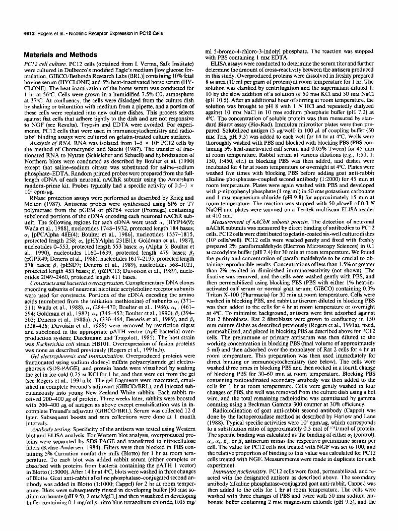
Figure I. Northern blot hybridization
P2 28s - -’ analysis of total RNA from PC1 2 cells. Total RNA was isolated from l-5 x
. ’ lo6 PC1 2 cells that were either not
18s -
p4 28s -
18s-
alkaline phosphatase was visualized by developing the cells at room temperature in the same buffer with freshly added nitro blue tetrazolium (1 mg/ml) and 5-bromo-4-chloro-3-indolyl phosphate (0.5 mg/ml). De- velopment was usually for 30-60 mitt, with fresh developing solution added every 15 min. To stop the reaction, PBS supplemented to 2 mM EDTA was added. Successful permeabilization of PC 12 cells was mon- itored in cells in adjacent wells with fluoresceinated anti-tubulin anti- body (Boehringer Mannheim; not shown).
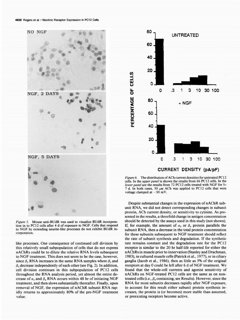
DNA synthesis by PC12 cells was visualized using immunocyto- chemistry for the incorporation of 5-bromo-2-deoxyuridine (BUdR). PC 12 cells were grown without NGF and in the presence of NGF for 2-5 d. For the final 24 hr of culture, BUdR (40 PM final) was added, and cells were rinsed with PBS, and fixed with 95% ethanol, 5% acetic acid for 30 min at room temperature. The cells were rinsed with distilled water and the DNA was denatured with 2 N HCl for 30 min. The cells were rinsed with PBS, placed in blocking PBS containing mouse anti- BUdR (1: 100; Becton-Dickinson) for 30 mitt, rinsed with PBS, placed in blocking PBS containing goat anti-mouse alkaline phosphatase-con- jugated secondary antibody (Cappel), and visualized as described above.
Electrophysiology. Sensitivity of PC12 cells to ACh was measured electrophysiologically using whole-cell patch-clamp techniques (Hamill et al., 198 1). All recordings were done with a List EPC-7 amplifier at 22-24°C. The recording electrodes typically had resistances of 2-6 MQ, and the current signal balanced to zero. During the experiments, mem- brane currents and voltages were filtered at 3 kHz, digitized at 44 kHz by a pulse code modulation unit (PCM 701, Sony Corp.), and stored on a Beta videocassette recorder. Round PC12 cells with clear nuclei were selected at random. In pilot experiments, we found that flat cells that adhered tightly to the culture surface were insensitive to ACh, and these were not examined further (not shown). For most PC 12 cells, the membrane potential was held at two potentials, -50 mV and - 100 mV, while ACh (ACh iodide, Sigma Chemical Co.) was applied to the cell body, as well as the proximal processes of the NGF-treated cells.
To measure the overall ACh current density of each PC12 cell, we applied the drug by pressure ejection from relatively large-tipped pi-
treated with NGF, treated for l-4 d with NGF, or treated with NGF for 4 d and then returned to NGF-free culture me- dium for l-3 d. Twenty micrograms of total RNA for each sample were elec- trophoresed through 1% agarose gels containing formaldehyde, transferred to Nytran blotting membrane, and then probed with full-length cDNA random- primed probes for each subunit as de- scribed in Materials and Methods. The exposure was for 14 hr at -7O”c, and the locations of the 28s and 18s ribo- somal RNA are indicated.
pettes (10-20 am), positioned at an appropriate distance (30-40 pm) from the PC12 cell, so that upon application of light pressure (usually 20-30 kPa) we could perfuse a large area (see Mandelzys et al., 1990). The cultures were continuously perfused at the rate of 1 mumin with extracellular solution (see below) to ensure that the drug did not accu- mulate during the experiment. The ACh pipettes were filled with 50 PM ACh dissolved in the extracellular solution. This concentration of ACh was chosen to ensure that we could detect cells with ACh-gated currents but avoid agonist-induced desensitization. The extracellular solution was composed of 140 mM NaCl, 5.4 mM KCl, 2.8 mM C&l,, 0.18 mM MgCl,, 10 mM HEPES, 5.6 mM glucose., 2 mM glutamine, 30 U/ml penicillin, and 30 &ml streptomycin. The intracellular solution con- sisted of 80 mM KF, 60 mM K-acetate, 5 mM NaCl, 1 mM MgCl,, 10 mM EGTA, and 10 mM HEPES. All solutions were adjusted to pH 7.2- 7.4 with NaOH for the extracellular solution and KOH for the intra- cellular solution. The limit of resolution in this study was determined to be 5-10 pA, which is equivalent to three to five receptors open simultaneously. Since the average cell capacitance is approximately 30 pF, our limit of detection is 0.3 pA/pF. PC12 cells with ACh-gated currents below this value were grouped as insensitive.
To determine the relationship between currents evoked by ACh and those evoked by cytisine (Aldrich Chemical Co.), we constructed double- barreled pipettes so that the tip of each barrel was fused side by side; each tip was 15-20 pm. One barrel contained 50 PM ACh, and the other barrel contained 50 PM cytisine. All ACh currents are normalized to cell capacitance by integrating the capacity current evoked by a 5 mV hyperpolarizing voltage step from a holding potential of - 50 mV. Un- treated PC1 2 cells had capacitances that ranged from 13 pF to 18 pF, whereas NGF-treated PC 12 cells had capacitances ranging from 22 pF to 30 pF. The distributions of ACh current densities represent values obtained in response to 50 PM ACh at a holding potential of -50 mV and were plotted on a log scale to cover the wide range of values (from 0.3 pA/pF to 28.1 pA/pF) that exist on different PC 12 cells. The zero column in each distribution reflects the proportion of neurons in which there was no detectable response. We used the Mann-Whitney U test
4614 Rogers et al. l Nicotinic Receptw Expression in PC12 Cells
a3 -
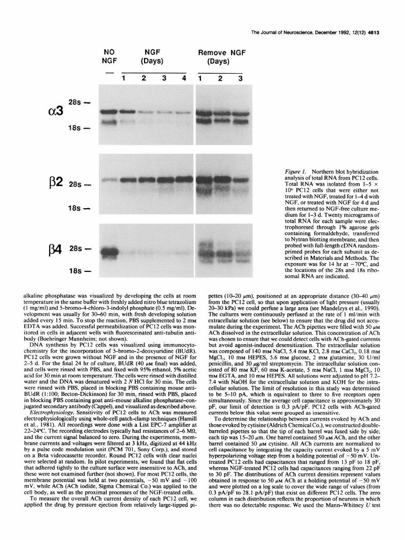
Figure 2 RNase protection of n4ChR subunits from total RNA prepared from PC12 cells treated with NGF. Total RNA from PC1 2 cells treated with NGF for the indicated time period was pre- pared and analyzed with subunit-spe- cific probes as described in Materials and Methods. Protected transcripts were observed for nAChR subunits a,, cz5, &, &, and @,, but not (Y* or LX,.
P2+
p3+ r,
P4
z Days with NGF 2 $ nAChR
‘0 2 5 6’ 5 5: subunit
and the x2 test to assess the sigpificance of differences between the current density distributions.
Results NGF alters nAChR subunit RNA expression RNA samples were prepared from PC 12 cells that were exposed to 50 rig/ml of NGF for l-6 d as described in Materials and Methods. At day 4, one group of cells was returned to culture media not supplemented with NGF. Northern blot analysis of 20 Mg of total RNA per lane is shown in Figure 1. CX, and & RNA decreased within 48 hr of exposure to NGF and decreased only slightly thereafter, but & RNA was observed to increase slowly over the 6 d period. The removal of NGF from the culture media subsequent to 4 d of NGF treatment resulted in the rapid return (within 24 hr) of all and j!& RNA to approximately 80% of the level seen in PC 12 cells never exposed to NGF. RNA for & also returned to pre-NGF treatment levels, but in contrast to aJ and & this return was gradual over the 3 d tested. Northern blot analysis of total RNA detects at least two forms of RNA for each subunit as reported previously (see Boulter et al., 1986, 1990). We found that the various transcript forms were altered similarly in response to NGF (see Fig. 1; quantitative data not shown), which suggests that NGF treatment and the regulation of nAChR subunit RNA do not occur through selectively al- tering the level of one RNA form relative to the other.
Days with NGF ii
2s nAChR 2 4 5 6’55 subunit '0
+P2
-3 P 734
The more sensitive method of RNase protection was applied to measure (Ye and to test for the presence of 1y2, (Ye, and & which are not observed using Northern blot analysis (Boulter et al., 1990; not shown). This method offers the additional advantage of allowing the simultaneous analysis of more than one nAChR transcript in an RNA preparation since probes of different lengths can be used. The results of these assays for (Ye, (Y,, a4, and (Ye and for &, /I,, and @., are shown in Figure 2. As expected from Northern blot analysis, the expression of 1~~ and & decreased rapidly following exposure to NGF and the expression of & increased slowly as seen above. The expression of cx5 decreased approximately 50% in response to NGF treatment. No protected species corresponding to (r2 or LY., was detected in PC1 2 cells. However, a protected species corresponding to & was observed, and the expression of this RNA was decreased subsequent to NGF treatment. These results demonstrate that PC 12 cells ex- press RNA corresponding to the nAChR subunits of (Y,, 01~, &, &, and & but not a2 and LY.,, and that the relative concentrations of these RNAs are altered by the presence NGF in the culture medium.
Antisera to neuronal nAChR subunits To determine the influence of NGF on the expression of nAChR subunit protein, we prepared rabbit polyclonal antisera against the putative cytoplasmic domains of the subunits a*, (Ye, (Y.,, as,
The Journal of Neuroscience, December 1992, f2(12) 4615
P,, p,, and p4, respectively (see Materials and Methods). As shown in Figure 3, Western blot analysis of these bacterially overproduced proteins indicates that each antiserum reacts only with the subunit protein to which it was made. Similarly, ELISA analysis showed that cross-reactivity between sera was not sig- nificant unless only low dilutions (less than 1:300) were used (data not shown). There remains the possibility that these sera could react with other subunits, although additional studies in- dicate that this is not the case. First, overlapping portions of each subunit were used for antigen, and there is essentially no sequence identity between these regions ofthe receptor subunits. Second, each antibody has been used to study Rat 2 cells that were transfected with either the subunit combination C& (Rog- ers et al., 199 1 a) or cu& (not shown). For the cell line expressing (YJ&, immunoprecipitation and immunocytochemistry were successful only with antisera to (Ye or & (Rogers et al., 1991a; not shown). Third, antisera to LYE or &, respectively, immuno- precipitate high-affinity 3H-nicotine binding sites from the rat brain. Immunoprecipitation is not altered by preclearing the membrane preparations with a3, (Ye, &, or p4 receptor subunit antisera, but it can be inhibited by preclearing with either an- tisera to (Ye or &, respectively (Flores et al., 1992). These results taken together with the results in the next section suggest that the antisera used in this study exhibit subunit specificity.
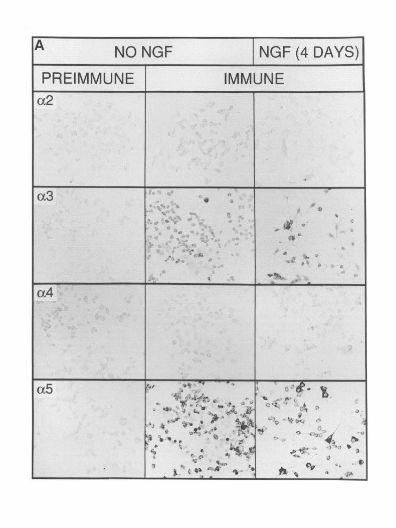
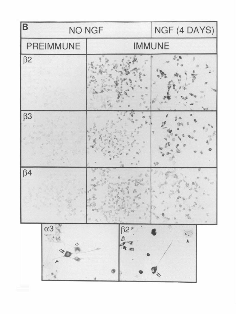
Immunocytochemistry reveals PC12 cell heterogeneity Immunocytochemical analysis of PC12 cells not treated with NGF or treated for 5 d is shown in Figure 4A for each antiserum. Immunostaining was observed in PC1 2 cells for antisera raised to a3, cr,, &, and p,. No staining was observed with antisera to either (Ye or LY+ Although immunoreactivity toward & is seen, the intensity of this staining varied between experiments. The data in Figure 4A illustrate the point that the treatment of PC1 2 cells with NGF does not obviously change either the number of cells expressing antigen or the amount of antigen present per cell (see below). As is seen in Figure 4B, immunoreactivity toward neuronal nAChR subunits (Ye and & reveals PC12 cell heterogeneity. At least two cell types can be distinguished based upon their relative adherence to the culture surface and their response to NGF. The first group (Fig. 4B) consists of flat cells that attach firmly to the culture dish, they fail to extend neurites in response to NGF, they rarely exhibit immunocytochemical staining, and they do not contain ACh-sensitive currents (see below). The relative abundance of these cells increases with prolonged culture and changes if culture conditions are altered (e.g., reduced serum concentrations or the use of trypsinization for subculture; not shown). In our experiments, these cells rep- resent less than 10% of the total cell population. The second group of cells are rounded and attach loosely to the dish; they extend neurites in response to NGF, they exhibit immunoreac- tivity toward neuronal nAChR-directed antisera, and they ex- hibit ACh-sensitive ion channels (Fig. 4B). Within this group of NGF-responsive PC 12 cells, there is consistently observed a subpopulation of cells that appear to be smaller and oval in shape, and to extend shorter neurites (Fig. 4B). They often stain less intensely for nAChR subunits than do PC12 cells that ex- tend extensive net&es. These cell types are also observed in Figure 4A.
Antigen appears to concentrate in the cell body (Fig. 4B). Although staining of neurite-like processes is observed occa- sionally, the majority remain unstained except for a punctate staining that is observed on some well-developed processes. At
present, we are uncertain if this punctate staining reflects clus- tering of the receptors.
PC12 cells treated with NGF generally cease cell division (Greene and Tischler, 1982; Rudkin et al., 1989), although DNA synthesis and cell division may persist within some cells of the population (Rudkin et al., 1989). To determine if PC12 cells that express nAChRs and respond to NGF by altering their morphology also stop cell division, DNA synthesis in PC 12 cells that were exposed to NGF for varying periods was measured. PC12 cells were distributed to culture dishes and cultured for 24 hr before adding 50 rig/ml of NGF for 2, 3, 4, and 5 d, respectively. For the final 24 hr of culture, the cells were grown in the presence of 40 PM BUdR (see Materials and Methods) and the nuclei of cells undergoing DNA syntheses were visu- alized using an antibody to BUdR as described in Materials and Methods. As shown in Figure 5 for PC12 cells cultured without NGF or in the presence of NGF for 2 or 5 d, the nuclei of cells that responded to NGF by extending processes failed to stain for BUdR immunoreactivity. In contrast, the nuclei of PC12 cells that did not extend processes in the presence of NGF stained for BUdR immunoreactivity. This result supports the conclu- sion that cells in our PC 12 cell population that fail to respond to NGF and fail to exhibit staining with nAChR subunit antisera correspond to those that also continue DNA synthesis (see Dis- cussion).
NGF does not alter neuronal nAChR subunit protein expression Immunocytochemistry reveals reactivity toward nAChR sub- units CY,, (Ye, &, and 0, in both NGF-treated and untreated PC 12 cells. Since there is a dramatic change in the relative mRNA levels of these subunits in response to NGF, we determined if corresponding changes in the protein concentrations of these subunits could be detected through measuring the direct binding of antibody to fixed and permeabilized PC 12 cells (see Materials and Methods). In three experiments, when binding was calcu- lated by subtracting (Y* immune binding and the results presented as a ratio to PC 12 cells not treated with NGF, no change between the ratio of radioactivity bound by PC 12 cells for up to 6 d was detected (Table 1). Similar results were obtained,when the bind- ing by preimmune serum was substituted for the binding to antisera to (Ye (not shown). The amount of binding or antibody to PC12 cells that were not permeabilized with Triton X-100 did not exceed 20% of the total counts (not shown). This binding probably reflects specific binding to the cytoplasmic domain of nAChR subunits since the immunoreactivity toward tubulin could be visualized in some PC12 cells fixed with 2% but not 4% paraformaldehyde (not shown). This result suggests that some membrane permeabilization occurs at the relatively low fixative concentration of 2% required to preserve immunoreac- tivity toward the nAChR subunits.
The sensitivity of this assay was determined using the (Ye subunit. Crude PC 12 membranes were prepared (see Rogers et al., 1991a,b), adsorbed against prediluted immune serum (in blocking PBS) overnight at 4°C removed by centrifugation, and the supematant was used for binding assays by radioiodinated antibody to fixed and permeabilized PC 12 cells as described in Materials and Methods. Membranes prepared from Rat 2 cul- tured fibroblasts were used to control adsorption to membranes not containing nAChRs, and a2 immune serum was used to measure background binding (not shown). These assays indi-
4616 Rogers et al. l Nicotinic Receptor Expression in PC1 2 Cells
M.W. (kD) Coomassie Blue Stain Western Blot
trpE cd2 cd a4 a!5 p2 p3 p4 trpE (~2 (~3 ad ~6 p2 p p
trpE Adsorbed
WE a2 a3 a4 05 p2 p3 p4
50 ---
39 ---
50 ---
39 ---
75 --
50 ---
39 ---
75 --
50 ---
39 ---
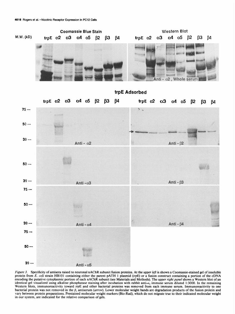
Figure 3. Specificity of antisera raised to neuronal nAChR subunit fusion proteins. At the upper left is shown a Coomassie-stained gel of insoluble protein from E. cofi strain HBlOl containing either the parent PATH 1 plasmid (trpE) or a fusion construct containing a portion of the cDNA encoding the putative cytoplasmic portion of each nAChR subunit (see Materials and Methods). The upper right panel shows a Western blot of an identical gel visualized using alkaline phosphatase staining after incubation with rabbit anti-a, immune serum diluted 1:3000. In the remaining Western blots, immunoreactivity toward trpE and other bacterial proteins was removed from each immune serum. Immunoreactivity to one bacterial protein was not removed in the & antiserum (arrow). Lower molecular weight bands are degradation products of the fusion protein and vary between protein preparations. Prestained molecular weight markers (Bio-Rad), which do not migrate true to their indicated molecular weight in our system, are indicated for the relative comparison of gels.
The Journal of Neuroscience, December 1992, 72(12) 4617
cated that the detection sensitivity of our assay is approximately 3 f 1 ng per lo6 cells (not shown).
ACh-activated currents are not altered by NGF treatment
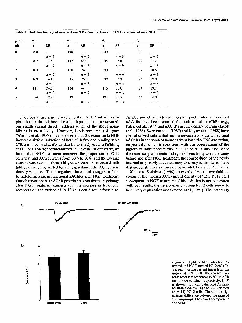
The above measurements indicate that the relative nAChR sub- unit protein concentration is not altered in response to NGF treatment despite significant changes in the respective subunit RNA levels. Nevertheless, it is well documented that NGF treat- ment of PC 12 cells increases ACh-activated currents. To ensure this was true of our PC1 2 cells and to study further the influence of NGF on expression of functional nAChRs, we used whole- cell patch-clamp techniques to compare the current densities and agonist sensitivities of nAChRs on untreated and NGF- treated PC12 cells. As noted in Materials and Methods, PC12 cells with a flat morphology were found to contain no detectable ACh-activated currents, consistent with immunocytochemical results, and they were not included in the following analysis. Figure 6 shows the ACh current density distribution for 66 PC 12 cells that were not treated with NGF. Thirty percent of the cells show ACh sensitivity, and their current densities range from 0.6 pA/pF to 28.9 pA/pF with a mean of 4.4 f 1.0. Figure 6 also shows the current distribution for 72 PC12 cells treated with NGF for 5-7 d. After treatment with NGF, approximately 60% of the cells respond to ACh and their current densities range from 0.3 pA/pF to 28.1 pA/pF with a mean of 2.9 + 0.6. These results demonstrate that exposure to NGF results in a twofold increase in the proportion (from 30% to 58%) of PC12 cells expressing functional nAChRs, but there is little effect on the current density of nAChRs on individual cells.
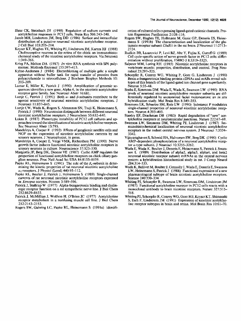
Recently, Luetje and Patrick (1991) observed that a,/& re- ceptors expressed in Xenopus oocytes were loo-fold less sen- sitive to the agonist cytisine relative to ACh. In contrast, cytisine was two- to threefold more potent than ACh on the LY,/& func- tional receptor. This observation suggests that cytisine may be used to infer the presence of & or & in functional receptors. An example of a typical response from an ACh-sensitive PC 12 cell is shown in Figure 7 for ACh and cytisine, respectively. The application of agonist causes a rapid inward current that peaks in 100-200 msec after the drug is applied, and the response shows a small degree of desensitization that occurs with a rel- atively slow time course. We found that cytisine produced a 2.4 * 0.3-fold increase in the evoked current from PC12 cells not exposed to NGF relative to ACh at equal concentrations (see Fig. 7). This result suggests that functional nAChRs on PC12 cells contain the p4 subunit or that it dominates this pharma- cology. Similarly, the cytisine:ACh ratio of receptors on PC12 cells treated with NGF is 2.6, which indicates that treatment with NGF does not alter the relative receptor sensitivity to the two agonists.
Discussion
In this study, we have analyzed the effect of NGF on the ex- pression of neuronal nAChR subunit RNA, subunit protein, and receptor function. We observed a rapid change in the amount of each nAChR subunit RNA expressed by PC 12 cells subse- quent to NGF exposure. RNA for & increased slowly over the 6 d NGF treatment period, but RNA for (Ye, +, /Is, and p4 decreased. This decrease appears to follow three courses: the rapid loss of approximately 75% of the RNA followed by a slow but continuous loss over the next 5 d of NGF exposure as observed for &,, a relatively rapid drop of approximately 50% followed by a period of essentially no change in the RNA level as observed for the (Ye and ,f3, subunits, and finally the loss of approximately 75% of the original RNA level over 3 d of NGF followed by a subsequent period of slow loss as observed for a3. These observations suggest that the RNA for each subunit is regulated independently subsequent to NGF exposure, but it is not known if this regulation is transcriptional or posttran- scriptional. These differences are also of interest since the genes encoding the nAChR subunits (Y,, (Ye, and p, that are tightly linked (Boulter et al., 1990).
Immunocytochemical staining of PC1 2 cells was observed for CQ, (Ye, &, p,, and p4, but the intensity of staining was particularly strong for (1~~ and &. Whether this staining reflects the amount of antigen present or if it is the result of the particular antiserum remains to be determined. Nevertheless, this suggests that the nAChR of PC 12 cells could be composed of multiple subunits, and the potential for substantial complexity exists. Further, re- cent reports of additional nAChR subunits have been made. These subunits include the preliminary reports of (Ye (Lamar et al., 1990) and the discovery of the a-bungarotoxin binding sub- units (Y, and (Y* from chick (Couturier et al., 1990; Schoepfer et al., 1990). At present, only the chick cY,-subunit has been re- ported to have function when introduced into Xenopus oocytes and this receptor is blocked by cY-bungarotoxin (Couturier et al., 1990). PC1 2 cells also express an a-bungarotoxin binding sub- unit (Patrick and Stallcup, 1977), but the relationship of this subunit to the toxin-binding protein of PC 12 remains unclear since cu-bungarotoxin does not block ACh or nicotine-elicited current in these cells (Patrick and Stallcup, 1977; Rogers et al., 1991a). We have observed RNA for the rat homolog of (Y, in our PC12 cells, and the RNA amount is decreased for these species when NGF is added to the culture (D. Johnson, J. Boul- ter, and S. Rogers, unpublished observation).
In this study, immunocytochemistry and electrophysiology revealed that nAChR expression in PC 12 cells was limited to those cells that responded to NGF by ceasing DNA synthesis and altering their morphology, particularly by extending neurite-
Figure 4. Immunocytochemistry of PC12 cells. A, PC12 cells that were either not treated with NGF or treated for 4 d were prepared for immunocytochemistry with either preimmune or immune rabbit anti-a,, -c+, -(Ye, -(Y>, -&, -&, or -j3, diluted 1:2000 as described in the Materials and Methods. Immunoreactivity of the preimmune sera toward PC12 cells exposed to NGF was similar to that presented for non-NGF-treated cells and is not shown. Immunoreactivity toward (r), (rs, &, &, and & is observed, but not toward (Y* or a0 B, Higher magnification of PC12 cells treated with NGF for 4 d that were stained for immunoreactivity toward nAChR subunit (Ye or &. PC12 cells that extend neurites in response to NGF (double arrows) or are smaller but are poorly attached to the dish surface and become elongated (single solid arrow) exhibit a3 and fi2 (double arrows) as well as (Ye and 8, (see above) immunoreactivity. Cells that adhere tightly to the culture dish but do not extend neurites in response to NGF fail to show immunoreactivity toward nAChR antisera (arrowheuds). A flat cell that is extending neurites and also shows (Ye immunoreactivity is indicated by the open arrow. Immunoreactivity is seen predominantly in the soma, and it is rarely observed in the extended processes. However, a fine “punctate” pattern of staining can be seen on the processes extended from the cells marked by double arrows. Scale: A, 10 mm = 50 pm; B, 10 mm = 12 pm.
4626 Rogers et al. * Nicotinic Receptor Expression in PC12 Cells
NO NGF _ ‘ UNffEA-l-ED
r ,i ’ / I/
‘, .
IL .3 1 3 10 30 100
NGF, 2 DAYS
+ NGF
\- ,‘j >”
0 .3 1 3 10 30 100
CURRENT DENSITY (pA@FJ
Figure 6. The distribution of ACh current densities for untreated PC12 cells. In the upper panel is shown the results from 66 PC 12 cells. In the lower panel are the results from 72 PC1 2 cells treated with NGF for 5- 7 d. In both cases, 50 PM ACh was applied to PC12 cells that were voltage clamped at -50 mV.
Despite substantial changes in the expression of nAChR sub- unit RNA, we did not detect corresponding changes in subunit protein, ACh current density, or sensitivity to cytisine. As pre- sented in the results, a threefold change in antigen concentration
Figure 5. Mouse anti-BUdR was used to visualize BUdR incorpora- should be detected by the assays used in this study (not shown). tion in to PC1 2 cells after 4 d of exposure to NGF. Cells that respond If, for example, the amount of (Ye or & protein parallels the to NGF by extending neurite-like processes do not exhibit BUdR in- subunit RNA, then a decrease in the total protein concentration corporation. for these subunits subsequent to NGF treatment should reflect
the rate of subunit synthesis and degradation. If the synthetic like processes. One consequence of continued cell division by rate remains constant and the degradation rate for the PC12 this relatively small subpopulation of cells that do not express receptor is similar to the 20 hr half-life reported for either the nAChRs could be to dilute the relative RNA levels subsequent nAChRs in muscle prior to innervation (Stanley and Drachman, to NGF treatment. This does not seem to be the case, however, 1983), in cultured muscle cells (Patrick et al., 1977), or in ciliary since & RNA increases in the same RNA samples where & and ganglia (Jacob et al., 1986) then as little as 5% of the original p4 decrease independently of each other (see Fig. 2). In addition, receptors at day 0 could be left after 6 d of NGF treatment. We cell division continues in this subpopulation of PC12 cells found that the whole-cell currents and agonist sensitivity of throughout the RNA analysis period, yet almost the entire de- nAChRs on NGF-treated PC12 cells are the same as on non- crease of aj and & RNA occurs within 48 hr of initiating NGF treated cells (i.e., & containing; see Results). However, since the treatment, and then slows substantially thereafter. Finally, upon RNA for most subunits decreases rapidly after NGF exposure, removal of NGF, the expression of nAChR subunit RNA rap- to account for this result either subunit protein synthesis in- idly returns to approximately 80% of the pre-NGF treatment creases, the protein is (or becomes) more stable than assumed, value. or preexisting receptors become active.
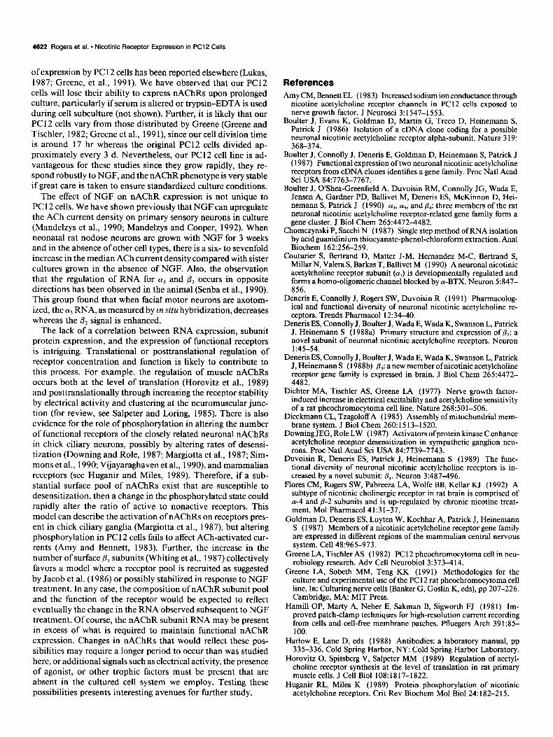
The Journal of Neuroscience, December 1992, 12(12) 4621
Table 1. Relative binding of neuronal nAChR subunit antisera to PC12 cells treated with NGF
NGF
(4
0
1
2
3
4
5
013 015 P2 A x SE x SE K SE R SE
100 - 100 - 100 - 100 - n=7 n=3 n=9 n=3
102 7.6 137 41.0 105 5.0 92 11.2 n=7 n=3 n=9 n=3
103 7.6 110 24.0 99 6.1 82 10.6 n=7 n=3 n=9 n=3
109 14.1 95 25.0 99 6.3 76 19.0 n=4 n=3 n=4 n=3
111 24.5 124 - 115 23.0 84 19.1 n=3 n=2 n=3 n&3
94 17.9 97 - 121 20.9 75 4.0 n=3 n=2 n=3 n=3
Since our antisera are directed to the nAChR subunit cyto- plasmic domain and the entire subunit protein pool is measured, our results cannot directly address which of the above possi- bilities is most likely. However, Lindstrom and colleagues (Whiting et al., 1987) have reported that a 3 d exposure to NGF induces a sixfold induction of both 86Rb flux and binding mAb 270, a monoclonal antibody that binds the & subunit (Whiting et al., 1990) on nonpermeabilized PC12 cells. In our study, we found that NGF treatment increased the proportion of PC1 2 cells that had ACh currents from 30% to 60%, and the average current was two- to threefold greater than on untreated cells (although when corrected for cell capacitance, the ACh current density was less). Taken together, these results suggest a four- to sixfold increase in functional nAChRs after NGF treatment. Our observation that nAChR protein does not detectably change after NGF treatment suggests that the increase in functional receptors on the surface of PC 12 cells could result from a re-
distribution of an internal receptor pool. Internal pools of nAChRs have been reported for both muscle nAChRs (e.g., Patrick et al., 1977) and nAChRs in chick ciliary neurons (Jacob et al., 1986). Swanson et al. (1987) and Keyser et al. (1988) have also observed substantial immunoreactivity toward neuronal nAChRs in the soma of neurons from both the CNS and retina, respectively, which is consistent with our observations of the pattern of immunoreactivity in PC12 cells. In any case, since the macroscopic currents and agonist sensitivity were the same before and after NGF treatment, the composition of the newly inserted or possibly activated receptors may be similar to those that are constitutively expressed by non-NGF-treated PC 12 cells.
Ifune and Steinbach (1990) observed a five- to sevenfold in- crease in the median ACh current density of their PC 12 cells subsequent to NGF treatment. Although this is not consistent with our results, the heterogeneity among PC12 cells seems to be a likely explanation (see Greene, et al., 199 1). The instability
50 UY hch A
so UY cyushle
UNTREATED . NGF
Figure 7. Cytisine:ACh ratio for un- treated and NGF-treated PC 12 cells. In A are shown two current traces from an untreated PC12 cell. The inward cur- rents represent responses to 50 PM ACh and 50 PM cytisine, respectively. In B is shown the mean cytisine:ACh ratio for untreated (n = 10) and NGF-treated (n = 13) PC12 cells. There is no sig- nificant difference between the ratio of the two groups. The error bars represent the SEM.
4622 Rogers et al. - Nicotinic Receptor Expression in PC12 Cells
of expression by PC 12 cells has been reported elsewhere (Lukas, 1987; Greene, et al., 199 1). We have observed that our PC12 cells will lose their ability to express nAChRs upon prolonged culture, particularly if serum is altered or trypsin-EDTA is used during cell subculture (not shown). Further, it is likely that our PC 12 cells vary from those distributed by Greene (Greene and Tischler, 1982; Greene et al., 199 l), since our cell division time is around 17 hr whereas the original PC12 cells divided ap- proximately every 3 d. Nevertheless, our PC12 cell line is ad- vantageous for these studies since they grow rapidly, they re- spond robustly to NGF, and the nAChR phenotype is very stable if great care is taken to ensure standardized culture conditions.
References Amy CM, Bennett EL (1983) Increased sodium ion conductance through
nicotine acetylcholine receptor channels in PC12 cells exposed to nerve growth factor. J Neurosci 3: 1547-l 553.
Boulter J, Evans K, Goldman D, Martin G, Treco D, Heinemann S, Patrick J (1986) Isolation of a cDNA clone coding for a possible neuronal nicotinic acetylcholine receptor alpha-subunit. Nature 3 19: 368-374.
Boulter J, Connolly J, Deneris E, Goldman D, Heinemann S, Patrick J (1987) Functional expression of two neuronal nicotinic acetylcholine receptors from cDNA clones identifies a gene family. Proc Nat1 Acad Sci USA 8417763-7767.
Boulter J, O’Shea-Greenfield A, Duvoisin RM, Connolly JG, Wada E, Jensen A, Gardner PD, Ballivet M, Deneris ES, McKinnon D, Hei- nemann S, Patrick J (1990) c+, (Ye, and p4: three members of the rat neuronal nicotinic acetylcholine receptor-related gene family form a gene cluster. J Biol Chem 265:4472-t482.
The effect of NGF on nAChR expression is not unique to PC1 2 cells. We have shown previously that NGF can upregulate the ACh current density on primary sensory neurons in culture (Mandelzys et al., 1990; Mandelzys and Cooper, 1992). When neonatal rat nodose neurons are grown with NGF for 3 weeks and in the absence of other cell types, there is a six- to sevenfold increase in the median ACh current density compared with sister cultures grown in the absence of NGF. Also, the observation that the regulation of RNA for oj and /I2 occurs in opposite directions has been observed in the animal (Senba et al., 1990). This group found that when facial motor neurons are axotom- ized, the (Ye RNA, as measured by in situ hybridization, decreases whereas the & signal is enhanced.
The lack of a correlation between RNA expression, subunit protein expression, and the expression of functional receptors is intriguing. Translational or posttranslational regulation of receptor concentration and function is likely to contribute to this process. For example, the regulation of muscle nAChRs occurs both at the level of translation (Horovitz et al., 1989) and posttranslationally through increasing the receptor stability by electrical activity and clustering at the neuromuscular junc- tion (for review, see Salpeter and Loting, 1985). There is also evidence for the role of phosphorylation in altering the number of functional receptors of the closely related neuronal nAChRs in chick ciliary neurons, possibly by altering rates of desensi- tization (Downing and Role, 1987; Margiotta et al., 1987; Sim- mons et al., 1990; Vijayaraghaven et al., 1990), and mammalian receptors (see Huganir and Miles, 1989). Therefore, if a sub- stantial surface pool of nAChRs exist that are susceptible to desensitization, then a change in the phosphorylated state could rapidly alter the ratio of active to nonactive receptors. This model can describe the activation of nAChRs on receptors pres- ent in chick ciliary ganglia (Margiotta et al., 1987), but altering phosphorylation in PC 12 cells fails to affect ACh-activated cur- rents (Amy and Bennett, 1983). Further, the increase in the number of surface & subunits (Whiting et al., 1987) collectively favors a model where a receptor pool is recruited as suggested by Jacob et al. (1986) or possibly stabilized in response to NGF treatment. In any case, the composition of nAChR subunit pool and the function of the receptor would be expected to reflect eventually the change in the RNA observed subsequent to NGF treatment. Of course, the nAChR subunit RNA may be present in excess of what is required to maintain functional nAChR expression. Changes in nAChRs that would reflect these pos- sibilities may require a longer period to occur than was studied here, or additional signals such as electrical activity, the presence of agonist, or other trophic factors must be present that are absent in the cultured cell system we employ. Testing these possibilities presents interesting avenues for further study.
Chomczynski P, Sacchi N ( 1987) Single step method of RNA isolation by acid guanidinium thiocyanate-phenol-chloroform extraction. Anal Biochem 162:256-259.
Couturier S, Bertrand D, Matter J-M, Hemandez M-C, Bertrand S, Millar N, Valera S, Barkas T, Ballivet M (1990) A neuronal nicotinic acetylcholine receptor subunit (o,) is developmentally regulated and forms a homo-oligomeric channel blocked by a-BTX. Neuron 5:847- 856.
Deneris E, Connolly J, Rogers SW, Duvoisin R (199 1) Pharmacolog- ical and functional diversity of neuronal nicotinic acetylcholine re- ceptors. Trends Phannacol 12:34-40.
Deneris ES, Connolly J, Boulter J, Wada E, Wada K, Swanson L, Patrick J, Heinemann S (1988a) Primarv structure and exnression of a,: a novel subunit of neuronal nicotinic acetylcholine receptors. Neuron 1:45-54.
Deneris ES, Connolly J, Boulter J, Wada E, Wada K, Swanson L, Patrick J, Heinemann S (198813) &: a new member of nicotinic acetylcholine receptor gene family is expressed in brain. J Biol Chem 265:4472- 4482.
Dichter MA, Tischler AS, Greene LA (1977) Nerve growth factor- induced increase in electrical excitability and acetylcholine sensitivity of a rat pheochromocytoma cell line. Nature 268:501-506.
Dieckmann CL, Tzagoloff A (1985) Assembly of mitochondrial mem- brane system. J Biol Chem 260: 15 13-l 520.
Downing JEG, Role LW ( 1987) Activators of protein kinase C enhance acetylcholine receptor desensitization in sympathetic ganglion neu- rons. Proc Nat1 Acad Sci USA 84~7739-7743.
Duvoisin R, Deneris ES, Patrick J, Heinemann S (1989) The func- tional diversity of neuronal nicotinic acetylcholine receptors is in- creased by a novel subunit: 6,. Neuron 3~487496.
Plores CM, Rogers SW, Pabreeza LA, Wolfe BB, Kellar KJ (1992) A subtype of nicotinic cholinergic receptor in rat brain is comprised of a-4 and D-2 subunits and is up-regulated by chronic nicotine treat- ment. Mol Pharmacol 41:31-37.
Goldman D, Deneris ES, Luyten W, Kochhar A, Patrick J, Heinemann S (1987) Members of a nicotinic acetylcholine receptor gene family are expressed in different regions of the mammalian central nervous system. Cell 48:965-973.
Greene LA, Tischler AS (1982) PC1 2 pheochromocytoma cell in neu- robiology research. Adv Cell Neurobiol 3:373-414.
Greene LA, Sobeth MM, Teng KK (1991) Methodologies for the culture and experimental use of the PC1 2 rat pheochromocytoma cell line. In: Culturing nerve cells (Banker G, Goslin K, eds), pp 207-226. Cambridge, MA: MIT Press.
Hamill OP, Marty A, Neher E, Sakman B, Sigworth FJ (198 1) Im- proved patch-clamp techniques for high-resolution current recording from cells and cell-free membrane patches. Pfluegers Arch 391:85- 100.
Harlow E, Lane D, eds (1988) Antibodies: a laboratory manual, pp 335-336. Cold Spring Harbor, NY: Cold Spring Harbor Laboratory.
Horovitz 0, Spitsberg V, Salpeter MM (1989) Regulation of acetyl- choline receptor synthesis at the level of translation in rat primary muscle cells. J Cell Biol 108:1817-1822.
Huganir RL, Miles K (1989) Protein phosphorylation of nicotinic acetylcholine receptors. Crit Rev Biochem Mol Bio124: 182-2 15.
The Journal of Neuroscience, December 1992, 12(12) 4923
Ifune CK, Steinbach JH (1990) Regulation of sodium currents and acetylcholine responses in PC12 cells. Brain Res 506:243-248.
Jacob MH. Lindstrom JM. Berg DD (1986) Surface and intracellular distribution of a putative necronal &co&tic acetylcholine receptor. J Cell Biol 103:205-214.
Keyser KT, Hughes TE, Whiting PJ, Lindstrom JM, Karten HJ (1988) Cholinoceptive neurons in the retina of the chick: an immunohisto- chemical study of the nicotinic acetylcholine receptors. Vis Neurosci 1:349-366.
Krieg PA, Melton DA (1987) In vitro RNA synthesis with SP6 poly- merase. Methods Enzymol 155:397-4 15.
Kyhse-Andersen J (1984) Electroblotting of multiple gels: a simple apparatus without buffer tank for rapid transfer of proteins from polyacrylamide to nitrocellulose. J Biochem Biophys Methods 10: 203-209.
Lamar E, Miller K, Patrick J (1990) Amplification of genomic se- quences identifies a new gene, Alpha 6, in the nicotinic acetylcholine receptor gene family. Sot Neurosci Abstr 16:68 1.
Luetje C, Patrick J (1991) Both Q- and b-subunits contribute to the agonist sensitivity of neuronal nicotinic acetylcholine receptors. J Neurosci 11:837-845.
Luetje CW, Wada K, Rogers S, Abramson SN, Tsuji K, Heinemann S, Patrick J (1990) Neurotoxins distinguish between different neuronal nicotinic acetylcholine receptors. J Neurochem 55:632-640.
Lukas R (1987) Phenotypic instability of PC12 cell cultures and ap- proaches toward the identification ofnicotinic acetylcholine receptors. Sot Neurosci Abstr 13:795.
Mandelzvs A. Cooner E f 1992) Effects of eanalionic satellite cells and NGF on the expression of nicotinic ac&yLholine currents by rat sensory neurons. J Neurophysiol, in press.
Mandelzys A, Cooper E, Verge VMK, Richardson PM (1990) Nerve growth factor induces functional nicotinic acetylcholine receptors in sensory neurons in culture. Neuroscience 37:523-530.
Margiotta JF, Berg DK, Dionne VE (1987) Cyclic AMP regulates the proportion of functional acetylcholine receptors on chick ciliary gan- glion neurons. Proc Nat1 Acad Sci USA 84:8 155-8 159.
Papke RL, Heinemann S (1991) The role of the &-subunit in deter- mining the kinetic properties of rat neuronal nicotinic acetylcholine cY,-receptors. J Physiol (Lond) 440:95-l 12.
Papke RL, Boulter J, Patrick J, Heinemann S (1989) Single-channel currents of rat neuronal nicotinic acetylcholine receptors expressed in Xenopus oocytes. Neuron 3:589-596.
Patrick J, Stallcup W (1977) Alpha-bungarotoxin binding and cholin- ergic receptor function on a rat sympathetic nerve line. J Biol Chem 252:8629-8633.
Patrick J, McMillian J, Wolfson H, O’Brien JC (1977) Acetylcholine receptor metabolism in a nonfusing muscle cell line. J Biol Chem 252:2143-2153.
Rogers SW, Gahring LC, Papke RL, Heinemann S (199la) Identifi-
cation of cultured cells expressing l&and-gated cationic channels. Pro- tein Expression Purification 2: 108-l 16.
Rogers SW, Hughes TE, Hollmann M, Gasic GP, Deneris ES, Heine- mann S (199 lb) The characterization and localization of the du- tamate receptor subunit GluR 1 in the rat brain. J Neurosci 11:27-l 3- 2724.
Rudkin BB, Lazarovici P, Levi BZ, Abe Y, Fujita K, Guroff G (1989) Cell cycle-specific action of nerve growth factor in PC1 2 cells: differ- entiation without proliferation. EMBO J 8:33 19-3325.
Salueter MM. Lorina RH (1985) Nicotinic acetvlcholine recenters in vertebrate muscle: properties, ‘distribution, and control. Prog Neu- robiol25:297-325.
Schoepfer R, Conroy WG, Whiting P, Gore G, Lindstrom J (1990) Brain a-bungarotoxin binding protein cDNAs and mABs reveal sub- types of this branch of the ligand-gated ion channel gene superfamily. Neuron 5:3548.
Senba E, Simmons DM, Wada E, Wada K, Swanson LW (1990) RNA levels of neuronal nicotinic acetylcholine receptor subunits are dif- ferentially regulated by axotomized facial motoneurons: an in situ hybridization study. Mol Brain Res 8:349-353.
Simmons LK, Schuetze SM, Role LW (1990) Substance P modulates single-channel properties of neuronal nicotinic acetylcholine recep- tors. Neuron 4:393403.
Stanley EF, Drachman DB (1983) Rapid degradation of “new” ace- tylcholine receptors at neuromuscularjunct&s. Nature 222:67-69.
Swanson LW, Simmons DM. Whitina PJ. Lindstrom J (1987) Im- munohistodhemical localization of neuronal nicotinic acetylcholine receptors in the rodent central nervous system. J Neurosci 7:3334- 3342.
Vijayaraghaven S, Schmid HA, Halvorsen SW, Berg DK (1990) Cyclic AMP-dependent phosphorylation of a neuronal acetylcholine recep- tor a-type subunit. J Neurosci 10:3255-3262.
Wada E, Wada K, Boulter J, Deneris E, Heinemann S, Patrick J, Swan- son L (1989) Distribution of alpha2, alpha3, alpha4, and beta2 neuronal nicotinic receptor subunit mRNAs in the central nervous system: a hybridization histochemical study in rat. J Comp Neurol 284:314-335.
Wada K, Ballivet M, Boulter J, Connolly J, Wada E, Deneris E, Swanson LW, Heinemann S, Patrick J (1988) Functional expression of a new pharmacological subtype of brain nicotinic acetylcholine receptors. Science 240:33&334.
Whiting PJ, Schoepfer R, Swanson LW, Simmons DM, Lindstrom JM (1987) Functional acetylcholine receptor in PC1 2 cells reacts with a monoclonal antibody to brain nicotinic receptors. Nature 327:5 15- 518.
Whiting PJ, Schoepfer R, Conroy WG, Gore MJ, Keyser KT, Shimasaki S, Esch F, Lindstrom JM (199 1) Expression of nicotinic acetylcho- line receptor subtypes in brain and retina. Mol Brain Res 10:6 l-70.