Embed Size (px)
Citation preview

Keratoconus Surgeryand
Cross-linking
Keratoconus Surgeryand
Cross-linking
®
JAYPEE BROTHERS MEDICAL PUBLISHERS (P) LTDNew Delhi • Ahmedabad • Bengaluru • Chennai • Hyderabad • Kochi
Kolkata • Lucknow • Mumbai • Nagpur • St Louis (USA)
Roberto Pinelli MD
Scientific DirectorIstituto Laser Microchirurgia Oculare
Brescia, ItalyPresident of Italian Refractive Surgery Society
Antonio Leccisotti MD PhD
Visiting ProfessorSchool of Biomedical Sciences
University of Ulster, Coleraine, UKand
DirectorDepartment of Ophthalmology
Casa di Cura RuganiSiena, Italy
ForewordStephen D Klyce

Published by
Jitendar P VijJaypee Brothers Medical Publishers (P) LtdCorporate Office4838/24 Ansari Road, Daryaganj, New Delhi - 110 002, India, +91-11-43574357 (30 lines)
Registered OfficeB-3 EMCA House, 23/23B Ansari Road, Daryaganj, New Delhi 110 002, IndiaPhones: +91-11-23272143, +91-11-23272703, +91-11-23282021,+91-11-23245672, Rel: +91-11-32558559 Fax: +91-11-23276490, +91-11-23245683e-mail: [email protected], Website: www.jaypeebrothers.comBranches• 2/B, Akruti Society, Jodhpur Gam Road Satellite
Ahmedabad 380 015 Phones: +91-79-26926233, Rel: +91-79-32988717Fax: +91-79-26927094 e-mail: [email protected]
• 202 Batavia Chambers, 8 Kumara Krupa Road, Kumara Park EastBengaluru 560 001 Phones: +91-80-22285971, +91-80-22382956, +91-80-22372664Rel: +91-80-32714073, Fax: +91-80-22281761 e-mail: [email protected]
• 282 IIIrd Floor, Khaleel Shirazi Estate, Fountain Plaza, Pantheon RoadChennai 600 008 Phones: +91-44-28193265, +91-44-28194897,Rel: +91-44-32972089 Fax: +91-44-28193231 e-mail: [email protected]
• 4-2-1067/1-3, 1st Floor, Balaji Building, Ramkote Cross RoadHyderabad 500 095 Phones: +91-40-66610020, +91-40-24758498, Rel:+91-40-32940929Fax:+91-40-24758499 e-mail: [email protected]
• No. 41/3098, B & B1, Kuruvi Building, St. Vincent RoadKochi 682 018, Kerala Phones: +91-484-4036109, +91-484-2395739, +91-484-2395740e-mail: [email protected]
• 1-A Indian Mirror Street, Wellington SquareKolkata 700 013 Phones: +91-33-22651926, +91-33-22276404, +91-33-22276415Rel: +91-33-32901926, Fax: +91-33-22656075, e-mail: [email protected]
• Lekhraj Market III, B-2, Sector-4, Faizabad Road, Indira NagarLucknow 226 016 Phones: +91-522-3040553, +91-522-3040554e-mail: [email protected]
• 106 Amit Industrial Estate, 61 Dr SS Rao Road, Near MGM Hospital, ParelMumbai 400012 Phones: +91-22-24124863, +91-22-24104532, Rel: +91-22-32926896Fax: +91-22-24160828 e-mail: [email protected]
• “KAMALPUSHPA” 38, Reshimbag, Opp. Mohota Science College, Umred RoadNagpur 440 009 (MS) Phone: Rel: +91-712-3245220,Fax: +91-712-2704275 e-mail: [email protected]
USA Office1745, Pheasant Run Drive, Maryland Heights (Missouri), MO 63043, USA, Ph: 001-636-6279734e-mail: [email protected], [email protected]
Keratoconus Surgery and Cross-linking© 2009, Jaypee Brothers Medical Publishers
All rights reserved. No part of this publication should be reproduced, stored in a retrieval system, or transmitted in any formor by any means: electronic, mechanical, photocopying, recording, or otherwise, without the prior written permission of theeditors and the publisher.
This book has been published in good faith that the material provided by contributors is original. Every effort is madeto ensure accuracy of material, but the publisher, printer and editors will not be held responsible for any inadvertenterror(s). In case of any dispute, all legal matters to be settled under Delhi jurisdiction only.
First Edition: 2009
ISBN 978-81-8448-650-6
Typeset at JPBMP typesetting unitPrinted at Ajanta Offset

To
My wife Elena
and
My twins Chiara and Francesco
For their patience during the
time spent away from them
— Roberto Pinelli
To
My beloved ones
— Antonio Leccisotti
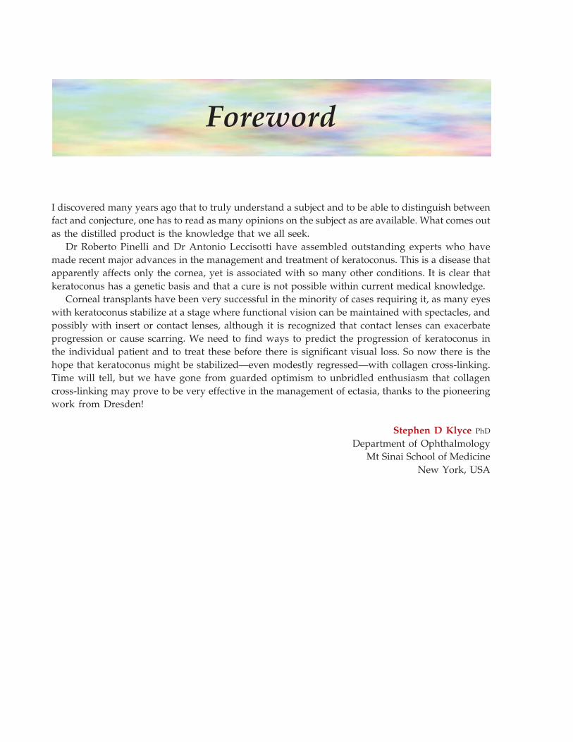
I discovered many years ago that to truly understand a subject and to be able to distinguish betweenfact and conjecture, one has to read as many opinions on the subject as are available. What comes outas the distilled product is the knowledge that we all seek.
Dr Roberto Pinelli and Dr Antonio Leccisotti have assembled outstanding experts who havemade recent major advances in the management and treatment of keratoconus. This is a disease thatapparently affects only the cornea, yet is associated with so many other conditions. It is clear thatkeratoconus has a genetic basis and that a cure is not possible within current medical knowledge.
Corneal transplants have been very successful in the minority of cases requiring it, as many eyeswith keratoconus stabilize at a stage where functional vision can be maintained with spectacles, andpossibly with insert or contact lenses, although it is recognized that contact lenses can exacerbateprogression or cause scarring. We need to find ways to predict the progression of keratoconus inthe individual patient and to treat these before there is significant visual loss. So now there is thehope that keratoconus might be stabilized—even modestly regressed—with collagen cross-linking.Time will tell, but we have gone from guarded optimism to unbridled enthusiasm that collagencross-linking may prove to be very effective in the management of ectasia, thanks to the pioneeringwork from Dresden!
Stephen D Klyce PhD
Department of OphthalmologyMt Sinai School of Medicine
New York, USA
Foreword


Contributors
Roberto Pinelli MDScientific Director, Istituto Laser MicrochirurgiaOculare, Brescia, ItalyPresident of Italian Refractive Surgery Society
Antonio Leccisotti MD PhDVisiting Professor, School of Biomedical SciencesUniversity of Ulster, Coleraine, UKDirector, Department of OphthalmologyCasa di Cura RuganiSiena, Italy
Ioannis M Aslanides MD PhDConsultant and Medical DirectorEmmetropia Mediterranean Eye InstituteHeraklion, Crete, Greece
H Burkhard Dick MDCenter for Vision ScienceRuhr University Eye HospitalBochum, Germany
Müriel Doors MDDepartment of OphthalmologyAcademic Hospital Maastricht, MaastrichtThe Netherlands
Daniel Elies MDCornea and Refractive Surgery UnitInstituto de Microcirugia OcularBarcelona, Spain
Michael J Endl MDDepartment of OphthalmologySUNY at Buffalo School of MedicineNY, USA
Tarek El Beltagi MDProfessor of OphthalmologyResearch Institute of OphthalmologyCairo, Egypt
Pierre Fournié MDService d’OphtalmologieCentre Hospitalier Universitaire ToulouseHôpital Purpan , Toulouse, France
Jose L Güell MD PhDAssociate Professor of the UniversitatAutonoma de BarcelonaBarcelona, Spain, andDirector of the Cornea and Refractive Surgery UnitInstituto de Microcirugia OcularBarcelona, Spain
Fritz Hengerer MDCenter for Vision ScienceRuhr University Eye HospitalBochum, Germany
Anne Hoyer MDDepartment of OphthalmologyUniversity Hospital Dresden, Germany
Stephen D Klyce PhDDepartment of OphthalmologyMt Sinai School of MedicineNew York, NY, USA
François Malecaze MD PhDProfessor at Service d’OphtalmologieCentre Hospitalier Universitaire ToulouseHôpital PurpanToulouse, France
Felicidad Manero MDCornea and Refractive Surgery UnitInstituto de Microcirugia OcularBarcelona, Spain
Colm McAlinden BSc (Hons) MCOptomSchool of Biomedical SciencesUniversity of Ulster, Coleraine, UK

Ali A Mearza MBBS FRCOphthConsultant OphthalmologistImperial College Healthcare NHS TrustCharing Cross HospitalLondon, UK
Johnny E Moore FRCOphth PhDVisiting ProfessorSchool of Biomedical SciencesUniversity of UlsterColeraine, UK, andDepartment of OphthalmologyMater Hospital Belfast Hospital Trust NorthernIreland, and Director of Leeson Eye InstituteDublin, Ireland
Merce Morral MDCornea and Refractive Surgery UnitInstituto de Microcirugia Ocular, BarcelonaSpainInstitut Clinic d’Oftalmologia, Hospital Clinic iProvincial de Barcelona, Barcelona, Spain
Rudy MMA Nuijts MD PhDAssociate Professor of OphthalmologyDepartment of OphthalmologyAcademic Hospital MaastrichtMaastricht, The Netherlands
Frederik Raiskup-Wolf MDDepartment of OphthalmologyUniversity Hospital Dresden, Germany
Chitra Sambare MS FRCSCornea and Anterior Segment FellowConsultant, Deenanath Hospital and ResearchCentre and Shashwat Hospital, Pune, India
Sunil Shah FRCOphth FRCSEd FBCLAVisiting Professor at the School of Life and HealthSciences, Aston University, Birmingham, UKVisiting Professor at the School of BiomedicalSciences, University of Ulster, Coleraine, UKMedical Director, Midland Eye Institute, SolihullUK, Consultant Ophthalmic SurgeonHeart of England Foundation Trust, Birmingham,UK, and Consultant Ophthalmic Surgeon,Birmingham and Midland Eye CentreBirmingham, UK
Eberhard Spoerl MDDepartment of OphthalmologyUniversity Hospital Dresden, Germany
Nayyirih G Tahzib MDDepartment of OphthalmologyAcademic Hospital MaastrichtMaastricht, The Netherlands
viii Keratoconus Surgery and Cross-linking

Many aspects contribute to the fact that keratoconus is an ideal link between refractive and cornealsurgery. The expansion of the options for the visual rehabilitation in keratoconus has partly originatedfrom the progress of diagnostic and therapeutical techniques in refractive surgery. Intrastromalrings and phakic IOLs, to mention a few, were conceived to correct myopia but have found a naturaland successful application in keratoconic eyes. But some of the new techniques discussed in thisbook were expressly ideated to strengthen (collagen cross-linking) or to replace (lamellar keratoplasty)a weakened corneal stroma. The subject of the surgery of keratoconus has therefore become so widethat no single surgeon can exhaustively cover all its aspects.
With this in mind, we wanted to gather the experience of some of the most valuable keratoconussurgeons in the world in a comprehensive book. Some of the contributors are old friends who didnot let us down in this daunting task, some are new friends who honoured us by believing in ourproject. Everyone is an outstanding surgeon and, above all, a brilliant medical writer, this latterquality being nowadays much rarer than the former. Thanks to such collaborations, the final resulthas reached the excellence we had in mind when the book was first planned.
We would like to thank the entire team of Istituto Laser Microchirurgia Oculare, Brescia—Italyand the Assistants of Dr Leccisotti for their blessings and motivation.
Roberto PinelliAntonio Leccisotti
Preface

Contents
1. Detecting Corneal Ectasia ................................................................................................... 1Michael J Endl, Stephen D Klyce
2. Technique of Collagen Cross-linking with Riboflavin and UVA-light ................. 13Anne Hoyer, Frederik Raiskup-Wolf, Eberhard Spoerl
3. Transepithelial Cross-linking for the Treatment of Keratoconus: Concepts ......... 21Roberto Pinelli
4. Transepithelial Corneal Cross-linking: Technique and Results .............................. 27Roberto Pinelli, Antonio Leccisotti, Tarek El Beltagi
5. Surgical Treatment of Keratoconus: An Overview ...................................................... 35Antonio Leccisotti, Roberto Pinelli
6. Intrastromal Ring Segments: Manual Implantation ................................................... 41Ali A Mearza, Ioannis M Aslanides
7. Intrastromal Ring Segments: Implantation by Femtosecond Laser ......................... 51Johnny E Moore, Colm McAlinden
8. Toric Phakic Intraocular Lenses for the Correction of RefractiveErrors in Keratoconus ........................................................................................................ 59Jose L Güell, Merce Morral, Daniel Elies, Felicidad Manero
9. Our Experience with Toric Phakic IOLs in Keratoconus ........................................... 73H Burkhard Dick, Fritz Hengerer
10. Refractive Lens Exchange in Keratoconus and after Keratoplasty ........................... 77Antonio Leccisotti, Roberto Pinelli
11. Automated Lamellar Therapeutic Keratoplasty in Keratoconus .............................. 85Chitra Sambare, Sunil Shah
12. Near-Descemetic Deep Anterior Lamellar Keratoplasty (DALK) ............................. 99Antonio Leccisotti
13. Penetrating Keratoplasty ................................................................................................ 129Pierre Fournié, François Malecaze
14. Refractive Surgery after Penetrating Keratoplasty .................................................... 151Jose L Güell, Merce Morral, Felicidad Manero
15. Toric Intraocular Lenses after Keratoplasty ................................................................ 169Rudy MMA Nuijts, Müriel Doors, Nayyirih G Tahzib
Index .................................................................................................................................... 185
2
Keratoconus Surgery and Cross-linking
IntroductionKeratoconus, pellucid marginal degeneration, and iatrogenic ectasia all exhibit an irregular form ofcorneal ‘bulging’ secondary to progressive stromal thinning. These appear to be unrelated to anyobvious inflammatory changes. The reported incidence of keratoconus among refractive surgicalcandidates when refractive surgery was first introduced was as high as 8 to 12 percent.1 This numberhas dropped to closer to one percent as refractive practices have become more established.2
Importantly, these percentages remain more than 10 times higher than the incidence of keratoconusin the general population (1.3-50 per 100,000 depending on ethnicity).3,4 Most of these patients withasymmetrical corneas experience poor vision quality with spectacle correction or are experiencing agrowing intolerance to ill-fitting contact lenses. For those reasons, patients with mild keratoconustend to seek out other modalities for correction, and excimer laser treatments are novel and attractivealternatives. Thus, it is particularly important for all refractive surgical candidates to undergo carefulscreening which specifically includes bilateral corneal topography examinations.
As LASIK approaches 1.4 million procedures annually in the United States alone, it has becomeimperative that the modern day refractive surgeon possess an increasingly expert skill set for thedetection of preoperative pathology such as keratoconus and pellucid marginal degeneration aswell as the development of post-surgical corneal ectatic changes. Corneal topographers have beenthe standard of care for preoperative screening of refractive surgical candidates since the early1990’s.5 In this chapter we will explore the basics of topographic pattern recognition essential to thedetection of preoperative keratoconus as well as some of the advancing technologies that maycomplement our current knowledge base.
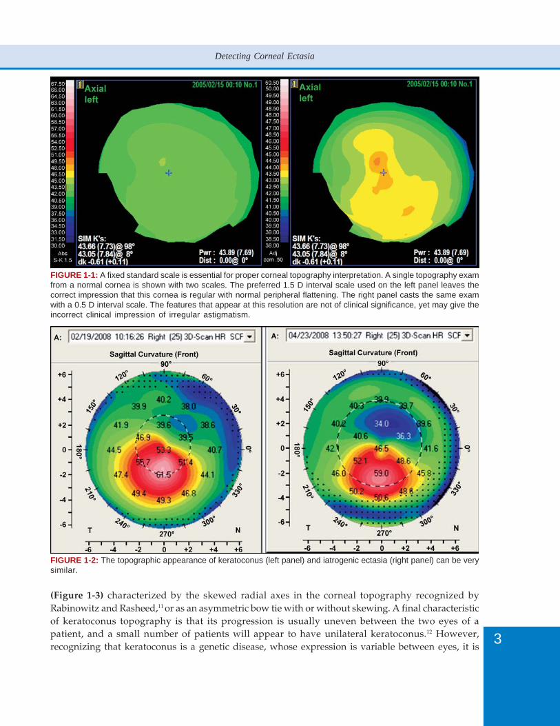
The mainstay for early detection, diagnosis and tracking of ectasia remains videokeratography,specifically the corneal topographic mapping of axial dioptric power with the color-coded contourmap developed in the late 1980’s.6 Utilizing Placido-based maps provides the most sensitive andreproducible method for the detection of early ectasia. The amount of information displayed inthese maps is determined in part by the topographic scale. However, without the use of a standardizedor absolute scale,7,8 certain irrelevant distortions can appear misleading or overemphasized(Figure 1-1).
Although modern corneal mapping systems can display upwards of 22,000 data points on asingle topographic map, a 1.5 D scale has sufficient resolution to detect all of the topographiccharacteristics necessary for diagnosing a variety of topographic abnormalities including: contactlens warpage, early and late keratoconus, penetrating keratoplasty, extracapsular cataract extraction,photorefractive keratotectomy, radial keratotomy, and epikeratophakia.9
Conversely, an ad hoc scale can cause the clinician to overlook early signs of irregular astigmatismor fail to detect progression of ectasia when following a keratoconus suspect. Such scales such as thenormalized scale are self-adapting to the power range present in individual topography examinations.This technique can obscure important detail on highly irregular corneas and over emphasizetopographic features irrelevant to diagnosis (see Figure 1-1).
The hallmark of corneal ectasia remains an irregular astigmatism which can take several forms.The result is several patterns of irregular astigmatism with which the eye care professional mustbecome familiar. Ectasia from keratoconus and ectasia following refractive surgery can be remarkablysimilar (Figure 1-2). Keratoconus is most often associated with an inferior localized steepening asshown in Figure 1-2, although the cone can be present centrally or even superiorly.10 However,keratoconus can also present as a central symmetric, but lopsided or ‘lazy eight’ bow tie
3
Detecting Corneal Ectasia
FIGURE 1-1: A fixed standard scale is essential for proper corneal topography interpretation. A single topography examfrom a normal cornea is shown with two scales. The preferred 1.5 D interval scale used on the left panel leaves thecorrect impression that this cornea is regular with normal peripheral flattening. The right panel casts the same examwith a 0.5 D interval scale. The features that appear at this resolution are not of clinical significance, yet may give theincorrect clinical impression of irregular astigmatism.
FIGURE 1-2: The topographic appearance of keratoconus (left panel) and iatrogenic ectasia (right panel) can be verysimilar.
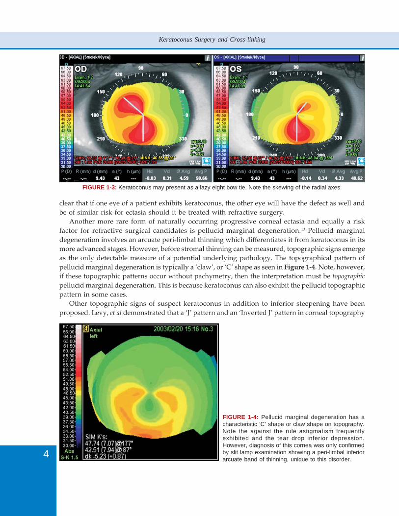
(Figure 1-3) characterized by the skewed radial axes in the corneal topography recognized byRabinowitz and Rasheed,11 or as an asymmetric bow tie with or without skewing. A final characteristicof keratoconus topography is that its progression is usually uneven between the two eyes of apatient, and a small number of patients will appear to have unilateral keratoconus.12 However,recognizing that keratoconus is a genetic disease, whose expression is variable between eyes, it is
4
Keratoconus Surgery and Cross-linking
FIGURE 1-3: Keratoconus may present as a lazy eight bow tie. Note the skewing of the radial axes.
clear that if one eye of a patient exhibits keratoconus, the other eye will have the defect as well andbe of similar risk for ectasia should it be treated with refractive surgery.
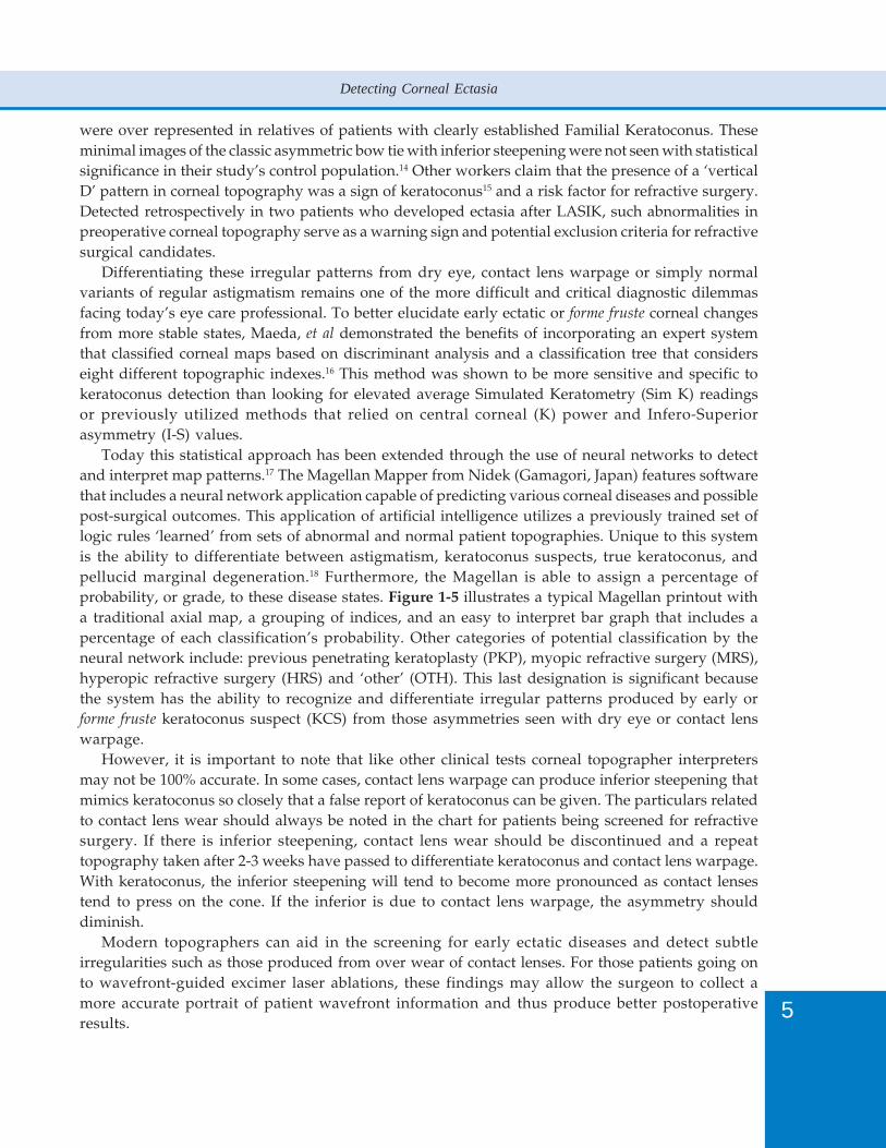
Another more rare form of naturally occurring progressive corneal ectasia and equally a riskfactor for refractive surgical candidates is pellucid marginal degeneration.13 Pellucid marginaldegeneration involves an arcuate peri-limbal thinning which differentiates it from keratoconus in itsmore advanced stages. However, before stromal thinning can be measured, topographic signs emergeas the only detectable measure of a potential underlying pathology. The topographical pattern ofpellucid marginal degeneration is typically a ‘claw’, or ‘C’ shape as seen in Figure 1-4. Note, however,if these topographic patterns occur without pachymetry, then the interpretation must be topographic
pellucid marginal degeneration. This is because keratoconus can also exhibit the pellucid topographicpattern in some cases.
Other topographic signs of suspect keratoconus in addition to inferior steepening have beenproposed. Levy, et al demonstrated that a ‘J’ pattern and an ‘Inverted J’ pattern in corneal topography
FIGURE 1-4: Pellucid marginal degeneration has acharacteristic ‘C’ shape or claw shape on topography.Note the against the rule astigmatism frequentlyexhibited and the tear drop inferior depression.However, diagnosis of this cornea was only confirmedby slit lamp examination showing a peri-limbal inferiorarcuate band of thinning, unique to this disorder.
5
Detecting Corneal Ectasia
were over represented in relatives of patients with clearly established Familial Keratoconus. Theseminimal images of the classic asymmetric bow tie with inferior steepening were not seen with statisticalsignificance in their study’s control population.14 Other workers claim that the presence of a ‘verticalD’ pattern in corneal topography was a sign of keratoconus15 and a risk factor for refractive surgery.Detected retrospectively in two patients who developed ectasia after LASIK, such abnormalities inpreoperative corneal topography serve as a warning sign and potential exclusion criteria for refractivesurgical candidates.
Differentiating these irregular patterns from dry eye, contact lens warpage or simply normalvariants of regular astigmatism remains one of the more difficult and critical diagnostic dilemmasfacing today’s eye care professional. To better elucidate early ectatic or forme fruste corneal changesfrom more stable states, Maeda, et al demonstrated the benefits of incorporating an expert systemthat classified corneal maps based on discriminant analysis and a classification tree that considerseight different topographic indexes.16 This method was shown to be more sensitive and specific tokeratoconus detection than looking for elevated average Simulated Keratometry (Sim K) readingsor previously utilized methods that relied on central corneal (K) power and Infero-Superiorasymmetry (I-S) values.
Today this statistical approach has been extended through the use of neural networks to detectand interpret map patterns.17 The Magellan Mapper from Nidek (Gamagori, Japan) features softwarethat includes a neural network application capable of predicting various corneal diseases and possiblepost-surgical outcomes. This application of artificial intelligence utilizes a previously trained set oflogic rules ‘learned’ from sets of abnormal and normal patient topographies. Unique to this systemis the ability to differentiate between astigmatism, keratoconus suspects, true keratoconus, andpellucid marginal degeneration.18 Furthermore, the Magellan is able to assign a percentage ofprobability, or grade, to these disease states. Figure 1-5 illustrates a typical Magellan printout witha traditional axial map, a grouping of indices, and an easy to interpret bar graph that includes apercentage of each classification’s probability. Other categories of potential classification by theneural network include: previous penetrating keratoplasty (PKP), myopic refractive surgery (MRS),hyperopic refractive surgery (HRS) and ‘other’ (OTH). This last designation is significant becausethe system has the ability to recognize and differentiate irregular patterns produced by early orforme fruste keratoconus suspect (KCS) from those asymmetries seen with dry eye or contact lenswarpage.
However, it is important to note that like other clinical tests corneal topographer interpretersmay not be 100% accurate. In some cases, contact lens warpage can produce inferior steepening thatmimics keratoconus so closely that a false report of keratoconus can be given. The particulars relatedto contact lens wear should always be noted in the chart for patients being screened for refractivesurgery. If there is inferior steepening, contact lens wear should be discontinued and a repeattopography taken after 2-3 weeks have passed to differentiate keratoconus and contact lens warpage.With keratoconus, the inferior steepening will tend to become more pronounced as contact lensestend to press on the cone. If the inferior is due to contact lens warpage, the asymmetry shoulddiminish.
Modern topographers can aid in the screening for early ectatic diseases and detect subtleirregularities such as those produced from over wear of contact lenses. For those patients going onto wavefront-guided excimer laser ablations, these findings may allow the surgeon to collect amore accurate portrait of patient wavefront information and thus produce better postoperativeresults.

6
Keratoconus Surgery and Cross-linking
FIGURE 1-5: Screen shot of the NIDEK Corneal Navigator automatically interpreting keratoconus (KC) with a 94.5%likelihood that the topography matches keratoconus. The severity (Keratoconus Severity Index, KSI) is given as 6.6%.
The above discussion centers on the use of the axial power map for displaying corneal topography.It is noted that there are three other common topography displays on most corneal topographers.These include the tangential or instantaneous power map, the refractive power map, and a height orelevation map. The former two are in units of diopters while the latter is in units of mm or microns.The elevation maps are presented as the difference between a best fit sphere and the measuredshape of a corneal surface. There are also scanning slit-based instruments (see below) that measurethe position of the two corneal surfaces to yield pachymetry maps which are an important adjunct tocorneal topography in the screening of patients for refractive surgery as well as differentiatingkeratoconus from pellucid marginal degeneration by the pattern of thinning.
Tangential power maps can be useful to show details of refractive surgery, in particular thecharacteristics of the transition zone after the correction of myopia. Abrupt transition between theoptical zone and the peripheral corneal can produce symptoms of haloes and monocular polyopia.Refractive power maps present the true refractive power of the corneal surface, but are not generally
7
Detecting Corneal Ectasia
useful for clinical diagnosis. In fact, refractive power maps can obscure mild inferior keratoconus orpellucid marginal degeneration, since with the refractive power display, the algorithms used tocalculate refractive power display a steepened peripheral cornea. The steep periphery in the refractivepower map can mask the peripheral steepening characteristic of mild keratoconus.
Slit Scanning and Scheimpflug ImagingBy providing anterior and posterior corneal surface information, along with whole cornealpachymetry, the Orbscan (Bausch & Lomb, Rochester, NY), the Pentacam (Oculus, Inc., Lynnwood,Washington), and the Galilei™ Dual Scheimpflug Analyzer (Ziemer Ophthalmic Systems AG, Port,Switzerland) imaging systems can further aid in the detection and progression of corneal ectasia.
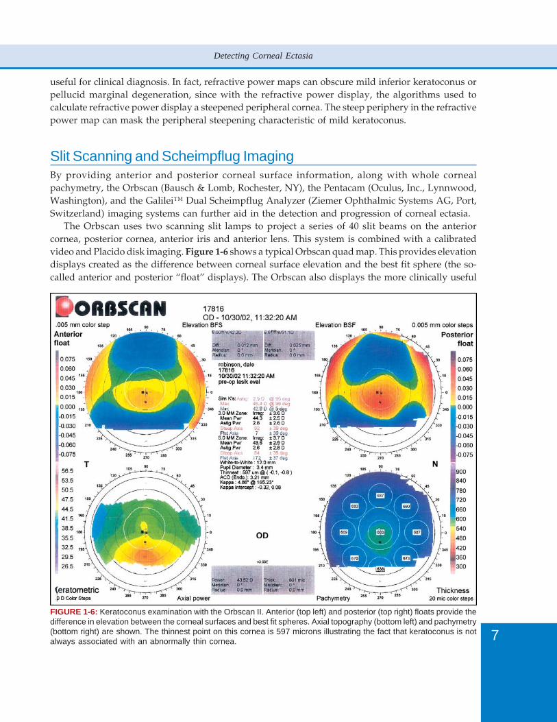
The Orbscan uses two scanning slit lamps to project a series of 40 slit beams on the anteriorcornea, posterior cornea, anterior iris and anterior lens. This system is combined with a calibratedvideo and Placido disk imaging. Figure 1-6 shows a typical Orbscan quad map. This provides elevationdisplays created as the difference between corneal surface elevation and the best fit sphere (the so-called anterior and posterior “float” displays). The Orbscan also displays the more clinically useful
FIGURE 1-6: Keratoconus examination with the Orbscan II. Anterior (top left) and posterior (top right) floats provide thedifference in elevation between the corneal surfaces and best fit spheres. Axial topography (bottom left) and pachymetry(bottom right) are shown. The thinnest point on this cornea is 597 microns illustrating the fact that keratoconus is notalways associated with an abnormally thin cornea.
8
Keratoconus Surgery and Cross-linking
traditional Placido disk keratometric topography and full corneal thickness measurements over abroad area of the cornea.
When screening refractive surgery candidates, several ‘red flags’ or indices have been associatedwith possible signs of early ectasia.19 With the Orbscan, these include: pachymetry readings with athinnest point less than a certain threshold (470 - 500 microns), a minimum peripheral corneal thicknessthat is not at least 20 microns greater than the central cornea, posterior float greater than 50 microns,20
high irregularity indices at the 3 mm and 5 mm zones, and the overall correlation of the highest/thinnest point coinciding on the anterior, posterior and pachymetry maps. Additional risk factorshave been identified by Randleman and colleagues.13 The strongest correlate was found to be abnormaltopography, but other risk factors included high myopia, reduced preoperative corneal thickness,reduced residual stromal bed after refractive surgery, and age. Note that these indications areregarded as warning signs that have been correlated with keratoconus; the signs of keratoconusseen in the axial power map from the Placido topography remains the most sensitive and reliableclue to the presence of keratoconus. There are many reports of large series of refractive surgicalpatients who have had one or more of these warning signs (but with normal topography) and whohave not developed iatrogenic ectasia. Again, apart from axial topography, none of these warningsigns provide a strong indicator of forme fruste keratoconus by themselves. In addition, topographicirregularities at both the 3 mm and 5 mm zones may simply point to the presence of increased higherorder aberrations without specific pathology. Further, the repeatability of peripheral slit-basedpachymetry measurements is somewhat controversial compared to the more consistent central cornealthickness values.21
Note also, that corneas with scarring or moderate dry eye will frequently produce inaccuratepachymetry maps as the scarred or dry surface may be interpreted as the posterior corneal surface.
With the Pentacam, by orienting the camera lens and lens plane at intersecting angles, theScheimpflug camera is able to record the corneal surfaces directly. Direct central recording isunavailable with the traditional Placido devices which places the viewing lens in the center of thePlacido mires. Similar to the Orbscan, Pentacam slit images are gathered that image both surfaces ofthe cornea as well as the surface of the crystalline lens. Although slit-based corneal front surfacedata is not as high in resolution as Placido imagery, in cases of advanced ectasia, Placido miresbecome obscured while the slit images can be used to present topography (Figure 1-7). This is amajor advantage of slit-based topography.
For screening purposes, it is claimed that Pentacam anterior elevation values between +12 to +15microns (above a reference sphere) are suspicious for ectasia and should prompt further investigation,whereas, a central deviation of the cornea’s anterior elevation of more than +15 microns is indicativeof keratoconus.22 However, these guidelines have not received rigorous experimental proof.
The Ziemer Galilei is a dual Scheimpflug instrument that, like the Orbscan, has also a Placidofunction. This instrument combines slit and Placido data in displaying corneal topography.
Wavefront SensingIt is becoming more apparent that wavefront data can further enhance our topographic diagnosticabilities. Maeda, et al showed that wavefront aberrometers may provide additional clues for thedetection of early corneal ectasia.23 The authors compared the total eye wavefront aberrations innormals to those in keratoconic eyes. An increase in the total higher order aberrations was noted inkeratoconus and attributed to the corneal shape. Coma-like aberrations were dominant and increased
9
Detecting Corneal Ectasia
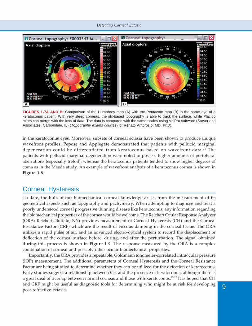
FIGURES 1-7A AND B: Comparison of the Humphrey map (A) with the Pentacam map (B) in the same eye of akeratoconus patient. With very steep corneas, the slit-based topography is able to track the surface, while Placidomires can merge with the loss of data. The data is compared with the same scales using VolPro software (Sarver andAssociates, Carbondale, IL) (Topography exams courtesy of Renato Ambrósio, MD, PhD).
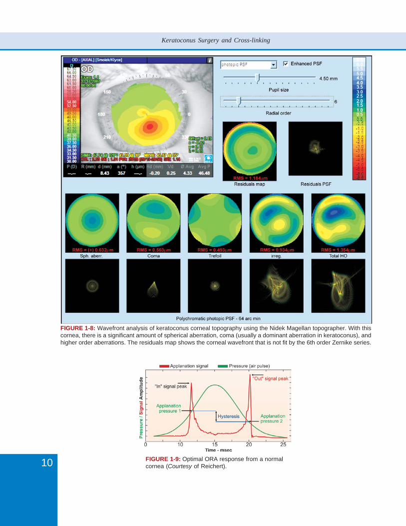
in the keratoconus eyes. Moreover, subsets of corneal ectasia have been shown to produce uniquewavefront profiles. Pepose and Applegate demonstrated that patients with pellucid marginaldegeneration could be differentiated from keratoconus based on wavefront data.24 Thepatients with pellucid marginal degeneration were noted to possess higher amounts of peripheralaberrations (especially trefoil), whereas the keratoconus patients tended to show higher degrees ofcoma as in the Maeda study. An example of wavefront analysis of a keratoconus cornea is shown inFigure 1-8.
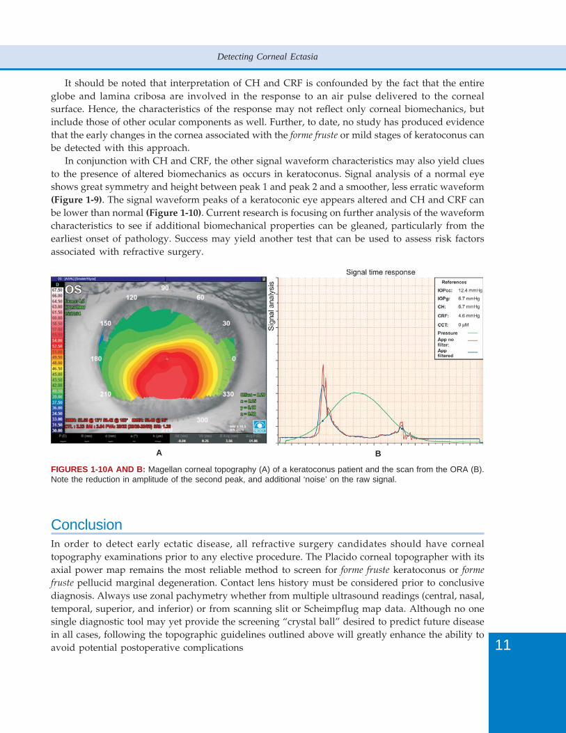
Corneal HysteresisTo date, the bulk of our biomechanical corneal knowledge arises from the measurement of itsgeometrical aspects such as topography and pachymetry. When attempting to diagnose and treat apoorly understood corneal progressive thinning disease like keratoconus, any information regardingthe biomechanical properties of the cornea would be welcome. The Reichert Ocular Response Analyzer(ORA; Reichert, Buffalo, NY) provides measurement of Corneal Hysteresis (CH) and the CornealResistance Factor (CRF) which are the result of viscous damping in the corneal tissue. The ORAutilizes a rapid pulse of air, and an advanced electro-optical system to record the displacement ordeflection of the corneal surface before, during, and after the perturbation. The signal obtainedduring this process is shown in Figure 1-9. The response measured by the ORA is a complexcombination of corneal and possibly other ocular biomechanical properties.
Importantly, the ORA provides a repeatable, Goldmann tonometer-correlated intraocular pressure(IOP) measurement. The additional parameters of Corneal Hysteresis and the Corneal ResistanceFactor are being studied to determine whether they can be utilized for the detection of keratoconus.Early studies suggest a relationship between CH and the presence of keratoconus, although there isa great deal of overlap between normal corneas and those with keratoconus.25-27 It is hoped that CHand CRF might be useful as diagnostic tools for determining who might be at risk for developingpost-refractive ectasia.
A B
10
Keratoconus Surgery and Cross-linking
FIGURE 1-8: Wavefront analysis of keratoconus corneal topography using the Nidek Magellan topographer. With thiscornea, there is a significant amount of spherical aberration, coma (usually a dominant aberration in keratoconus), andhigher order aberrations. The residuals map shows the corneal wavefront that is not fit by the 6th order Zernike series.
FIGURE 1-9: Optimal ORA response from a normalcornea (Courtesy of Reichert).
11
Detecting Corneal Ectasia
It should be noted that interpretation of CH and CRF is confounded by the fact that the entireglobe and lamina cribosa are involved in the response to an air pulse delivered to the cornealsurface. Hence, the characteristics of the response may not reflect only corneal biomechanics, butinclude those of other ocular components as well. Further, to date, no study has produced evidencethat the early changes in the cornea associated with the forme fruste or mild stages of keratoconus canbe detected with this approach.
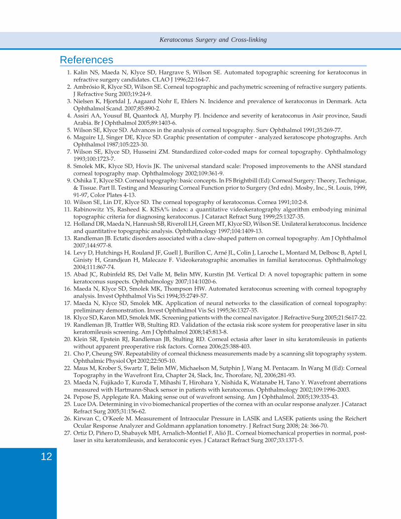
In conjunction with CH and CRF, the other signal waveform characteristics may also yield cluesto the presence of altered biomechanics as occurs in keratoconus. Signal analysis of a normal eyeshows great symmetry and height between peak 1 and peak 2 and a smoother, less erratic waveform(Figure 1-9). The signal waveform peaks of a keratoconic eye appears altered and CH and CRF canbe lower than normal (Figure 1-10). Current research is focusing on further analysis of the waveformcharacteristics to see if additional biomechanical properties can be gleaned, particularly from theearliest onset of pathology. Success may yield another test that can be used to assess risk factorsassociated with refractive surgery.
FIGURES 1-10A AND B: Magellan corneal topography (A) of a keratoconus patient and the scan from the ORA (B).Note the reduction in amplitude of the second peak, and additional ‘noise’ on the raw signal.
ConclusionIn order to detect early ectatic disease, all refractive surgery candidates should have cornealtopography examinations prior to any elective procedure. The Placido corneal topographer with itsaxial power map remains the most reliable method to screen for forme fruste keratoconus or forme
fruste pellucid marginal degeneration. Contact lens history must be considered prior to conclusivediagnosis. Always use zonal pachymetry whether from multiple ultrasound readings (central, nasal,temporal, superior, and inferior) or from scanning slit or Scheimpflug map data. Although no onesingle diagnostic tool may yet provide the screening “crystal ball” desired to predict future diseasein all cases, following the topographic guidelines outlined above will greatly enhance the ability toavoid potential postoperative complications
A B
12
Keratoconus Surgery and Cross-linking
References1. Kalin NS, Maeda N, Klyce SD, Hargrave S, Wilson SE. Automated topographic screening for keratoconus in
refractive surgery candidates. CLAO J 1996;22:164-7.2. Ambrósio R, Klyce SD, Wilson SE. Corneal topographic and pachymetric screening of refractive surgery patients.
J Refractive Surg 2003;19:24-9.3. Nielsen K, Hjortdal J, Aagaard Nohr E, Ehlers N. Incidence and prevalence of keratoconus in Denmark. Acta
Ophthalmol Scand. 2007;85:890-2.4. Assiri AA, Yousuf BI, Quantock AJ, Murphy PJ. Incidence and severity of keratoconus in Asir province, Saudi
Arabia. Br J Ophthalmol 2005;89:1403-6.5. Wilson SE, Klyce SD. Advances in the analysis of corneal topography. Surv Ophthalmol 1991;35:269-77.6. Maguire LJ, Singer DE, Klyce SD. Graphic presentation of computer - analyzed keratoscope photographs. Arch
Ophthalmol 1987;105:223-30.7. Wilson SE, Klyce SD, Husseini ZM. Standardized color-coded maps for corneal topography. Ophthalmology
1993;100:1723-7.8. Smolek MK, Klyce SD, Hovis JK. The universal standard scale: Proposed improvements to the ANSI standard
corneal topography map. Ophthalmology 2002;109:361-9.9. Oshika T, Klyce SD. Corneal topography: basic concepts. In FS Brightbill (Ed): Corneal Surgery: Theory, Technique,
& Tissue. Part II. Testing and Measuring Corneal Function prior to Surgery (3rd edn). Mosby, Inc., St. Louis, 1999,91-97, Color Plates 4-13.
10. Wilson SE, Lin DT, Klyce SD. The corneal topography of keratoconus. Cornea 1991;10:2-8.11. Rabinowitz YS, Rasheed K. KISA% index: a quantitative videokeratography algorithm embodying minimal
topographic criteria for diagnosing keratoconus. J Cataract Refract Surg 1999;25:1327-35.12. Holland DR, Maeda N, Hannush SB, Riveroll LH, Green MT, Klyce SD, Wilson SE. Unilateral keratoconus. Incidence
and quantitative topographic analysis. Ophthalmology 1997;104:1409-13. 13. Randleman JB. Ectatic disorders associated with a claw-shaped pattern on corneal topography. Am J Ophthalmol
2007;144:977-8.14. Levy D, Hutchings H, Rouland JF, Guell J, Burillon C, Arné JL, Colin J, Laroche L, Montard M, Delbosc B, Aptel I,
Ginisty H, Grandjean H, Malecaze F. Videokeratographic anomalies in familial keratoconus. Ophthalmology2004;111:867-74.
15. Abad JC, Rubinfeld RS, Del Valle M, Belin MW, Kurstin JM. Vertical D: A novel topographic pattern in somekeratoconus suspects. Ophthalmology 2007;114:1020-6.
16. Maeda N, Klyce SD, Smolek MK, Thompson HW. Automated keratoconus screening with corneal topographyanalysis. Invest Ophthalmol Vis Sci 1994;35:2749-57.
17. Maeda N, Klyce SD, Smolek MK. Application of neural networks to the classification of corneal topography:preliminary demonstration. Invest Ophthalmol Vis Sci 1995;36:1327-35.
18. Klyce SD, Karon MD, Smolek MK. Screening patients with the corneal navigator. J Refractive Surg 2005;21:S617-22.19. Randleman JB, Trattler WB, Stulting RD. Validation of the ectasia risk score system for preoperative laser in situ
keratomileusis screening. Am J Ophthalmol 2008;145:813-8.20. Klein SR, Epstein RJ, Randleman JB, Stulting RD. Corneal ectasia after laser in situ keratomileusis in patients
without apparent preoperative risk factors. Cornea 2006;25:388-403.21. Cho P, Cheung SW. Repeatability of corneal thickness measurements made by a scanning slit topography system.
Ophthalmic Physiol Opt 2002;22:505-10.22. Maus M, Krober S, Swartz T, Belin MW, Michaelson M, Sutphin J, Wang M. Pentacam. In Wang M (Ed): Corneal
Topography in the Wavefront Era, Chapter 24, Slack, Inc, Thorofare, NJ, 2006;281-93.23. Maeda N, Fujikado T, Kuroda T, Mihashi T, Hirohara Y, Nishida K, Watanabe H, Tano Y. Wavefront aberrations
measured with Hartmann-Shack sensor in patients with keratoconus. Ophthalmology 2002;109:1996-2003.24. Pepose JS, Applegate RA. Making sense out of wavefront sensing. Am J Ophthalmol. 2005;139:335-43.25. Luce DA. Determining in vivo biomechanical properties of the cornea with an ocular response analyzer. J Cataract
Refract Surg 2005;31:156-62.26. Kirwan C, O’Keefe M. Measurement of Intraocular Pressure in LASIK and LASEK patients using the Reichert
Ocular Response Analyzer and Goldmann applanation tonometry. J Refract Surg 2008; 24: 366-70.27. Ortiz D, Piñero D, Shabayek MH, Arnalich-Montiel F, Alió JL. Corneal biomechanical properties in normal, post-
laser in situ keratomileusis, and keratoconic eyes. J Cataract Refract Surg 2007;33:1371-5.
Please cite figure 2-2 in the textand clear query on page 17
14
Keratoconus Surgery and Cross-linking
IntroductionThe reduced mechanical stability of the cornea in keratoconus for example may be increased byphotooxidative cross-linking of the corneal collagen.
Keratoconus is a corneal degeneration which often leads to a severe visual impairment due to anincreasing corneal irregularity associated with a worsening of the optical abilities. The biomechanicalcharacteristics of the cornea result from the collagen scaffold, the collagen compound and theirbonding with the collagen fibrils. Even though the total amount of collagen lamellas is not significantlylower in keratoconic corneas7 the stability is lowered by factor 0.7.1 A decreased thickness16 inaddition to a modified configuration of the stromal collagen lamellas5, 13, 15 are seen. Furthermore inkeratoconic corneas twice as much hydroxyproline can be dissolved by pepsin.1 Hydroxyprolinestabilizes in a great amount the collagen’s triplehelix. Moreover enzymatical alterations have beenobserved with an increased expression of lysosomal and proteolytic enzymes.8, 16, 18, 35 as well as adecreased concentration of protease inhibitors.8, 9 These facts indicate a disturbed cross-linking withinor/and among the collagen molecules.
By photooxidative collagen cross-linking with riboflavin and ultraviolet (UV)-light additionalcovalent bindings between the collagen molecules can be achieved which consequently stabilize thecollagen scaffold.
Photooxidative Collagen Cross-linking with Riboflavin andUltraviolet (UV)-lightWith the photooxidative collagen cross-linking with riboflavin and ultraviolet (UV)-light we areable to treat a limited area within a short time.
Riboflavin is a vitamin (vitamin B2), nontoxic and available as a drug. It has two important functions:the absorption of the UV-irradiation and as photosensitizer the generation of reactive oxygen species(singlet oxygen). In combination with UV-light, riboflavin creates free radicals which induce newchemical bonds. A significant increase in mechanical stiffness can be achieved through the cross-linking.
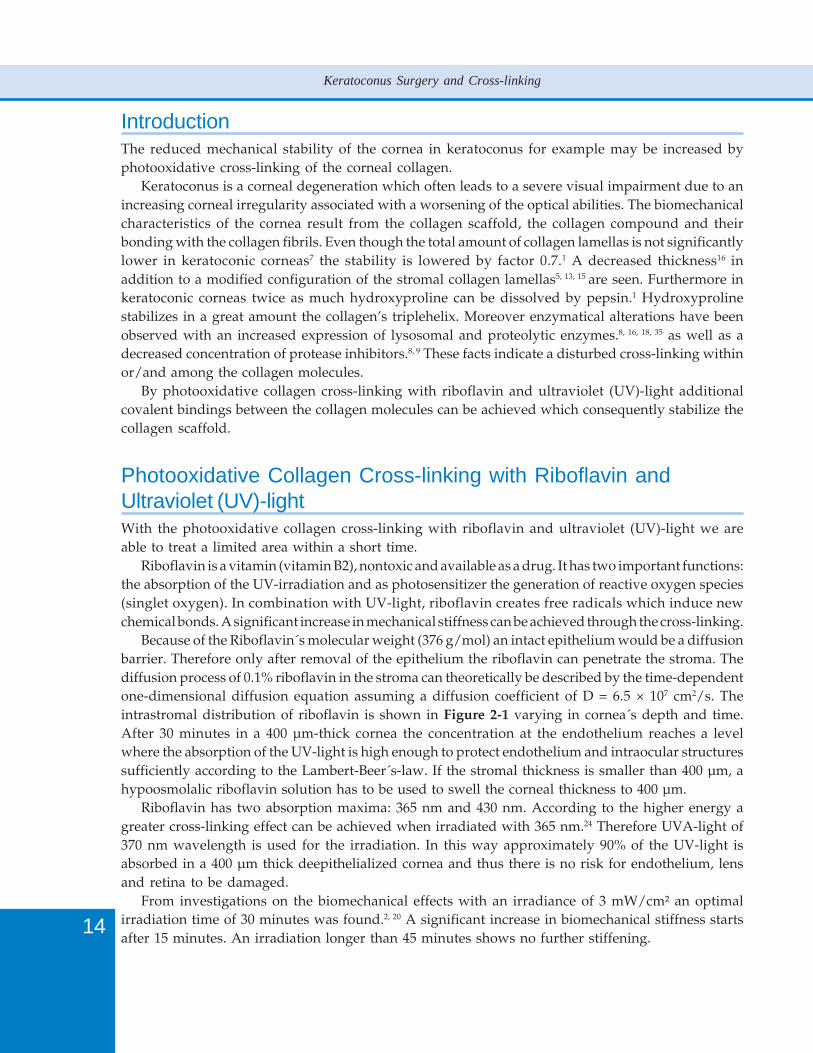
Because of the Riboflavin´s molecular weight (376 g/mol) an intact epithelium would be a diffusionbarrier. Therefore only after removal of the epithelium the riboflavin can penetrate the stroma. Thediffusion process of 0.1% riboflavin in the stroma can theoretically be described by the time-dependentone-dimensional diffusion equation assuming a diffusion coefficient of D = 6.5 × 107 cm2/s. Theintrastromal distribution of riboflavin is shown in Figure 2-1 varying in cornea´s depth and time.After 30 minutes in a 400 µm-thick cornea the concentration at the endothelium reaches a levelwhere the absorption of the UV-light is high enough to protect endothelium and intraocular structuressufficiently according to the Lambert-Beer´s-law. If the stromal thickness is smaller than 400 µm, ahypoosmolalic riboflavin solution has to be used to swell the corneal thickness to 400 µm.
Riboflavin has two absorption maxima: 365 nm and 430 nm. According to the higher energy agreater cross-linking effect can be achieved when irradiated with 365 nm.24 Therefore UVA-light of370 nm wavelength is used for the irradiation. In this way approximately 90% of the UV-light isabsorbed in a 400 µm thick deepithelialized cornea and thus there is no risk for endothelium, lensand retina to be damaged.
From investigations on the biomechanical effects with an irradiance of 3 mW/cm² an optimalirradiation time of 30 minutes was found.2, 20 A significant increase in biomechanical stiffness startsafter 15 minutes. An irradiation longer than 45 minutes shows no further stiffening.
15
Technique of Collagen Cross-linking with Riboflavin and UVA-light
FIGURE 2-1: Intrastromal distribution of 0.1% Riboflavin in thecorneal stroma at different times after riboflavin application atthe surface.
Safety for Endothelium and LensAccording to industrial safety guidelines for an unprotected eye a daily UVA-irradiation of 1 mW/cm2 without a photosensitizer is permitted.23 35% of this are absorbed in the cornea and theendothelium is strained with 0.65 mW/cm2. For a 30 minutes irradiation without photosensitizer invivo experiments showed a damage threshold for the endothelium of 4 mW/cm2 .30 By cross-linkingwith riboflavin and UV-light in a 400 µm cornea 94% of the UV-irradiation is absorbed and theendothelium gets only 0.18 mW/cm2.26 This value is below the determined damage threshold30 andbelow the value from the industrial safety guidelines. Therefore there is no risk for lens and retina.
Another reason for its safety lies in the small distance between the irradiation source and eyeand the irradiation´s divergency. Thus no UV-light is focussed on the retina.
Effect and Evidence of Cross-linkingPhotochemical induced cross-linking cannot be visualized directly by staining or other microscopicaltechniques. However cross-linking causes modification of numerous physicochemical qualities ofthe stromal collagen which are indirect evidences for cross-linking. Below a few of these changesare pointed out.
INCREASE IN STIFFNESS AND BENDING STIFFNESS, INCREASE IN ELASTICITY MODULUSThe cross-linked cornea is stiffer by factor 1.8 than normal cornea. With it the reduced stability in akeratoconic cornea is compensated.10, 24, 29 The stiffening effect is stronger in corneas with a higheramount of collagen respectively in elder corneas.2, 29
RAISING OF SHRINKING TEMPERATUREThe cornea´s shrinking temperature is raised from 63°C to 70°C. The shrinking temperature correlatespositively with the degree of cross-linking.27
DECREASE OF SWELLINGCross-linked collagen shows significantly less tendency for swelling.25, 33
16
Keratoconus Surgery and Cross-linking
INCREASED THICKNESS OF THE COLLAGEN LAMELLASThe diameter of collagen lamellas increases by 12% in the anterior stroma and by 4.5% in the posterior.32
ENHANCED RESISTANCE AGAINST PROTEOLYTIC ENZYMESIn patients with keratoconus a higher expression of lysosomal and proteolytic enzymes as well as adecreased concentration of protease inhibitors was found in the lacrimal fluid.8, 9, 12, 16, 18, 22, 35 These mayaccount for the stromal thinning. The cross-linked cornea shows an enhanced resistance againstproteolytic enzymes.28 This decelerated degradation process also results in an extension of thecollagen´s turn-over-time.
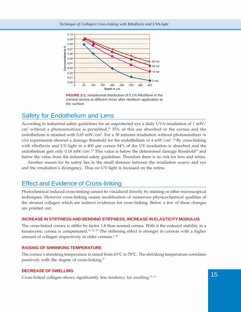
Stronger Cross-linking in the Anterior StromaThe cross-linking effect is distributed inhomogeneous over the corneal depth. The stiffening effectis concentrated on the anterior 200 to 300 µm of the cornea.10, 19 The UV-intensity is getting lower indeeper tissue due to its high absorbance. As a marginal increase of the collagen lamellas´ diameter,30
an increased enzymatic degradation28 and a stronger shrinking effect27 were observed in the posteriorstroma, a minor cross-linking effect can be assumed for the posterior parts.
This reduced cross-linking effect in deeper tissues can be explained by the above describedintrastromal distribution of riboflavin (Figures 2-1 and 2-2) and by the exponentially declined UV-intensity according to the Lambert-Beer´s-law. 65% of the UV-light is absorbed in the anterior 200µm, only 25 to 30% in the further 200 µm. Since the cross-linking effect predominantly depends onthe UV-irradiance and not as much on the riboflavin concentration20 the stiffening effect is strongestin the anterior 200 to 300 µm of the cornea.
FIGURE 2-2: Decrease of UV-intensity by depth in a400 µm cornea without (left) and with (right) riboflavin.
Treatment ProcedureThe cross-linking is performed as an outpatient treatment.
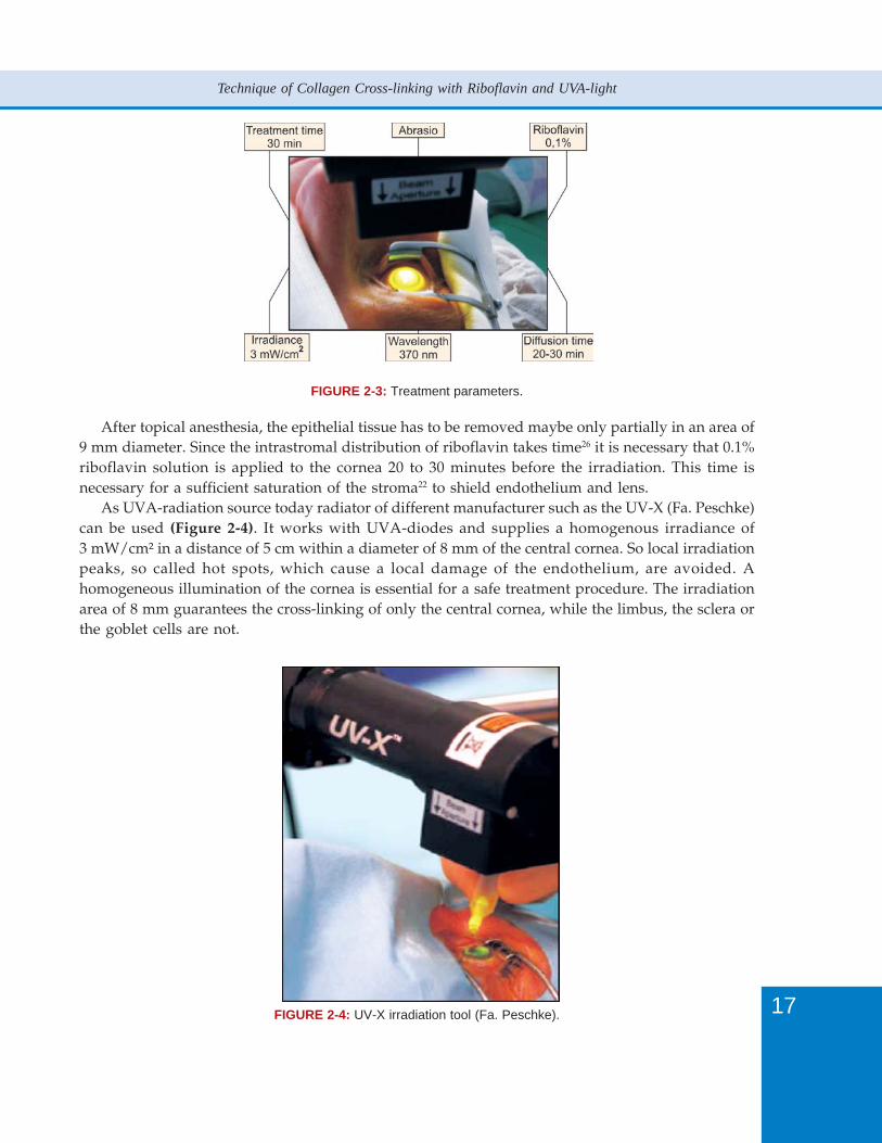
The treatment parameter and protocol is shown in Figure 2-3.
17
Technique of Collagen Cross-linking with Riboflavin and UVA-light
FIGURE 2-3: Treatment parameters.
After topical anesthesia, the epithelial tissue has to be removed maybe only partially in an area of9 mm diameter. Since the intrastromal distribution of riboflavin takes time26 it is necessary that 0.1%riboflavin solution is applied to the cornea 20 to 30 minutes before the irradiation. This time isnecessary for a sufficient saturation of the stroma22 to shield endothelium and lens.
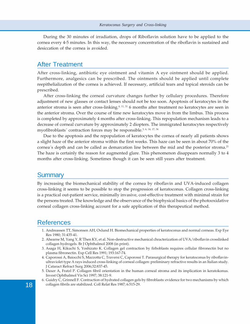
As UVA-radiation source today radiator of different manufacturer such as the UV-X (Fa. Peschke)can be used (Figure 2-4). It works with UVA-diodes and supplies a homogenous irradiance of3 mW/cm² in a distance of 5 cm within a diameter of 8 mm of the central cornea. So local irradiationpeaks, so called hot spots, which cause a local damage of the endothelium, are avoided. Ahomogeneous illumination of the cornea is essential for a safe treatment procedure. The irradiationarea of 8 mm guarantees the cross-linking of only the central cornea, while the limbus, the sclera orthe goblet cells are not.
FIGURE 2-4: UV-X irradiation tool (Fa. Peschke).
18
Keratoconus Surgery and Cross-linking
During the 30 minutes of irradiation, drops of Riboflavin solution have to be applied to thecornea every 4-5 minutes. In this way, the necessary concentration of the riboflavin is sustained anddesiccation of the cornea is avoided.
After TreatmentAfter cross-linking, antibiotic eye ointment and vitamin A eye ointment should be applied.Furthermore, analgesics can be prescribed. The ointments should be applied until completereepithelialization of the cornea is achieved. If necessary, artificial tears and topical steroids can beprescribed.
After cross-linking the corneal curvature changes further by cellulary procedures. Thereforeadjustment of new glasses or contact lenses should not be too soon. Apoptosis of keratocytes in theanterior stroma is seen after cross-linking.4, 11, 31 6 months after treatment no keratocytes are seen inthe anterior stroma. Over the course of time new keratocytes move in from the limbus. This processis completed by approximately 4 months after cross-linking. This repopulation mechanism leads to adecrease of corneal curvature by approximately 2 diopters. The immigrated keratocytes respectivelymyofibroblasts´ contraction forces may be responsible.3, 6, 14, 17, 34
Due to the apoptosis and the repopulation of keratocytes the cornea of nearly all patients showsa slight haze of the anterior stroma within the first weeks. This haze can be seen in about 70% of thecornea´s depth and can be called as demarcation line between the mid and the posterior stroma.21
The haze is certainly the reason for augmented glare. This phenomenon disappears normally 3 to 4months after cross-linking. Sometimes though it can be seen still years after treatment.
SummaryBy increasing the biomechanical stability of the cornea by riboflavin and UVA-induced collagencross-linking it seems to be possible to stop the progression of keratoconus. Collagen cross-linkingis a practical out-patient service, minimally invasive, cost-effective treatment with minimal strain forthe persons treated. The knowledge and the observance of the biophysical basics of the photooxidativecorneal collagen cross-linking account for a safe application of this therapeutical method.
References1. Andreassen TT, Simonsen AH, Oxlund H. Biomechanical properties of keratoconus and normal corneas. Exp Eye
Res 1980; 31:435-41.2. Ahearne M, Yang Y, R´Then KY, et al. Non-destructive mechanical chracterization of UVA/riboflavin crosslinked
collagen hydrogels. Br J Ophthalmol 2008 (in print).3. Asaga H, Kikuchi S, Yoshizato K. Collagen gel contraction by firboblasts requires cellular fibronectin but no
plasma fibronectin. Exp Cell Res 1991; 193:167-74.4. Caporossi A, Baiocchi S, Mazzotta C, Traversi C, Caporossi T. Parasurgical therapy for keratoconus by riboflavin-
ultraviolet type A rays induced cross-linking of corneal collagen: preliminary refractive results in an Italian study.J Cataract Refract Surg 2006;32:837-45.
5. Daxer A, Fratzl P. Collagen fibril orientation in the human corneal stroma and its implication in keratokonus.Invest Ophthalmol Vis Sci 1997; 38:121-9.
6. Guidry C, Grinnell F. Contraction of hydrated collagen gels by fibroblasts: evidence for two mechanisms by whichcollagen fibrils are stabilized. Coll Relat Res 1987; 6:515-29.
19
Technique of Collagen Cross-linking with Riboflavin and UVA-light
7. Ihme A, Krieg T; Müller RK, et al. Biochemical investigation of cells from keratoconus and normal corneas. Exp EyeRes 1983; 36:625-31.
8. Kao WWY, Vergnes JP, Ebert J, Sundar-Ray CV, Brown SI. Increased collagenase and gelatinase activities inkeratoconus. Biochem Biophys Res Commun 1982; 107:929-36.
9. Kenney MC, Nesburn AB, Burgeson RE, Butkowski RJ, Ljubimov AV. Abnormalities of the extracellular matrix inkeratoconus corneas. Cornea 1997; 16:345-51.
10. Kohlhaas M, Spoerl E, Schilde T, et al. Biomechanical evidence of the distribution of cross-links in corneas treatedwith riboflavin and ultraviolet A light. J Cataract Refract Surg 2006;32:279-83.
11. Mazzotta C, Balestrazzi A, Baiocchi S, et al. Stromal haze after combined riboflavin-UVA corneal cross-linking inkeratoconus: in vivo confocal microscopic evaluation. Clin Exp Ophthalomol 2007; 35: 580-2.
12. Mackiewicz Z, Määttä M, Stenman M, et al. Collagenolytic proteinases in keratoconus. Cornea 2006; 25:603-10.13. Meek KM, Tuft SJ, Huang Y. Changes in collagen orientation and distribution in keratoconuc corneas. Invest
Ophthalmol Vis Sci 2005; 46:1948-56.14. Netto MV, Mohan RR, Sinha S, et al. Stromal haze, myofibroblasts, and surface irregularity after PRK. Exp Eye Res
2006; 82:788-97.15. Radner W, Zehetmayer M, Skorpik C, Mallinger R. Zur Anordnung der kollagenen Lamellen beim Keratokonus.
Spektrum Augenheilkd 1996;10:156-60.16. Rehany U, Lahav M, Shoshan S. Collagenolytic activity in keratoconus. Ann Ophthalmol 1982;107:1507-10.17. Reichl S, Müller-Goymann CC. Entwicklung eines organtypischen Korneakonstrukts als ein in-vitro Modell für
Permeationsstudien. Ophthalmologe 2001; 98:853-8.18. Sawaguchi S, Yue BYJT, Sugar J, Gilboy J. Lysosomal enzyme activities in keratoconus. Arch Ophthalmol
1989;107:929-36.19. Schilde T, Kohlhaas M, Spoerl E, et al. Enzymatischer Nachweis der Tiefenabhängigkeit der Vernetzungswirkung
von Riboflavin/UVA an der Hornhaut. Ophthalmologe 2007 (in print).20. Schreiber J. Verfestigung der Hornhaut durch UVA 365 nm und Riboflavin oder durch Glutaraldehyd. Dissertation
Dresden: TU Dresden, 2003.21. Seiler T, Hafezi F. Corneal cross-linking induced stromal demarcation line. Cornea 2006; 25: 1057-9.22. Seppälä HP, Määttä M, Rautia M, et al. EMMPRIN and MMP-1 in keratoconus. Cornea 2006; 25: 325-30.23. Sliney D, Aron-Rosa D, DeLori F, et al. Adjustment of guidelines for exposure of the eye to optical radiation from
ocular instruments: Statement from a task group of the International Commission on Non-Ionizing RadiationProtection (ICNIRP). Applied Optics 2005; 44:2162-76.
24. Spoerl E, Huhle M, Seiler T. Induction of cross-links in corneal tissue, Exp Eye Res 1998; 66:97-103.25. Spörl E, Huhle M, Seiler T. The swelling behaviour of the cornea after artificial cross-linking. Invest Ophthalmol Vis
Sci 1997; 38: 507.26. Spoerl E, Mrochen M, Sliney D, Trokel S, Seiler T. Safety of UVA-riboflavin cross-linking of the cornea. Cornea
2007; 26:385-9.27. Spoerl E, Wollensak G, Dittert DD, et al. Thermomechanical behavior of collagen-crosslinked porcine cornea.
Ophthalmologica 2004; 218:136-40.28. Spoerl E, Wollensak G, Seiler T. Increased resistance of crosslinked cornea against enzymatic digestion. Curr Eye
Res 2004: 29:35-40.29. Wollensak G, Spoerl E, Seiler T. Riboflavin/ultraviolet-A-induced cross-linking for the treatment of keratoconus.
Am J Ophthalmol 2003; 135:620-7.30. Wollensak G, Spoerl E, Wilsch M, et al. Endothelial cell damage after riboflavin-ultraviolet-A-treatment in the
rabbit. J Cataract Ref Surg 2003; 29: 1786-90.31. Wollensak G, Spoerl E, Wilsch M, et al. Keratocyte apoptosis after corneal collagen cross-linking using riboflavin-
UVA treatment. Cornea 2004; 23: 43-9.32. Wollensak G, Wilsch M, Spoerl E. Collagen fiber diameter in the rabbit cornea after corneal cross-linking by
riboflavin/UVA. Cornea 2004; 23: 503-7.33. Wollensak G, Aurich H, Pham DT, et al. Hydration behaviour of porcine cornea crosslinked with riboflavin and
ultraviolet A. J Cataract Refract Surg 2007; 33: 516-21.34. Wollensak G, Iomdina E, Dittert DD, et al. Wound healing in the rabbit cornea after corneal cross-linking with
riboflavin and UVA. Cornea 2007; 26: 600-5.35. Zhou L, Sawaguchi S, Twining SS, et al. Expression of degradative enzymes and protease inhibitors in corneas with
keratoconus. Invest Ophthalmol Vis Sci 1998; 39:1117-24.

22
Keratoconus Surgery and Cross-linking
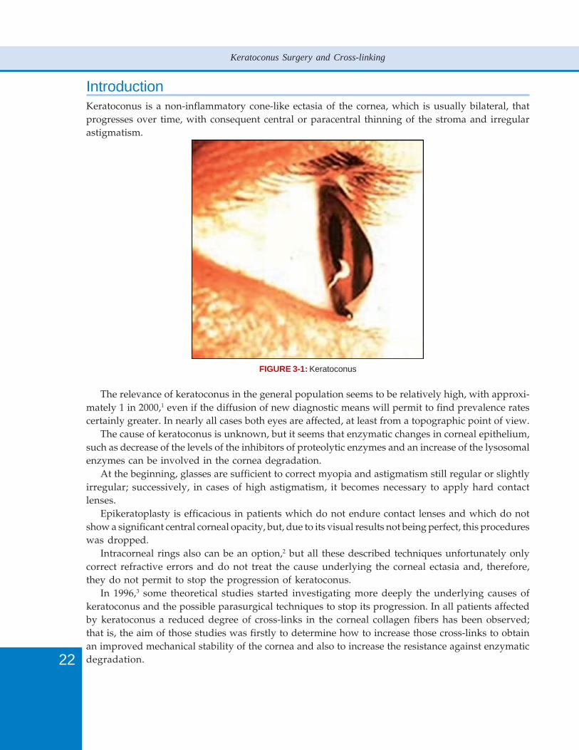
IntroductionKeratoconus is a non-inflammatory cone-like ectasia of the cornea, which is usually bilateral, thatprogresses over time, with consequent central or paracentral thinning of the stroma and irregularastigmatism.
FIGURE 3-1: Keratoconus
The relevance of keratoconus in the general population seems to be relatively high, with approxi-mately 1 in 2000,1 even if the diffusion of new diagnostic means will permit to find prevalence ratescertainly greater. In nearly all cases both eyes are affected, at least from a topographic point of view.
The cause of keratoconus is unknown, but it seems that enzymatic changes in corneal epithelium,such as decrease of the levels of the inhibitors of proteolytic enzymes and an increase of the lysosomalenzymes can be involved in the cornea degradation.
At the beginning, glasses are sufficient to correct myopia and astigmatism still regular or slightlyirregular; successively, in cases of high astigmatism, it becomes necessary to apply hard contactlenses.
Epikeratoplasty is efficacious in patients which do not endure contact lenses and which do notshow a significant central corneal opacity, but, due to its visual results not being perfect, this procedureswas dropped.
Intracorneal rings also can be an option,2 but all these described techniques unfortunately onlycorrect refractive errors and do not treat the cause underlying the corneal ectasia and, therefore,they do not permit to stop the progression of keratoconus.
In 1996,3 some theoretical studies started investigating more deeply the underlying causes ofkeratoconus and the possible parasurgical techniques to stop its progression. In all patients affectedby keratoconus a reduced degree of cross-links in the corneal collagen fibers has been observed;that is, the aim of those studies was firstly to determine how to increase those cross-links to obtainan improved mechanical stability of the cornea and also to increase the resistance against enzymaticdegradation.
23
Transepithelial Cross-linking for the Treatment of Keratoconus: Concepts
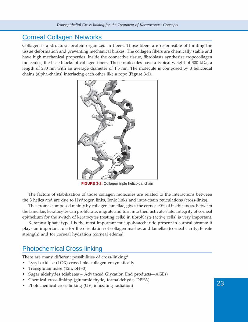
Corneal Collagen NetworksCollagen is a structural protein organized in fibers. Those fibers are responsible of limiting thetissue deformation and preventing mechanical brakes. The collagen fibers are chemically stable andhave high mechanical properties. Inside the connective tissue, fibroblasts synthesize tropocollagenmolecules, the base blocks of collagen fibers. Those molecules have a typical weight of 300 kDa, alength of 280 nm with an average diameter of 1.5 nm. The molecule is composed by 3 helicoidalchains (alpha-chains) interlacing each other like a rope (Figure 3-2).
FIGURE 3-2: Collagen triple helicoidal chain
The factors of stabilization of those collagen molecules are related to the interactions betweenthe 3 helics and are due to Hydrogen links, Ionic links and intra-chain reticulations (cross-links).
The stroma, composed mainly by collagen lamellae, gives the cornea 90% of its thickness. Betweenthe lamellae, keratocytes can proliferate, migrate and turn into their activate state. Integrity of cornealepithelium for the switch of keratocytes (resting cells) in fibroblasts (active cells) is very important.
Keratansulphate type I is the most important mucopolysaccharide present in corneal stroma: itplays an important role for the orientation of collagen mashes and lamellae (corneal clarity, tensilestrength) and for corneal hydration (corneal edema).
Photochemical Cross-linkingThere are many different possibilities of cross-linking:4
• Lysyl oxidase (LOX) cross-links collagen enzymatically• Transglutaminase (12h, pH=3)• Sugar aldehydes (diabetes – Advanced Glycation End products—AGEs)• Chemical cross-linking (glutaraldehyde, formaldehyde, DPPA)• Photochemical cross-linking (UV, ionizating radiation)
24
Keratoconus Surgery and Cross-linking
The interaction between organic tissues and radiation depends on the type of radiation used.The ionizing radiation has enough energy to turn out electrons from the atoms of the tissues. Othertypes of radiation, i.e. UV radiation, have not enough energy to turn out electrons but to make themjump to higher energy levels (exciting radiation).
In the human biologic tissues, water molecules are present at a rate of 70 to 90%, so it is clearlythe main target of radiation. During the water radiolysis process, the energy applied to watermolecules ionizes them and generates free radicals. Free radicals are continuously produced intissues and quickly inactivated by chemical or enzymatic transformation.
In the eye, ascorbic acid absorbs UV radiation (at cornea, lens and vitreous body areas); it is acofactor of several enzymes, the best known of which are prolyne hydroxylase, enzymes involvedin byosinthesis of collagen. In vitreous body, after cataract surgery (absence of glutathione), ascorbicacid (in ascorbate form) absorbs UV not stopped by the lens, resulting in the formation of freeradicals, disaggregation of hyaluronic acid and increase in cross-linking of collagen fiber networks.
Riboflavin-UVA TreatmentA photo sensitizer is a substance which is activated by the absorption of light at a given wavelightand which can induce free radical reactions in its activate form. This substance can amplify lightradiation effect on biologic tissues.
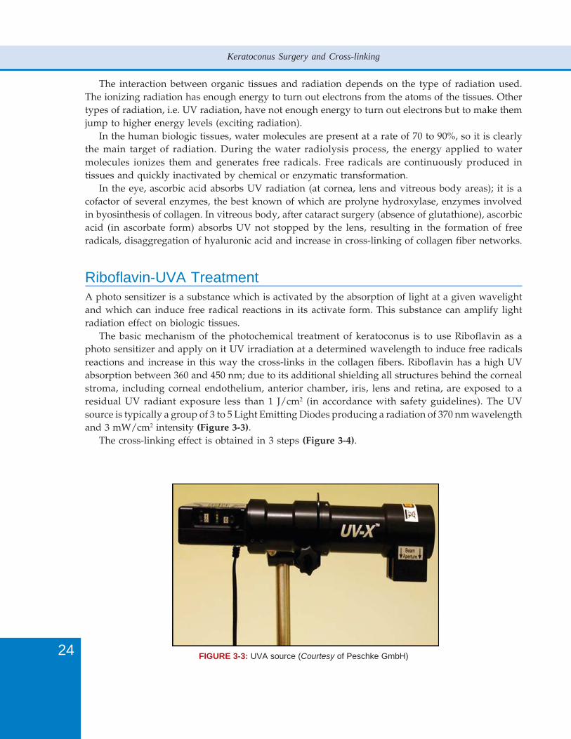
The basic mechanism of the photochemical treatment of keratoconus is to use Riboflavin as aphoto sensitizer and apply on it UV irradiation at a determined wavelength to induce free radicalsreactions and increase in this way the cross-links in the collagen fibers. Riboflavin has a high UVabsorption between 360 and 450 nm; due to its additional shielding all structures behind the cornealstroma, including corneal endothelium, anterior chamber, iris, lens and retina, are exposed to aresidual UV radiant exposure less than 1 J/cm2 (in accordance with safety guidelines). The UVsource is typically a group of 3 to 5 Light Emitting Diodes producing a radiation of 370 nm wavelengthand 3 mW/cm2 intensity (Figure 3-3).
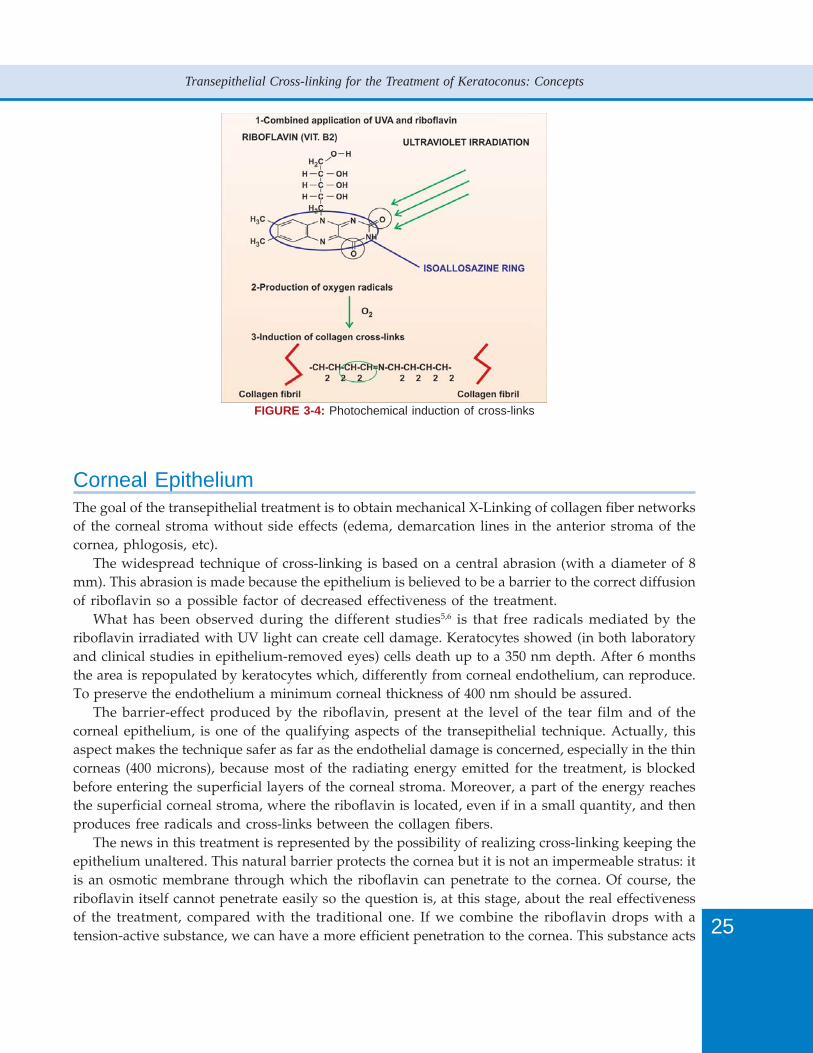
The cross-linking effect is obtained in 3 steps (Figure 3-4).
FIGURE 3-3: UVA source (Courtesy of Peschke GmbH)
25
Transepithelial Cross-linking for the Treatment of Keratoconus: Concepts
FIGURE 3-4: Photochemical induction of cross-links
Corneal EpitheliumThe goal of the transepithelial treatment is to obtain mechanical X-Linking of collagen fiber networksof the corneal stroma without side effects (edema, demarcation lines in the anterior stroma of thecornea, phlogosis, etc).
The widespread technique of cross-linking is based on a central abrasion (with a diameter of 8mm). This abrasion is made because the epithelium is believed to be a barrier to the correct diffusionof riboflavin so a possible factor of decreased effectiveness of the treatment.
What has been observed during the different studies5,6 is that free radicals mediated by theriboflavin irradiated with UV light can create cell damage. Keratocytes showed (in both laboratoryand clinical studies in epithelium-removed eyes) cells death up to a 350 nm depth. After 6 monthsthe area is repopulated by keratocytes which, differently from corneal endothelium, can reproduce.To preserve the endothelium a minimum corneal thickness of 400 nm should be assured.
The barrier-effect produced by the riboflavin, present at the level of the tear film and of thecorneal epithelium, is one of the qualifying aspects of the transepithelial technique. Actually, thisaspect makes the technique safer as far as the endothelial damage is concerned, especially in the thincorneas (400 microns), because most of the radiating energy emitted for the treatment, is blockedbefore entering the superficial layers of the corneal stroma. Moreover, a part of the energy reachesthe superficial corneal stroma, where the riboflavin is located, even if in a small quantity, and thenproduces free radicals and cross-links between the collagen fibers.
The news in this treatment is represented by the possibility of realizing cross-linking keeping theepithelium unaltered. This natural barrier protects the cornea but it is not an impermeable stratus: itis an osmotic membrane through which the riboflavin can penetrate to the cornea. Of course, theriboflavin itself cannot penetrate easily so the question is, at this stage, about the real effectivenessof the treatment, compared with the traditional one. If we combine the riboflavin drops with atension-active substance, we can have a more efficient penetration to the cornea. This substance acts
26
Keratoconus Surgery and Cross-linking
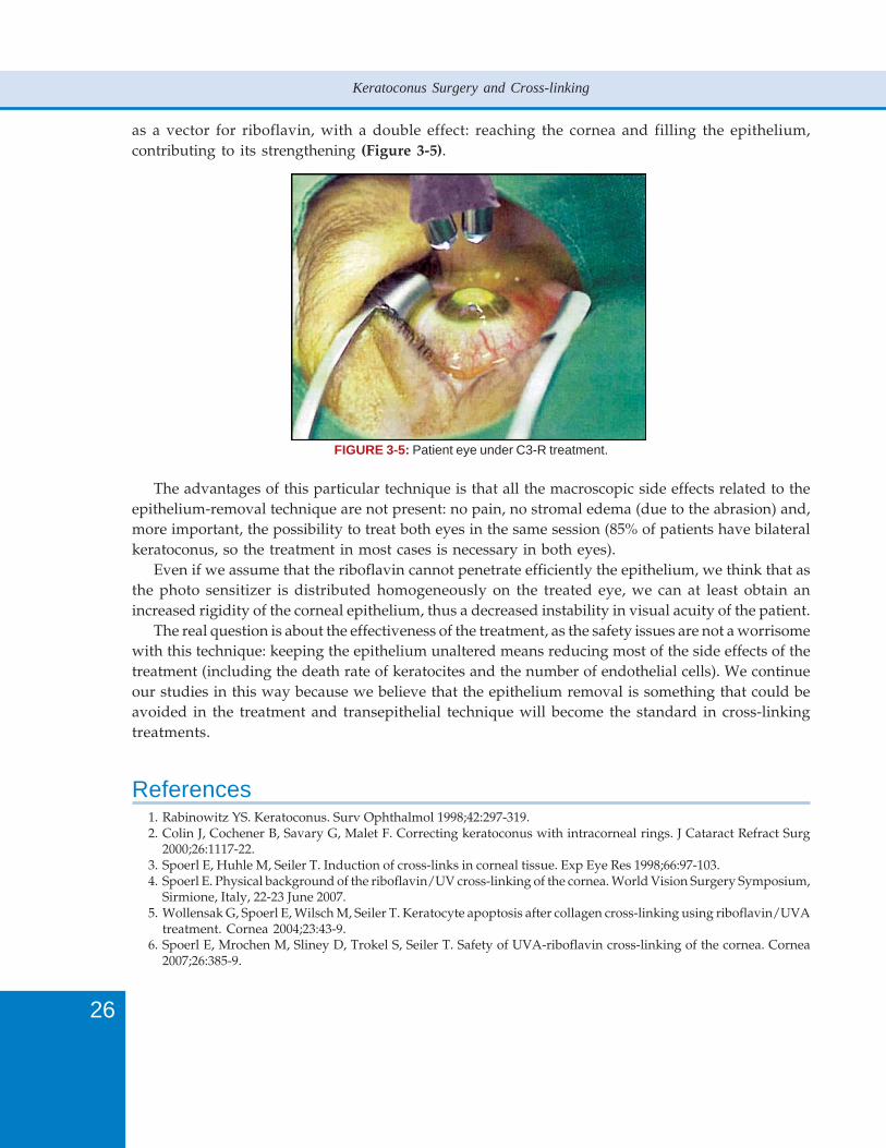
as a vector for riboflavin, with a double effect: reaching the cornea and filling the epithelium,contributing to its strengthening (Figure 3-5).
FIGURE 3-5: Patient eye under C3-R treatment.
The advantages of this particular technique is that all the macroscopic side effects related to theepithelium-removal technique are not present: no pain, no stromal edema (due to the abrasion) and,more important, the possibility to treat both eyes in the same session (85% of patients have bilateralkeratoconus, so the treatment in most cases is necessary in both eyes).
Even if we assume that the riboflavin cannot penetrate efficiently the epithelium, we think that asthe photo sensitizer is distributed homogeneously on the treated eye, we can at least obtain anincreased rigidity of the corneal epithelium, thus a decreased instability in visual acuity of the patient.
The real question is about the effectiveness of the treatment, as the safety issues are not a worrisomewith this technique: keeping the epithelium unaltered means reducing most of the side effects of thetreatment (including the death rate of keratocites and the number of endothelial cells). We continueour studies in this way because we believe that the epithelium removal is something that could beavoided in the treatment and transepithelial technique will become the standard in cross-linkingtreatments.
References1. Rabinowitz YS. Keratoconus. Surv Ophthalmol 1998;42:297-319.2. Colin J, Cochener B, Savary G, Malet F. Correcting keratoconus with intracorneal rings. J Cataract Refract Surg
2000;26:1117-22.3. Spoerl E, Huhle M, Seiler T. Induction of cross-links in corneal tissue. Exp Eye Res 1998;66:97-103.4. Spoerl E. Physical background of the riboflavin/UV cross-linking of the cornea. World Vision Surgery Symposium,
Sirmione, Italy, 22-23 June 2007.5. Wollensak G, Spoerl E, Wilsch M, Seiler T. Keratocyte apoptosis after collagen cross-linking using riboflavin/UVA
treatment. Cornea 2004;23:43-9.6. Spoerl E, Mrochen M, Sliney D, Trokel S, Seiler T. Safety of UVA-riboflavin cross-linking of the cornea. Cornea
2007;26:385-9.
QueriesPlease cited ref 1 in the textkindly clear the query in Table 4-1, page 30
28
Keratoconus Surgery and Cross-linking
IntroductionThe term cross-linking indicates a medical intervention; it was originally used in specialties such asdentistry and orthopedics. Theo Seiler, MD, PhD, of Switzerland, was the first to suggest applyingthis principle to ophthalmology, more specifically cross-linking corneal collagen fibers.
After researching this idea, Professor Seiler and his colleagues studied the use of riboflavin(vitamin B2) and UVA irradiation, noting that the combination induced a strengthening of the cornealstroma. This effect was obtained by creating new bonds between the collagen fibers—where unstableriboflavin molecules produced these bonds after irradiation with UVA. This early research provedan effective treatment for keratoconus; however, one problem was standardizing the main parametersof the treatment, including riboflavin concentration and penetration, UV fluence, and time of exposure.This standardization was necessary to render the treatment safe and effective.
PresentThe corneal collagen cross-linking (CXL) treatment initially required epithelial debridement to improveriboflavin penetration in the stroma; however, now the treatment may be performed with or withoutdeepithelialization. There are different opinions regarding epithelial debridement, but we mustremember that most complications of the procedure (infections, slow healing, subepithelial haze)arise from deepithelialization. Epithelial healing in keratoconic corneas is indeed much slower thanin healthy corneas, and may take several weeks after CXL in some eyes (personal observation).Some surgeons argue that leaving the epithelium on the stroma is less efficacious because it slowsthe penetration of the riboflavin on the stroma; however, our experiences demonstrate the opposite.
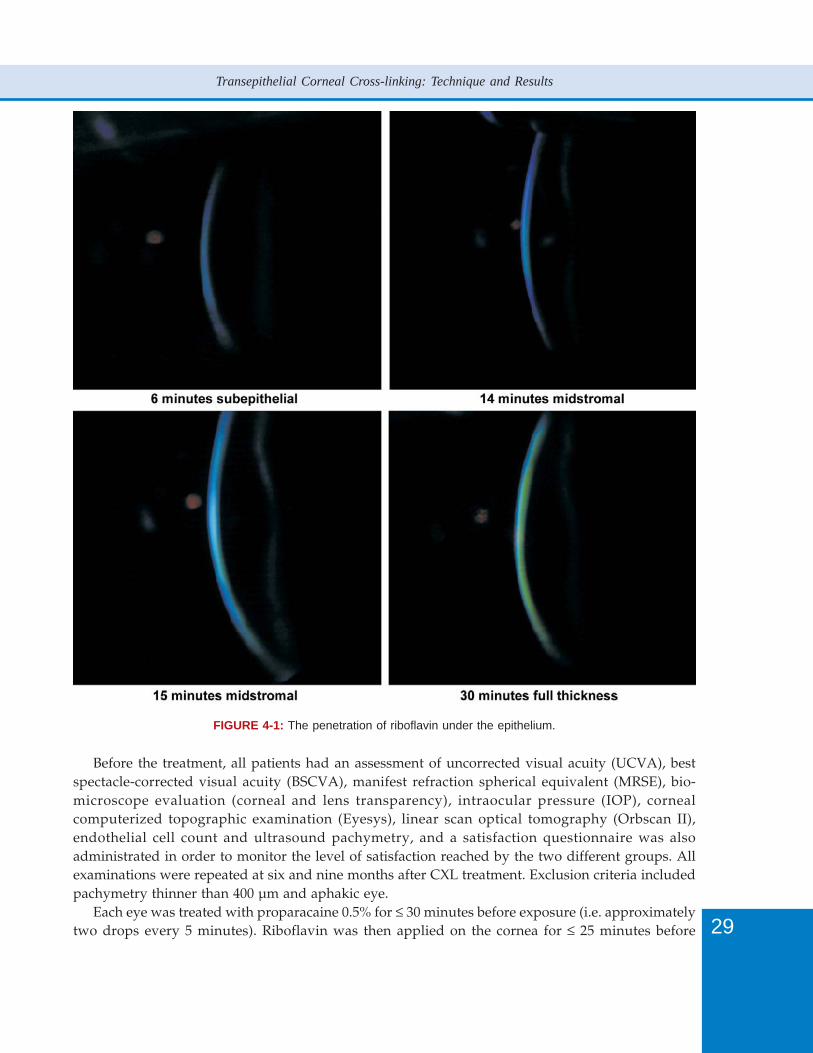
Recently, Pinelli, et al1 used fluoroscopy to observe the absorption of riboflavin in the absence ofepithelial debridement (Figure 4-1). Riboflavin 0.1% was applied to the cornea via a saturated Merocelsponge and left on the eye for 5 minutes before the start of UVA light administration. We repeatedriboflavin applications every 3 minutes. After 6 minutes, the riboflavin penetrated under theepithelium; after 14 minutes, it penetrated the middle of the stroma; and after 30 minutes, weobserves its full diffusion. Our research demonstrated that during CXL treatments, leaving theepithelium intact does not significantly limit the penetration of the riboflavin.
Personal ExperienceObserving via fluoroscopy the riboflavin absorption without epithelial removal, we noticed that theepithelium does not restrict significantly the riboflavin penetration.
Riboflavin 0.1% (PriaLight®, PriaVision, Menlo Park, CA, USA) was applied on the cornea via asaturated Merocel sponge for 5 minutes before the start of UVA light administration. The riboflavinis then applied every 3 minutes during the whole procedure.
After 6 minutes the riboflavin penetrates under the epithelium; after 14 minutes it penetrates inthe middle stroma and after 30 minutes we can observe its full diffusion (Figure 4-1).
On this basis, we conducted a comparative study to evaluate the difference between C3-R withand without deepithelialization on patients affected by keratoconus.
They were divided into two groups (A and B) each was composed of five patients.Group A was treated monocularly with CXL without deepithelialization; group B was treated
monocularly with CXL with deepithelialization.
29
Transepithelial Corneal Cross-linking: Technique and Results
FIGURE 4-1: The penetration of riboflavin under the epithelium.
Before the treatment, all patients had an assessment of uncorrected visual acuity (UCVA), bestspectacle-corrected visual acuity (BSCVA), manifest refraction spherical equivalent (MRSE), bio-microscope evaluation (corneal and lens transparency), intraocular pressure (IOP), cornealcomputerized topographic examination (Eyesys), linear scan optical tomography (Orbscan II),endothelial cell count and ultrasound pachymetry, and a satisfaction questionnaire was alsoadministrated in order to monitor the level of satisfaction reached by the two different groups. Allexaminations were repeated at six and nine months after CXL treatment. Exclusion criteria includedpachymetry thinner than 400 µm and aphakic eye.
Each eye was treated with proparacaine 0.5% for ≤ 30 minutes before exposure (i.e. approximatelytwo drops every 5 minutes). Riboflavin was then applied on the cornea for ≤ 25 minutes before
30
Keratoconus Surgery and Cross-linking
irradiation and was then activated by a 30-minute exposure to the UVA light (i.e. 370 nm fluence at3 mW/cm2). Riboflavin solution was reapplied on the cornea every 3 minutes during the UVAirradiation.
ResultsBefore the treatment, UCVA ranged from 0.1 to 0.3, BSCVA from 0.4 to 0.7, medium K value rangedfrom 45 to 49 D, and corneal thickness from 432 to 463 microns.
At six and nine months postoperatively there were no significant differences in the analyzedparameters between the deepithelialized group and the non-deepithelialized one.
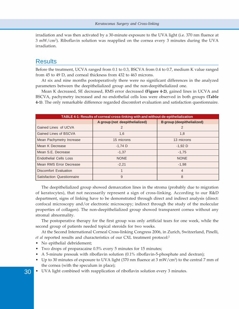
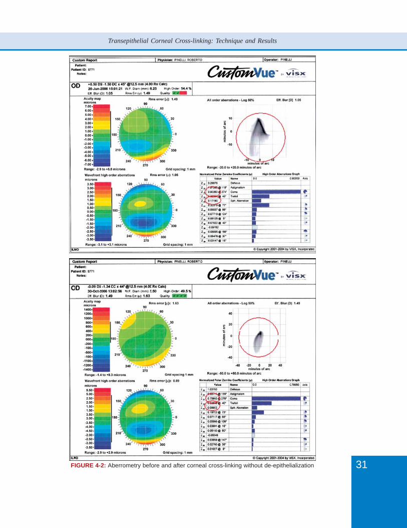
Mean K decreased, SE decreased, RMS error decreased (Figure 4-2), gained lines in UCVA andBSCVA, pachymetry increased and no endothelial cells loss were observed in both groups (Table4-1). The only remarkable difference regarded discomfort evaluation and satisfaction questionnaire.
TABLE 4-1: Results of corneal cross-linking with and without de-epithelialization A group (not deepithelialized) B group (deepithelialized)Gained Lines of UCVA 2 2
Gained Lines of BSCVA 1,6 1,8
Mean Pachymetry Increase 15 microns 13 microns
Mean K Decrease -1,74 D -1,92 D
Mean S.E. Decrease -1,37 -1,75
Endothelial Cells Loss NONE NONE
Mean RMS Error Decrease -2,21 -1,98
Discomfort Evaluation 1 4
Satisfaction Questionnaire 9 8
The deepithelialized group showed demarcation lines in the stroma (probably due to migrationof keratocytes), that not necessarily represent a sign of cross-linking. According to our R&Ddepartment, signs of linking have to be demonstrated through direct and indirect analysis (direct:confocal microscopy and/or electronic microscopy; indirect through the study of the molecularproperties of collagen). The non-deepithelialized group showed transparent cornea without anystromal abnormality.
The postoperative therapy for the first group was only artificial tears for one week, while thesecond group of patients needed topical steroids for two weeks.
At the Second International Corneal Cross-linking Congress 2006, in Zurich, Switzerland, Pinelli,et al reported results and characteristics of our CXL treatment protocol:2
• No epithelial debridement;• Two drops of proparacaine 0.5% every 5 minutes for 15 minutes;• A 5-minute presoak with riboflavin solution (0.1% riboflavin-5-phosphate and dextran);• Up to 30 minutes of exposure to UVA light (370 nm fluence at 3 mW/cm2) to the central 7 mm of
the cornea (with the speculum in place);• UVA light combined with reapplication of riboflavin solution every 3 minutes.
31
Transepithelial Corneal Cross-linking: Technique and Results
FIGURE 4-2: Aberrometry before and after corneal cross-linking without de-epithelialization
32
Keratoconus Surgery and Cross-linking
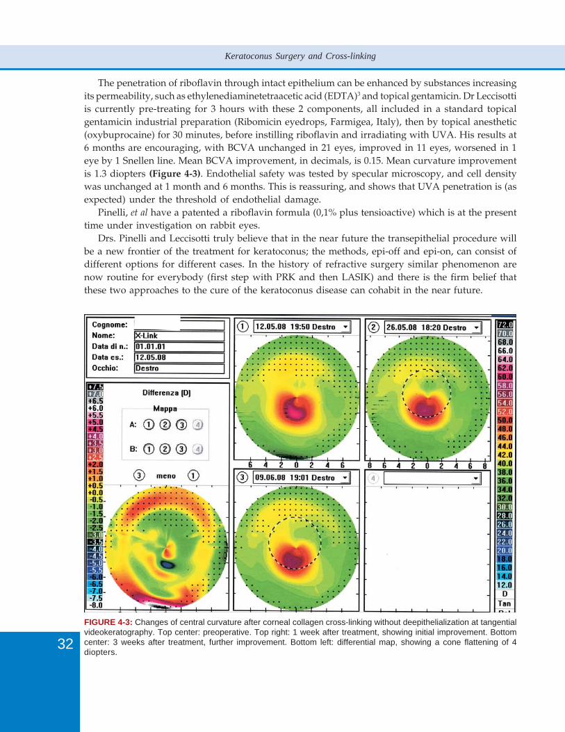
The penetration of riboflavin through intact epithelium can be enhanced by substances increasingits permeability, such as ethylenediaminetetraacetic acid (EDTA)3 and topical gentamicin. Dr Leccisottiis currently pre-treating for 3 hours with these 2 components, all included in a standard topicalgentamicin industrial preparation (Ribomicin eyedrops, Farmigea, Italy), then by topical anesthetic(oxybuprocaine) for 30 minutes, before instilling riboflavin and irradiating with UVA. His results at6 months are encouraging, with BCVA unchanged in 21 eyes, improved in 11 eyes, worsened in 1eye by 1 Snellen line. Mean BCVA improvement, in decimals, is 0.15. Mean curvature improvementis 1.3 diopters (Figure 4-3). Endothelial safety was tested by specular microscopy, and cell densitywas unchanged at 1 month and 6 months. This is reassuring, and shows that UVA penetration is (asexpected) under the threshold of endothelial damage.
Pinelli, et al have a patented a riboflavin formula (0,1% plus tensioactive) which is at the presenttime under investigation on rabbit eyes.
Drs. Pinelli and Leccisotti truly believe that in the near future the transepithelial procedure willbe a new frontier of the treatment for keratoconus; the methods, epi-off and epi-on, can consist ofdifferent options for different cases. In the history of refractive surgery similar phenomenon arenow routine for everybody (first step with PRK and then LASIK) and there is the firm belief thatthese two approaches to the cure of the keratoconus disease can cohabit in the near future.
FIGURE 4-3: Changes of central curvature after corneal collagen cross-linking without deepithelialization at tangentialvideokeratography. Top center: preoperative. Top right: 1 week after treatment, showing initial improvement. Bottomcenter: 3 weeks after treatment, further improvement. Bottom left: differential map, showing a cone flattening of 4diopters.
33
Transepithelial Corneal Cross-linking: Technique and Results
FutureAlthough ophthalmologists are still debating whether to remove or keep the epithelium intact beforeC3-R treatment, we prefer to avoid deepithelialization and its associated discomfort, especially untila scientific method or new technology in vivo will demonstrate the opposite.
In our opinion, the C3-R treatment in the future will be a less invasive, painless technique thatdoes not require deepithelialization. A bilateral option may also be psychologically easier and moreaccepted by our patients. Thus far, C3-R treatments are effective, and results and follow-up are veryencouraging. The numerous studies on C3-R and its impending CE mark demonstrate its safety.
References1. Pinelli R. Corneal collagen cross-linking with riboflavin (C3-R) treatment opens new frontiers for keratoconus and
corneal ectasia. Eyeworld, May 2007, Pages 34-40.2. Pinelli R. The Italian Refractive Surgery Society (SICR) results using C3-R, paper presented at the Second International
Congress of Corneal Cross Linking (CCL), Zurich, 2006.3. Nakamura T, et al. Electrophysiological characterization of tight junctional pathway of rabbit cornea treated with
ophthalmic ingredients. Biol Pharm Bull 2007;30:2360-4.

Please provide reference 19 and cite reference 20 in the text
36
Keratoconus Surgery and Cross-linking
Principles of Surgical Treatment of KeratoconusThe first consideration about keratoconus (KC) is that most keratoconic eyes do not need any surgicalor parasurgical treatment, because of minimal deformation, of acceptable vision with contact lensesor even spectacles, and of stability.1 However, the advent of collagen cross-linking has introducedan element of prevention of further corneal deformation even in those eyes in which no surgicaltreatment was considered in the past.2 Therefore, we should clearly separate the indications tocross-linking from those to surgery. Cross-linking is indicated in earlier stages of keratoconus,showing progression, and with no scarring; we should not hope that this technique avoid surgery ineyes with advanced keratoconus and poor vision with contact lenses.
Surgical treatment of KC has several options: the simple correction of the associated refractiveerror without touching the cornea, by means of phakic or pseudophakic IOLs; the attempt to reshapethe cornea, by insertion of intrastromal rings; the selective transplantation of the stroma, at differentdepths; finally, the full transplantation of the cornea.
Several techniques that have not passed the test of time or that are still under investigation willnot be covered by this chapter. Procedures causing corneal weakening, such as excimer laser,3 incisionsand circular keratotomy4 can have disastrous consequences and are therefore not advised onkeratoconic eyes. Epikeratophakia, consisting in the application of a donor lenticule over the cornea5
has been abandoned because of poor predictability and interface opacity.
Phakic IOLsKC is often associated with a myopic refraction. When the cornea is not excessively irregular, themyopic error can be addressed by the implantation an phakic IOL,6 with a toric component if theastigmatism is significant.7 Since the same results can be achieved by refractive lens exchange (RLE),phakic IOLs are usually reserved to patients younger than 40, because their advantage over RLE isessentially the preservation of accommodation. Phakic IOLs do cause complications, may needexplantation because of cataract or age-related shallowing of the anterior chamber (AC) and, whenimplanted in the AC, require a lifetime endothelial surveillance.
Several models of phakic IOLs are available, and their detailed description is beyond the purposesof this chapter. Angle-supported IOLs are currently falling out of favour, so the main phakic IOLmodels now in use are iris-fixated and posterior chamber. Iris-fixated IOLs have the advantage ofrotational stability, and are therefore ideal toric IOLs. AC depth, lens rise (i.e. the convexity of theiris-lens plane) and endothelial status are of paramount importance in preoperative evaluation, asmost complications are caused by the IOL contact with the iris (pigment dispersion, chroniciridocyclitis), the lens (cataract), and the endothelium (corneal decompensation).8 Although the surgicalmaneuvers to implant a phakic IOL should disturb the vitreous less than those occurring duringphacoemulsification, phakic IOLs are associated with retinal detachment, and this risk should bediscussed with the patient. The difficulties in obtaining a reliable refraction, necessary to IOL powercalculation, can explain some cases of significant overcorrection or undercorrection. In such cases,being unadvisable excimer laser fine-tuning, the phakic IOL should be exchanged.
Refractive Lens Exchange (RLE)The indications of RLE are the same as phakic IOLs’, but the loss of accommodation following RLErestricts this procedure to patients in the presbyopic age or younger patients that for some reason
37
Surgical Treatment of Keratoconus: An Overview
may not undergo phakic IOL implantation. RLE is based upon phacoemulsification of the crystallinelens and implantation in the capsular bag of a pseudophakic IOL,9 which may bear a toric componentto correct corneal astigmatism.10 Multifocal IOLs are obviously contraindicated in KC because cornealirregularity affects multifocality and cause aberrations and dysphotopsia.
The calculation of the IOL power is less reliable than in ordinary RLE; it has been advised touse videokeratographic K readings,11 but the idea of using prepupillary corneal curvature appearsmore appropriate. If the cornea is not too irregular, intraoperative autorefraction can be helpful todirectly calculate the IOL power (in highly myopic eyes) or to verify the power of a just implantedIOL.9
The main side effects of RLE in KC are under- or overcorrection, dysphotopsia, posterior capsuleopacification, and vitreoretinal complications. Under- and over corrections can be treated by promptIOL exchange (easier in the first postoperative weeks) or by piggyback implantation of a sulcus IOL(normally, for each diopter [D] of myopia the 1st IOL power should be reduced by 1 D, and for eachD of hyperopia, the 1st IOL power should be augmented by 1.5 D). Dysphotopsia is generally welltolerated, because KC patients are used to glare and halos induced by KC itself or by contact lenses.Vitreoretinal complications include posterior vitreous detachment and retinal detachment, and arecaused by the changes in the AC pressure during phacoemulsification.
Intrastromal RingsIntrastromal ring segments reshape the keratoconic cornea by supporting the tissue and by shorteningthe corneal arc length.12-14 Their implantation can be combined with cross-linking.15 The indicationsto intrastromal rings are a clear cornea, contact lens intolerance, minimal central thickness of 400micron. Their effect on refraction is a reduction of myopia and a regularization of astigmatism, buttheir predictability is limited in KC because of the variable tissue response. Two rings segments ora single segment can be implanted. Manual implantation is sometimes difficult; femtosecond laser istherefore often used to create the intrastromal tunnels.
The effects of intrastromal ring segments is reverted after explantation, and the cornea returnsto the previous shape. The main drawbacks of intrastromal ring segments are poor predictabilityand initial irregular astigmatism.
Anterior Lamellar KeratoplastyKC is essentially a stromal disease; therefore, partial or total replacement of the anterior centralstroma (anterior lamellar keratoplasty—ALK) is theoretically its rational cure. Unfortunately, anteriorlamellar keratoplasty often yields suboptimal visual results, and is therefore limited to cases inwhich contact lenses are not tolerated, spectacle-corrected visual acuity is not sufficient for thepatient’s activities, and the cornea is too thin or irregular for intrastromal rings (but several surgeonsdo not even consider the option of rings). The main drawback of anterior lamellar keratoplasty isastigmatism, which can be relevant and/or irregular. While mean astigmatism is 2.5 to 3 D in mostseries,16 unexpected cases of frustrating high astigmatism may occur even after perfectly conductedsurgery. The other complications include stromal rejection and suture-related inflammations, whichcan be however adequately tackled if early addressed, and intraoperative complications such asperforation, which can lead to penetrating keratoplasty (PKP).
38
Keratoconus Surgery and Cross-linking
Anterior lamellar keratoplasty is usually classified according to the depth of dissection. Somesurgical techniques such as the big-bubble may bare the Descemet’s membrane (total ALK or maximumdepth ALK); in other cases, only 20-50 microns of stromal bed are left (deep ALK);17 finally, inautomated ALK, a microkeratome removes a 250 micron thick free corneal cap, replaced by ananalogue but thicker donor flap (usually 350 micron thick). In all cases, the donor tissue is a centralcorneal button deprived of the endothelium and the Descemet’s membrane, and of the same diameterof the excised host button (or 0.25 mm larger). The choice of the technique is essentially surgeondependent; in general deeper dissections are technically more challenging but achieve best visualresults because of no interface opacity.18 Deep ALK is a good compromise between an acceptableintraoperative safety and a good final vision.
The advantages of ALK (especially of total and deep ALK) over PKP are enormous. The healthyperennial host endothelium is preserved; the endothelial rejection is impossible; surgery is not opensky; astigmatism is normally lower; finally, donor tissue is more readily available, because ALKdoes not need the endothelial standards required for PKP. Even the strongest advocates of PKP areprogressively accepting the fact that visual acuity after ALK is comparable with PKP.19
Penetrating KeratoplastyCurrently, the indications for PKP in KC are rather restricted. Obviously, a previously failed PKP isthe first indication. Unexpected perforation during ALK sometimes requires intraoperative conversionto PKP. Finally, extremely thin corneas are at high chance of intraoperative perforation during ALK,and therefore some surgeons directly perform PKP in such cases. We think that ALK should beattempted even in thin and protruding corneas, because the success rate is more than 50% in thesecases; obviously, everything (adequate tissue included) should be ready for a PKP.
This said, we must remember that PKP has been the golden standard for advanced KC untilrecent years, and its merits are not questionable. The absence of donor-host interface, the fastlearning curve, the manageability of rejection, its repeatability are well known.20 Our main concernswith PKP are the duration of donor endothelium and open sky surgery, with the inherent risk ofincreased vitreous pressure.
ConclusionThe term KC entails a wide spectrum of corneal deformations: the approach to visual rehabilitationin KC is therefore based upon careful staging and realistic expectations. Contact lenses have a definiterole in improving the quality of life and postponing or avoiding surgery in many keratoconic eyes.When corneal irregularity is limited, the spherical and astigmatic refractive error can be addressedby phakic or pseudophakic IOL implantation. Intrastromal ring segments can regularize the cornealshape in selected cases, and can be combined with IOL implantation. When corneal deformation isrelevant, ALK in its variants is the most rational intervention, with most cases however resulting ina refractive error that will require spectacles, contact lenses or further surgical correction.
References1. Rabinowitz YS. Keratoconus. Surv Ophthalmol 1998; 42:297-319.
39
Surgical Treatment of Keratoconus: An Overview
2. Wollensak G, Spoerl E, Seiler T. Riboflavin/ultraviolet-A-induced collagen crosslinking for the treatment ofkeratoconus. Am J Ophthalmol 2003; 135:620-7.
3. Kremer I, Shochot Y, Kaplan A, Blumenthal M. Three year results of photoastigmatic refractive keratectomy formild and atypical keratoconus. J Cataract Refract Surg 1998;24:1581-8.
4. Leccisotti A. Effect of circular keratotomy on keratoconus. J Cataract Refract Surg 2006;32:2139-41.5. Halliday BL. Epikeratophakia for aphakia, keratoconus and myopia. Br J Ophthalmol 1990;74:66-72.6. Leccisotti A, Fields SV. Angle-supported phakic intraocular lenses in myopia and keratoconus. J Cataract Refract
Surg 2003;29:1530-6.7. Budo C, Bartels MC, van Rij G. Implantation of Artisan toric phakic intraocular lenses for the correction of
astigmatism and spherical errors in patients with keratoconus. J Refract Surg 2005;21:218-22.8. Baikoff G, Bourgeon G, Jitsuo Jodai H, Fontaine A, Viera Lellis F, Trinquet L. Pigment dispersion and Artisan phakic
intraocular lenses. Crystalline lens rise as a safety criterion. J Cataract Refract Surg 2005;31:674-80.9. Leccisotti A. Refractive lens exchange in keratoconus. J Cataract Refract Surg 2006;32:742-6.
10. Sauder G, Jonas JB. Treatment of keratoconus by toric foldable intraocular lenses. Eur J Ophthalmol 2003;13:577-9.11. Celikkol L, Ahn D, Celikkol G, Feldman ST. Calculating intraocular lens power in eyes with keratoconus using
videokeratography. J Cataract Refract Surg 1996;22:497-500.12. Kwitko S, Souto Severo N. Ferrara intracorneal ring segments for keratoconus. J Cataract Refract Surg 2004;30:
812-20.13. Aliò JL, Artola A, Hassanein A, Haroun H, Galal A. One or 2 Intacs segments for the correction of keratoconus. J
Cataract Refract Surg 2005;31:943-53.14. Boxer Wachler BS, Christie JP, Chandra NS, Chou B, Korn T, Nepomuceno R. Intacs for keratoconus. Ophthalmology
2003; 110:1031-40.15. Kamburoglu G, Ertan A. Intacs implantation with sequential collagen cross-linking treatment in post-operative
LASIK ectasia. J Refract Surg 2008;24:S726-9.16. Noble BA, Agrawal A, Collins C, Saldana M, Brogden PR, Zuberbuhler B. Deep Anterior Lamellar Keratoplasty
(DALK): visual outcome and complications for a heterogeneous group of corneal pathologies. Cornea 2007;26:59-64.
17. Anwar M, Teichmann KD. Deep lamellar keratoplasty. Surgical techniques for anterior lamellar keratoplasty withand without baring of Descemet’s membrane. Cornea 2002;21:374-83.
18. Ardjomand N, Hau S, McAlister JC, et al. Quality of vision and graft thickness in deep anterior lamellar andpenetrating keratoplasty. Am J Ophthalmol 2007;143:228-35.
19. Borderie VM, Werthel AL, Touzeau O, Allouch C, Boutboul S, Laroche L. Comparison of techniques used forremoving the recipient stroma in anterior lamellar keratoplasty. Arch Ophthalmol 2008;126:31-7.
20. Muraine M, Sanchez C, Watt L, Retout A, Brasseur G. Long-term results of penetrating keratoplasty. A 10-yearplus retrospective study. Graefes Arch Clin Exp Ophthalmol 2003;241:571-6.

42
Keratoconus Surgery and Cross-linking
IntroductionIntrastromal ring segments (ISRS) were originally developed to treat low levels of myopia in healthyeyes. By adding material to the peripheral cornea, they alter the curvature of the anterior corneainducing a central flattening and thereby reducing myopia.
With increasing safety and accuracy of laser refractive surgery for vision correction, the use ofISRS for this indication has reduced dramatically in the last 10 years.
However, a new role for these segments in the treatment of corneal ectatic disorders has nowbeen firmly established with numerous published articles to support their use.1-13
The purpose of this chapter is to provide an in-depth review of these ring segments for thetreatment of keratoconus.
Although there are several types of ISRS, IntacsTM (Addition Technology, Inc, Fremont, California,USA) are the most commonly studied and used although implantation techniques and complicationsare similar between different types.
The implanted ring segments are two, clear, arcs of variable thickness up to 450 microns made ofpolymethylmethacrylate (PMMA), each with an arc length of 150 degrees. The outer diameter ofeach segment is 8.1 mm and the inner diameter measures 6.8 mm (Figure 6-1).
The segments are implanted in the peripheral cornea at the 7 mm optical zone. The rationale fortreatment of conditions such as keratoconus is that they regularize the corneal surface essentially byproviding a scaffold for the cornea.
As the segments are inserted peripherally, they have the effect of flattening the central corneawith the effect proportional to the thickness of the segments. As such, they reduce the amount ofastigmatism, regularize the anterior corneal surface and induce a hyperopic shift.
In many cases, they can delay or obviate the need for a corneal graft and can allow patients toresume contact lens wear where they were intolerant before.
IndicationsIndications for the use of ICRS include keratoconus,1-13 pellucid marginal degeneration14-16 and othercauses of corneal ectasia, e.g. refractive surgery induced.17-20
In keratoconus, ICRS are used in those patients intolerant to contact lenses, have a clear centralcornea and who have a minimal central corneal thickness of at least 400 microns.
There are several algorithms available to determine where the initial incision should be and whatthickness segment should be used. Ultimately, treatment is individualized to each patient based ontheir refraction, keratometry readings and their corneal topography.
Manual Implantation TechniqueSurgery is performed either under topical anesthesia or general anesthesia according to patient andsurgeon preference.
STEP-BY-STEP TECHNIQUE1. The operative eye is prepped and draped in the usual fashion ensuring the lashes are well away
from the surgical field.
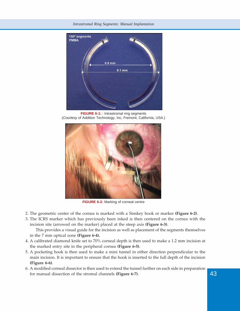
43
Intrastromal Ring Segments: Manual Implantation
2. The geometric center of the cornea is marked with a Sinskey hook or marker (Figure 6-2).3. The ICRS marker which has previously been inked is then centered on the cornea with the
incision site (arrowed on the marker) placed at the steep axis (Figure 6-3).This provides a visual guide for the incision as well as placement of the segments themselves
in the 7 mm optical zone (Figure 6-4).4. A calibrated diamond knife set to 70% corneal depth is then used to make a 1.2 mm incision at
the marked entry site in the peripheral cornea (Figure 6-5).5. A pocketing hook is then used to make a mini tunnel in either direction perpendicular to the
main incision. It is important to ensure that the hook is inserted to the full depth of the incision(Figure 6-6).
6. A modified corneal dissector is then used to extend the tunnel further on each side in preparationfor manual dissection of the stromal channels (Figure 6-7).
FIGURE 6-1: : Intrastromal ring segments(Courtesy of Addition Technology, Inc, Fremont, California, USA.)
FIGURE 6-2: Marking of corneal centre
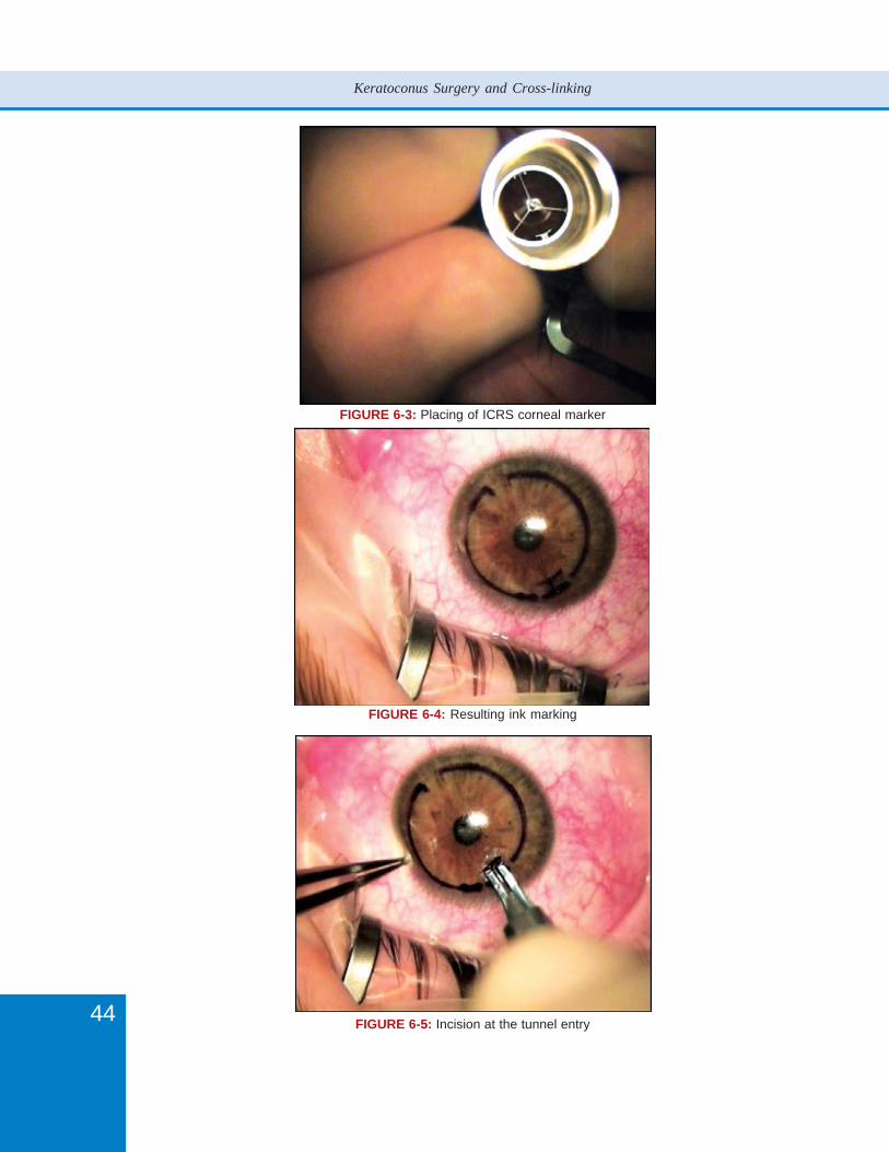
44
Keratoconus Surgery and Cross-linking
FIGURE 6-3: Placing of ICRS corneal marker
FIGURE 6-4: Resulting ink marking
FIGURE 6-5: Incision at the tunnel entry
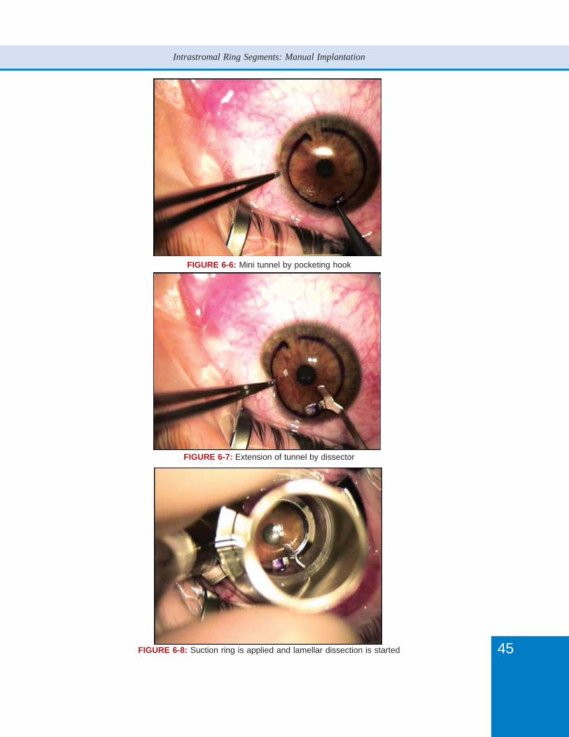
45
Intrastromal Ring Segments: Manual Implantation
FIGURE 6-6: Mini tunnel by pocketing hook
FIGURE 6-7: Extension of tunnel by dissector
FIGURE 6-8: Suction ring is applied and lamellar dissection is started
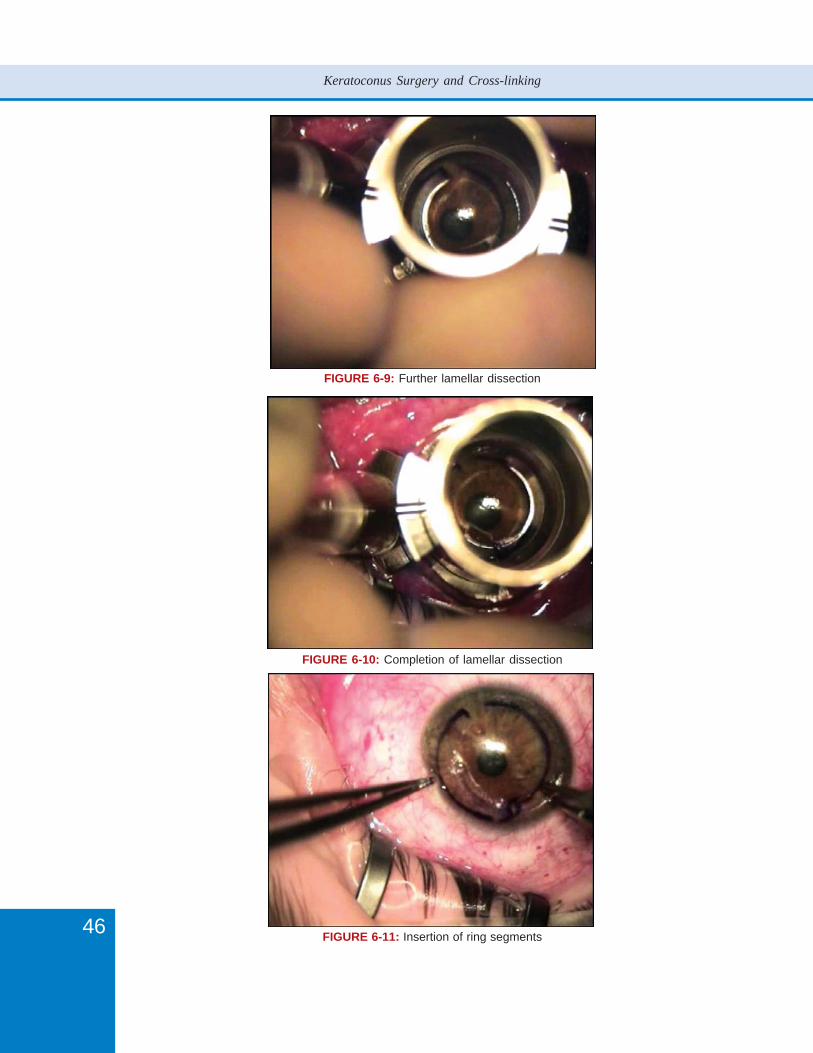
46
Keratoconus Surgery and Cross-linking
FIGURE 6-9: Further lamellar dissection
FIGURE 6-10: Completion of lamellar dissection
FIGURE 6-11: Insertion of ring segments
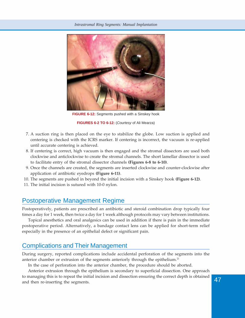
47
Intrastromal Ring Segments: Manual Implantation
FIGURE 6-12: Segments pushed with a Sinskey hook
FIGURES 6-2 TO 6-12: (Courtesy of Ali Mearza)
7. A suction ring is then placed on the eye to stabilize the globe. Low suction is applied andcentering is checked with the ICRS marker. If centering is incorrect, the vacuum is re-applieduntil accurate centering is achieved.
8. If centering is correct, high vacuum is then engaged and the stromal dissectors are used bothclockwise and anticlockwise to create the stromal channels. The short lamellar dissector is usedto facilitate entry of the stromal dissector channels (Figures 6-8 to 6-10).
9. Once the channels are created, the segments are inserted clockwise and counter-clockwise afterapplication of antibiotic eyedrops (Figure 6-11).
10. The segments are pushed in beyond the initial incision with a Sinskey hook (Figure 6-12).11. The initial incision is sutured with 10-0 nylon.
Postoperative Management RegimePostoperatively, patients are prescribed an antibiotic and steroid combination drop typically fourtimes a day for 1 week, then twice a day for 1 week although protocols may vary between institutions.
Topical anesthetics and oral analgesics can be used in addition if there is pain in the immediatepostoperative period. Alternatively, a bandage contact lens can be applied for short-term reliefespecially in the presence of an epithelial defect or significant pain.
Complications and Their ManagementDuring surgery, reported complications include accidental perforation of the segments into theanterior chamber or extrusion of the segments anteriorly through the epithelium.21
In the case of perforation into the anterior chamber, the procedure should be aborted.Anterior extrusion through the epithelium is secondary to superficial dissection. One approach
to managing this is to repeat the initial incision and dissection ensuring the correct depth is obtainedand then re-inserting the segments.
48
Keratoconus Surgery and Cross-linking
Other complications occurring at the time of surgery are relatively mild and resolve very quickly.These include epithelial defects, conjunctival chemosis and hemorrhage.
Perforation either anteriorly or posteriorly as well as ring decentration can all be prevented bymeticulous surgical technique paying particular attention to centration and tunnel depth.
Wound complications include wound dehiscence with extrusion of the implants, epithelial ingrowthand new vessel formation.
With wound dehiscence and extrusion, the implant can be repositioned and the wound re-sutured.If there is new vessel formation, this is likely to progress and the implants should be removed.
Epithelial ingrowth is usually self limiting but if progressive, the implants may to be removedand the ingrowing epithelium debrided.
Deposits around the segments themselves are common and typically develop within the first sixmonths of surgery. Their etiology is unclear but they typically have no effect on vision. If the depositsare marked, then removal of the segments are indicated.22,23
Infectious keratitis has also been reported with ICRS implantation and should be managed in theusual manner with a low threshold for segment removal. In some recalcitrant cases, penetratingkeratoplasty has been required as a measure to control the infection.24-28
Glare and haloes have been reported and are sometimes debilitating enough to require segmentremoval. In one study1 the ICRS had to removed in 12% of eyes because of visual symptoms.
Overall, complications are rare and as long as they are recognized and managed appropriately,there is minimal compromise to the patient’s vision.
Clinical ResultsThere is now a wealth of published material on ICRS use for keratoconus and other causes of cornealectasia.
Colin1 in his landmark European clinical evaluation paper prospectively studied 57 keratoconiceyes who were contact lens intolerant and underwent ICRS implantation. At the 6 month time gate,78% patients had an improvement in uncorrected visual acuity (UCVA) of 2 lines or more. Bestcorrected vision acuity (BCVA) improved from 53% of patients pre-operatively seeing 20/40 orbetter to 74% patients postoperatively. Keratometry readings were also reduced by a mean of 4.3+/- 2.8 D. In 12% of eyes, the ICRS were removed because of visual complaints, typically glare andhaloes but there were no complications associated with their removal.
Colin and Malet2 recently published their 2 year prospective results in 100 keratoconic eyes. Theyfound that the UCVA and BCVA improved in 80.5% and 68.3% of eyes at the 2 year time gate as wellas a significant reduction in keratometry values. Overall, they concluded that significant and sustainedimprovements in vision occurred in most cases.
Other authors have also found significant improvements in keratoconic patients.Aliò et al3 looked at 3 year results in their small series of 13 eyes and found that mean BCVA
increased from 20/50 preoperatively to 20/30 postoperatively at the 3 year time gate. They alsonoted decreased inferior-superior asymmetry by a mean of 2.81 Diopters which was sustained atthe 3 year time gate.
Aliò’s group4 also published an interesting paper on potential factors that influenced whether theoutcome was good or poor with ICRS implantation. They concluded that ICRS provided betterresults in those eyes with relatively low mean K values (less than or equal to 53 D) compared to eyeswith advanced keratoconus with mean K’s greater than or equal to 55 D.
49
Intrastromal Ring Segments: Manual Implantation
The longest follow up data regarding the use of ICRS to date has been published by Kymioniset al.5 They looked at 17 eyes with keratoconus in their 5 year retrospective study and found UCVA,BCVA and refraction was improved in the majority of patients (59%) with no evidence of progressivesight threatening complications.
DiscussionIn patients with keratoconus, ICRS are a good stop gap prior to corneal grafting in contact lensintolerant patients. They not only improve UCVA and BCVA but also restore contact lens tolerancein the majority of patients.
Whereas in the past, the only option for contact lens intolerant patients with keratoconus was acorneal transplant procedure, there are now several modalities of treatment that can be employedprior to resorting to a corneal graft.
ICRS implantation is being made easier and more accurate with the use of the femtosecond laserto create the channels and the results have been shown to be comparable with the manualtechnique.17,29,30
In addition, ICRS can be combined with other modalities of treatment to further improve the visualoutcome. There have been several published reports of ICRS combined with phakic intraocular lensesperformed in a sequential manner to correct any residual refractive error with good results.31,32
The procedure has also been combined with the latest modality of treatment, collagen cross-linking in order to prevent further progression of keratoconus but published results on this combinedapproach are scarce at present.33
In the future, ICRS will be further refined to reliably predict the effect and outcome in keratoconicand other ectatic conditions. Addition Technology have already produced a new type of ICRS calledIntacs SKTM. This implant has been specifically designed to be used in severe keratoconic caseswhere the K’s are extremely high and standard implants would be deemed ineffective.
The implants are elliptical in cross section and are designed to be inserted in the 6 mm opticalzone, hence providing increased effect. Anecdotal evidence seems to show good results and furtherdata is awaited with interest.34
ConclusionsIntracorneal ring segments are an important part of the surgical repertoire now available to treatkeratoconus.
Patients no longer need to proceed automatically to a corneal graft procedure which involves aprolonged route to visual rehabilitation.Intracorneal ring segments provide a safe and effective alternative in selected patients and are likelyto become more efficacious in the future with continuing research and development.
References1. Colin J. European clinical evaluation: use of Intacs for the treatment of keratoconus. J Cataract Refract Surg
2006;32:747-55. 2. Colin J, Malet FJ. Intacs for the correction of keratoconus: Two-year follow-up. J Cataract Refract Surg 2007;33:69-74.3. Aliò JL, Shabayek MH, Artola A. Intracorneal ring segments for keratoconus correction: long term follow up.
J Cataract Refract Surg 2006;32:978-85.
50
Keratoconus Surgery and Cross-linking
4. Aliò JL, Shabayek MH, Belda JI, Correas P, Feijoo ED. Analysis of results related to good and bad outcomes ofIntacs implantation fro keratoconus correction. J Cataract Refract Surg 2006;32:756-61.
5. Kymionis GD, Siganos CS, Tsiklis NS, et al. Long-term follow-up of Intacs in keratoconus. Am J Ophthalmol2007;143:236-44.
6. Levinger S, Pokroy R. Keratoconus managed by Intacs: one year results. Arch Ophthalmol 2005;123:1308-14.7. Hellstedt T, Makela J, Uusitalo R, et al. Treating keratoconus with Intacs corneal ring segments. J Refract Surg
2005;21:236-46.8. Boxer-Wachler BS, Christie JP, et al. Intacs for keratoconus. Ophthalmology 2003;110:1031-40.9. Siganos CS, Kymionis GD, Kartakis N, et al. Management if keratoconus with Intacs. Am J Ophthalmol
2003;135:64-70.10. Colin J, Cochener B, Savary G, et al. Intacs inserts for treating keratoconus: one year results. Ophthalmology
2001;108:1409-14.11. Colin J, Cochener B, Savary G, et al. Correcting keratoconus with intracorneal rings. J Cataract Refract Surg
2000;26:1117-22.12. Koch DD. Refractive surgery for keratoconus: a new approach. J Cataract Refract Surg 2000;26:1099-1100.13. Siganos D, Ferrara P, Chatzinikolas K, et al. Intrastromal corneal rings for the correction of keratoconus. J Cataract
Refract Surg 2002;28:1947-51.14. Barbara A, Shehadeh-Masha’our R, Zvi F, et al. Management of pellucid marginal degeneration with intracorneal
ring segments. J Refract Surg 2005;21(3):296-8.15. Mularoni A, Torreggiani A, di Biase A, et al. Conservative treatment of early and moderate pellucid marginal
degeneration: a new refractive approach with intracorneal rings. Ophthalmology 2005;112:660-6.16. Kymionis GD, Aslanides IM, Siganos CS, et al. Intacs for early pellucid marginal degeneration. J Cataract Refract
Surg 2004;30:230-3.17. Carrasquillo KG, Rand J, Talamo JH. Intacs for keratoconus and post-LASIK ectasia:mechanical versus femtosecond
laser-assisted channel creation. Cornea 2007;26:956-62.18. Kymionis GD, Tsiklis NS, Pallikaris AI, et al. Long-term follow-up of Intacs for post-LASIK corneal ectasia.
Ophthalmology 2006;113:1909-17.19. Sharma M, Boxer Wachler BS. Comparison of single-segment and double-segment Intacs for keratoconus and
post-LASIK ectasia. Am J Ophthalmol 2006;141:891-5.20. Pokroy R, Levinger S, Hirsh A. Single Intacs segment for post-laser in situ keratomilieusis keratectasia. J Cataract
Refract Surg 2004;30:1685-95.21. Schanzlin DJ, Abbott RL, Asbell PA, et al. Two year outcomes of intrastromal corneal ring segments for the
correction of myopia. Ophthalmology 2001;108:1688-94.22. Fink AM, Gore C, Rosen ES. Corneal changes associated with intrastromal corneal ring segments. Arch Ophthalmol
1999;117:282.23. Quantock AJ, Assil KK, Schanzlin DJ. Electron microscopic evaluation of intrastromal corneal rings explanted from
nonfunctional human eyes. J Refract Corneal Surg 1994;10:142-8.24. Hashemi H, Ghaffari R, Mohammadi M, et al. Microbial keratitis after INTACS implantation with loose suture.
J Refract Surg 2008;24:551-2.25. Slade DS, Johnson JT, Tabin G. Acanthamoeba and fungal keratitis in a woman with a history of Intacs corneal
implants. Eye Contact Lens 2008;34:185-7.26. Shehadeh-Masha’our R, Modi N, Barbara A, et al. Keratitis after implantation of intrastromal corneal segments.
J Cataract Refract Surg 2004;30:1802-4.27. Hofling-Lima AL, Branco BC, Romano AC, et al. Corneal infections after implantation of intracorneal ring segments.
Cornea 2004;23:547-9.28. Bourcier T, Borderie V, Laroche L. Late bacterial keratitis after implantation of intrastromal corneal ring segments.
J Cataract Refract Surg 2003;29:407-9.29. Rabinowitz YS. Intacs for keratoconus. Curr Opin Ophthalmol 2007;18:279-83.30. Ertan A, Kamburoglu G, Bahadir M. Intacs insertion with the femtosecond laser for the management of keratoconus:
one year results. J Cataract Refract Surg 2006;32:2039-42.31. Coskunseven E, Onder M, Kymionis GD, et al. Combined Intacs and posterior chamber toric implantable collamer
lens implantation for keratoconic patients with extreme myopia. Am J Ophthalmol 2007;144:387-9.32. El-Raggal TM, Abdel Fattah AA. Sequential Intacs and Verisyse phakic intraocular lens for refractive improvement
in keratoconic eyes. J Cataract Refract Surg 2007;33:966-70.33. Chan CC, Sharma M, Wachler BS. Effect of inferior segment Intacs with and without C3-R on keratoconus.
J Cataract Refract Surg 2007;33:75-80.34. Personal Communication with Kestrel Ophthalmics LTD, Addition Technology distributors, UK.
Please cite reference 15 in the text and provide reference 16
52
Keratoconus Surgery and Cross-linking
IntroductionKeratoconus is a disease of unknown etiology or pathogenesis which results in progressive bilateralthinning and protrusion of the cornea. Various genetic factors have been implicated in its causationresulting in increased protease activity and possibly aberrant interleukin-11 expression resulting inboth collagen loss or apoptosis of keratocytes.2
The initial mainstay of treatment consists of spectacles and then contact lens wear—progressingfrom soft contact lenses to hard. Once a person has become contact lens intolerant until recently themainstay of treatment has been to carryout a penetrating keratoplasty (PKP). This is obviously aninvasive treatment with both significant risks but also one requiring intensive follow-up with attentionto suture removal, astigmatism management and anti-rejection treatment.
Where there is central opacification surgical intervention either in the form of PKP or lamellarsurgery is a pre-requisite to clear the visual axis and rehabilitate the eyes visual function. Where thecentral cornea is transparent but the patient is intolerant of contact lens wear a variety of proceduresare now becoming available to modify the corneal shape or otherwise improve visual function withoutthe need for penetrating keratoplasty.
The ever expanding treatment options include:1. IntacsTM (Addition Technology, Inc, Fremont, California, USA) corneal reshaping2. IntacsTM plus cross-linking3. Phototherapeutic keratectomy plus IntacsTM where there is central corneal scarring4. Excimer laser plus corneal collagen cross-linking (with strict selection criteria)5. Toric phakic intraocular lens (IOL)6. IntacsTM plus toric phakic IOL7. Cross-linking plus toric phakic IOL8. Lamellar keratoplasty9. Lamellar keratoplasty plus posterior cornea repositioning.
IntacsTM treatments were first instituted by Colin in 1997 on patients with keratoconus who hada central clear cornea but were contact lens intolerant. The aim of this treatment is to modify theshape of the cornea to improve vision rather than arresting the condition. These forms of treatmentmay be of particular interest in those patients where more invasive forms of management are moreat risk such as in highly atopic individuals and in Down syndrome patients.
The treatment consists of inserting semicircular plastic rings within the peripheral corneal stromain order to shorten the arc length of the anterior cornea and therefore force the anterior cornea toflatten. The usual treatment consists of either placing one or two IntacsTM within the cornea. Mostcommonly two IntacsTM are used and the thicker segment is placed inferiorly to thicken thedegenerated area of cornea in an attempt to push the cone upwards. Increasingly often groups areplacing only one inferior segment. Colin reported in the literature the first year results of IntacsTM
treatment to manage keratoconus in 20003 where they described a flattening of overall cone reductionin astigmatism. In addition most patients reported improved uncorrected and best corrected visualacuity. Although Boxer Wachler has reported improvements in overall shape and visual acuityin patients with central scarring treated with IntacsTM, most other studies have demonstratedimproved clinical responses in those with no central scarring and only moderate keratoconic shapechange.4-6 Insertion of IntacsTM can be carried out by mechanical dissection or by femtosecond laserdissection.
53
Intrastromal Ring Segments: Implantation by Femtosecond Laser
Preoperative EvaluationIt is important to carry out a full ophthalmologic examination with uncorrected visual acuity andbest corrected with both glasses and contact lenses. Slit-lamp examination indicating evidence andposition of corneal opacities, folds and cone position. Corneal topography will provide detailedinformation on cone position and cone steepness. Corneal thickness can be determined with thePentacam or Orbscan topographer or via ultrasonic pachymetry. An evaluation of contact lens fittingand tolerance is imperative.
Insertion of the IntacsTM by Femtosecond Laser DissectionA femtosecond is equivalent to 10-15 of a second. The laser works in the infrared spectrum ofelectromagnetic radiation at wavelength 1053 nm. Unlike the excimer laser which exerts its energyon the surface of the cornea to ablate corneal tissue, the femtosecond is focussed at an exact predefineddepth within the corneal tissue. Each tissue pulse causes plasma photodisruption to the precise areaof corneal tissue upon which it is focussed. The tissue affected is vaporized and produces smallbubbles within the corneal stroma which expand and cause a 5-12 µm microsplit in the corneal tissueat this point. Using thousands of these focussed spots it is possible to create three dimensionalshapes within the corneal stroma. The bubbles of gas are gradually removed from the tissue by theendothelial cell pump.
The major advantages of the femtosecond laser are the ability to create the track from the insideout thus reducing the postoperative chances of infection. In addition, it provides the ability to makea consistent depth track at 70-80% of corneal thickness.
PRIOR TO INTRALASE DISSECTION OF INTACSTM TRACKS
1. Preoperative pachymetry, topography and refraction2. Mark pupil center and center Intralase on that point3. Decide on incision site (avoid superior as increased risk of neovascularization)4. Choose thickness of Intacs™ based upon spherical equivalent5. Often place two Intacs™ even if only one thought to be required—easy to remove one
postoperatively.
Intacs™ are made of polymethylmethacrylate and have a crescent-shaped arc length of 150 degreeswith an inner diameter of 6.8 mm and an outer diameter of 8.1 mm. The thickness ranges from 0.25to 0.45 mm.
NOMOGRAMS
Various nomograms exist:6-8 These are related to both the diameter of the tract and size of the Intacs™.Most groups vary between using an inner diameter of 6.6 mm minimum and a maximum outerdiameter of 8.2 mm. The tighter the diameter of the track, the greater the effect gained from theprocedure. Complications (peripheral haze, tract deposits and displacement of the Intacs™) however,have been increasingly found with tighter tracts compared to those which are wider. Though intuitivethat adjustments should be made in the thickness of the tract to suit the thickness of the individualIntacs™ used. Most studies use one standard size of tract irrespective of the size of Intacs™—thismay have an effect upon the efficacy of each treatment.
54
Keratoconus Surgery and Cross-linking
CHOICE OF SIZES
Oval cones: asymmetric segmentsNipple cone: symmetric segmentsMild cone: single segment
Increasingly one large segment is often used, i.e. 0.45 mm inferiorly—this has the function tothicken the thinned area of cornea and push the cone upwards. Various groups have published thebenefits in improving postoperative vision by removing the superior segment in those cases withdual segment insertion. A recent report of two cases with IntacsTM placed in a vertical positionrather than a horizontal position to thicken the temporally thinned cornea and displaced cone assistedin pushing the cone nasally. This also demonstrates the flexibility and ease of positioning of theimplants using the femtosecond laser.9
Surgical OutcomesColin reported the results from a 100 cases of consecutive eyes at two years. 80.5% and 68.3%showed improvement in uncorrected and best corrected visual acuity respectively. Mean sphericalequivalent improved from -6.9+/- 3.9 to -3.8 +/-2.7 D at year two.10 A recent study of 118 eyes of 69patients by Ertan demonstrated at one year 81% improvement in UCVA and 74% improvement inBCVA with mean spherical equivalent improved from -7.6 D to -3.7 D.11 A head to head analysis ofresults from the mechanical compared to the femtosecond insertion of Intacs™ demonstrated betterresults using the femtosecond over the mechanical method, most of these benefits did not howeverreach statistical significance.12
ComplicationsUNDERCORRECTION
Induced regular astigmatism in early period which usually regresses by 6 months.Residual myopia requiring CL fitting or possibly amenable to phakic IOL.Surface laser ablation with or without cross-linking is a possible adjunctive therapy to those not
tolerant of CL wear.
OVERCORRECTION
It is possible to induce astigmatism by a large superior segment causing over flattening to the superiorsegment. The easiest way to address this is to remove the superior segment.
NEOVASCULARIZATION
Limbal neovascularization is common in cases of chronic contact lens wear, this is often more apparentin the superior cornea (Figure 7-1). It is best to avoid this region for insertion of the Intacs™ toavoid inducing inflammation in this point and possible subsequent neovascularization.
MIGRATION OF AN INTACS™ SEGMENT
Intacs™ can move particularly in the early period after insertion. It is important to ensure that thesegment is moved adequately from the insertion site to prevent pouting of the wound and possibleextrusion or induced inflammation.
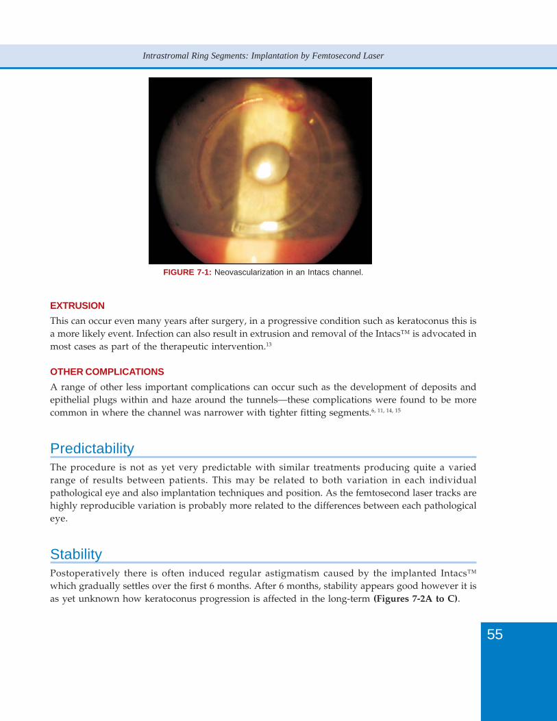
55
Intrastromal Ring Segments: Implantation by Femtosecond Laser
FIGURE 7-1: Neovascularization in an Intacs channel.
EXTRUSION
This can occur even many years after surgery, in a progressive condition such as keratoconus this isa more likely event. Infection can also result in extrusion and removal of the Intacs™ is advocated inmost cases as part of the therapeutic intervention.13
OTHER COMPLICATIONS
A range of other less important complications can occur such as the development of deposits andepithelial plugs within and haze around the tunnels—these complications were found to be morecommon in where the channel was narrower with tighter fitting segments.6, 11, 14, 15
PredictabilityThe procedure is not as yet very predictable with similar treatments producing quite a variedrange of results between patients. This may be related to both variation in each individualpathological eye and also implantation techniques and position. As the femtosecond laser tracks arehighly reproducible variation is probably more related to the differences between each pathologicaleye.
StabilityPostoperatively there is often induced regular astigmatism caused by the implanted Intacs™which gradually settles over the first 6 months. After 6 months, stability appears good however it isas yet unknown how keratoconus progression is affected in the long-term (Figures 7-2A to C).
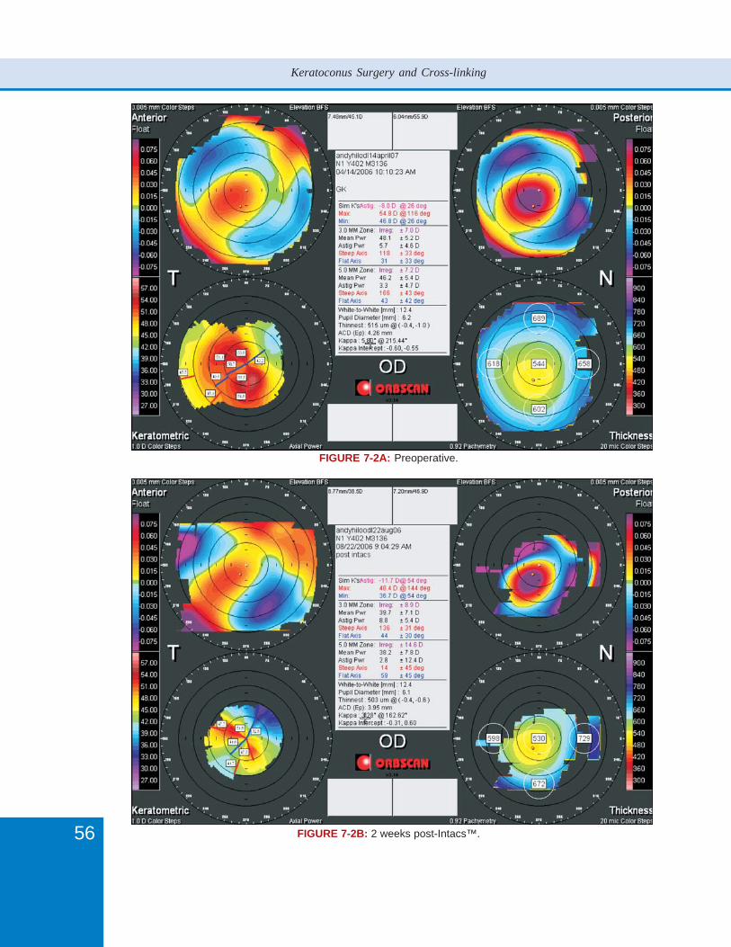
56
Keratoconus Surgery and Cross-linking
FIGURE 7-2A: Preoperative.
FIGURE 7-2B: 2 weeks post-Intacs™.
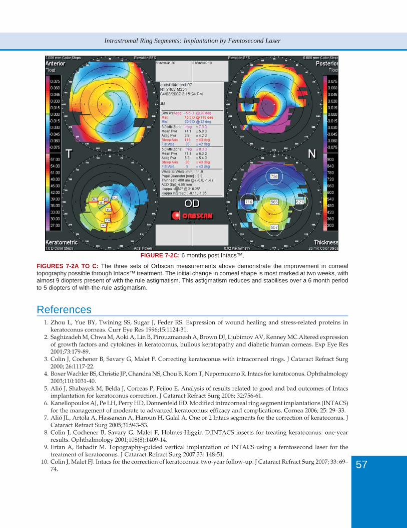
57
Intrastromal Ring Segments: Implantation by Femtosecond Laser
FIGURE 7-2C: 6 months post Intacs™.
FIGURES 7-2A TO C: The three sets of Orbscan measurements above demonstrate the improvement in cornealtopography possible through Intacs™ treatment. The initial change in corneal shape is most marked at two weeks, withalmost 9 diopters present of with the rule astigmatism. This astigmatism reduces and stabilises over a 6 month periodto 5 diopters of with-the-rule astigmatism.
References1. Zhou L, Yue BY, Twining SS, Sugar J, Feder RS. Expression of wound healing and stress-related proteins in
keratoconus corneas. Curr Eye Res 1996;15:1124-31.2. Saghizadeh M, Chwa M, Aoki A, Lin B, Pirouzmanesh A, Brown DJ, Ljubimov AV, Kenney MC.Altered expression
of growth factors and cytokines in keratoconus, bullous keratopathy and diabetic human corneas. Exp Eye Res2001;73:179-89.
3. Colin J, Cochener B, Savary G, Malet F. Correcting keratoconus with intracorneal rings. J Cataract Refract Surg2000; 26:1117-22.
4. Boxer Wachler BS, Christie JP, Chandra NS, Chou B, Korn T, Nepomuceno R. Intacs for keratoconus. Ophthalmology2003;110:1031-40.
5. Alió J, Shabayek M, Belda J, Correas P, Feijoo E. Analysis of results related to good and bad outcomes of Intacsimplantation for keratoconus correction. J Cataract Refract Surg 2006; 32:756-61.
6. Kanellopoulos AJ, Pe LH, Perry HD, Donnenfeld ED. Modified intracorneal ring segment implantations (INTACS)for the management of moderate to advanced keratoconus: efficacy and complications. Cornea 2006; 25: 29–33.
7. Alió JL, Artola A, Hassanein A, Haroun H, Galal A. One or 2 Intacs segments for the correction of keratoconus. JCataract Refract Surg 2005;31:943-53.
8. Colin J, Cochener B, Savary G, Malet F, Holmes-Higgin D.INTACS inserts for treating keratoconus: one-yearresults. Ophthalmology 2001;108(8):1409-14.
9. Ertan A, Bahadir M. Topography-guided vertical implantation of INTACS using a femtosecond laser for thetreatment of keratoconus. J Cataract Refract Surg 2007;33: 148-51.
10. Colin J, Malet FJ. Intacs for the correction of keratoconus: two-year follow-up. J Cataract Refract Surg 2007; 33: 69–74.

58
Keratoconus Surgery and Cross-linking
11. Ertan A, Kamburoðlu G, Akgün U. Comparison of outcomes of 2 channel sizes for intrastromal ring segmentimplantation with a femtosecond laser in eyes with keratoconus. J Cataract Refract Surg 2007; 33:648-53.
12. Rabinowitz YS, Li X, Ignacio TS, Maguen EJ. INTACS inserts using the femtosecond laser compared to the mechanicalspreader in the treatment of keratoconus. J Refract Surg 2006;22:764-71.
13. Hashemi H, Ghaffari R, Mohammadi M, Moghimi S, Miraftaab M. Microbial keratitis after INTACS implantationwith loose suture. J Refract Surg 2008;24:551-2.
14. Rodriguez-Prats J, Galal A, Garcia-Lledo M, De La Hoz F, Alió JL. Intracorneal rings for correction of pellucidmarginal degeneration. J Cataract Refract Surg 2003; 29:1421-4.
15. Barbara A, Shehadeh-Masha’our R, Zvi F, Garzozi HJ. Management of pellucid marginal degeneration withintracorneal ring segments. J Refract Surg 2005; 21:296-8.
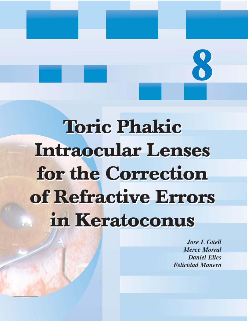

60
Keratoconus Surgery and Cross-linking
IntroductionKeratoconus is a non-inflammatory corneal disease that develops progressive thinning and anteriorbulging of the cornea. Corneal ectasia frequently induces varied degrees of myopia and/orastigmatism.1 In early stages, spectacles and contact lenses are the treatment of choice.2-4 However,a considerable amount of patients with progressive keratoconus have not only reduced visual acuitywith spectacles due to irregular astigmatism,5,6 but also reduced tolerance to contact lenses.7-10
Before the advent of modern refractive surgery techniques, penetrating keratoplasty (PKP) ordeep anterior lamellar keratoplasty (DALK) were the treatment of choice when a patient withkeratoconus became contact lens intolerant or had poor best-spectacle corrected visual acuity (BSCVA).This is still true in advanced stages of the disease, where severe thinning and/or corneal scarringoccurs. Because refractive anisometropia and high postoperative astigmatism are common problemsafter penetrating keratoplasty (PKP), visual rehabilitation and return to binocular function may beslow.11-15 Moreover, complications related to corneal transplant surgery itself, such as endophthalmitisor rejection episodes, should also be taken into account.16-21 Therefore, in early stages of keratoconus,when the central cornea remains clear, other options should be considered to avoid or delaykeratoplasty.
With the exception of some anecdotal reports, corneal incisional (radial keratotomy, or arquatekeratotomy) and ablational refractive approaches such as PRK or LASIK are contraindicated inkeratoconus, as they increase the risk of progressive, irreversible corneal ectasia.22-25 Availablerefractive procedures include intrastromal corneal ring segments, and toric phakic intraocular lenses(TPIOLs). The main goal of these procedures is to provide enough BSCVA, and sometimes uncorrectedvisual acuity (UCVA), to postpone the need for a corneal transplantation.
Intracorneal rings provide structural reinforcement of the cornea, and reshape and center cornea’soptical zone, improving topographic abnormalities (irregular astigmatism), quality of vision, andvisual acuity (Figure 8.1).26-29 Intracorneal rings act as spacing elements that shorten the arc of lengthof the anterior corneal surface and flatten the central cornea, which reduce myopic sphericalequivalent.30-32 However, they only correct a limited range of myopia, and high refractive errorsmay remain. Residual refractive errors may be corrected with spectacles or soft contact lenses.33
TPIOLs have also been implanted after ICRS.34-36
PIOLs have been used to correct moderate and high myopia, hyperopia and /or astigmatism.There are also a few reports on their use in patients with keratoconus.37-39 The implantation of PIOLshas been reported to be a stable, predictable and safe refractive procedure.40-50 This chapter dealswith the use of TPIOLs for the correction of spherical errors (usually myopia) and astigmatism inpatients with keratoconus.
IndicationsRecommended inclusion and exclusion criteria are summarized in Table 8-1. Being sure thatkeratoconus is stable is not an easy issue. Keratoconus is a progressive disease that starts aroundpuberty, generally progresses slowly during early years, and usually stabilizes in the third andfourth decade of life.1 However, it may move at any moment throughout life.51 It has been generallyaccepted that a patient older than 25 years old who shows stability on keratometry and refractionfor more than six months can be considered for PIOL implantation.39 Moreover, if refraction changesin the future, PIOL might be exchanged for another one.
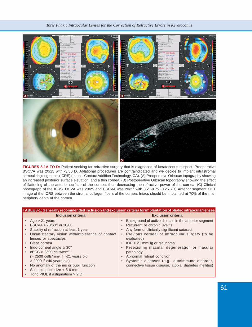
61
Toric Phakic Intraocular Lenses for the Correction of Refractive Errors in Keratoconus
FIGURES 8-1A TO D: Patient seeking for refractive surgery that is diagnosed of keratoconus suspect. PreoperativeBSCVA was 20/25 with -3.50 D. Ablational procedures are contraindicated and we decide to implant intrastromalcorneal ring segments (ICRS) (Intacs, Contact Addition Technology, CA). (A) Preoperative Orbscan topography showingan increased posterior surface elevation, and a thin cornea. (B) Postoperative Orbscan topography showing the effectof flattening of the anterior surface of the cornea, thus decreasing the refractive power of the cornea. (C) Clinicalphotograph of the ICRS. UCVA was 20/25 and BSCVA was 20/27 with 85° -0.75 -0.25. (D) Anterior segment OCTimage of the ICRS between the stromal collagen fibers of the cornea. Intacs should be implanted at 70% of the mid-periphery depth of the cornea.
TABLE 8-1: Generally recommended inclusion and exclusion criteria for implantation of phakic intraocular lensesInclusion criteria
• Age > 21 years• BSCVA > 20/6039 or 20/80• Stability of refraction at least 1 year• Unsatisfactory vision with/intolerance of contact
lenses or spectacles• Clear cornea• Irido-corneal angle ≥ 30°• cECC > 2300 cells/mm2:
(> 2500 cells/mm2 if >21 years old,> 2000 if >40 years old)
• No anomaly of the iris or pupil function• Scotopic pupil size < 5-6 mm• Toric PIOL if astigmatism > 2 D
Exclusion criteria• Background of active disease in the anterior segment• Recurrent or chronic uveitis• Any form of clinically significant cataract• Previous corneal or intraocular surgery (to be
evaluated)• IOP > 21 mmHg or glaucoma• Preexisting macular degeneration or macular
pathology• Abnormal retinal condition• Systemic diseases (e.g., autoimmune disorder,
connective tissue disease, atopia, diabetes mellitus)
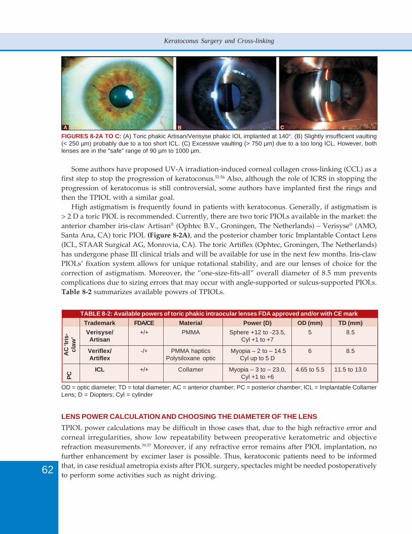
62
Keratoconus Surgery and Cross-linking
Some authors have proposed UV-A irradiation-induced corneal collagen cross-linking (CCL) as afirst step to stop the progression of keratoconus.52-56 Also, although the role of ICRS in stopping theprogression of keratoconus is still controversial, some authors have implanted first the rings andthen the TPIOL with a similar goal.
High astigmatism is frequently found in patients with keratoconus. Generally, if astigmatism is> 2 D a toric PIOL is recommended. Currently, there are two toric PIOLs available in the market: theanterior chamber iris-claw Artisan® (Ophtec B.V., Groningen, The Netherlands) – Verisyse® (AMO,Santa Ana, CA) toric PIOL (Figure 8-2A), and the posterior chamber toric Implantable Contact Lens(ICL, STAAR Surgical AG, Monrovia, CA). The toric Artiflex (Ophtec, Groningen, The Netherlands)has undergone phase III clinical trials and will be available for use in the next few months. Iris-clawPIOLs’ fixation system allows for unique rotational stability, and are our lenses of choice for thecorrection of astigmatism. Moreover, the “one-size-fits-all” overall diameter of 8.5 mm preventscomplications due to sizing errors that may occur with angle-supported or sulcus-supported PIOLs.Table 8-2 summarizes available powers of TPIOLs.
FIGURES 8-2A TO C: (A) Toric phakic Artisan/Verisyse phakic IOL implanted at 140°. (B) Slightly insufficient vaulting(< 250 µm) probably due to a too short ICL. (C) Excessive vaulting (> 750 µm) due to a too long ICL. However, bothlenses are in the “safe” range of 90 µm to 1000 µm.
TABLE 8-2: Available powers of toric phakic intraocular lenses FDA approved and/or with CE mark
Trademark FDA/CE Material Power (D) OD (mm) TD (mm)
Verisyse/ +/+ PMMA Sphere +12 to -23.5, 5 8.5Artisan Cyl +1 to +7
Veriflex/ -/+ PMMA haptics Myopia – 2 to – 14.5 6 8.5Artiflex Polysiloxane optic Cyl up to 5 D
ICL +/+ Collamer Myopia – 3 to – 23.0, 4.65 to 5.5 11.5 to 13.0Cyl +1 to +6
OD = optic diameter; TD = total diameter; AC = anterior chamber; PC = posterior chamber; ICL = Implantable CollamerLens; D = Diopters; Cyl = cylinder
AC
‘Iri
s-cl
aw’
PC
LENS POWER CALCULATION AND CHOOSING THE DIAMETER OF THE LENS
TPIOL power calculations may be difficult in those cases that, due to the high refractive error andcorneal irregularities, show low repeatability between preoperative keratometric and objectiverefraction measurements.39,57 Moreover, if any refractive error remains after PIOL implantation, nofurther enhancement by excimer laser is possible. Thus, keratoconic patients need to be informedthat, in case residual ametropia exists after PIOL surgery, spectacles might be needed postoperativelyto perform some activities such as night driving.
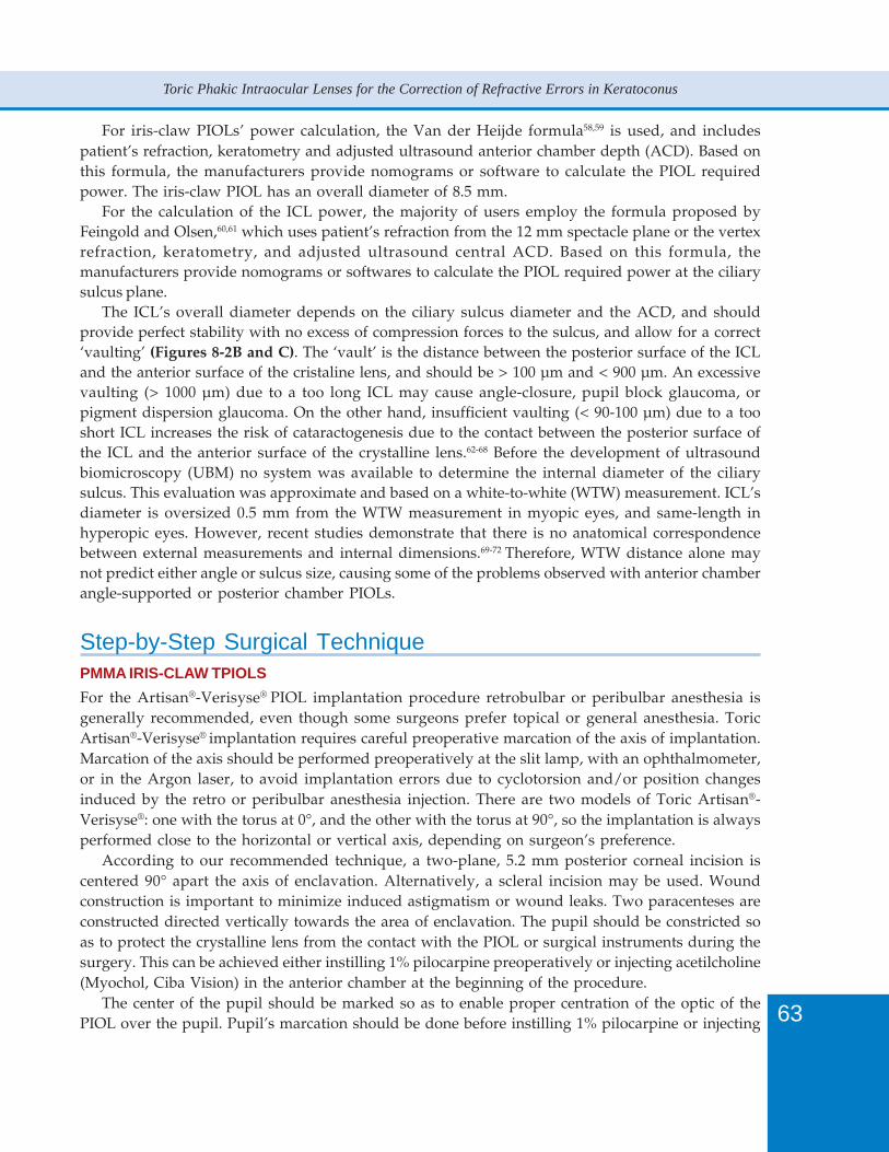
63
Toric Phakic Intraocular Lenses for the Correction of Refractive Errors in Keratoconus
For iris-claw PIOLs’ power calculation, the Van der Heijde formula58,59 is used, and includespatient’s refraction, keratometry and adjusted ultrasound anterior chamber depth (ACD). Based onthis formula, the manufacturers provide nomograms or software to calculate the PIOL requiredpower. The iris-claw PIOL has an overall diameter of 8.5 mm.
For the calculation of the ICL power, the majority of users employ the formula proposed byFeingold and Olsen,60,61 which uses patient’s refraction from the 12 mm spectacle plane or the vertexrefraction, keratometry, and adjusted ultrasound central ACD. Based on this formula, themanufacturers provide nomograms or softwares to calculate the PIOL required power at the ciliarysulcus plane.
The ICL’s overall diameter depends on the ciliary sulcus diameter and the ACD, and shouldprovide perfect stability with no excess of compression forces to the sulcus, and allow for a correct‘vaulting’ (Figures 8-2B and C). The ‘vault’ is the distance between the posterior surface of the ICLand the anterior surface of the cristaline lens, and should be > 100 µm and < 900 µm. An excessivevaulting (> 1000 µm) due to a too long ICL may cause angle-closure, pupil block glaucoma, orpigment dispersion glaucoma. On the other hand, insufficient vaulting (< 90-100 µm) due to a tooshort ICL increases the risk of cataractogenesis due to the contact between the posterior surface ofthe ICL and the anterior surface of the crystalline lens.62-68 Before the development of ultrasoundbiomicroscopy (UBM) no system was available to determine the internal diameter of the ciliarysulcus. This evaluation was approximate and based on a white-to-white (WTW) measurement. ICL’sdiameter is oversized 0.5 mm from the WTW measurement in myopic eyes, and same-length inhyperopic eyes. However, recent studies demonstrate that there is no anatomical correspondencebetween external measurements and internal dimensions.69-72 Therefore, WTW distance alone maynot predict either angle or sulcus size, causing some of the problems observed with anterior chamberangle-supported or posterior chamber PIOLs.
Step-by-Step Surgical TechniquePMMA IRIS-CLAW TPIOLS
For the Artisan®-Verisyse® PIOL implantation procedure retrobulbar or peribulbar anesthesia isgenerally recommended, even though some surgeons prefer topical or general anesthesia. ToricArtisan®-Verisyse® implantation requires careful preoperative marcation of the axis of implantation.Marcation of the axis should be performed preoperatively at the slit lamp, with an ophthalmometer,or in the Argon laser, to avoid implantation errors due to cyclotorsion and/or position changesinduced by the retro or peribulbar anesthesia injection. There are two models of Toric Artisan®-Verisyse®: one with the torus at 0°, and the other with the torus at 90°, so the implantation is alwaysperformed close to the horizontal or vertical axis, depending on surgeon’s preference.
According to our recommended technique, a two-plane, 5.2 mm posterior corneal incision iscentered 90° apart the axis of enclavation. Alternatively, a scleral incision may be used. Woundconstruction is important to minimize induced astigmatism or wound leaks. Two paracenteses areconstructed directed vertically towards the area of enclavation. The pupil should be constricted soas to protect the crystalline lens from the contact with the PIOL or surgical instruments during thesurgery. This can be achieved either instilling 1% pilocarpine preoperatively or injecting acetilcholine(Myochol, Ciba Vision) in the anterior chamber at the beginning of the procedure.
The center of the pupil should be marked so as to enable proper centration of the optic of thePIOL over the pupil. Pupil’s marcation should be done before instilling 1% pilocarpine or injecting
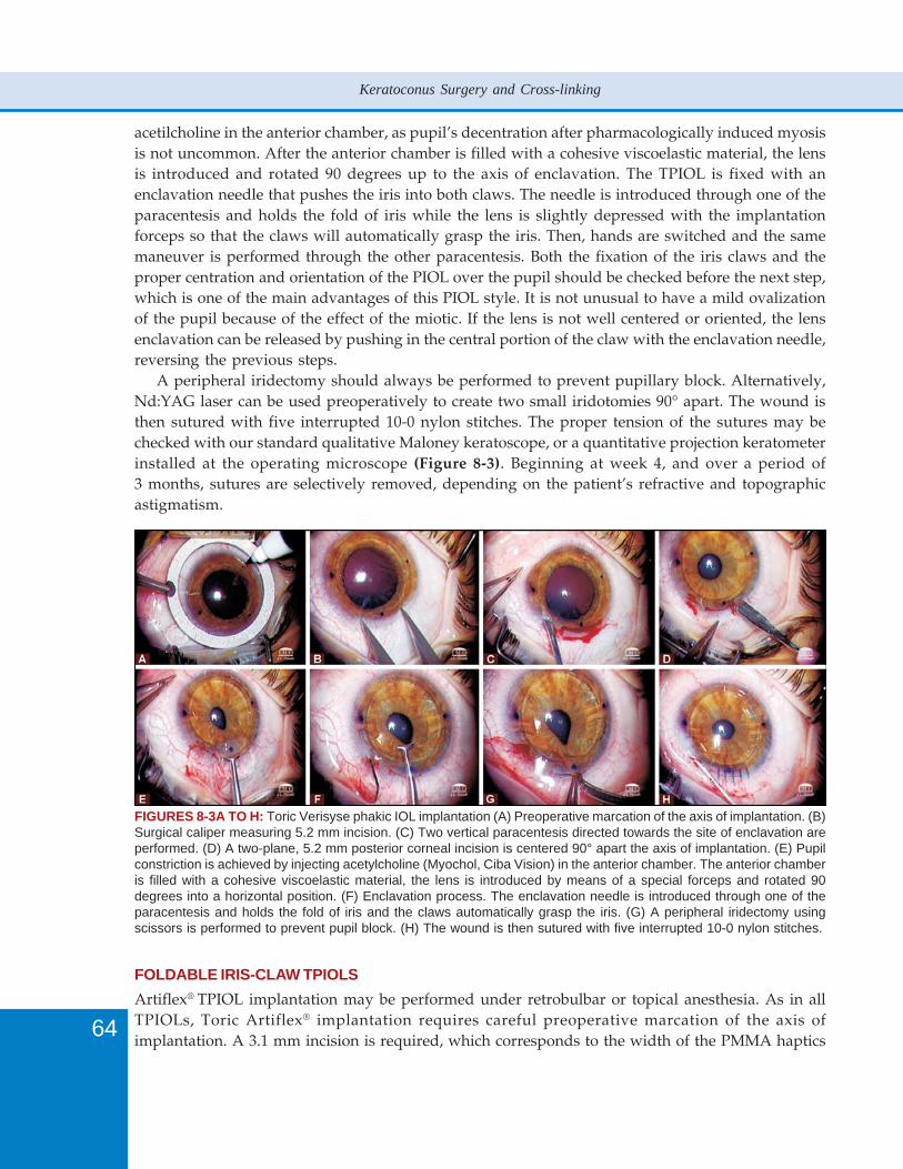
64
Keratoconus Surgery and Cross-linking
acetilcholine in the anterior chamber, as pupil’s decentration after pharmacologically induced myosisis not uncommon. After the anterior chamber is filled with a cohesive viscoelastic material, the lensis introduced and rotated 90 degrees up to the axis of enclavation. The TPIOL is fixed with anenclavation needle that pushes the iris into both claws. The needle is introduced through one of theparacentesis and holds the fold of iris while the lens is slightly depressed with the implantationforceps so that the claws will automatically grasp the iris. Then, hands are switched and the samemaneuver is performed through the other paracentesis. Both the fixation of the iris claws and theproper centration and orientation of the PIOL over the pupil should be checked before the next step,which is one of the main advantages of this PIOL style. It is not unusual to have a mild ovalizationof the pupil because of the effect of the miotic. If the lens is not well centered or oriented, the lensenclavation can be released by pushing in the central portion of the claw with the enclavation needle,reversing the previous steps.
A peripheral iridectomy should always be performed to prevent pupillary block. Alternatively,Nd:YAG laser can be used preoperatively to create two small iridotomies 90° apart. The wound isthen sutured with five interrupted 10-0 nylon stitches. The proper tension of the sutures may bechecked with our standard qualitative Maloney keratoscope, or a quantitative projection keratometerinstalled at the operating microscope (Figure 8-3). Beginning at week 4, and over a period of3 months, sutures are selectively removed, depending on the patient’s refractive and topographicastigmatism.
FIGURES 8-3A TO H: Toric Verisyse phakic IOL implantation (A) Preoperative marcation of the axis of implantation. (B)Surgical caliper measuring 5.2 mm incision. (C) Two vertical paracentesis directed towards the site of enclavation areperformed. (D) A two-plane, 5.2 mm posterior corneal incision is centered 90° apart the axis of implantation. (E) Pupilconstriction is achieved by injecting acetylcholine (Myochol, Ciba Vision) in the anterior chamber. The anterior chamberis filled with a cohesive viscoelastic material, the lens is introduced by means of a special forceps and rotated 90degrees into a horizontal position. (F) Enclavation process. The enclavation needle is introduced through one of theparacentesis and holds the fold of iris and the claws automatically grasp the iris. (G) A peripheral iridectomy usingscissors is performed to prevent pupil block. (H) The wound is then sutured with five interrupted 10-0 nylon stitches.
FOLDABLE IRIS-CLAW TPIOLS
Artiflex® TPIOL implantation may be performed under retrobulbar or topical anesthesia. As in allTPIOLs, Toric Artiflex® implantation requires careful preoperative marcation of the axis ofimplantation. A 3.1 mm incision is required, which corresponds to the width of the PMMA haptics
65
Toric Phakic Intraocular Lenses for the Correction of Refractive Errors in Keratoconus
(3.0 mm). The incision is placed 90° apart the site of enclavation, and two vertical paracenteses aredirected towards the area of enclavation. The Artiflex® lens is inserted using a specially designedspatula, and the process of enclavation is the same as for the PMMA lens. The only difference is thatthe lens is grasped with the implantation forceps by the base of the haptic instead of by the edge ofthe optic. Iridectomy should also be performed. The incision is usually water-tight, but we prefer tosuture it with a 10/0 Nylon suture (Figure 8-4).
IMPLANTABLE COLLAMER LENS
The correct loading of the ICL in the cartridge and the injector is essential for a correct and easyimplantation. Using a modified McPherson forceps, or Aus der Ar coaxial forceps, with long, blunt,curved tips, the lens is carefully grasped and checked under the operating microscope. The ICL hastwo tiny holes on the footplates (distal-right and proximal-left) that allow correct anterior-posteriororientation of the lens. The cartridge is filled with viscoelastic material. The lens is loaded with thedome up, taking special care in correctly position the haptics so as not to rupture them during theinjection. A piece of soft material, the Staar Foam-tip, is positioned to protect the ICL from contactwith the plunger of the shooter. Additionally, some surgeons recommend inserting the tip of a wetsurgical microsponge between the foam-tip and the ICL to further protect the optic and the haptics.
As with the other TPIOLs, Toric ICL implantation requires careful preoperative marcation of theaxis of implantation (Figure 8-5). Broad pharmacological mydriasis is essential for implantation. ForToric ICL’s, a temporal sub-3.00 mm incision is preferred. One side-port incision of about 1 mm andseparated 90° from the main incision is created. Some surgeons prefer two paracentesis to enableeasier implantation of the haptics in the sulcus. The anterior chamber is filled with a cohesive, low-viscosity viscoelastic to protect the endothelium and the crystalline lens from surgical trauma, andto create enough space in the anterior chamber for surgical manipulation. The cartridge is insertedbevel-down, and the lens is then carefully injected. It is essential to control the unfolding of the lens,so as to twist the bevel right or left to assure the correct orientation of the lens. Finally, the hapticsare gently pushed under the iris with a blunt spatula. When the correct centration of the ICL and theposition of the haptics in the ciliary sulcus is checked, acetylcholine is injected in the anterior chamber
FIGURES 8-4A AND B: Enclavation process of the Artiflex/Veriflex phakic IOL. (A) The enclavationneedle is introduced through one of the paracentesis and holds the fold of iris while the lens isgrasped with the implantation forceps by the base of the haptic. (B) Hands are switched and thesame maneuver is repeated to enclavate the other claw. The white line shows the axis of enclavation.
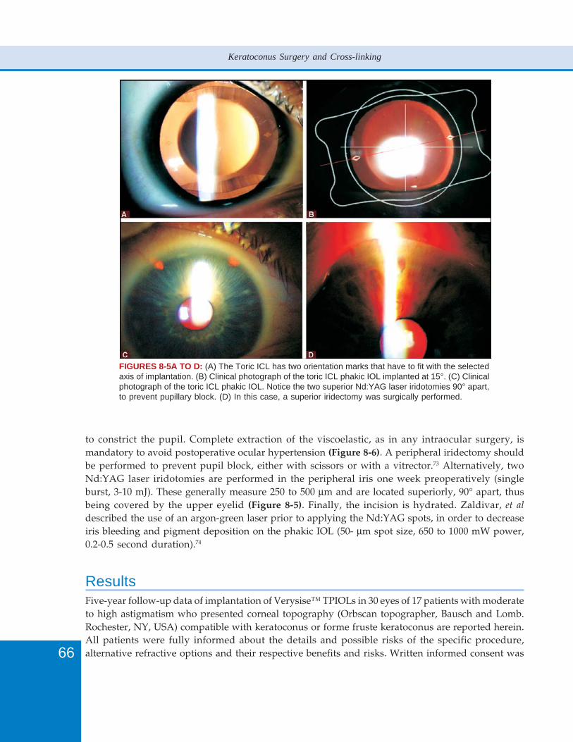
66
Keratoconus Surgery and Cross-linking
FIGURES 8-5A TO D: (A) The Toric ICL has two orientation marks that have to fit with the selectedaxis of implantation. (B) Clinical photograph of the toric ICL phakic IOL implanted at 15°. (C) Clinicalphotograph of the toric ICL phakic IOL. Notice the two superior Nd:YAG laser iridotomies 90° apart,to prevent pupillary block. (D) In this case, a superior iridectomy was surgically performed.
to constrict the pupil. Complete extraction of the viscoelastic, as in any intraocular surgery, ismandatory to avoid postoperative ocular hypertension (Figure 8-6). A peripheral iridectomy shouldbe performed to prevent pupil block, either with scissors or with a vitrector.73 Alternatively, twoNd:YAG laser iridotomies are performed in the peripheral iris one week preoperatively (singleburst, 3-10 mJ). These generally measure 250 to 500 μm and are located superiorly, 90° apart, thusbeing covered by the upper eyelid (Figure 8-5). Finally, the incision is hydrated. Zaldivar, et aldescribed the use of an argon-green laser prior to applying the Nd:YAG spots, in order to decreaseiris bleeding and pigment deposition on the phakic IOL (50- μm spot size, 650 to 1000 mW power,0.2-0.5 second duration).74
ResultsFive-year follow-up data of implantation of Verysise™ TPIOLs in 30 eyes of 17 patients with moderateto high astigmatism who presented corneal topography (Orbscan topographer, Bausch and Lomb.Rochester, NY, USA) compatible with keratoconus or forme fruste keratoconus are reported herein.All patients were fully informed about the details and possible risks of the specific procedure,alternative refractive options and their respective benefits and risks. Written informed consent was
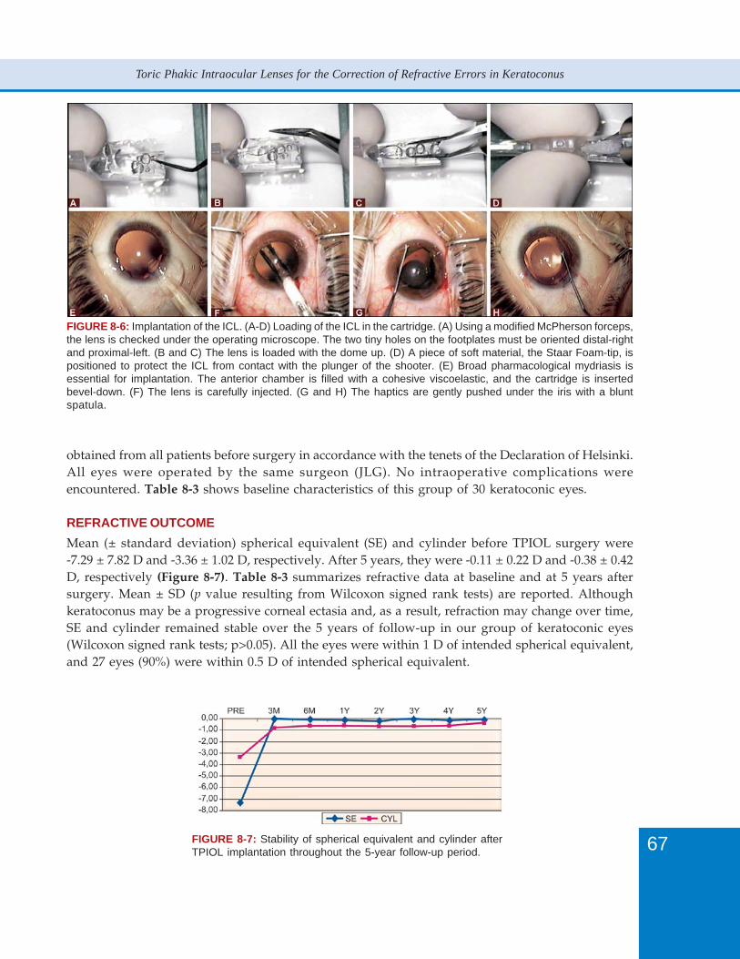
67
Toric Phakic Intraocular Lenses for the Correction of Refractive Errors in Keratoconus
FIGURE 8-6: Implantation of the ICL. (A-D) Loading of the ICL in the cartridge. (A) Using a modified McPherson forceps,the lens is checked under the operating microscope. The two tiny holes on the footplates must be oriented distal-rightand proximal-left. (B and C) The lens is loaded with the dome up. (D) A piece of soft material, the Staar Foam-tip, ispositioned to protect the ICL from contact with the plunger of the shooter. (E) Broad pharmacological mydriasis isessential for implantation. The anterior chamber is filled with a cohesive viscoelastic, and the cartridge is insertedbevel-down. (F) The lens is carefully injected. (G and H) The haptics are gently pushed under the iris with a bluntspatula.
obtained from all patients before surgery in accordance with the tenets of the Declaration of Helsinki.All eyes were operated by the same surgeon (JLG). No intraoperative complications wereencountered. Table 8-3 shows baseline characteristics of this group of 30 keratoconic eyes.
REFRACTIVE OUTCOME
Mean (± standard deviation) spherical equivalent (SE) and cylinder before TPIOL surgery were-7.29 ± 7.82 D and -3.36 ± 1.02 D, respectively. After 5 years, they were -0.11 ± 0.22 D and -0.38 ± 0.42D, respectively (Figure 8-7). Table 8-3 summarizes refractive data at baseline and at 5 years aftersurgery. Mean ± SD (p value resulting from Wilcoxon signed rank tests) are reported. Althoughkeratoconus may be a progressive corneal ectasia and, as a result, refraction may change over time,SE and cylinder remained stable over the 5 years of follow-up in our group of keratoconic eyes(Wilcoxon signed rank tests; p>0.05). All the eyes were within 1 D of intended spherical equivalent,and 27 eyes (90%) were within 0.5 D of intended spherical equivalent.
FIGURE 8-7: Stability of spherical equivalent and cylinder afterTPIOL implantation throughout the 5-year follow-up period.
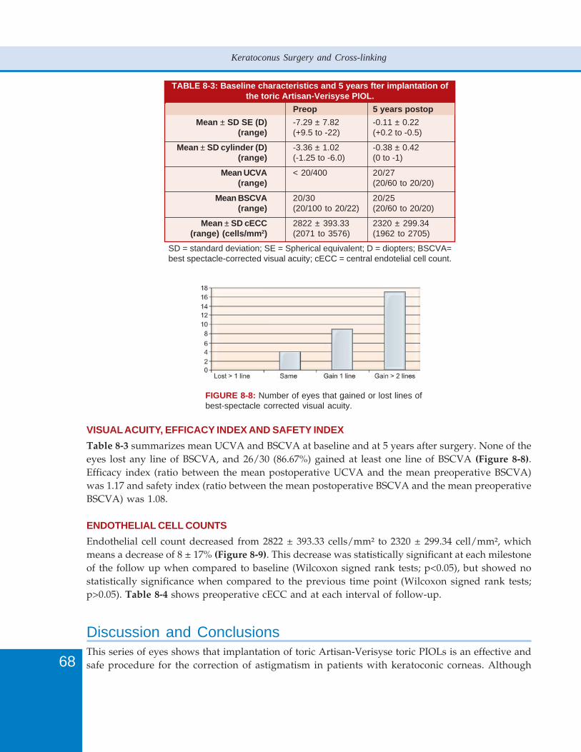
68
Keratoconus Surgery and Cross-linking
TABLE 8-3: Baseline characteristics and 5 years fter implantation ofthe toric Artisan-Verisyse PIOL.
Preop 5 years postop
Mean ± SD SE (D) -7.29 ± 7.82 -0.11 ± 0.22(range) (+9.5 to -22) (+0.2 to -0.5)
Mean ± SD cylinder (D) -3.36 ± 1.02 -0.38 ± 0.42(range) (-1.25 to -6.0) (0 to -1)
Mean UCVA < 20/400 20/27(range) (20/60 to 20/20)
Mean BSCVA 20/30 20/25(range) (20/100 to 20/22) (20/60 to 20/20)
Mean ± SD cECC 2822 ± 393.33 2320 ± 299.34(range) (cells/mm2) (2071 to 3576) (1962 to 2705)
SD = standard deviation; SE = Spherical equivalent; D = diopters; BSCVA=best spectacle-corrected visual acuity; cECC = central endotelial cell count.
FIGURE 8-8: Number of eyes that gained or lost lines ofbest-spectacle corrected visual acuity.
VISUAL ACUITY, EFFICACY INDEX AND SAFETY INDEX
Table 8-3 summarizes mean UCVA and BSCVA at baseline and at 5 years after surgery. None of theeyes lost any line of BSCVA, and 26/30 (86.67%) gained at least one line of BSCVA (Figure 8-8).Efficacy index (ratio between the mean postoperative UCVA and the mean preoperative BSCVA)was 1.17 and safety index (ratio between the mean postoperative BSCVA and the mean preoperativeBSCVA) was 1.08.
ENDOTHELIAL CELL COUNTS
Endothelial cell count decreased from 2822 ± 393.33 cells/mm² to 2320 ± 299.34 cell/mm², whichmeans a decrease of 8 ± 17% (Figure 8-9). This decrease was statistically significant at each milestoneof the follow up when compared to baseline (Wilcoxon signed rank tests; p<0.05), but showed nostatistically significance when compared to the previous time point (Wilcoxon signed rank tests;p>0.05). Table 8-4 shows preoperative cECC and at each interval of follow-up.
Discussion and ConclusionsThis series of eyes shows that implantation of toric Artisan-Verisyse toric PIOLs is an effective andsafe procedure for the correction of astigmatism in patients with keratoconic corneas. Although
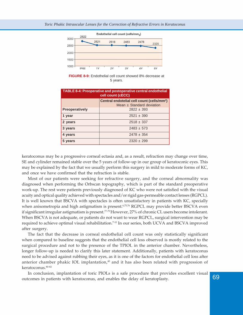
69
Toric Phakic Intraocular Lenses for the Correction of Refractive Errors in Keratoconus
TABLE 8-4: Preoperative and postoperative central endothelialcell count (cECC)
Central endotelial cell count (cells/mm2)Mean ± Standard deviation
Preoperatively 2822 ± 393
1 year 2521 ± 390
2 years 2518 ± 337
3 years 2483 ± 573
4 years 2478 ± 354
5 years 2320 ± 299
FIGURE 8-9: Endothelial cell count showed 8% decrease at5 years.
keratoconus may be a progressive corneal ectasia and, as a result, refraction may change over time,SE and cylinder remained stable over the 5 years of follow-up in our group of keratoconic eyes. Thismay be explained by the fact that we usually perform this surgery in mild to moderate forms of KC,and once we have confirmed that the refraction is stable.
Most of our patients were seeking for refractive surgery, and the corneal abnormality wasdiagnosed when performing the Orbscan topography, which is part of the standard preoperativework-up. The rest were patients previously diagnosed of KC who were not satisfied with the visualacuity and optical quality achieved with spectacles and/or rigid gas-permeable contact lenses (RGPCL).It is well known that BSCVA with spectacles is often unsatisfactory in patients with KC, speciallywhen anisometropia and high astigmatism is present.6,75,76 RGPCL may provide better BSCVA evenif significant irregular astigmatism is present.77-79 However, 27% of chronic CL users become intolerant.When BSCVA is not adequate, or patients do not want to wear RGPCL, surgical intervention may berequired to achieve optimal visual rehabilitation.7-10 In our series, both UCVA and BSCVA improvedafter surgery.
The fact that the decrease in corneal endothelial cell count was only statistically significantwhen compared to baseline suggests that the endothelial cell loss observed is mostly related to thesurgical procedure and not to the presence of the TPIOL in the anterior chamber. Nevertheless,longer follow-up is needed to clarify this later statement. Additionally, patients with keratoconusneed to be advised against rubbing their eyes, as it is one of the factors for endothelial cell loss afteranterior chamber phakic IOL implantation,40 and it has also been related with progression ofkeratoconus.80-82
In conclusion, implantation of toric PIOLs is a safe procedure that provides excellent visualoutcomes in patients with keratoconus, and enables the delay of keratoplasty.
70
Keratoconus Surgery and Cross-linking
References1. Rabinowitz YS. Keratoconus. Surv Ophthalmol 1998;42:297-319.2. Riddle HK, Parker DA, Price FW. Management of postkeratoplasty astigmatism. Curr Opin Ophthalmol 1998;9:15-
28.3. Chastang PJ, Borderie VM, Carvajal-Gonzalez S, et al. Prediction of spectacle-corrected visual acuity using
videokeratography. J Refract Surg 1999;15:572-9.4. Speaker MG, Cohen EJ, Edelhauser HF, et al. Effect of gas-permeable contact lenses on the endothelium of corneal
transplants. Arch Ophthalmol 1991;109:1703-6.5. Busin M, Arffa RC, Zambiachi L, et al. Effect of hinged lamellar keratotomy on postkeratoplasty eyes. Ophthalmology
2001;108:1845-51; discussion 1851-2.6. Vajpayee RB, Sharma N, Sinha R, et al. Laser in-situ keratomileusis after penetrating keratoplasty. Surv Ophthalmol
2003;48:503-14.7. Lim N, Vogt U. Characteristics and functional outcomes of 130 patients with keratoconus attending specialist
contact lens clinic. Eye 2002;16:54-9.8. Geerards AJ, Vreugdebhil W, Khazen A. Incidence of rigid gas-permeable contact lens wear after keratoplasty for
keratoconus. Eye Contact Lens 2006;32:207-10.9. Espana EM, Tseng SC. Analysis of contact lens intolerance by exploring neuroanatomic integration of ocular
surface defense. Cont Lens Anterior Eye 2003;26:131-7.10. Glasson MJ, Stapleton F, Keay L, et al. Invest Ophthalmol Vis Sci 2003;44:5116-24.11. Thompson RW Jr, Price MO, Bowers PJ, Price FW. Long-term graft survival after penetrating keratoplasty.
Ophthalmology 2003;110:1396-1402.12. Lim L, Pesudovs K, Coster DJ. Penetrating keratoplasty for keratoconus: visual outcome and success. Ophthalmology
2000;107:1125-31.13. Brooks SF, Johnson D, Fischer N. Anisometropia and binocularity. Ophthalmology 1996;103(7):1139-43.14. Perlman EM. An analysis and interpretation of refractive errors after penetrating keratoplasty. Ophthalmology
1981;88:39-45.15. Perl T, Charlton KH, Binder PS. Disparate diameter grafting. Astigmatism, intraocular pressure, and visual acuity.
Ophthalmology 1981;88:774-81.16. Kruse FE, Cursiefen C. Surgery of the cornea: corneal, limbal stem cell and amniotic membrane transplantation.
Dev Ophthalmol 2008;41:159-70.17. Frost NA, Wu J, Lai TF, Coster DJ. A review of randomized controlled trials of penetrating keratoplasty techniques.
Ophthalmology 2006;113(6):942-9.18. Chang DH, Hardten DR. Refractive surgery after corneal transplantation. Curr Opin Ophthalmol 2005;16(4):251-
5.19. Taban M, Behrens A, Newcomb RL, et al. Incidence of acute endophthalmitis following penetrating keratoplasty:
a systematic review. Arch Ophthalmol 2005;123(5):605-9.20. Patel HY, Omonde S, Brookes NH, et al. The indications and outcome of paediatric corneal transplantation in New
Zealand: 1991-2003. Br J Ophthalmol 2005;89(4):404-8.21. Panda A, Vanathi M, Kumar A, et al. Corneal graft rejection. Surv Ophthalmol 2007;52(4):375-96.22. Randleman JB, Woodward M, Lynn MJ, Stulting RD. Risk assessment for ectasia after corneal refractive surgery.
Ophthalmology 2008;115:37-50.23. Randleman JB, Russell B, Ward MA, et al. Risk factors and prognosis for corneal ectasia after LASIK. Ophthalmology
2003;110:267-75.24. Randleman JB, Trattler WB, Stulting RD. Validation of the Ectasia Risk Score System for preoperative laser in situ
keratomileusis screening. Am J Ophthalmol 2008;145:813-8.25. Binder PS. Analysis of ectasia after laser in situ keratomileusis : risk factors. J Cataract Refract Surg 2007;33:1530-8.26. Colin J. European clinical evaluation: use of Intacs for the treatment of keratoconus. J Cataract Refract Surg
2006;32:747-55.27. Colin J, Cochener B, Savary G, Malet F. Correcting keratoconus with intracorneal rings. J Cataract Refract Surg
2000;26:1117-22.28. Colin J, Cochener B, Savary G, et al. INTACS inserts for treating keratoconus; one-year results. Ophthalmology
2001;108:1409-14.29. Siganos D, Ferrara P, Chatzinikolas K, et al. Ferrara intrastromal corneal rings for the correction of keratoconus.
J Cataract Refract Surg 2002;28:1947-51.30. Holmes-Higgin DK, Burris TE. Corneal surface topography and associated visual performance with Intacs for
myopia: phase III clinical trials results. The Intacs Study Group. Ophthalmology 2000;107:2061-71.

71
Toric Phakic Intraocular Lenses for the Correction of Refractive Errors in Keratoconus
31. Schanzlin DJ, Asbell PA, Burris TE, Durrie DS. The intrastromal corneal ring segments; phase II results for thecorrection of myopia. Ophthalmology 1997;104:1067-78.
32. Pokroy R, Levinger S. Intacs adjustment surgery for keratoconus. J Cataract Refract Surg 2006;32:986-92.33. Ucakhan OO, Kanpolat A, Ozdemir O. Contact lens fitting for keratoconus after Intacs placement. Eye contact lens
2006;32:75-7.34. Kamburoglu G, Etan A, Bahadir M. Implantation of Artisan toric phakic intraocular lens following Intacs in a
patient with keratoconus. J Cataract Refract Surg 2007;33:528-30.35. Colin J, Velou S. Implantation of Intacs and a refractive intraocular lens to correct keratoconus. J Cataract Refract
Surg 2003;29:832-4.36. Coskunseven E, Onder M, Kymionis GD, et al. Combined Intacs and posterior chamber toric implantable collamer
lens implantation for keratoconic patients with extreme myopia. Am J Ophthalmol 2007;144:387-9.37. Moshirfar M, Gregoire FJ, Mirzaian G, et al. Use of Verisyse iris-supported phakic intraocular lens for myopia in
keratoconic patients. J Cataract Refract Surg 2006;32:1227-32.38. Leccisotti A, Fields SV. Angle-supported phakic intraocular lenses in eyes with keratoconus and myopia. J Cataract
Refract Surg 2003;29:1530-6.39. Budo C, Bartels MC, van Rij G. Implantation of Artisan toric phakic intraocular lenses for the correction of
astigmatism and spherical errors in patients with keratoconus. J Refract Surg 2005;21:218-22.40. Güell JL, Morral M, Gris O, et al. Five-year follow-up of 399 phakic Artisan®-Verisyse® implantation for myopia,
hyperopia and/or astigmatism. Ophthalmology 2008;115(6):1002-12.41. Budo C, Hessloehl JC, Izak M, et al. Multicenter study of the Artisan phakic intraocular lens. J Cataract Refract Surg
2000;26(8):1163-71.42. Stulting RD, John ME, Maloney RK. US Verisyse Study Group. Three-Year results of Artisan/Verisyse phakic
intraocular lens implantation results of the United States Food and Drug Administration Clinical Trial.Ophthalmology 2008;115(3):464-72.
43. Pop M, Payette Y. Initial results of endothelial cell counts after Artisan lens for phakic eyes: an evaluation of theUnited States Food and Drug Administration Ophtec Study. Ophthalmology 2004;111(2):309-17.
44. Maloney RK, Nguyen LH, John ME. Artisan phakic intraocular lens for myopia: short-term results of a prospective,multicenter study. Ophthalmology 2002;109(9):1631-41.
45. Gimbel HV, Ziemba SL. Management of myopic astigmatism with phakic intraocular lens implantation. J CataractRefract Surg 2002;28:883-6.
46. Uusitalo RJ, Aine E, Sen NH, et al. Implantable contact lens for high myopia. J Cataract Refract Surg 2002;28:29-36.47. Pesando PM, Ghiringhello MP, Di Meglio G, Fanton G. Posterior chamber phakic intraocular lens (ICL) for hyperopia:
ten-year follow-up. J Cataract Refract Surg. 2007;33(9):1579-84.48. Sanders DR, Doney K, Poco M. United States Food and Drug Administration clinical trial of the Implantable
Collamer Lens (ICL) for moderate to high myopia: three-year follow-up. Ophthalmology 2004;111:1683-92.49. Zaldivar R, Davidorf JM, Oscherow S. Posterior chamber phakic intraocular lens for myopia of -8 to -19 diopters.
J Refract Surg 1998;14:294-305.50. Davidorf JM, Zaldivar R, Oscherow S. Posterior chamber phakic intraocular lens for hyperopia of +4 to +11
diopters. J Refract Surg 1998;14:306-11.51. Tuft SJ, Moodaley LC, Gregory WM, et al. Prognostic factors for the progression of keratoconus. Ophthalmology
1994;101:439-47.52. Wollensak G. Crosslinking treatment of progressive keratoconus: new hope. Curr Opin Ophthalmol 2006;17:356-
60.53. Wollensak G, Spoerl E, Seiler T. Riboflavin/ultraviolet-a-induced collagen crosslinking for the treatment of
keratoconus. Am J Ophthalmol 2003;135:620-7.54. Caporossi A, Baiocchi S, Mazzotta C, et al. Parasurgical therapy for keratoconus by riboflavin-ultraviolet type A
rays induced cross-linking of corneal collagen: preliminary refractive results in an Italian study. J Cataract RefractSurg 2006;32:837-45.
55. Tan DT, Por YM. Current treatment options for corneal ectasia. Curr Opin Ophthalmol 2007;18:284-9.56. Kanellopoulos AJ, Binder PS. Collagen cross-linking (CCL) with sequential topography-guided PRK: a temporizing
alternative for keratoconus to penetrating keratoplasty. Cornea 2007;26:891-5.57. Davis LJ, Schechtman KB, Begley CG, et al. Repeatability of refraction and corrected visual acuity in keratoconus.
The CLEK Study Group. Collaborative Longitudinal Evaluation of Keratoconus. Optom Vis Sci 1998;75:887-96.58. Van der Heijde GL. Some optical aspects of implantation of an intraocular lens in a myopia eye. Eur J Implant
Refract Surg 1989;1:245-8.59. Fechner PU, Van der Heijde GL, Worst JGF. The correction of myopia by lens implantation into phakic eyes. Am
J Ophthalmol 1989;107:659-63.
72
Keratoconus Surgery and Cross-linking
60. Olsen T, Corydon L, Gimbel H. Intraocular lens power calculation with an improved anterior chamber depthprediction algorithm. J Cataract Refract Surg 1995;21:313-9.
61. Olsen T, Thim K, Corydon L. Accuracy of the newer generation intraocular lens power calculation formulas in longand short eyes. J Cataract Refract Surg 1995;17:187-93.
62. Lovisolo CF, Reinstein DZ. Phakic intraocular lenses. Surv Ophthalmol 2005;50(6):549-87.63. Zaldivar R, Ricur G, Oscherow S. The phakic intraocular lens implant: in-depth focus on posterior chamber phakic
IOLs. Curr Opin Ophthalmol 2000;11:22-34.64. Smallman DS, Probst L, Rafuse PE. Pupil block glaucoma secondary to posterior chamber phakic intraocular lens
implantation for high myopia. J Cataract Refract Surg 2004;30:905-7.65. Sanders DR, Vukich JA. Incidence of lens opacities and clinically significant cataracts with the implantable contact
lens: Comparison of two lens designs. J Refract Surg 2002;18:673-82.66. Gonvers M, Bornet C, Othenin-Girard P. Implantable contact lens for moderate to high myopia: relationship of
vaulting to cataract formation. J Cataract Refract Surg 2003;29:918-24.67. Kohnen T, Kasper T, Buhren J, Fechner PU. Ten-year follow-up of a ciliary sulcus-fixated silicone phakic posterior
chamber intraocular lens. J Cataract Refract Surg 2004;30: 2431-4.68. Sanders DR, Vukich JA, Doney K, Gaston M. US Food and Drug Administration clinical trial of the Implantable
Contact Lens for moderate to high myopia. Ophthalmology 2003;110:255-66.69. Apple DJ, Werner L. Complications of cataract and refractive surgery: a clinicopathological documentation. Trans
Am Ophthalmol Soc 2001;99:95-107; discussion 107-9.70. Werner L, Apple DJ, Pandey SK, et al. Phakic posterior chamber intraocular lenses. Int Ophthalmol Clin 2001;41(3):
153-74.71. Werner L, Izak AM, Pandey SK, Apple DJ, Trivedi RH, Schmidbauer JM. Correlation between different
measurements within the eye relative to phakic intraocular lens implantation. J Cataract Refract Surg 2004;30:1982-8.
72. Pop M, Payette Y, Mansour M. Predicting sulcus size using ocular measurements. J Cataract Refrat Surg 2001;27:1033-8.
73. Davidorf JM, Zaldivar R, Oscherow S. Posterior chamber phakic intraocular lens for hyperopia of +4 to +11diopters. J Refract Surg 1998;14:306-11.
74. Zaldivar R, Davidorf JM, Oscherow S. The intraocular contact lens. In: Buratto L, Brint SF (Eds). LASIK. Principlesand Techniques. Thorofare, NJ: Slack 1997;401-13.
75. Pesudovs K, Garamendi E, Elliott DB. A quality of life comparison of people wearing spectacles or contact lenses orhaving undergone refractive surgery. J Refract Surg 2006;22(1):19-27.
76. Wietharn BE, Driebe WT Jr. Fitting contact lenses for visual rehabilitation after penetrating keratoplasty. EyeContact Lens 2004;30(1):31-3.
77. Riddle HK, Parker DA, Price FW. Management of postkeratoplasty astigmatism. Curr Opin Ophthalmol 1998;9:15-28.
78. Chastang PJ, Borderie VM, Carvajal-Gonzalez S, et al. Prediction of spectacle-corrected visual acuity usingvideokeratography. J Refract Surg 1999;15:572-9.
79. Speaker MG, Cohen EJ, Edelhauser HF, et al. Effect of gas-permeable contact lenses on the endothelium of cornealtransplants. Arch Ophthalmol 1991;109:1703-6.
80. McMonnies CW. Abnormal rubbing and keratectasia. Eye Contact Lens 2007;33(6 Pt 1):265-71.81. McMonnies CW. The biomechanics of keratoconus and rigid contact lenses. Eye Contact Lens 2005;31(2):80-92.82. Krachmer JH, Feder RS, Belin MW. Keratoconus and related noninflammatory corneal thinning disorders. Surv
Ophthalmol 1984;28(4):293-322.
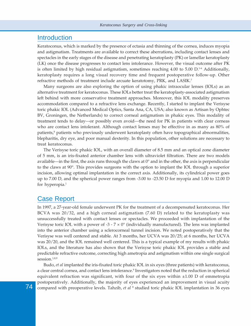
74
Keratoconus Surgery and Cross-linking
IntroductionKeratoconus, which is marked by the presence of ectasia and thinning of the cornea, induces myopiaand astigmatism. Treatments are available to correct these aberrations, including contact lenses andspectacles in the early stages of the disease and penetrating keratoplasty (PK) or lamellar keratoplasty(LK) once the disease progresses to contact lens intolerance. However, the visual outcome after PKis often limited by high residual astigmatism, sometimes reaching 4.00 to 5.00 D.1-6 Additionally,keratoplasty requires a long visual recovery time and frequent postoperative follow-up. Otherrefractive methods of treatment include arcuate keratotomy, PRK, and LASIK.7
Many surgeons are also exploring the option of using phakic intraocular lenses (IOLs) as analternative treatment for keratoconus. These IOLs better treat the keratoplasty-associated astigmatismleft behind with more conservative treatment approaches. Moreover, this IOL modality preservesaccommodation compared to a refractive lens exchange. Recently, I started to implant the Verisysetoric phakic IOL (Advanced Medical Optics, Santa Ana, CA, USA; also known as Artisan by OphtecBV, Groningen, the Netherlands) to correct corneal astigmatism in phakic eyes. This modality oftreatment tends to delay—or possibly even avoid—the need for PK in patients with clear corneaswho are contact lens intolerant. Although contact lenses may be effective in as many as 80% ofpatients,8 patients who previously underwent keratoplasty often have topographical abnormalities,blepharitis, dry eye, and poor manual dexterity. In this population, other solutions are necessary totreat keratoconus.
The Verisyse toric phakic IOL, with an overall diameter of 8.5 mm and an optical zone diameterof 5 mm, is an iris-fixated anterior chamber lens with ultraviolet filtration. There are two modelsavailable—in the first, the axis runs through the claws at 0° and in the other, the axis is perpendicularto the claws at 90°. This provides surgeons with the option to implant the IOL through a superiorincision, allowing optimal implantation in the correct axis. Additionally, its cylindrical power goesup to 7.00 D, and the spherical power ranges from -3.00 to -23.50 D for myopia and 1.00 to 12.00 Dfor hyperopia.1
Case ReportIn 1997, a 27-year-old female underwent PK for the treatment of a decompensated keratoconus. HerBCVA was 20/32, and a high corneal astigmatism (7.60 D) related to the keratoplasty wasunsuccessfully treated with contact lenses or spectacles. We proceeded with implantation of theVerisyse toric IOL with a power of -3 - 7 × 0° (individually manufactured). The lens was implantedinto the anterior chamber using a sclerocorneal tunnel incision. We noted postoperatively that theVerisyse was well centered and stable. At 3 months, her UCVA was 20/25; at 6 months, her UCVAwas 20/20, and the IOL remained well centered. This is a typical example of my results with phakicIOLs, and the literature has also shown that the Verisyse toric phakic IOL provides a stable andpredictable refractive outcome, correcting high ametropia and astigmatism within one single surgicalsession.1,8,9
Budo, et al implanted the iris-fixated toric phakic IOL in six eyes (three patients) with keratoconus,a clear central cornea, and contact lens intolerance.9 Investigators noted that the reduction in sphericalequivalent refraction was significant, with four of the six eyes within ±1.00 D of emmetropiapostoperatively. Additionally, the majority of eyes experienced an improvement in visual acuitycompared with preoperative levels. Tahzib, et al 8 studied toric phakic IOL implantation in 36 eyes
75
Our Experience with Toric Phakic IOLs in Keratoconus
(35 patients) with contact lens intolerance. In their study, despite two patients with irreversiblegraft rejections and one with gradual endothelial decompensation, the vast majority of patientsexperienced a reduction of refractive astigmatism (88.8% ± 29.5%) and ametropia (77.8% ± 19.3%).On a scale of zero to 10, patient satisfaction increased from 3.6 preoperatively to 8.0 postoperatively.
Although we do not expect patients’ refraction to remain stable after phakic IOL implantation,the need for PK is delayed. Additionally, the use of a phakic IOL, including a toric model, does nothinder the results of the eventual need for PK or LK for those patients with keratoconus and contactlens intolerance.10-12
LimitationsThe use of a toric phakic IOL for the treatment of keratoconus does have its limitations. For instance,if a patient has high myopia, use of a toric phakic IOL may induce halos and glare when the patientis confronted with dim-light conditions.1 Additionally, Budo, et al9 concluded that although thepredictability for spherical errors is high, the predictability for astigmatic errors is only moderate.Endothelial cell count is also a concern when implanting a toric phakic IOL. We choose not to implantthis lens in patients whose endothelial cell count is below 2000 mm2.
Another drawback to use the toric phakic IOL is incision size. The IOL only fits through anincision of at least 5.5 mm, which may induce astigmatism. My colleagues and I were able to showthat the surgically induced astigmatism after toric phakic IOL implantation in nondiseased eyes was0.53 D.9
I would suggest only using this method of treatment in an eye with stable keratometry andsubjective refraction. If the patient does not meet these requirements, such as patients with recentlydiagnosed keratoconus or those with progressive keratoconus, consider other options for thetreatment of keratoconus, including LASIK,13 intracorneal rings and segments,14 epikeratoplasty,15
and lamellar keratoplasty.16 Be aware, however, that keratoconus is a contraindication of LASIKand, therefore, its use for this purpose is limited. Furthermore, although intracorneal rings or segmentsreduce astigmatism, the marked reduction is not as great as what toric phakic IOLs offer.1
ConclusionIn some cases, patients may benefit from the use of spectacles or contact lenses to correct astigmatism;however, when patients become contact lens intolerant, I frequently use the iris-fixated toric phakicIOL (Verisyse) as an alternative treatment. In my experience, this IOL reduces the patient’s refractiveerror and astigmatism to a more manageable level and provides patients with a speedy visualrehabilitation.
In conclusion, I believe that toric phakic Verisyse implantation is the natural first step to thetreatment of keratoconus and should be done before performing PK, especially in patients withassociated myopia. The risk of endothelial cell loss should not be taken lightly, however, and long-term follow-up is necessary for patients.
References1. Budo C, Bartels MC, van Rij G. Implantation of Artisan toric phakic intraocular lenses for the correction of
astigmatism and spherical errors in patients with keratoconus. J Refract Surg 2005;21:218-22.
76
Keratoconus Surgery and Cross-linking
2. Lim L, Pesudovs K, Coster DJ. Penetrating keratoplasty for keratoconus: visual outcome and success. Ophthalmology2000;107:1125-31.
3. Webber SK, Lawless MA, Sutton GL, Rogers CM. LASIK for post penetrating keratoplasty astigmatism andmyopia. Br J Ophthalmol. 1999;83:1013-8.
4. Troutman RC, Lawless MA. Penetrating keratoplasty for keratoconus. Cornea 1987;6:298-305.5. Binder PS. The effect of suture removal on postkeratoplasty astigmatism. Am J Ophthalmol 1988;106:507.6. Seitz B, Langenbucher A, Jus MM, et al. Nonmechanical corneal trephination with the excimer laser improves
outcome after penetrating keratoplasty. Ophthalmology 1999;106:1156-64.7. Tehrani M, Dick HB. Implantation of an Artisan toric phakic intraocular lens to correct high astigmatism after
penetrating keratoplasty. Klin Monatsbl Augenheilkd 2002;219:159-63.8. Tahzib NG, Cheng YYY, Nuijts RMMA. Three-year follow-up analysis of Artisan toric lens implantation for
correction of postkeratoplasty ametropia in phakic and pseudophakic eyes. Ophthalmology 2006;113:976-84.9. Dick HB, Alio J, Bianchetti M, Budo C, Christiaans BJ, El-Danasoury MA, Güell JL, Krumeich J, Landesz M, Loureiro
F, Luyten GP, Marinho A, Rahhal SM, Schwenn O, Spirig R, Thomann U, Venter J. Toric phakic intraocular lens:European multicenter study. Ophthalmology 2003;110:150-62.
10. Brierly SC, Izquierdo L Jr, Mannis MJ. Penetrating keratoplasty for keratoconus. Cornea 2000;19:329-32.11. Kirkness CM, Ficker LA, Steele AD, Rice NS. The success of penetrating keratoplasty for I-III stage-specific surgical
treatment of keratoconus. Klin Monatsbl Augenheilkd 1997;211:94-100.12. Buzard KA, Tuengler A, Febbraro JL. Treatment of mild to moderate keratoconus with laser in situ keratomileusis.
J Cataract Refract Surg 1999;25:1600-09.13. Colin J, Cochener B, Savary G, Malet F, Holmes-Higgin D. Intacs inserts for treating keratoconus: one-year results.
Ophthalmology 2001;108:1409-14.14. Siganos D, Ferrara P, Chatzinikolas K, Bessis N, Papastergiou G. Ferrara intrastromal corneal rings for the correction
of keratoconus. J Cataract Refract Surg 2002;28:1947-51.15. Wagoner MD, Smith SD, Rademaker WJ, Mahmood MA. Penetrating keratoplasty vs epikeratoplasty for the
surgical treatment of keratoconus. J Refract Surg 2001;17:138-46.16. Richard JM, Paton D, Gasset AR. A comparison of penetrating keratoplasty and lamellar keratoplasty in the
surgical management of keratoconus. Am J Ophthalmol 1978;86:807-11.
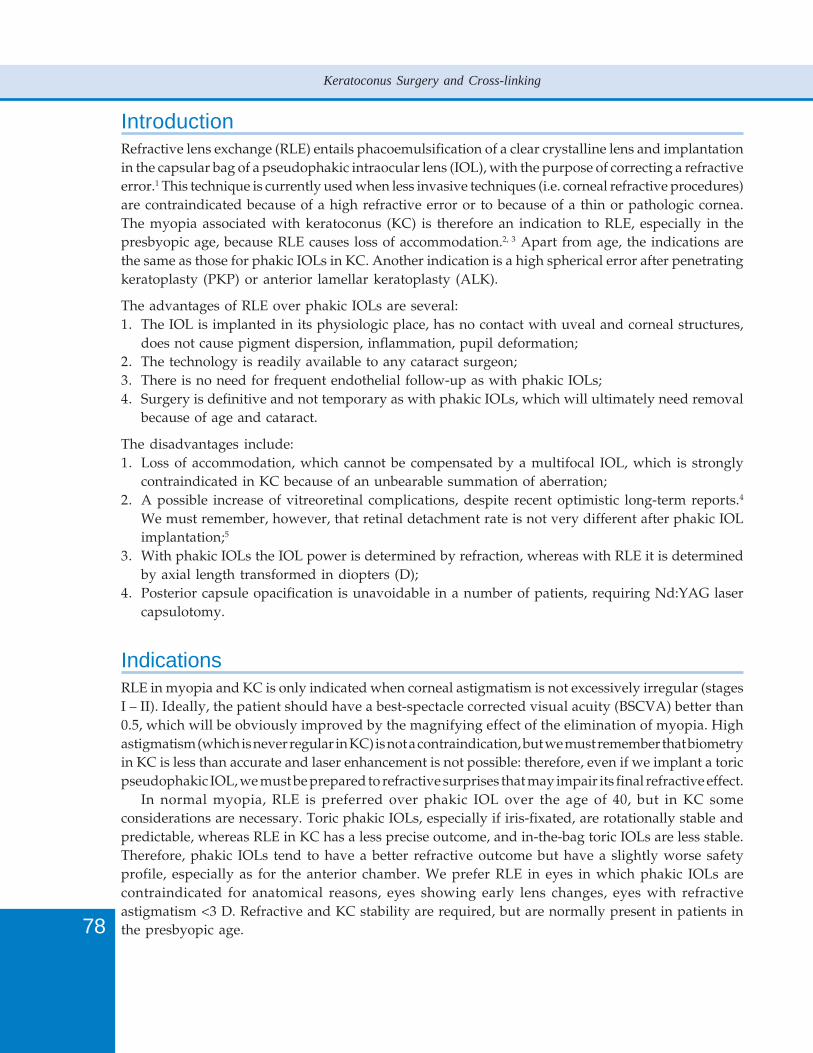
78
Keratoconus Surgery and Cross-linking
IntroductionRefractive lens exchange (RLE) entails phacoemulsification of a clear crystalline lens and implantationin the capsular bag of a pseudophakic intraocular lens (IOL), with the purpose of correcting a refractiveerror.1 This technique is currently used when less invasive techniques (i.e. corneal refractive procedures)are contraindicated because of a high refractive error or to because of a thin or pathologic cornea.The myopia associated with keratoconus (KC) is therefore an indication to RLE, especially in thepresbyopic age, because RLE causes loss of accommodation.2, 3 Apart from age, the indications arethe same as those for phakic IOLs in KC. Another indication is a high spherical error after penetratingkeratoplasty (PKP) or anterior lamellar keratoplasty (ALK).
The advantages of RLE over phakic IOLs are several:1. The IOL is implanted in its physiologic place, has no contact with uveal and corneal structures,
does not cause pigment dispersion, inflammation, pupil deformation;2. The technology is readily available to any cataract surgeon;3. There is no need for frequent endothelial follow-up as with phakic IOLs;4. Surgery is definitive and not temporary as with phakic IOLs, which will ultimately need removal
because of age and cataract.
The disadvantages include:1. Loss of accommodation, which cannot be compensated by a multifocal IOL, which is strongly
contraindicated in KC because of an unbearable summation of aberration;2. A possible increase of vitreoretinal complications, despite recent optimistic long-term reports.4
We must remember, however, that retinal detachment rate is not very different after phakic IOLimplantation;5
3. With phakic IOLs the IOL power is determined by refraction, whereas with RLE it is determinedby axial length transformed in diopters (D);
4. Posterior capsule opacification is unavoidable in a number of patients, requiring Nd:YAG lasercapsulotomy.
IndicationsRLE in myopia and KC is only indicated when corneal astigmatism is not excessively irregular (stagesI – II). Ideally, the patient should have a best-spectacle corrected visual acuity (BSCVA) better than0.5, which will be obviously improved by the magnifying effect of the elimination of myopia. Highastigmatism (which is never regular in KC) is not a contraindication, but we must remember that biometryin KC is less than accurate and laser enhancement is not possible: therefore, even if we implant a toricpseudophakic IOL, we must be prepared to refractive surprises that may impair its final refractive effect.
In normal myopia, RLE is preferred over phakic IOL over the age of 40, but in KC someconsiderations are necessary. Toric phakic IOLs, especially if iris-fixated, are rotationally stable andpredictable, whereas RLE in KC has a less precise outcome, and in-the-bag toric IOLs are less stable.Therefore, phakic IOLs tend to have a better refractive outcome but have a slightly worse safetyprofile, especially as for the anterior chamber. We prefer RLE in eyes in which phakic IOLs arecontraindicated for anatomical reasons, eyes showing early lens changes, eyes with refractiveastigmatism <3 D. Refractive and KC stability are required, but are normally present in patients inthe presbyopic age.
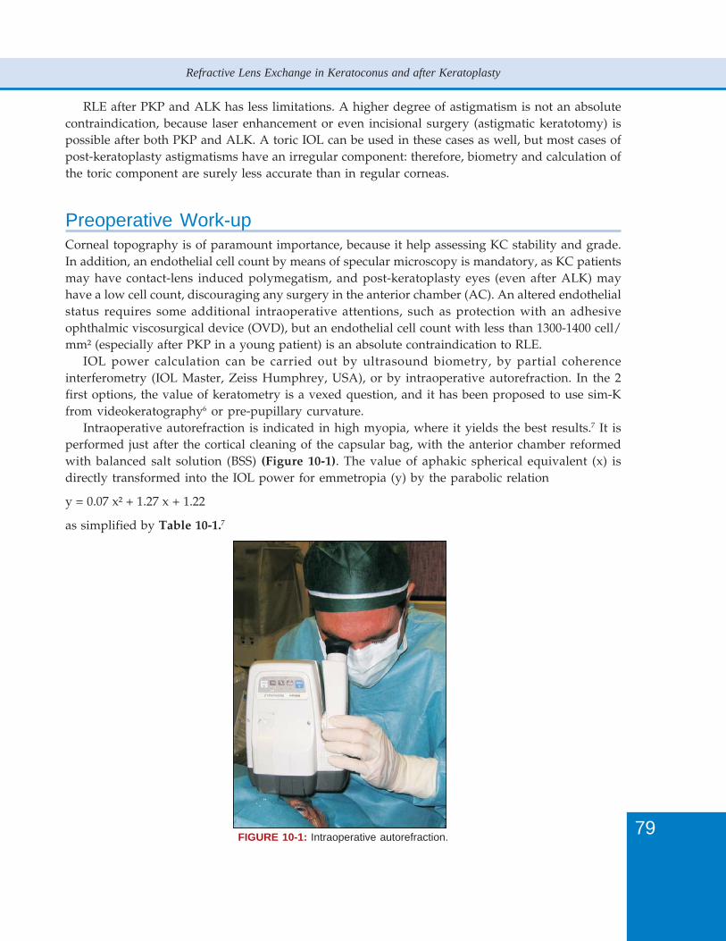
79
Refractive Lens Exchange in Keratoconus and after Keratoplasty
RLE after PKP and ALK has less limitations. A higher degree of astigmatism is not an absolutecontraindication, because laser enhancement or even incisional surgery (astigmatic keratotomy) ispossible after both PKP and ALK. A toric IOL can be used in these cases as well, but most cases ofpost-keratoplasty astigmatisms have an irregular component: therefore, biometry and calculation ofthe toric component are surely less accurate than in regular corneas.
Preoperative Work-upCorneal topography is of paramount importance, because it help assessing KC stability and grade.In addition, an endothelial cell count by means of specular microscopy is mandatory, as KC patientsmay have contact-lens induced polymegatism, and post-keratoplasty eyes (even after ALK) mayhave a low cell count, discouraging any surgery in the anterior chamber (AC). An altered endothelialstatus requires some additional intraoperative attentions, such as protection with an adhesiveophthalmic viscosurgical device (OVD), but an endothelial cell count with less than 1300-1400 cell/mm² (especially after PKP in a young patient) is an absolute contraindication to RLE.
IOL power calculation can be carried out by ultrasound biometry, by partial coherenceinterferometry (IOL Master, Zeiss Humphrey, USA), or by intraoperative autorefraction. In the 2first options, the value of keratometry is a vexed question, and it has been proposed to use sim-Kfrom videokeratography6 or pre-pupillary curvature.
Intraoperative autorefraction is indicated in high myopia, where it yields the best results.7 It isperformed just after the cortical cleaning of the capsular bag, with the anterior chamber reformedwith balanced salt solution (BSS) (Figure 10-1). The value of aphakic spherical equivalent (x) isdirectly transformed into the IOL power for emmetropia (y) by the parabolic relation
y = 0.07 x² + 1.27 x + 1.22
as simplified by Table 10-1.7
FIGURE 10-1: Intraoperative autorefraction.
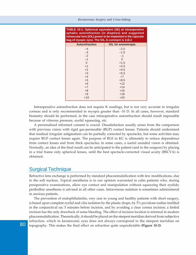
80
Keratoconus Surgery and Cross-linking
TABLE 10-1: Spherical equivalent (SE) at intraoperativeaphakic autorefraction (in dioptres) and suggestedintraocular lens (IOL) power to be implanted in the capsularbag of myopic eyes. The IOL A-constant is 118.2
Autorefraction IOL for emmetropia
–4 –2.5–3 –1.5–2 –1–1 00 +1.5
+1 +2.5+2 +4.5+3 +5.5+4 +7+5 +8.5+6 +12+7 +14+8 +16+9 +18
+10 +20
Intraoperative autorefraction does not require K readings, but is not very accurate in irregularcorneas and is only recommended in myopia greater than -10 D. In all cases, however, standardbiometry should be performed, in the case intraoperative autorefraction should result impossiblebecause of vitreous pressure, eyelid squeezing, etc.
A personalized informed consent is crucial. Dissatisfaction usually arises from the comparisonwith previous vision with rigid gas-permeable (RGP) contact lenses. Patients should understandthat residual irregular astigmatism can be partially corrected by spectacles, but some activities mayrequire RGP contact lenses again. The purpose of RLE in KC is ultimately to reduce dependencefrom contact lenses and from thick spectacles; in some cases, a useful unaided vision is obtained.Normally, an idea of the final result can be anticipated to the patient (and to the surgeon) by placingin a trial frame only spherical lenses, until the best spectacle-corrected visual acuity (BSCVA) isobtained.
Surgical TechniqueRefractive lens exchange is performed by standard phacoemulsification with few modifications, dueto the soft nucleus. Topical anesthesia is in our opinion warranted in calm patients who, duringpreoperative examinations, allow eye contact and manipulation without squeezing their eyelids;peribulbar anesthesia is advised in all other cases. Intravenous sedation is sometimes administeredin anxious patients.
The prevention of endophthalmitis, very rare in young and healthy patients with short surgery,is based upon complete eyelid and cilia isolation by the plastic drape, by 5% povidone-iodine instilledin the conjunctival sac 5 minutes before incision, and by avoiding a clear cornea incision; a limbalincision has the only drawback of some bleeding. The effect of incision location is minimal in modernphacoemulsification. Theoretically, it should be placed on the steepest meridian derived from subjectiverefraction, which in keratoconic eyes does not always correspond to the steepest meridian ontopography. This makes the final effect on refraction quite unpredictable (Figure 10-2).
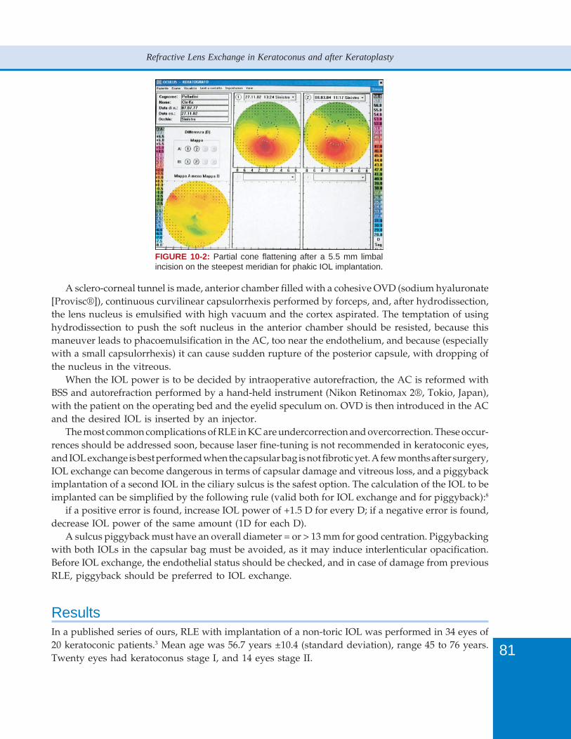
81
Refractive Lens Exchange in Keratoconus and after Keratoplasty
FIGURE 10-2: Partial cone flattening after a 5.5 mm limbalincision on the steepest meridian for phakic IOL implantation.
A sclero-corneal tunnel is made, anterior chamber filled with a cohesive OVD (sodium hyaluronate[Provisc®]), continuous curvilinear capsulorrhexis performed by forceps, and, after hydrodissection,the lens nucleus is emulsified with high vacuum and the cortex aspirated. The temptation of usinghydrodissection to push the soft nucleus in the anterior chamber should be resisted, because thismaneuver leads to phacoemulsification in the AC, too near the endothelium, and because (especiallywith a small capsulorrhexis) it can cause sudden rupture of the posterior capsule, with dropping ofthe nucleus in the vitreous.
When the IOL power is to be decided by intraoperative autorefraction, the AC is reformed withBSS and autorefraction performed by a hand-held instrument (Nikon Retinomax 2®, Tokio, Japan),with the patient on the operating bed and the eyelid speculum on. OVD is then introduced in the ACand the desired IOL is inserted by an injector.
The most common complications of RLE in KC are undercorrection and overcorrection. These occur-rences should be addressed soon, because laser fine-tuning is not recommended in keratoconic eyes,and IOL exchange is best performed when the capsular bag is not fibrotic yet. A few months after surgery,IOL exchange can become dangerous in terms of capsular damage and vitreous loss, and a piggybackimplantation of a second IOL in the ciliary sulcus is the safest option. The calculation of the IOL to beimplanted can be simplified by the following rule (valid both for IOL exchange and for piggyback):8
if a positive error is found, increase IOL power of +1.5 D for every D; if a negative error is found,decrease IOL power of the same amount (1D for each D).
A sulcus piggyback must have an overall diameter = or > 13 mm for good centration. Piggybackingwith both IOLs in the capsular bag must be avoided, as it may induce interlenticular opacification.Before IOL exchange, the endothelial status should be checked, and in case of damage from previousRLE, piggyback should be preferred to IOL exchange.
ResultsIn a published series of ours, RLE with implantation of a non-toric IOL was performed in 34 eyes of20 keratoconic patients.3 Mean age was 56.7 years ±10.4 (standard deviation), range 45 to 76 years.Twenty eyes had keratoconus stage I, and 14 eyes stage II.
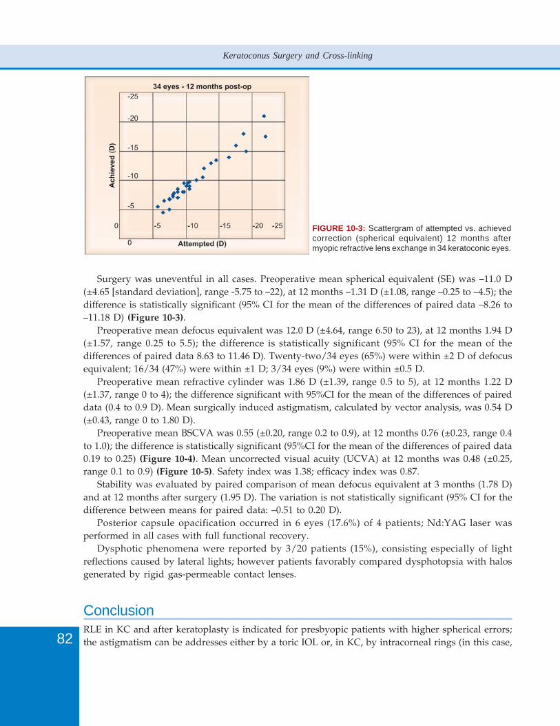
82
Keratoconus Surgery and Cross-linking
FIGURE 10-3: Scattergram of attempted vs. achievedcorrection (spherical equivalent) 12 months aftermyopic refractive lens exchange in 34 keratoconic eyes.
Surgery was uneventful in all cases. Preoperative mean spherical equivalent (SE) was –11.0 D(±4.65 [standard deviation], range -5.75 to –22), at 12 months –1.31 D (±1.08, range –0.25 to –4.5); thedifference is statistically significant (95% CI for the mean of the differences of paired data –8.26 to–11.18 D) (Figure 10-3).
Preoperative mean defocus equivalent was 12.0 D (±4.64, range 6.50 to 23), at 12 months 1.94 D(±1.57, range 0.25 to 5.5); the difference is statistically significant (95% CI for the mean of thedifferences of paired data 8.63 to 11.46 D). Twenty-two/34 eyes (65%) were within ±2 D of defocusequivalent; 16/34 (47%) were within ±1 D; 3/34 eyes (9%) were within ±0.5 D.
Preoperative mean refractive cylinder was 1.86 D (±1.39, range 0.5 to 5), at 12 months 1.22 D(±1.37, range 0 to 4); the difference significant with 95%CI for the mean of the differences of paireddata (0.4 to 0.9 D). Mean surgically induced astigmatism, calculated by vector analysis, was 0.54 D(±0.43, range 0 to 1.80 D).
Preoperative mean BSCVA was 0.55 (±0.20, range 0.2 to 0.9), at 12 months 0.76 (±0.23, range 0.4to 1.0); the difference is statistically significant (95%CI for the mean of the differences of paired data0.19 to 0.25) (Figure 10-4). Mean uncorrected visual acuity (UCVA) at 12 months was 0.48 (±0.25,range 0.1 to 0.9) (Figure 10-5). Safety index was 1.38; efficacy index was 0.87.
Stability was evaluated by paired comparison of mean defocus equivalent at 3 months (1.78 D)and at 12 months after surgery (1.95 D). The variation is not statistically significant (95% CI for thedifference between means for paired data: –0.51 to 0.20 D).
Posterior capsule opacification occurred in 6 eyes (17.6%) of 4 patients; Nd:YAG laser wasperformed in all cases with full functional recovery.
Dysphotic phenomena were reported by 3/20 patients (15%), consisting especially of lightreflections caused by lateral lights; however patients favorably compared dysphotopsia with halosgenerated by rigid gas-permeable contact lenses.
ConclusionRLE in KC and after keratoplasty is indicated for presbyopic patients with higher spherical errors;the astigmatism can be addresses either by a toric IOL or, in KC, by intracorneal rings (in this case,
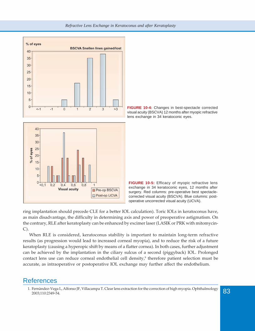
83
Refractive Lens Exchange in Keratoconus and after Keratoplasty
FIGURE 10-4: Changes in best-spectacle correctedvisual acuity (BSCVA) 12 months after myopic refractivelens exchange in 34 keratoconic eyes.
FIGURE 10-5: Efficacy of myopic refractive lensexchange in 34 keratoconic eyes, 12 months aftersurgery. Red columns: pre-operative best spectacle-corrected visual acuity (BSCVA). Blue columns: post-operative uncorrected visual acuity (UCVA).
ring implantation should precede CLE for a better IOL calculation). Toric IOLs in keratoconus have,as main disadvantage, the difficulty in determining axis and power of preoperative astigmatism. Onthe contrary, RLE after keratoplasty can be enhanced by excimer laser (LASIK or PRK with mitomycin-C).
When RLE is considered, keratoconus stability is important to maintain long-term refractiveresults (as progression would lead to increased corneal myopia), and to reduce the risk of a futurekeratoplasty (causing a hyperopic shift by means of a flatter cornea). In both cases, further adjustmentcan be achieved by the implantation in the ciliary sulcus of a second (piggyback) IOL. Prolongedcontact lens use can reduce corneal endothelial cell density,9 therefore patient selection must beaccurate, as intraoperative or postoperative IOL exchange may further affect the endothelium.
References1. Fernàndez-Vega L, Alfonso JF, Villacampa T. Clear lens extraction for the correction of high myopia. Ophthalmology
2003;110:2349-54.

84
Keratoconus Surgery and Cross-linking
2. Sauder G, Jonas JB. Treatment of keratoconus by toric foldable intraocular lenses. Eur J Ophthalmol 2003;13:577-9.
3. Leccisotti A. Refractive lens exchange in keratoconus. J Cataract Refract Surg 2006;32:742-6.4. Neuhann IM, et al. Retinal detachment after phacoemulsification in high myopia: analysis of 2356 cases. J Cataract
Refract Surg 2008;34:1644-57.5. Ongucci T, Kojima T, Azar DT. 10-year review of the literature on complications of incisional, thermal, and
lenticular refractive procedures. In Aliò LJ, Azar DT (Eds). Management of Complications in Refractive Surgery.Springer: Berlin, 2008;339-50.
6. Celikkol L, Ahn D, Celikkol G, Feldman ST. Calculating intraocular lens power in eyes with keratoconus usingvideokeratography. J Cataract Refract Surg 1996;22:497-500.
7. Leccisotti A. Intraocular lens calculation by intraoperative autorefraction in myopic eyes. Graefes Archive Clin ExpOphthalmol 2008;246:729-33.
8. Holladay JT. Piggyback intraocular lenses. Ann Ophthalmol 1998;30:203-4.9. Esgin H, Erda N. Corneal endothelial polymegethism and pleomorphism induced by daily-wear rigid gas-permeable
contact lenses. CLAO J 2002;28:40-3.
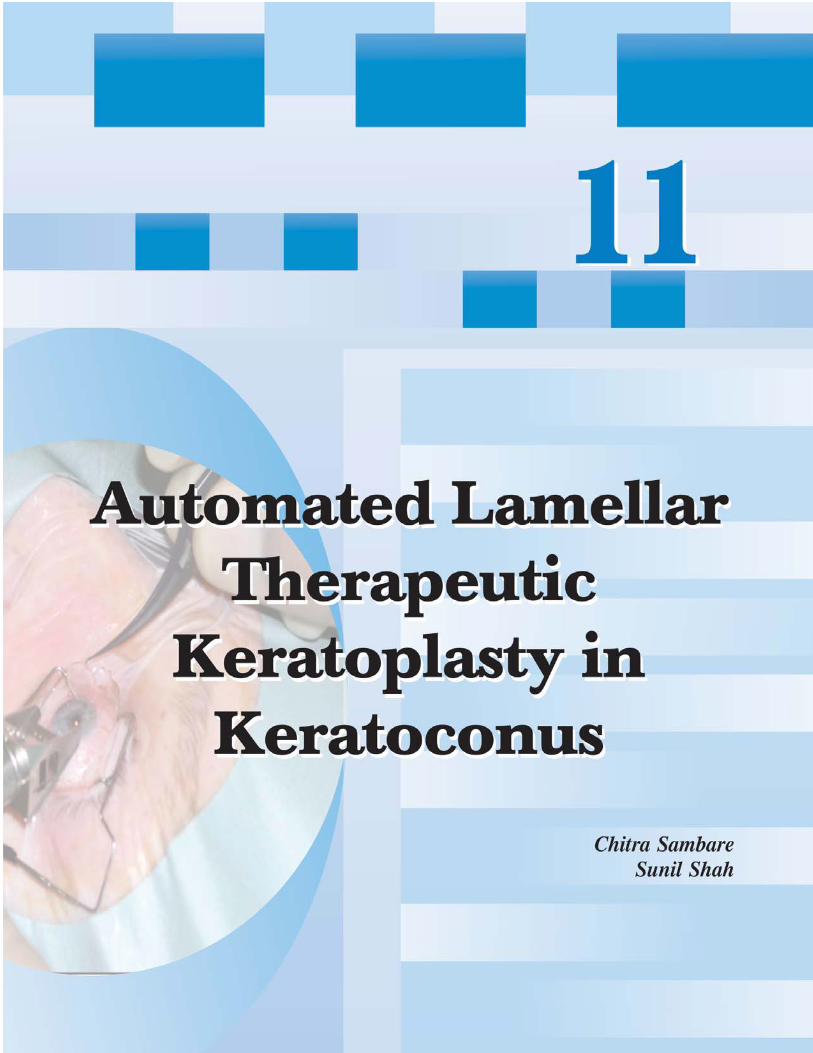
86
Keratoconus Surgery and Cross-linking
IntroductionThe advent of automated lamellar therapeutic keratoplasty (ALTK) using an automated microkeratomeand artificial anterior chamber to obtain lamellar cuts of repeatable quality and thickness has changedlamellar corneal transplantation completely. In addition, the recent introduction of femtosecondlasers for the creation of lamellar cuts has offered further opportunities.Lamellar surgery has beenpracticed in various forms for many years, however ALTK has reduced the dependence on individualsurgeon expertise and has made lamellar keratoplasty more available.
The benefits of lamellar corneal surgery are well recognized and include reduced rejection rates,increased speed of rehabilitation, reduced suture related problems, reduced postoperativeastigmatism and higher wound tensile strength.
ALTK is relatively simple, reversible, extraocular surgery and has reduced risks of immunologicreaction. The visual results of lamellar surgery in the form of epikeratophakia have been reported tobe nearly equal or often superior to those of penetrating keratoplasty (PKP).1,2 However, there are asparsity of reports on ALTK.
Most techniques involving lamellar surgery for keratoconus involve using manual dissection.They include Anwar’s big bubble technique,3-6 viscoelastic separation of lamellae7 and Melles’technique.8 However, all of these techniques have the drawback of an irregular interface (exceptwith good baring of Descemet in the big bubble technique) and the risk of perforation. An irregularinterface can result in visual compromise. All of these procedures are dependant on the skill of thesurgeon. Even in the hands of experienced surgeons, these procedures can be complicated by microor macro perforation in a relatively high number of cases (up to 1 in 3) – which can make conversionto a PKP necessary.
ALTK triumphs over all these methods in this respect. It is useful for superficial scars,epithelial and stromal dystrophies. However, it suffers from the need for relatively expensiveinstrumentation. However, this instrumentation is becoming cheaper and more available because ofits use for posterior automated lamellar transplantation (Descemet stripping automated endothelialkeratoplasty).
ALTK can be used to produce a lenticule similar to that from epikeratophakia but has manyof the same problems as epikeratophakia such as interface haze, melting and suture relatedproblems, etc.
In ALTK for keratoconus, the interface remains clear if there was no scarring preoperatively,although, it is difficult to produce adequate flattening of the ectasia without suturing tightly. Whilstthis can give good results, there is also a fairly high incidence of peripheral corneal melting relatedto the tight sutures. The addition of tissue to the ectatic cornea hence has the ability to thicken thecornea, add some rigidity and flatten the cornea.
Massimo Busin has used this technique for keratoconus and modified it further to obtain desirablevisual outcome in advanced keratoconus.10 According to him, microkeratome-assisted lamellarkeratoplasty for keratoconus has the advantage of being a standardized, technically easy procedure,which yields smooth dissected surfaces compatible with 20/20 vision. He has modified the originaltechnique of just adding tissue by perforating the residual base after the microkeratome cut in acircular fashion but just leaving 3 bridging points to hold the tissue in place. This technique has theadvantage of allowing increased flattening of the ectatic area. He has reported ALTK in patientswith keratoconus with an average keratometry reading of up to 57 D.
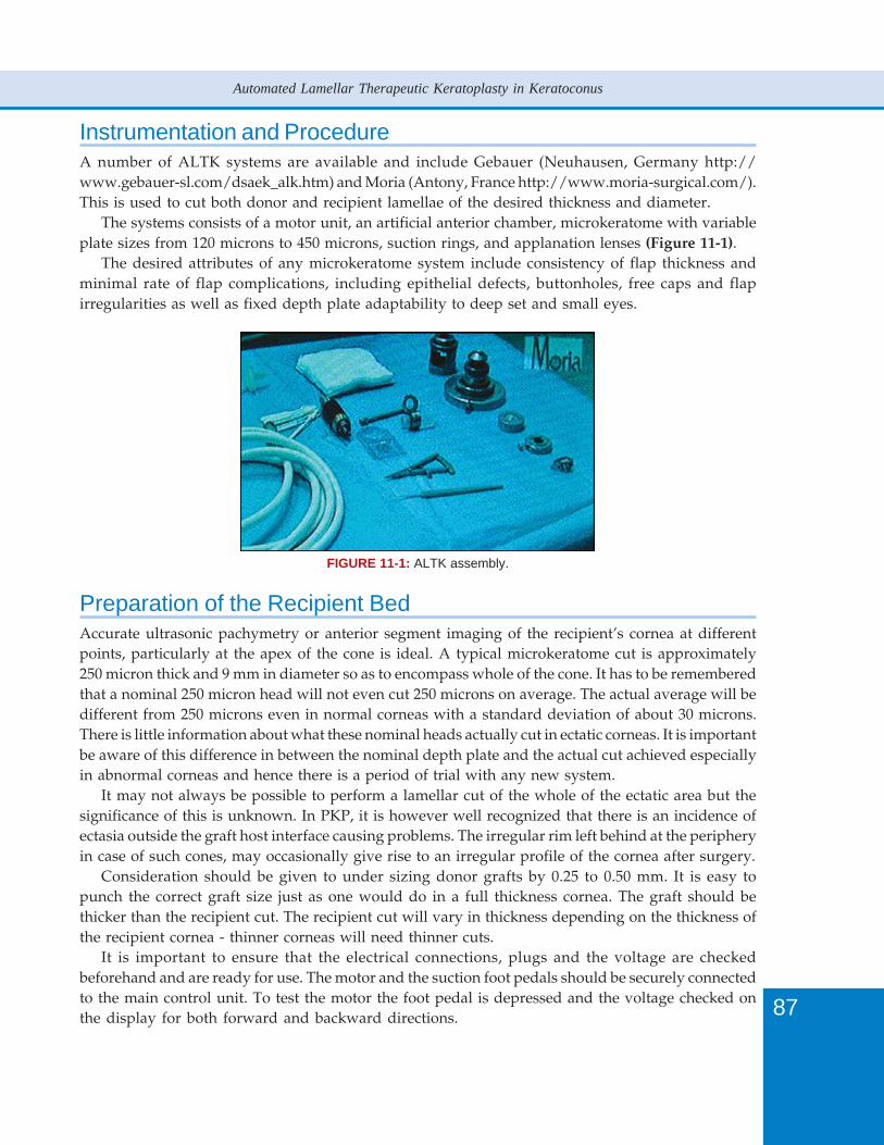
87
Automated Lamellar Therapeutic Keratoplasty in Keratoconus
Instrumentation and ProcedureA number of ALTK systems are available and include Gebauer (Neuhausen, Germany http://www.gebauer-sl.com/dsaek_alk.htm) and Moria (Antony, France http://www.moria-surgical.com/).This is used to cut both donor and recipient lamellae of the desired thickness and diameter.
The systems consists of a motor unit, an artificial anterior chamber, microkeratome with variableplate sizes from 120 microns to 450 microns, suction rings, and applanation lenses (Figure 11-1).
The desired attributes of any microkeratome system include consistency of flap thickness andminimal rate of flap complications, including epithelial defects, buttonholes, free caps and flapirregularities as well as fixed depth plate adaptability to deep set and small eyes.
FIGURE 11-1: ALTK assembly.
Preparation of the Recipient BedAccurate ultrasonic pachymetry or anterior segment imaging of the recipient’s cornea at differentpoints, particularly at the apex of the cone is ideal. A typical microkeratome cut is approximately250 micron thick and 9 mm in diameter so as to encompass whole of the cone. It has to be rememberedthat a nominal 250 micron head will not even cut 250 microns on average. The actual average will bedifferent from 250 microns even in normal corneas with a standard deviation of about 30 microns.There is little information about what these nominal heads actually cut in ectatic corneas. It is importantbe aware of this difference in between the nominal depth plate and the actual cut achieved especiallyin abnormal corneas and hence there is a period of trial with any new system.
It may not always be possible to perform a lamellar cut of the whole of the ectatic area but thesignificance of this is unknown. In PKP, it is however well recognized that there is an incidence ofectasia outside the graft host interface causing problems. The irregular rim left behind at the peripheryin case of such cones, may occasionally give rise to an irregular profile of the cornea after surgery.
Consideration should be given to under sizing donor grafts by 0.25 to 0.50 mm. It is easy topunch the correct graft size just as one would do in a full thickness cornea. The graft should bethicker than the recipient cut. The recipient cut will vary in thickness depending on the thickness ofthe recipient cornea - thinner corneas will need thinner cuts.
It is important to ensure that the electrical connections, plugs and the voltage are checkedbeforehand and are ready for use. The motor and the suction foot pedals should be securely connectedto the main control unit. To test the motor the foot pedal is depressed and the voltage checked onthe display for both forward and backward directions.
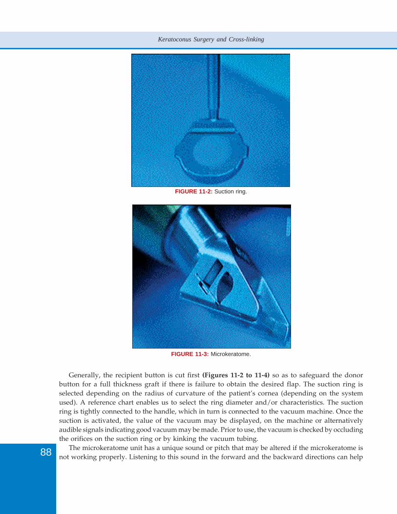
88
Keratoconus Surgery and Cross-linking
FIGURE 11-2: Suction ring.
FIGURE 11-3: Microkeratome.
Generally, the recipient button is cut first (Figures 11-2 to 11-4) so as to safeguard the donorbutton for a full thickness graft if there is failure to obtain the desired flap. The suction ring isselected depending on the radius of curvature of the patient’s cornea (depending on the systemused). A reference chart enables us to select the ring diameter and/or characteristics. The suctionring is tightly connected to the handle, which in turn is connected to the vacuum machine. Once thesuction is activated, the value of the vacuum may be displayed, on the machine or alternativelyaudible signals indicating good vacuum may be made. Prior to use, the vacuum is checked by occludingthe orifices on the suction ring or by kinking the vacuum tubing.
The microkeratome unit has a unique sound or pitch that may be altered if the microkeratome isnot working properly. Listening to this sound in the forward and the backward directions can help
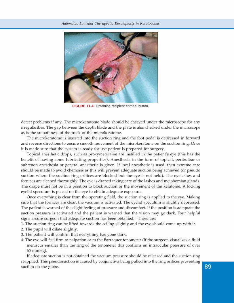
89
Automated Lamellar Therapeutic Keratoplasty in Keratoconus
detect problems if any. The microkeratome blade should be checked under the microscope for anyirregularities. The gap between the depth blade and the plate is also checked under the microscopeas is the smoothness of the track of the microkeratome.
The microkeratome is inserted into the suction ring and the foot pedal is depressed in forwardand reverse directions to ensure smooth movement of the microkeratome on the suction ring. Onceit is made sure that the system is ready for use patient is prepared for surgery.
Topical anesthetic drops, such as proxymetacaine are instilled in the patient’s eye (this has thebenefit of having some lubricating properties). Anesthesia in the form of topical, peribulbar orsubtenon anesthesia or general anesthetic is given. If local anesthetic is used, then extreme careshould be made to avoid chemosis as this will prevent adequate suction being achieved (or pseudosuction where the suction ring orifices are blocked but the eye is not held). The eyelashes andfornices are cleaned thoroughly. The eye is draped taking care of the lashes and meiobomian glands.The drape must not be in a position to block suction or the movement of the keratome. A lockingeyelid speculum is placed on the eye to obtain adequate exposure.
Once everything is clear from the operating field, the suction ring is applied to the eye. Makingsure that the fornices are clear, the vacuum is activated. The eyelid speculum is slightly depressed.The patient is warned of the slight feeling of pressure and discomfort. If the position is adequate thesuction pressure is activated and the patient is warned that the vision may go dark. Four helpfulsigns assure surgeon that adequate suction has been obtained.11 These are:1. The suction ring can be lifted towards the ceiling slightly and the eye should come up with it.2. The pupil will dilate slightly.3. The patient will confirm that everything has gone dark.4. The eye will feel firm to palpation or to the Barraquer tonometer (If the surgeon visualizes a fluid
meniscus smaller than the ring of the tonometer this confirms an intraocular pressure of over65 mmHg).If adequate suction is not obtained the vacuum pressure should be released and the suction ring
reapplied. This pseudosuction is caused by conjunctiva being pulled into the ring orifices preventingsuction on the globe.
FIGURE 11-4: Obtaining recipient corneal button.
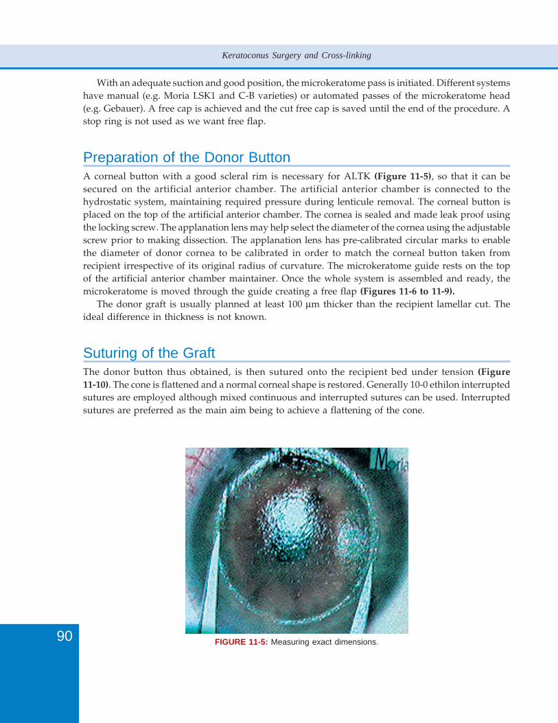
90
Keratoconus Surgery and Cross-linking
With an adequate suction and good position, the microkeratome pass is initiated. Different systemshave manual (e.g. Moria LSK1 and C-B varieties) or automated passes of the microkeratome head(e.g. Gebauer). A free cap is achieved and the cut free cap is saved until the end of the procedure. Astop ring is not used as we want free flap.
Preparation of the Donor ButtonA corneal button with a good scleral rim is necessary for ALTK (Figure 11-5), so that it can besecured on the artificial anterior chamber. The artificial anterior chamber is connected to thehydrostatic system, maintaining required pressure during lenticule removal. The corneal button isplaced on the top of the artificial anterior chamber. The cornea is sealed and made leak proof usingthe locking screw. The applanation lens may help select the diameter of the cornea using the adjustablescrew prior to making dissection. The applanation lens has pre-calibrated circular marks to enablethe diameter of donor cornea to be calibrated in order to match the corneal button taken fromrecipient irrespective of its original radius of curvature. The microkeratome guide rests on the topof the artificial anterior chamber maintainer. Once the whole system is assembled and ready, themicrokeratome is moved through the guide creating a free flap (Figures 11-6 to 11-9).
The donor graft is usually planned at least 100 μm thicker than the recipient lamellar cut. Theideal difference in thickness is not known.
Suturing of the GraftThe donor button thus obtained, is then sutured onto the recipient bed under tension (Figure11-10). The cone is flattened and a normal corneal shape is restored. Generally 10-0 ethilon interruptedsutures are employed although mixed continuous and interrupted sutures can be used. Interruptedsutures are preferred as the main aim being to achieve a flattening of the cone.
FIGURE 11-5: Measuring exact dimensions.
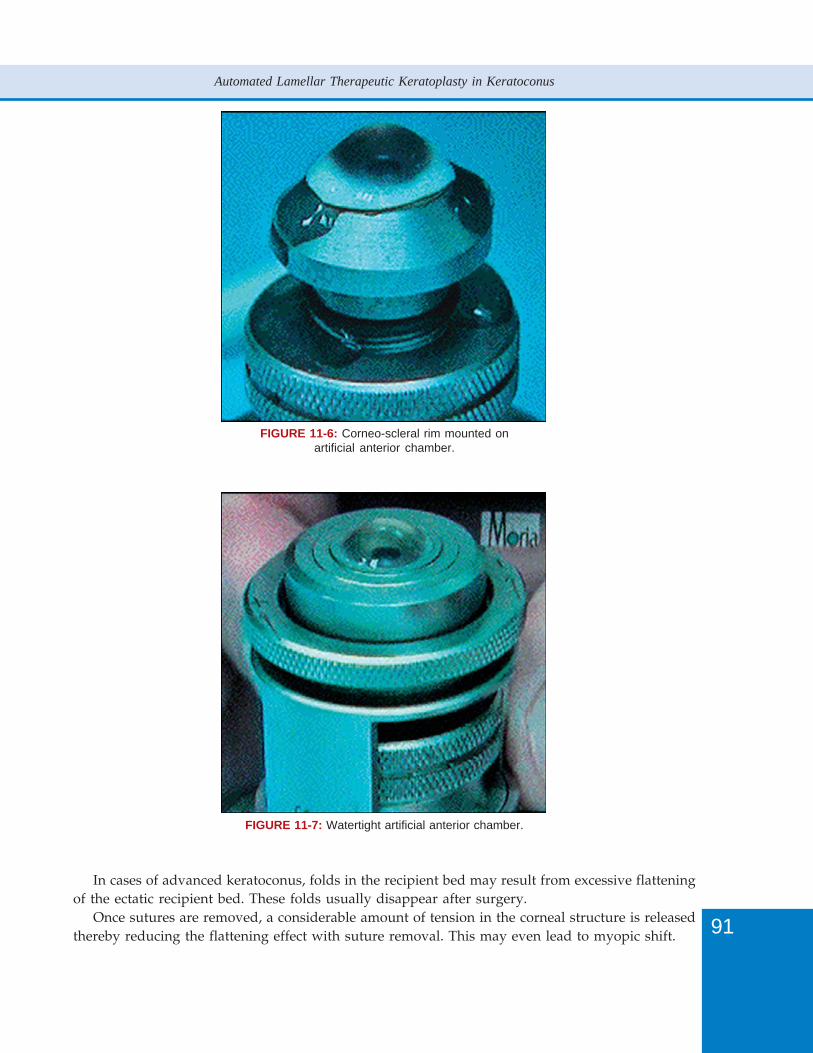
91
Automated Lamellar Therapeutic Keratoplasty in Keratoconus
FIGURE 11-6: Corneo-scleral rim mounted onartificial anterior chamber.
FIGURE 11-7: Watertight artificial anterior chamber.
In cases of advanced keratoconus, folds in the recipient bed may result from excessive flatteningof the ectatic recipient bed. These folds usually disappear after surgery.
Once sutures are removed, a considerable amount of tension in the corneal structure is releasedthereby reducing the flattening effect with suture removal. This may even lead to myopic shift.
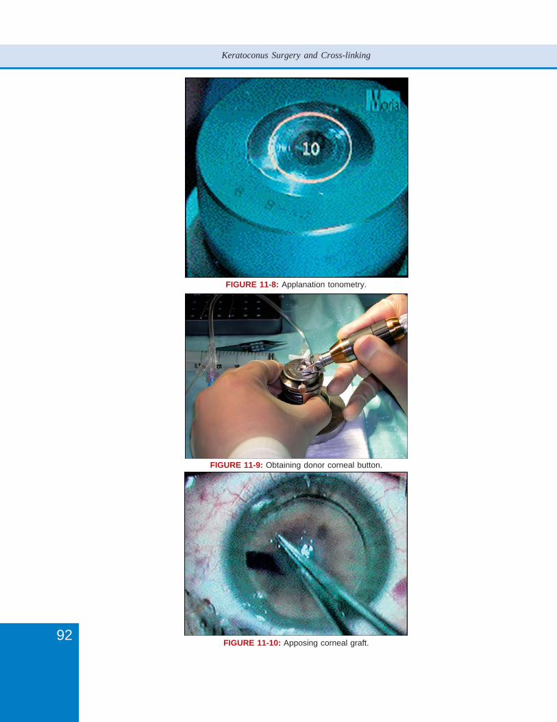
92
Keratoconus Surgery and Cross-linking
FIGURE 11-8: Applanation tonometry.
FIGURE 11-9: Obtaining donor corneal button.
FIGURE 11-10: Apposing corneal graft.
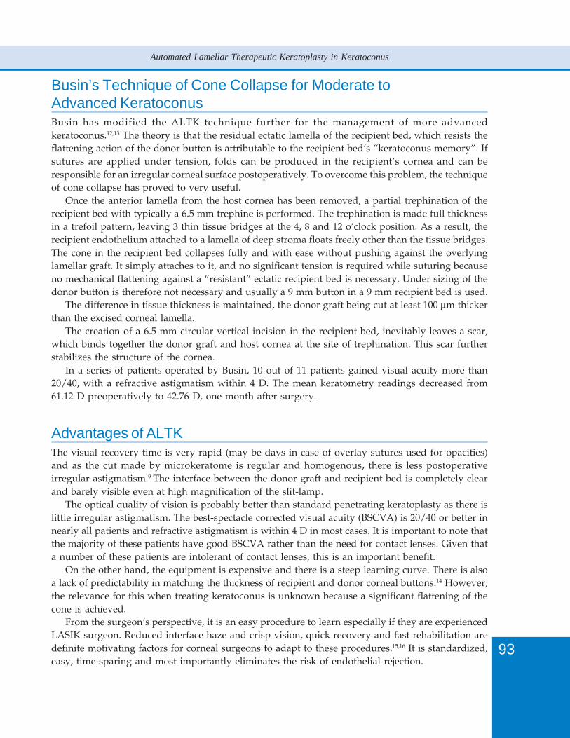
93
Automated Lamellar Therapeutic Keratoplasty in Keratoconus
Busin’s Technique of Cone Collapse for Moderate toAdvanced KeratoconusBusin has modified the ALTK technique further for the management of more advancedkeratoconus.12,13 The theory is that the residual ectatic lamella of the recipient bed, which resists theflattening action of the donor button is attributable to the recipient bed’s “keratoconus memory”. Ifsutures are applied under tension, folds can be produced in the recipient’s cornea and can beresponsible for an irregular corneal surface postoperatively. To overcome this problem, the techniqueof cone collapse has proved to very useful.
Once the anterior lamella from the host cornea has been removed, a partial trephination of therecipient bed with typically a 6.5 mm trephine is performed. The trephination is made full thicknessin a trefoil pattern, leaving 3 thin tissue bridges at the 4, 8 and 12 o’clock position. As a result, therecipient endothelium attached to a lamella of deep stroma floats freely other than the tissue bridges.The cone in the recipient bed collapses fully and with ease without pushing against the overlyinglamellar graft. It simply attaches to it, and no significant tension is required while suturing becauseno mechanical flattening against a “resistant” ectatic recipient bed is necessary. Under sizing of thedonor button is therefore not necessary and usually a 9 mm button in a 9 mm recipient bed is used.
The difference in tissue thickness is maintained, the donor graft being cut at least 100 μm thickerthan the excised corneal lamella.
The creation of a 6.5 mm circular vertical incision in the recipient bed, inevitably leaves a scar,which binds together the donor graft and host cornea at the site of trephination. This scar furtherstabilizes the structure of the cornea.
In a series of patients operated by Busin, 10 out of 11 patients gained visual acuity more than20/40, with a refractive astigmatism within 4 D. The mean keratometry readings decreased from61.12 D preoperatively to 42.76 D, one month after surgery.
Advantages of ALTKThe visual recovery time is very rapid (may be days in case of overlay sutures used for opacities)and as the cut made by microkeratome is regular and homogenous, there is less postoperativeirregular astigmatism.9 The interface between the donor graft and recipient bed is completely clearand barely visible even at high magnification of the slit-lamp.
The optical quality of vision is probably better than standard penetrating keratoplasty as there islittle irregular astigmatism. The best-spectacle corrected visual acuity (BSCVA) is 20/40 or better innearly all patients and refractive astigmatism is within 4 D in most cases. It is important to note thatthe majority of these patients have good BSCVA rather than the need for contact lenses. Given thata number of these patients are intolerant of contact lenses, this is an important benefit.
On the other hand, the equipment is expensive and there is a steep learning curve. There is alsoa lack of predictability in matching the thickness of recipient and donor corneal buttons.14 However,the relevance for this when treating keratoconus is unknown because a significant flattening of thecone is achieved.
From the surgeon’s perspective, it is an easy procedure to learn especially if they are experiencedLASIK surgeon. Reduced interface haze and crisp vision, quick recovery and fast rehabilitation aredefinite motivating factors for corneal surgeons to adapt to these procedures.15,16 It is standardized,easy, time-sparing and most importantly eliminates the risk of endothelial rejection.
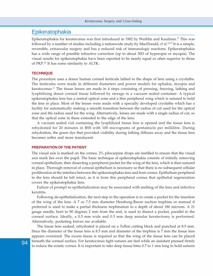
94
Keratoconus Surgery and Cross-linking
EpikeratophakiaEpikeratophakia for keratoconus was first introduced in 1982 by Werblin and Kaufman.17 This wasfollowed by a number of studies including a nationwide study by MacDonald, et al.18,19 It is a simple,reversible, extraocular surgery and has a reduced risk of immunologic reactions. Epikeratophakiahas a wide range of possible refractive correction (up to about 30D of hyperopia or myopia). Thevisual results for epikeratophakia have been reported to be nearly equal or often superior to thoseof PKP.20 It has some similarity to ALTK.
TECHNIQUE
The procedure uses a donor human corneal lenticule lathed to the shape of lens using a cryolathe.The lenticules were made in different diameters and power models for aphakia, myopia andkeratoconus.11 The tissue lenses are made in 4 steps consisting of pressing, freezing, lathing andlyophilizing donor corneal tissue followed by storage in a vacuum sealed container. A typicalepikeratophakia lens has a central optical zone and a thin peripheral wing which is sutured to holdthe lens in place. Most of the lenses were made with a specially developed cryolathe which has afacility for automatically making a smooth transition between the radius of cut used for the opticalzone and the radius used for the wing. Alternatively, lenses are made with a single radius of cut, sothat the optical zone in these extended to the edge of the lens.
A vacuum sealed vial containing the lyophilized tissue lens is opened and the tissue lens isrehydrated for 20 minutes in BSS with 100 micrograms of gentamicin per millilitre. Duringrehydration, the green dye that provided visibility during lathing diffuses away and the tissue lensbecomes softer and more translucent.
PREPARATION OF THE PATIENT
The visual axis is marked on the cornea. 2% pilocarpine drops are instilled to ensure that the visualaxis mark lies over the pupil. The basic technique of epikeratophakia consists of initially removingcorneal epithelium, then dissecting a peripheral pocket for the wing of the lens, which is then suturedin place. Thorough removal of corneal epithelium is necessary so that there is no subsequent cellularproliferation at the interface between the epikeratophakia lens and host cornea. Epithelium peripheralto the lens should be left intact, as it is from this peripheral cornea that epithelial regenerationcovers the epikeratophakia lens.
Failure of prompt re-epithelialization may be associated with melting of the lens and infectivekeratitis.
Following de-epithelialization, the next step in the operation is to create a pocket for the insertionof the wing of the lens. A 7 or 7.5 mm diameter Hessburg-Baron suction trephine or manual ifpreferred is used to make a partial thickness trephination to a depth of about 180 microns. A 21gauge needle, bent to 90 degrees 2 mm from the end, is used to dissect a pocket, parallel to thecorneal surface. Ideally, a 0.5 mm wide and 0.3 mm deep annular keratectomy is performed.Alternatively, pocketing knives are available.
The tissue lens soaked, rehydrated is placed on a Teflon cutting block and punched at 8.5 mm.Since the diameter of the tissue lens is 8.5 mm and diameter of the trephine is 7 mm the tissue lensappears oversized. The excess tissue is required so that the wing of the tissue lens can be placedbeneath the corneal surface. For keratoconus tight sutures are tied while an assistant pressed firmlyto reduce the ectatic cornea. It is important to take deep tissue bites 0.7 to 1 mm long to hold sutures
95
Automated Lamellar Therapeutic Keratoplasty in Keratoconus
tight. The wing of the tissue lens is pushed below the recipient lip so that it lies in the peripherallamellar keratectomy created by the lamellar dissector. The sutures are left in place unless theybecame loose or are inducing astigmatism in case of keratoconus.
COMPLICATIONS
The major complications of epikeratophakia are related to delay in epithelialization. With acompromised epithelium and nonviable donor cap, patients are at risk of developing infections, andpoor epithelialization can lead to melting of the tissue. Persistent refractive errors and infiltrates atthe recipient-lenticule interface are other important issues. A rare complication is dehiscence of thegraft. Persistent haze is another postoperative issue.
In a series of patients at Moorfields Eye Hospital,21 patients who were intolerant to contact lensesunderwent epikeratophakia, six out of seven patients (86%) with more than two months’ follow-upachieved a good result with BSCVA of 6/9 or better. The remaining patient achieved 6/12 but witha very high cylinder and required penetrating keratoplasty. Results of 1-year follow-up findings on42 of the first epikeratophakia procedures performed for keratoconus at the Helsinki UniversityCentral Hospital showed encouraging results.22 The mean flattening by keratometry readings was9.8 diopters (D) and the mean decrease in myopia in terms of spherical equivalent was 5.3 D. Thedegree of irregular astigmatism was measured in five cases showed that the mean preoperativeirregular astigmatism of 3.9 D was reduced to 1.3 D in the long-term analysis.
The noninvasive nature of epikeratophakia makes it a safe and desirable option for the treatmentof keratoconus.23 In properly selected patients, epikeratophakia can effectively be used to treatkeratoconus and thus avoid potential intraocular surgical complications and immunogenic phenomena.However, ALTK has overshadowed this technique because the availability of the lamellar graft ismuch simpler.
Femtosecond LaserThe femtosecond laser is possibly the single biggest innovation in corneal surgery in the past thirtyyears.24-28 The main principle of femtosecond is the use of ultrashort infrared laser pulses causingintrastromal microdissection without ablation. Femtosecond employs a solid state laser with a1053 nm wavelength and uses brief pulses to cause disruption in the lamellar plane. Examples offemtosecond include
Intralase (AMO Santa Ana, USA http://www.amo-inc.com/products/refractive/ilasik/intralase-fs-laser),
Femtec (20/10 Perfect Vision, Heidelberg, Germany http://www.2010pv.com/2010-pv/products/femtec-femtosecond-laser-workstation_100003_0.html),
Visumax (Carl Zeiss Meidtec http://www.meditec.zeiss.com/visumax) andFemtoLDV (Zeimer, Port, Switzerland http://www.ziemergroup.ch/?id=33).As the lamellar cut is controlled by software, a variety of shape and configuration of incisions
may be easily programmed and can enable us improve indications in corneal surgery. The advantagesof femtosecond laser corneal dissection include:1. Precise cuts2. Improved mechanical stability3. Better wound healing4. Early suture removal5. Reduced astigmatism due to better apposition.
96
Keratoconus Surgery and Cross-linking
Different geometric patterns have been used by different surgeons across the world to obtainperfect apposition of the graft in PKP and in lamellar corneal surgery. Angled incision edge facilitatesstromal excision with scissors. Interlocking wound configuration facilitates matching anterior donorand recipient surfaces.
Anterior Lamellar Keratoplasty has been performed by Sheraz Daya in an initial series of fourteenpatients using zigzag incisions within seventy microns of Descemet’s membrane.
Whilst the technology is available to perform lamellar transplants for keratoconus, this is not inroutine use as yet.
References1. Tan D, Ang LPK. Automated lamellar therapeutic keratoplasty for post-PRK corneal scarring and thinning. Am J
Ophthalmol 2004; 138: 1067-9.2. Jimenez-Alfaro I, Perez-Santonja. Therapeutic lamellar keratoplasty with an automated microkeratome. J Cataract
Refract Surg 2001;27:1161–5.3. Anwar M. Dissection technique in lamellar keratoplasty. Br J Ophthalmol 1972;56:711-3.4. Anwar M. Techniques in lamellar keratoplasty. Trans Ophthalmol Soc UK 1974; 94:163-71.5. Archila EA. Deep lamellar keratoplasty dissection of host tissue with intrastromal air injection. Cornea 1984/
85;3:217-8.6. Price FW Jr. Air lamellar keratoplasty. Refract Corneal Surg 1989;5:240-3.7. Manche EE, Holland GN, Maloney RK. Deep lamellar keratoplasty using viscoelastic dissection. Arch Ophthalmol
1999;117:1561-5.8. Melles GRJ, Lander F, Rietveld FJR, Remeijer L, Beekhuis WH, Binder PS. A new surgical technique for deep,
anterior lamellar keratoplasty. Br J Ophthalmol 1999;83:327-33.9. Vajpayee RB. Automated lamellar therapeutic keratoplasty (ALTK) the treatment of anterior to mid-stromal
corneal pathologies. Acta Ophthalmol Scand 2006;84:771-3.10. Busin M, Zambianchi L, Arffa RC. Microkeratome-assisted lamellar keratoplasty for the surgical treatment of
keratoconus. Ophthalmology 2005;112:987-97.11. Agarwal S, Agarwal A, Apple DJ, Buratto L, Aliò JL, Pandey SK, Agarwal A. Textbook of Ophthalmology, vol 2.
Lippincott: Philadelphia, 2002.12. Busin M. New techniques for the surgical treatment of keratoconus. Ophthalmology Times Europe, January 2005.13. Tan DT, Ang LP. Modified automated lamellar therapeutic keratoplasty for keratoconus: a new technique. Cornea
2006;25:1217-9.14. Giaconi JA, Culbertson W. Predictability of donor lamellar graft dimensions in an artificial anterior chamber
system with the Moria ALTK microkeratome. Invest Ophthalmol Vis Sci 2003;44: E-Abstract 4682.15. Tan DT, Mehta JS. Future directions in lamellar corneal transplantation. Cornea 2007;26:S21-S28.16. Busin M, Arffa RC. Atlas of Microkeratome Assisted Lamellar Keratoplasty. Slack: Thorofare (USA), 2006.17. Tamaki K, Yamaguchi T, McDonald MB, Kaufman HE. Epikeratophakia for aphakia, keratoconus, and myopia. Br
J Ophthalmol 1990;74:67–72.18. McDonald MB, Kaufman HE, Aquavella JV, et al. The nationwide study of epikeratophakia for myopia. Am J
Ophthalmol 1987; 103:375-83.19. McDonald MB, Kaufman HE, Durrie DS. Epikeratophakia for keratoconus, the nationwide study. Arch Ophthalmol
1986;104:1294-300.20. Busin M, Bechrakis-Böker I, Denninger U. Surgical therapy of keratoconus. Epikeratophakia versus penetrating
keratoplasty. Fortschr Ophthalmol 1991;88:794-6.21. Halliday BL. Epikeratophakia for aphakia, keratoconus, and myopia. Br J Ophthalmol 1990;74: 67–72.22. Spitznas M, Eckert J, Frising M, Eter N. Long-term functional and topographic results seven years after
epikeratophakia for keratoconus. Graefes Arch Clin Exp Ophthalmol 2002;240:639-43.23. Dietze TR, Durrie DS. Indications and treatment of keratoconus using epikeratophakia. Ophthalmology 1988;95:
236-46.24. Sarayba MA, Maguen E, Salz J, Rabinowitz Y, Ignacio TS. Femtosecond laser keratome creation of partial thickness
donor corneal buttons for lamellar keratoplasty. J Refract Surg 2007;23:58-65.

97
Automated Lamellar Therapeutic Keratoplasty in Keratoconus
25. Jones YJ, Goins KM, Sutphin JE, Mullins R, Skeie JM. Comparison of the femtosecond laser (IntraLase) versusmanual microkeratome (Moria ALTK) in dissection of the donor in endothelial keratoplasty: initial study in eyebank eyes. J Fr Ophtalmol 2007;30:689-94.
26. Hoffart L, Proust H, Matonti F, Ridings B, Conrath J. Short-term results of penetrating keratoplasty performedwith the Femtec femtosecond laser. Am J Ophthalmol 2008;146:50-55.
27. Mehta JS, Por YM, Cajucom-Uy H, Parthasarathy A, Tan DT. Femtosecond laser for endothelial keratoplasty.J Cataract Refract Surg 2007;33:1141.
28. Suwan-Apichon O, Reyes JM, Griffin NB, Barker J, Gore P, Chuck RS. Microkeratome versus femtosecond laserpredissection of corneal grafts for anterior and posterior lamellar keratoplasty. Cornea 2006;25:966-8.

100
Keratoconus Surgery and Cross-linking
Why Near-Descemetic?Deep Anterior Lamellar Keratoplasty (DALK) has only recently become the standard of care foradvanced keratoconus (KC). This is surprising, because we already had the technology for modernDALK at least 20 years ago, and because the many advantages of DALK over penetrating keratoplasty(PKP) in keratoconus are obvious: preserved endothelium, rejection virtually absent, no opening ofthe anterior chamber (AC), better control of astigmatism. What has really changed is the surgeon’smindset, together with the observation that the ‘Holy Grail’ of Descemet’s membrane baring is notnecessary to achieve a good visual result.
Two myths have indeed delayed (and still hamper) the diffusion of DALK as the rational substituteof PKP in KC: the first is that PKP yields better visual results, the second is that DALK is a difficultprocedure, reserved to few excellent surgeons, because, without the exposure of Descemet’smembrane, the interface recipient stroma/donor stroma would become hazy and cause visualaberrations. Fortunately, both prejudices have been demolished by studies demonstrating that DALKcan achieve the same visual results as PKP1 and that avoidance of Descemet’s membrane baring doesnot affect final visual acuity.1-3
Several techniques have been proposed for DALK, using air, viscoelastic, hydration to helpdissection.4-7 One of the most interesting and popular method is the Big-Bubble technique,8, 9 inwhich, after partial trephination, air is forcefully injected into the stroma by a needle, to create a bigair bubble separating the posterior stroma from the Descemet’s membrane. After the excision ofmost of the stromal tissue, the bubble is anteriorly perforated, and the residual stroma is removed.The Big-Bubble technique has some drawbacks:1. The big bubble does not occur in 20-50% of cases, depending on surgeons experience, depth of
trephination, etc.2. The last phase of Descemet’s membrane exposure carries a high risk of perforation.
The same authors have described an alternative technique (“near-Descemetic” DALK) to usewhen a big bubble did not form or in the presence of a damaged Descemet’s membrane:10 with thismethod, after partial trephination and intrastromal air injection, stromal emphysema is used to helpmanual deep dissection only to a pre-Descemetic stromal plane. This variant is safer and technicallyeasier than Big-Bubble, and has become my preferred technique for DALK in KC.
IndicationsDALK is indicated in all cases of KC in which best-corrected visual acuity by spectacles or contactlenses is not satisfactory, and cannot be improved by less invasive surgical techniques. When thecornea is not particularly deformed, myopia associated with KC can be corrected by phakic intraocularlenses (IOLs) in younger patients,11 and by refractive lens exchange in the presbyopic age;12 theadvent of toric IOLs has improved the visual prognosis of these techniques. Corneal collagen cross-linking by ultraviolet and riboflavin has recently emerged as a para-surgical technique that canimprove earlier stages of KC.13 Intrastromal ring segments do not involve tissue transplantation, canbe implanted with femtosecond laser, are reversible and have few complications.14
I generally consider DALK when contact lens cannot be worn or yield a corrected visual acuity<0.5, and best-spectacle corrected visual acuity is <0.5. Endothelial cell count is not relevant, as longas the stroma is transparent. The status of Descemet’s membrane is equally not significant if weadopt a near-Descemetic approach. Corneal thickness is important but does not preclude DALK,
101
Near-Descemetic Deep Anterior Lamellar Keratoplasty (DALK)
because when facing a thin cornea, we should prepare (surgeon, instruments and tissue) for PKP,but we should first attempt DALK.
Stromal scars, especially at the cone apex, do not represent an absolute contraindication to DALK,especially if superficial (contact lens induced).
Preoperative Work-upIf a correct preoperative assessment is the key for all surgery, this is even more true for DALK inKC. Poor results and dissatisfaction often stem from poor preoperative evaluation, poor planning,poor communication with the patient.
History is mandatory. Amblyopia is not rare in keratoconic eyes, which sometimes start asastigmatic eyes. Best corrected visual acuity with contact lenses, spectacles, and pin-hole (which willoften indicate the achievable postoperative visual acuity) are obviously needed before any decision.Undilated and dilated anterior segment slit-lamp examination, intraocular pressure (IOP) measurementand dilated funduscopy should be performed in all cases.
The evaluation of keratoconus is best carried out by corneal topography (videokeratography).Orbscan and Pentacam can be helpful to assess the posterior surface and to obtain a pachymetry mapin suspect KC, but in advanced KC these techniques are highly unreliable. Therefore, Placido-basedtopography remains the golden standard to assess the corneal deformation, the KC stage, and thevariations over time. Usually, axial topography gives an overall impression of the KC, tangentialtopography should be used in contact lens warpage and post-surgical ectasia, and the altitudinalmap help localizing the cone apex. When the cornea is so altered that no color coded image ispossible, an image of Placido rings should be enclosed in the patient chart.
Corneal thickness is of paramount importance, and should be measured by ultrasounds. Ultrasoundpachymetry is fast, inexpensive and highly reliable in all kind of corneas (scarred, edematous, thin);the best pachymetry map is obtained checking at the slit-lamp the position of the probe tip. The importantpoints to measure are the corneal geometrical center, the presumed apex, and the 4 cardinal pointsat 4 mm from the center, where the trephination will take place. In lid squeezers or in poorly collabo-rating patients, pachymetry should be repeated in the operating theatre after the induction of anesthesia.
The evaluation of corneal endothelium by specular microscopy should be done, but in myexperience is of a limited value, apart from the rare association keratoconus/endothelial dystrophy,15
in which the endothelial cell density can be so poor that a PKP is directly planned. In such cases, theendothelium has the classical beaten silver appearance at slit-lamp, and specular microscopy willconfirm the finding by showing dark rounded areas (corresponding to guttae) circumscribed by athin brighter net (Figure 12-1). In plain KC, dark or defocused images can result from severelydeformed corneas (Figure 12-2), but, as long as the stroma is transparent, a near-Descemetic DALKcan be performed, even in the presence of Descemet’s ruptures, despite a sub-normal endothelialcell density. A DALK with a postoperative cell density of 1200 cell/mm2 is always better than anyPKP, and, should such a cornea decompensate, a PKP can be performed later with the same successrate and at an elder age.
Ultrasound biometry can be used to predict, with a large approximation, the postoperative sphericalequivalent (SE). Eyes with a normal axial length normally end between -1 and -3 diopters (D) SE. Ineyes with long axial length a myopic outcome should be anticipated to the patient, thus helping him/her to understand the need for postoperative spectacles and contact lenses.
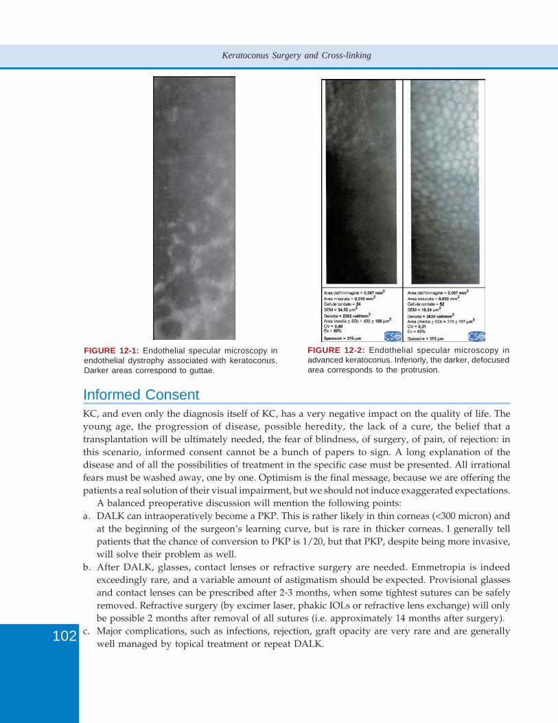
102
Keratoconus Surgery and Cross-linking
FIGURE 12-1: Endothelial specular microscopy inendothelial dystrophy associated with keratoconus.Darker areas correspond to guttae.
FIGURE 12-2: Endothelial specular microscopy inadvanced keratoconus. Inferiorly, the darker, defocusedarea corresponds to the protrusion.
Informed ConsentKC, and even only the diagnosis itself of KC, has a very negative impact on the quality of life. Theyoung age, the progression of disease, possible heredity, the lack of a cure, the belief that atransplantation will be ultimately needed, the fear of blindness, of surgery, of pain, of rejection: inthis scenario, informed consent cannot be a bunch of papers to sign. A long explanation of thedisease and of all the possibilities of treatment in the specific case must be presented. All irrationalfears must be washed away, one by one. Optimism is the final message, because we are offering thepatients a real solution of their visual impairment, but we should not induce exaggerated expectations.
A balanced preoperative discussion will mention the following points:a. DALK can intraoperatively become a PKP. This is rather likely in thin corneas (<300 micron) and
at the beginning of the surgeon’s learning curve, but is rare in thicker corneas. I generally tellpatients that the chance of conversion to PKP is 1/20, but that PKP, despite being more invasive,will solve their problem as well.
b. After DALK, glasses, contact lenses or refractive surgery are needed. Emmetropia is indeedexceedingly rare, and a variable amount of astigmatism should be expected. Provisional glassesand contact lenses can be prescribed after 2-3 months, when some tightest sutures can be safelyremoved. Refractive surgery (by excimer laser, phakic IOLs or refractive lens exchange) will onlybe possible 2 months after removal of all sutures (i.e. approximately 14 months after surgery).
c. Major complications, such as infections, rejection, graft opacity are very rare and are generallywell managed by topical treatment or repeat DALK.
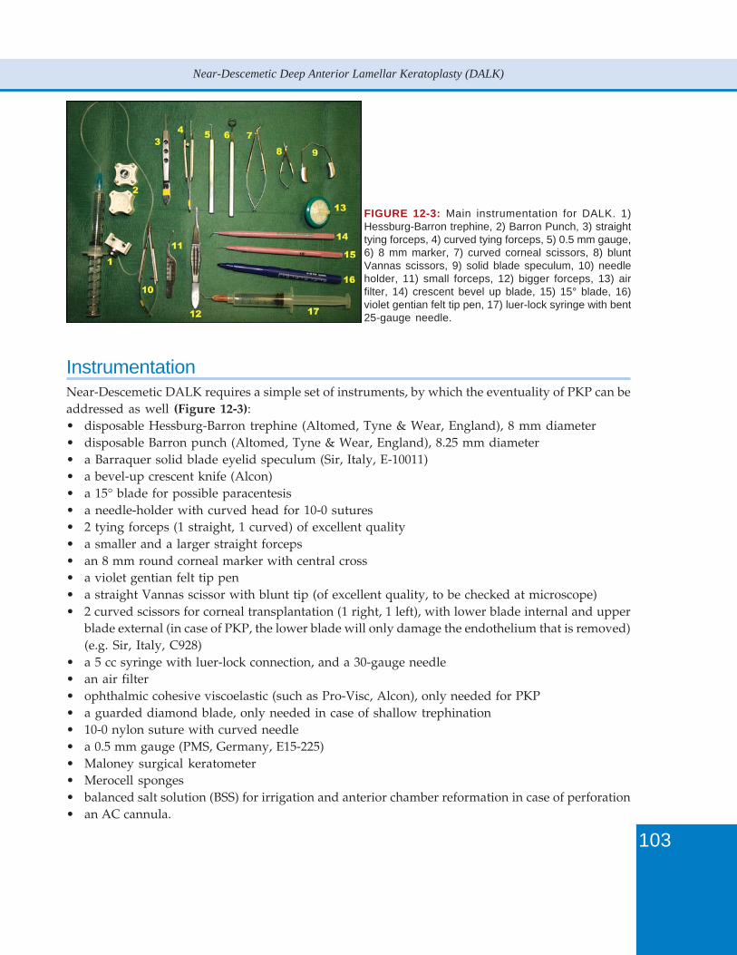
103
Near-Descemetic Deep Anterior Lamellar Keratoplasty (DALK)
InstrumentationNear-Descemetic DALK requires a simple set of instruments, by which the eventuality of PKP can beaddressed as well (Figure 12-3):• disposable Hessburg-Barron trephine (Altomed, Tyne & Wear, England), 8 mm diameter• disposable Barron punch (Altomed, Tyne & Wear, England), 8.25 mm diameter• a Barraquer solid blade eyelid speculum (Sir, Italy, E-10011)• a bevel-up crescent knife (Alcon)• a 15° blade for possible paracentesis• a needle-holder with curved head for 10-0 sutures• 2 tying forceps (1 straight, 1 curved) of excellent quality• a smaller and a larger straight forceps• an 8 mm round corneal marker with central cross• a violet gentian felt tip pen• a straight Vannas scissor with blunt tip (of excellent quality, to be checked at microscope)• 2 curved scissors for corneal transplantation (1 right, 1 left), with lower blade internal and upper
blade external (in case of PKP, the lower blade will only damage the endothelium that is removed)(e.g. Sir, Italy, C928)
• a 5 cc syringe with luer-lock connection, and a 30-gauge needle• an air filter• ophthalmic cohesive viscoelastic (such as Pro-Visc, Alcon), only needed for PKP• a guarded diamond blade, only needed in case of shallow trephination• 10-0 nylon suture with curved needle• a 0.5 mm gauge (PMS, Germany, E15-225)• Maloney surgical keratometer• Merocell sponges• balanced salt solution (BSS) for irrigation and anterior chamber reformation in case of perforation• an AC cannula.
FIGURE 12-3: Main instrumentation for DALK. 1)Hessburg-Barron trephine, 2) Barron Punch, 3) straighttying forceps, 4) curved tying forceps, 5) 0.5 mm gauge,6) 8 mm marker, 7) curved corneal scissors, 8) bluntVannas scissors, 9) solid blade speculum, 10) needleholder, 11) small forceps, 12) bigger forceps, 13) airfilter, 14) crescent bevel up blade, 15) 15° blade, 16)violet gentian felt tip pen, 17) luer-lock syringe with bent25-gauge needle.
104
Keratoconus Surgery and Cross-linking
Donor TissueOne of the main advantages of DALK is tissue availability. Donor tissue with endothelium that isunusable for PKP can be safely destined to DALK. The problem at the beginning of the surgeon’slearning curve is the possibility of conversion to PKP. The suggestion of planning in the same sessiona DALK and a PKP is highly unpractical, especially because PKPs will hopefully decline in the future.It is therefore necessary, at least for the first cases, to use a donor tissue with an endothelial statusalso appropriate for PKP.
The donor age is, in any case, not relevant.
AnesthesiaExcluding as absolutely inappropriate topical anesthesia, the choice is between general and infiltrativeanesthesia. The surgical procedure is particularly long in the first cases (my first DALK lasted 3hours), becoming only a little faster with experience (never less than 1 hour). In addition, eye andhead movements should be avoided by all means in many delicate phases. The preference goestherefore to general anesthesia, which reduces patient’s and surgeon anxiety. A peribulbar blockcan still be employed in selected cases: expert surgeon and very calm patient, or contraindications togeneral anesthesia.
Surgical Technique1. The periocular skin is cleaned with 10% povidone iodine, and 2 drops of 5% povidone iodine
are instilled in the conjunctival sac and over the cornea. The eyelids and cilia are isolated by thesurgical adhesive drape and by adjunctive adhesive strips. The cornea should be well exposedand in a vertical gaze position.
2. An 8 mm circular marker tinted with gentian violet is centered on the geometrical center of thecornea and applied to the epithelium (Figure 12-4).
3. The Hessburg-Barron trephine is inspected under the microscope, and its blade (inner metalcircle) is aligned with the suction ring (intermediate metal circle) (Figure 12-5), and then turnedback (anti-clockwise) of a full turn, to avoid immediate contact of the blade with the cornea.
4. The syringe connected to the trephine is held by the assistant, air is expelled and the plunger iskept pushed. The cornea must be not too wet, nor too dry.
5. The surgeon puts the trephine on the cornea, holding it vertical with the thumb and the forefingerof the non-dominant hand, checking its centration with the ink marks under the microscope(Figure 12-6).
6. The assistant releases the plunger and suction is checked by slow movements of the trephine. Ifcentration is incorrect or suction is not valid, steps 4 and 5 must be repeated.
7. With the forefinger of the dominant hand, the trephine is slowly turned clockwise. The first fullturn will bring the blade in contact with the cornea, then each subsequent full turn will producea circular cut of the depth of 250 micron. With a peripheral thickness of 500 micron, a full turnand a half (approximately 400 micron) are advised. The center of the cornea is constantly checked,and if a drop of fluid (aqueous) is unexpectedly seen, the suction is immediately released.
8. After completing the partial trephination, suction is released, and the depth of trephination ischecked by a blunt instrument, such as a large cyclodialysis spatula or a 0.5 mm gauge. If the cut
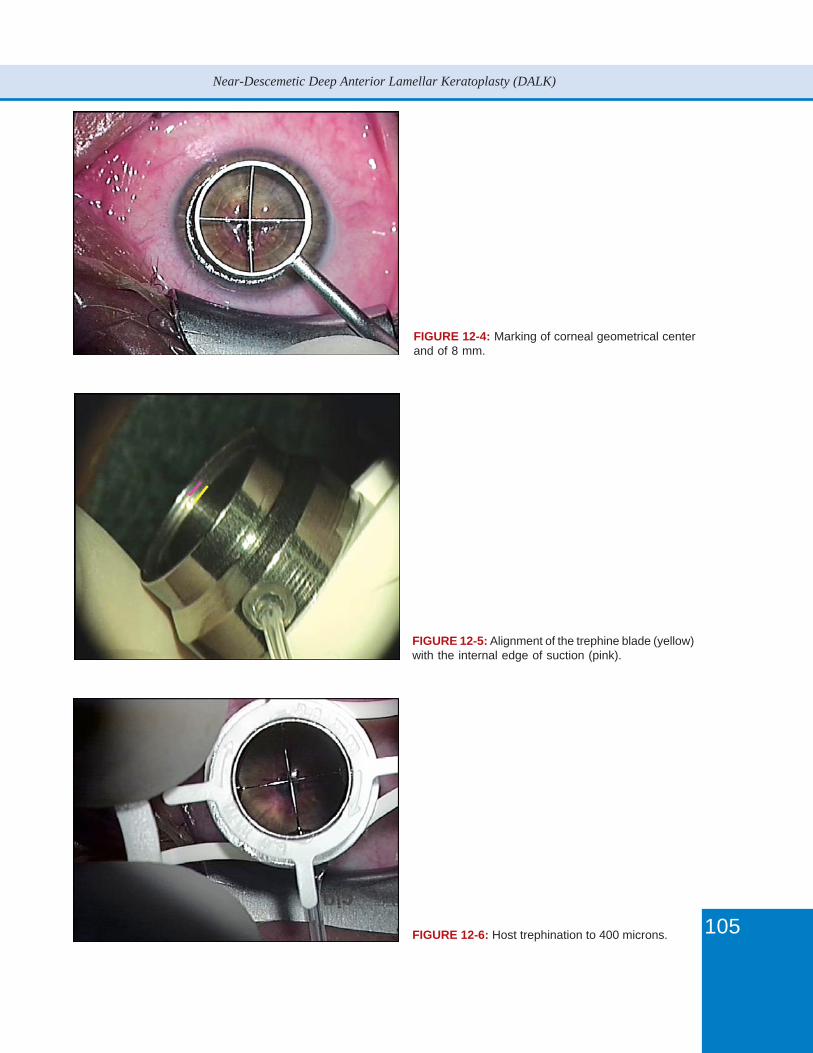
105
Near-Descemetic Deep Anterior Lamellar Keratoplasty (DALK)
FIGURE 12-4: Marking of corneal geometrical centerand of 8 mm.
FIGURE 12-5: Alignment of the trephine blade (yellow)with the internal edge of suction (pink).
FIGURE 12-6: Host trephination to 400 microns.
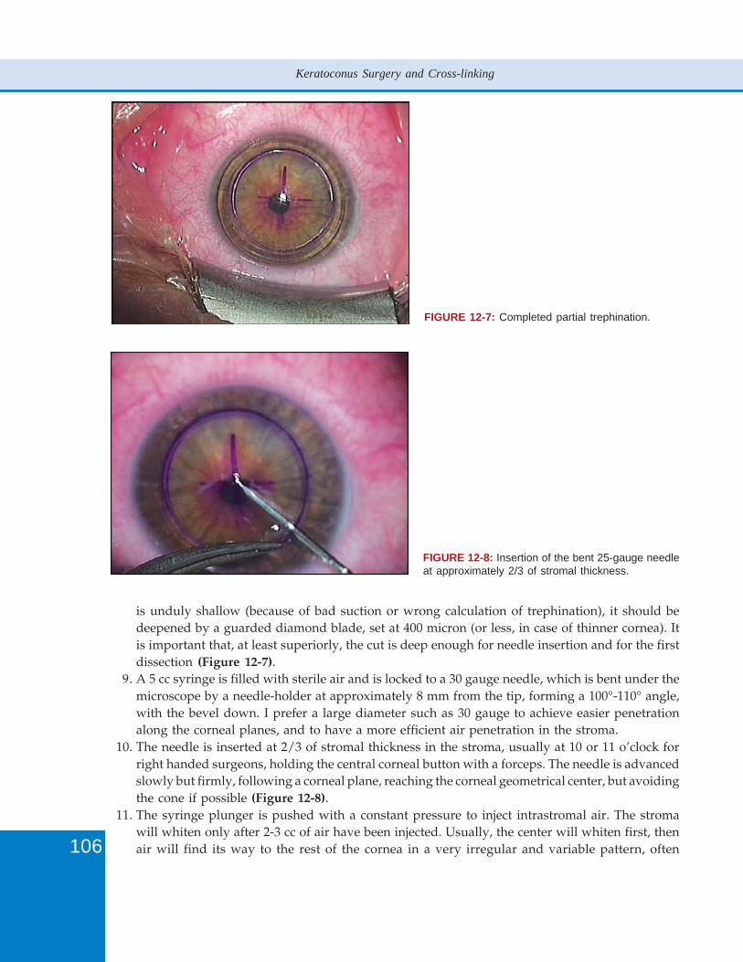
106
Keratoconus Surgery and Cross-linking
is unduly shallow (because of bad suction or wrong calculation of trephination), it should bedeepened by a guarded diamond blade, set at 400 micron (or less, in case of thinner cornea). Itis important that, at least superiorly, the cut is deep enough for needle insertion and for the firstdissection (Figure 12-7).
9. A 5 cc syringe is filled with sterile air and is locked to a 30 gauge needle, which is bent under themicroscope by a needle-holder at approximately 8 mm from the tip, forming a 100°-110° angle,with the bevel down. I prefer a large diameter such as 30 gauge to achieve easier penetrationalong the corneal planes, and to have a more efficient air penetration in the stroma.
10. The needle is inserted at 2/3 of stromal thickness in the stroma, usually at 10 or 11 o’clock forright handed surgeons, holding the central corneal button with a forceps. The needle is advancedslowly but firmly, following a corneal plane, reaching the corneal geometrical center, but avoidingthe cone if possible (Figure 12-8).
11. The syringe plunger is pushed with a constant pressure to inject intrastromal air. The stromawill whiten only after 2-3 cc of air have been injected. Usually, the center will whiten first, thenair will find its way to the rest of the cornea in a very irregular and variable pattern, often
FIGURE 12-7: Completed partial trephination.
FIGURE 12-8: Insertion of the bent 25-gauge needleat approximately 2/3 of stromal thickness.
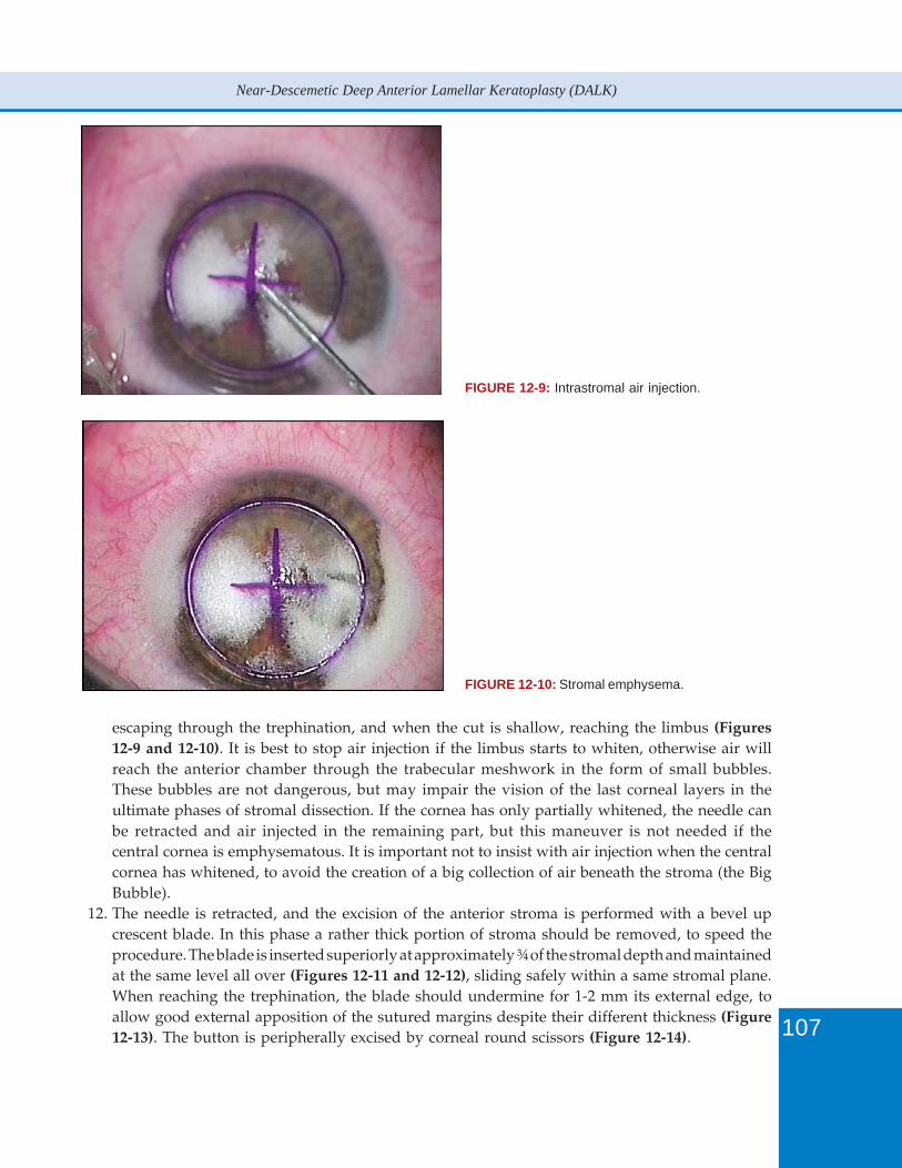
107
Near-Descemetic Deep Anterior Lamellar Keratoplasty (DALK)
FIGURE 12-9: Intrastromal air injection.
FIGURE 12-10: Stromal emphysema.
escaping through the trephination, and when the cut is shallow, reaching the limbus (Figures12-9 and 12-10). It is best to stop air injection if the limbus starts to whiten, otherwise air willreach the anterior chamber through the trabecular meshwork in the form of small bubbles.These bubbles are not dangerous, but may impair the vision of the last corneal layers in theultimate phases of stromal dissection. If the cornea has only partially whitened, the needle canbe retracted and air injected in the remaining part, but this maneuver is not needed if thecentral cornea is emphysematous. It is important not to insist with air injection when the centralcornea has whitened, to avoid the creation of a big collection of air beneath the stroma (the BigBubble).
12. The needle is retracted, and the excision of the anterior stroma is performed with a bevel upcrescent blade. In this phase a rather thick portion of stroma should be removed, to speed theprocedure. The blade is inserted superiorly at approximately ¾ of the stromal depth and maintainedat the same level all over (Figures 12-11 and 12-12), sliding safely within a same stromal plane.When reaching the trephination, the blade should undermine for 1-2 mm its external edge, toallow good external apposition of the sutured margins despite their different thickness (Figure12-13). The button is peripherally excised by corneal round scissors (Figure 12-14).
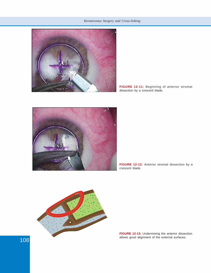
108
Keratoconus Surgery and Cross-linking
FIGURE 12-11: Beginning of anterior stromaldissection by a crescent blade.
FIGURE 12-12: Anterior stromal dissection by acrescent blade.
FIGURE 12-13: Undermining the anterior dissectionallows good alignment of the external surfaces.
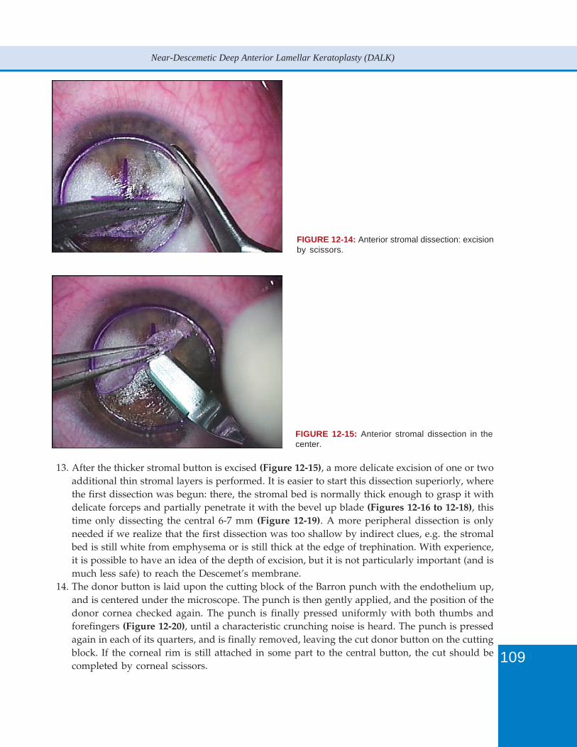
109
Near-Descemetic Deep Anterior Lamellar Keratoplasty (DALK)
13. After the thicker stromal button is excised (Figure 12-15), a more delicate excision of one or twoadditional thin stromal layers is performed. It is easier to start this dissection superiorly, wherethe first dissection was begun: there, the stromal bed is normally thick enough to grasp it withdelicate forceps and partially penetrate it with the bevel up blade (Figures 12-16 to 12-18), thistime only dissecting the central 6-7 mm (Figure 12-19). A more peripheral dissection is onlyneeded if we realize that the first dissection was too shallow by indirect clues, e.g. the stromalbed is still white from emphysema or is still thick at the edge of trephination. With experience,it is possible to have an idea of the depth of excision, but it is not particularly important (and ismuch less safe) to reach the Descemet’s membrane.
14. The donor button is laid upon the cutting block of the Barron punch with the endothelium up,and is centered under the microscope. The punch is then gently applied, and the position of thedonor cornea checked again. The punch is finally pressed uniformly with both thumbs andforefingers (Figure 12-20), until a characteristic crunching noise is heard. The punch is pressedagain in each of its quarters, and is finally removed, leaving the cut donor button on the cuttingblock. If the corneal rim is still attached in some part to the central button, the cut should becompleted by corneal scissors.
FIGURE 12-14: Anterior stromal dissection: excisionby scissors.
FIGURE 12-15: Anterior stromal dissection in thecenter.
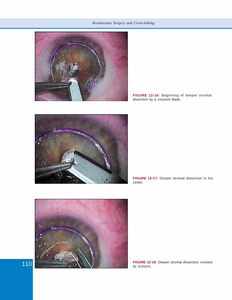
110
Keratoconus Surgery and Cross-linking
FIGURE 12-16: Beginning of deeper stromaldissection by a crescent blade.
FIGURE 12-17: Deeper stromal dissection in thecenter.
FIGURE 12-18: Deeper stromal dissection: excisionby scissors.
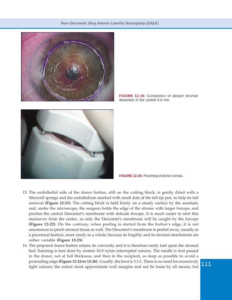
111
Near-Descemetic Deep Anterior Lamellar Keratoplasty (DALK)
15. The endothelial side of the donor button, still on the cutting block, is gently dried with aMerocell sponge and the endothelium marked with small dots of the felt tip pen, to help its fullremoval (Figure 12-21). The cutting block is held firmly on a steady surface by the assistant,and, under the microscope, the surgeon holds the edge of the stroma with larger forceps, andpinches the central Descemet’s membrane with delicate forceps. It is much easier to start thismaneuver from the center, as only the Descemet’s membrane will be caught by the forceps(Figure 12-22). On the contrary, when peeling is started from the button’s edge, it is notuncommon to pinch stromal tissue as well. The Descemet’s membrane is peeled away, usually ina piecemeal fashion, more rarely as a whole, because its fragility and its stromal attachments arerather variable (Figure 12-23).
16. The prepared donor button retains its convexity and it is therefore easily laid upon the stromalbed. Suturing is best done by sixteen 10-0 nylon interrupted sutures. The needle is first passedin the donor, not at full thickness, and then in the recipient, as deep as possible to avoid aprotruding edge (Figure 12-24 to 12-26). Usually, the knot is 3:1:1. There is no need for excessivelytight sutures; the suture must approximate well margins and not be loose by all means, but
FIGURE 12-19: Completion of deeper stromaldissection in the central 5-6 mm.
FIGURE 12-20: Punching of donor cornea.
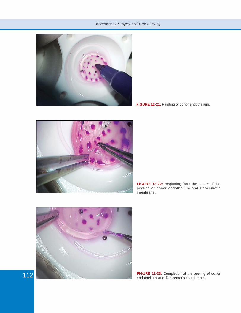
112
Keratoconus Surgery and Cross-linking
FIGURE 12-21: Painting of donor endothelium.
FIGURE 12-22: Beginning from the center of thepeeling of donor endothelium and Descemet’smembrane.
FIGURE 12-23: Completion of the peeling of donorendothelium and Descemet’s membrane.
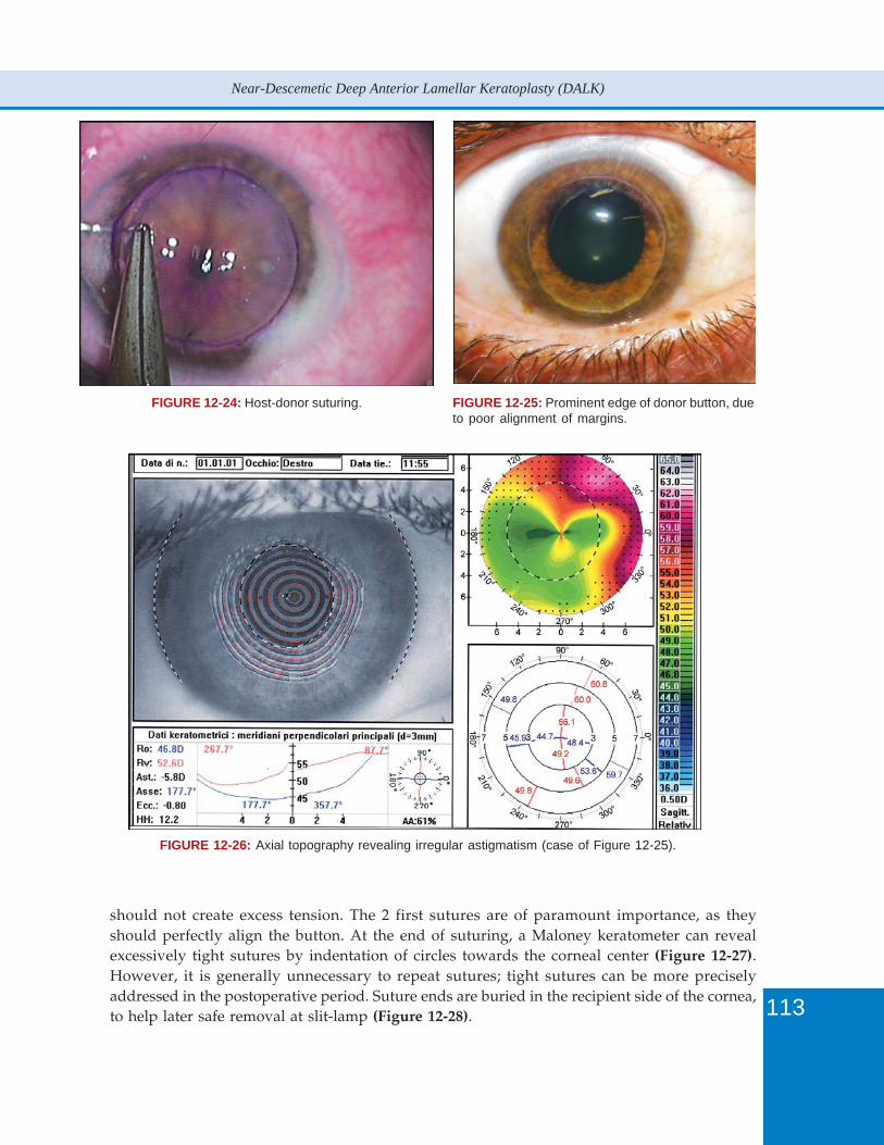
113
Near-Descemetic Deep Anterior Lamellar Keratoplasty (DALK)
FIGURE 12-24: Host-donor suturing. FIGURE 12-25: Prominent edge of donor button, dueto poor alignment of margins.
FIGURE 12-26: Axial topography revealing irregular astigmatism (case of Figure 12-25).
should not create excess tension. The 2 first sutures are of paramount importance, as theyshould perfectly align the button. At the end of suturing, a Maloney keratometer can revealexcessively tight sutures by indentation of circles towards the corneal center (Figure 12-27).However, it is generally unnecessary to repeat sutures; tight sutures can be more preciselyaddressed in the postoperative period. Suture ends are buried in the recipient side of the cornea,to help later safe removal at slit-lamp (Figure 12-28).
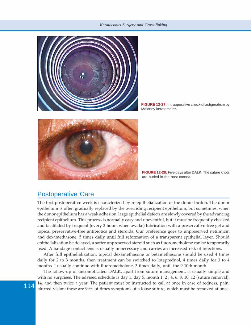
114
Keratoconus Surgery and Cross-linking
FIGURE 12-27: Intraoperative check of astigmatism byMaloney keratometer.
FIGURE 12-28: Five days after DALK. The suture knotsare buried in the host cornea.
Postoperative CareThe first postoperative week is characterized by re-epithelialization of the donor button. The donorepithelium is often gradually replaced by the overriding recipient epithelium, but sometimes, whenthe donor epithelium has a weak adhesion, large epithelial defects are slowly covered by the advancingrecipient epithelium. This process is normally easy and uneventful, but it must be frequently checkedand facilitated by frequent (every 2 hours when awake) lubrication with a preservative-free gel andtopical preservative-free antibiotics and steroids. Our preference goes to unpreserved netilmicinand dexamethasone, 5 times daily until full reformation of a transparent epithelial layer. Shouldepithelialization be delayed, a softer unpreserved steroid such as fluorometholone can be temporarilyused. A bandage contact lens is usually unnecessary and carries an increased risk of infections.
After full epithelialization, topical dexamethasone or betamethasone should be used 4 timesdaily for 2 to 3 months, then treatment can be switched to loteprednol, 4 times daily for 3 to 4months. I usually continue with fluorometholone, 3 times daily, until the 9-10th month.
The follow-up of uncomplicated DALK, apart from suture management, is usually simple andwith no surprises. The advised schedule is day 1, day 5, month 1, 2 , 4, 6, 8, 10, 12 (suture removal),14, and then twice a year. The patient must be instructed to call at once in case of redness, pain,blurred vision: these are 99% of times symptoms of a loose suture, which must be removed at once.
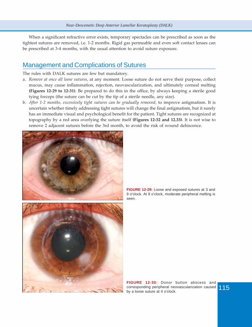
115
Near-Descemetic Deep Anterior Lamellar Keratoplasty (DALK)
When a significant refractive error exists, temporary spectacles can be prescribed as soon as thetightest sutures are removed, i.e. 1-2 months. Rigid gas permeable and even soft contact lenses canbe prescribed at 3-4 months, with the usual attention to avoid suture exposure.
Management and Complications of SuturesThe rules with DALK sutures are few but mandatory.a. Remove at once all loose sutures, at any moment. Loose suture do not serve their purpose, collect
mucus, may cause inflammation, rejection, neovascularization, and ultimately corneal melting(Figures 12-29 to 12-31). Be prepared to do this in the office, by always keeping a sterile goodtying forceps (the suture can be cut by the tip of a sterile needle, any size).
b. After 1-2 months, excessively tight sutures can be gradually removed, to improve astigmatism. It isuncertain whether timely addressing tight sutures will change the final astigmatism, but it surelyhas an immediate visual and psychological benefit for the patient. Tight sutures are recognized attopography by a red area overlying the suture itself (Figures 12-32 and 12.33). It is not wise toremove 2 adjacent sutures before the 3rd month, to avoid the risk of wound dehiscence.
FIGURE 12-29: Loose and exposed sutures at 3 and9 o’clock. At 9 o’clock, moderate peripheral melting isseen.
FIGURE 12-30: Donor button abscess andcorresponding peripheral neovascularization causedby a loose suture at 4 o’clock.
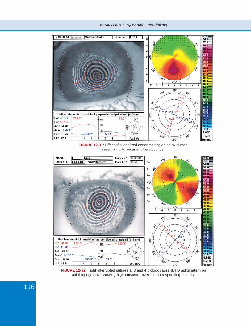
116
Keratoconus Surgery and Cross-linking
FIGURE 12-31: Effect of a localized donor melting on an axial map,resembling to recurrent keratoconus.
FIGURE 12-32: Tight interrupted sutures at 3 and 4 o’clock cause 8.4 D astigmatism onaxial topography, showing high curvature over the corresponding sutures.
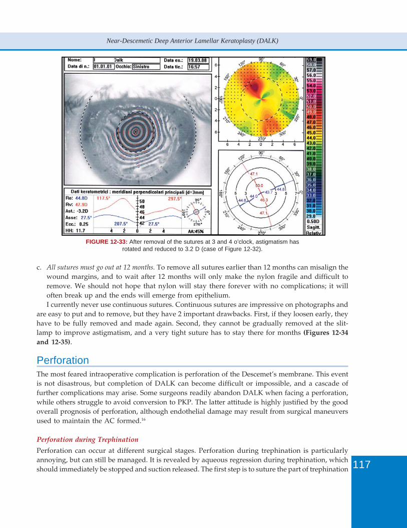
117
Near-Descemetic Deep Anterior Lamellar Keratoplasty (DALK)
c. All sutures must go out at 12 months. To remove all sutures earlier than 12 months can misalign thewound margins, and to wait after 12 months will only make the nylon fragile and difficult toremove. We should not hope that nylon will stay there forever with no complications; it willoften break up and the ends will emerge from epithelium.I currently never use continuous sutures. Continuous sutures are impressive on photographs and
are easy to put and to remove, but they have 2 important drawbacks. First, if they loosen early, theyhave to be fully removed and made again. Second, they cannot be gradually removed at the slit-lamp to improve astigmatism, and a very tight suture has to stay there for months (Figures 12-34and 12-35).
PerforationThe most feared intraoperative complication is perforation of the Descemet’s membrane. This eventis not disastrous, but completion of DALK can become difficult or impossible, and a cascade offurther complications may arise. Some surgeons readily abandon DALK when facing a perforation,while others struggle to avoid conversion to PKP. The latter attitude is highly justified by the goodoverall prognosis of perforation, although endothelial damage may result from surgical maneuversused to maintain the AC formed.16
Perforation during Trephination
Perforation can occur at different surgical stages. Perforation during trephination is particularlyannoying, but can still be managed. It is revealed by aqueous regression during trephination, whichshould immediately be stopped and suction released. The first step is to suture the part of trephination
FIGURE 12-33: After removal of the sutures at 3 and 4 o’clock, astigmatism hasrotated and reduced to 3.2 D (case of Figure 12-32).
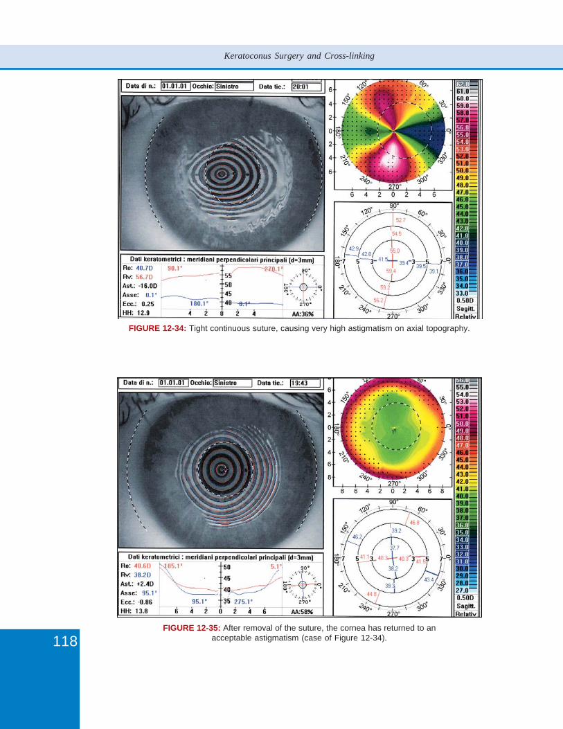
118
Keratoconus Surgery and Cross-linking
FIGURE 12-34: Tight continuous suture, causing very high astigmatism on axial topography.
FIGURE 12-35: After removal of the suture, the cornea has returned to anacceptable astigmatism (case of Figure 12-34).
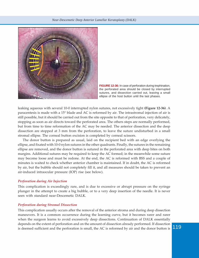
119
Near-Descemetic Deep Anterior Lamellar Keratoplasty (DALK)
leaking aqueous with several 10-0 interrupted nylon sutures, not excessively tight (Figure 12-36). Aparacentesis is made with a 15° blade and AC is reformed by air. The intrastromal injection of air isstill possible, but it should be carried out from the site opposite to that of perforation, very delicately,stopping as soon as air directs toward the perforated area. The others steps are normally performed,but from time to time reformation of the AC may be needed. The anterior dissection and the deepdissection are stopped at 3 mm from the perforation, to leave the suture undisturbed in a smallstromal ellipse. The corneal button excision is completed by corneal scissors.
The donor button is prepared as usual, laid on the recipient bed with an edge overlying theellipse, and fixated with 10-0 nylon sutures in the other quadrants. Finally, the sutures in the remainingellipse are removed, and the donor button is sutured in the perforated area with deep bites on bothmargins. Additional sutures may be required to keep the AC formed; in the meanwhile some suturemay become loose and must be redone. At the end, the AC is reformed with BSS and a couple ofminutes is waited to check whether anterior chamber is maintained. If in doubt, the AC is reformedby air, but the bubble should not completely fill it, and all measures should be taken to prevent anair-induced intraocular pressure (IOP) rise (see below).
Perforation during Air Injection
This complication is exceedingly rare, and is due to excessive or abrupt pressure on the syringeplunger in the attempt to create a big bubble, or to a very deep insertion of the needle. It is neverseen with standard near-Descemetic DALK.
Perforation during Stromal Dissection
This complication usually occurs after the removal of the anterior stroma and during deep dissectionmaneuvers. It is a common occurrence during the learning curve, but it becomes rarer and rarerwhen the surgeon learns to avoid excessively deep dissections. Continuation of DALK essentiallydepends on the extent of perforation and on the amount of dissection already performed. If dissectionis deemed sufficient and the perforation is small, the AC is reformed by air and the donor button is
FIGURE 12-36: In case of perforation during trephination,the perforated area should be closed by interruptedsutures, and dissection carried out, leaving a smallellipse of the host button until the last phases.
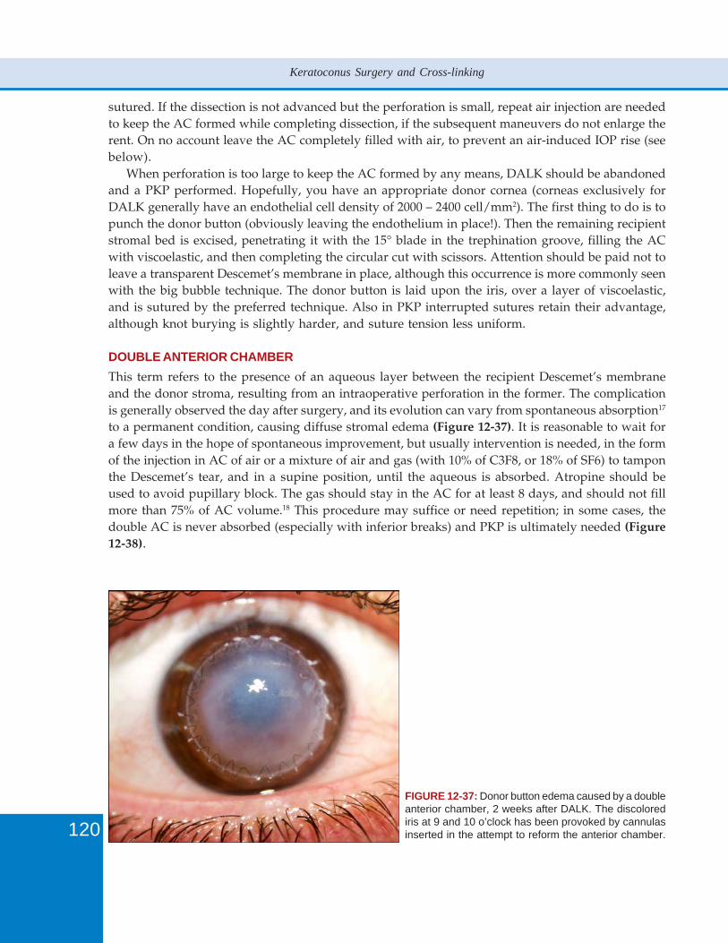
120
Keratoconus Surgery and Cross-linking
sutured. If the dissection is not advanced but the perforation is small, repeat air injection are neededto keep the AC formed while completing dissection, if the subsequent maneuvers do not enlarge therent. On no account leave the AC completely filled with air, to prevent an air-induced IOP rise (seebelow).
When perforation is too large to keep the AC formed by any means, DALK should be abandonedand a PKP performed. Hopefully, you have an appropriate donor cornea (corneas exclusively forDALK generally have an endothelial cell density of 2000 – 2400 cell/mm2). The first thing to do is topunch the donor button (obviously leaving the endothelium in place!). Then the remaining recipientstromal bed is excised, penetrating it with the 15° blade in the trephination groove, filling the ACwith viscoelastic, and then completing the circular cut with scissors. Attention should be paid not toleave a transparent Descemet’s membrane in place, although this occurrence is more commonly seenwith the big bubble technique. The donor button is laid upon the iris, over a layer of viscoelastic,and is sutured by the preferred technique. Also in PKP interrupted sutures retain their advantage,although knot burying is slightly harder, and suture tension less uniform.
DOUBLE ANTERIOR CHAMBER
This term refers to the presence of an aqueous layer between the recipient Descemet’s membraneand the donor stroma, resulting from an intraoperative perforation in the former. The complicationis generally observed the day after surgery, and its evolution can vary from spontaneous absorption17
to a permanent condition, causing diffuse stromal edema (Figure 12-37). It is reasonable to wait fora few days in the hope of spontaneous improvement, but usually intervention is needed, in the formof the injection in AC of air or a mixture of air and gas (with 10% of C3F8, or 18% of SF6) to tamponthe Descemet’s tear, and in a supine position, until the aqueous is absorbed. Atropine should beused to avoid pupillary block. The gas should stay in the AC for at least 8 days, and should not fillmore than 75% of AC volume.18 This procedure may suffice or need repetition; in some cases, thedouble AC is never absorbed (especially with inferior breaks) and PKP is ultimately needed (Figure12-38).
FIGURE 12-37: Donor button edema caused by a doubleanterior chamber, 2 weeks after DALK. The discolorediris at 9 and 10 o’clock has been provoked by cannulasinserted in the attempt to reform the anterior chamber.
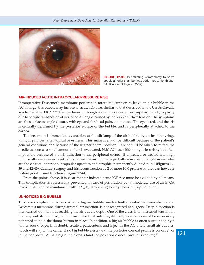
121
Near-Descemetic Deep Anterior Lamellar Keratoplasty (DALK)
AIR-INDUCED ACUTE INTRAOCULAR PRESSURE RISE
Intraoperative Descemet’s membrane perforation forces the surgeon to leave an air bubble in theAC. If large, this bubble may induce an acute IOP rise, similar to that described in the Urrets-Zavaliasyndrome after PKP.16, 19 The mechanism, though sometimes referred as pupillary block, is partlydue to peripheral adhesion of iris to the AC angle, caused by the bubble surface tension. The symptomsare those of acute angle closure, with eye and forehead pain, and nausea. The eye is red, and the irisis centrally deformed by the posterior surface of the bubble, and is peripherally attached to thecornea.
The treatment is immediate evacuation at the slit-lamp of the air bubble by an insulin syringewithout plunger, after topical anesthesia. This maneuver can be difficult because of the patient’sgeneral conditions and because of the iris peripheral position. Care should be taken to retract theneedle as soon as a small amount of air is evacuated. Nd:YAG laser iridotomy is less risky but oftenimpossible because of the iris adhesion to the peripheral cornea. If untreated or treated late, highIOP usually resolves in 12-24 hours, when the air bubble is partially absorbed. Long-term sequelaeare the classical anterior subcapsular opacities and atrophic, permanently dilated pupil (Figures 12-39 and 12-40). Cataract surgery and iris reconstruction by 2 or more 10-0 prolene sutures can howeverrestore good visual function (Figure 12-41).
From the points above, it is clear that air-induced acute IOP rise must be avoided by all means.This complication is successfully prevented, in case of perforation, by: a) moderate use of air in CA(avoid if AC can be maintained with BSS); b) atropine; c) hourly check of pupil dilation.
UNNOTICED BIG BUBBLE
This rare complication occurs when a big air bubble, inadvertently created between stroma andDescemet’s membrane during stromal air injection, is not recognized at surgery. Deep dissection isthen carried out, without reaching the air bubble depth. One of the clues is an increased tension onthe recipient stromal bed, which can make final suturing difficult, as sutures must be excessivelytightened to hold the donor button in place. In addition, a big air bubble is often surrounded by awhiter round edge. If in doubt, create a paracentesis and inject in the AC a few small air bubbles,which will stay in the center if no big bubble exists (and the posterior corneal profile is concave), orin the peripheral AC if a big bubble exists (and the posterior corneal profile is convex).20
FIGURE 12-38: Penetrating keratoplasty to solvedouble anterior chamber was performed 1 month afterDALK (case of Figure 12-37).
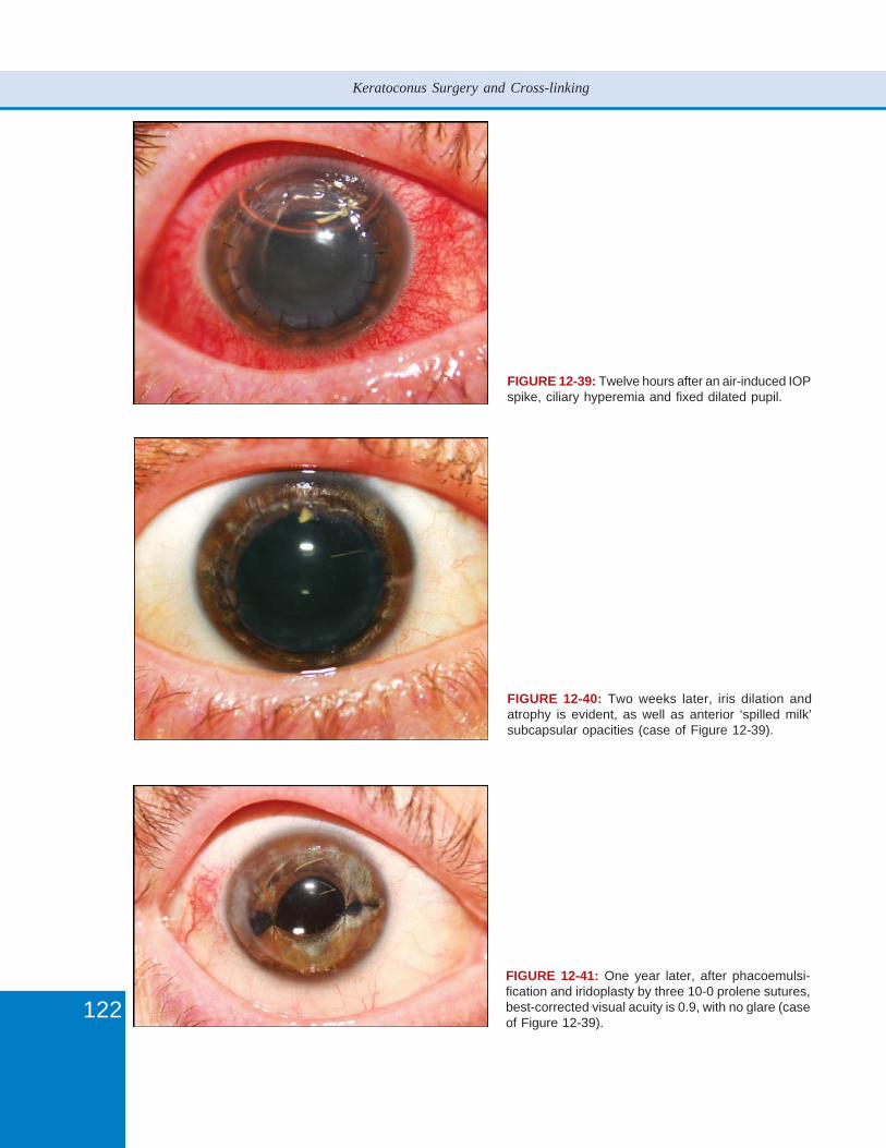
122
Keratoconus Surgery and Cross-linking
FIGURE 12-39: Twelve hours after an air-induced IOPspike, ciliary hyperemia and fixed dilated pupil.
FIGURE 12-40: Two weeks later, iris dilation andatrophy is evident, as well as anterior ‘spilled milk’subcapsular opacities (case of Figure 12-39).
FIGURE 12-41: One year later, after phacoemulsi-fication and iridoplasty by three 10-0 prolene sutures,best-corrected visual acuity is 0.9, with no glare (caseof Figure 12-39).
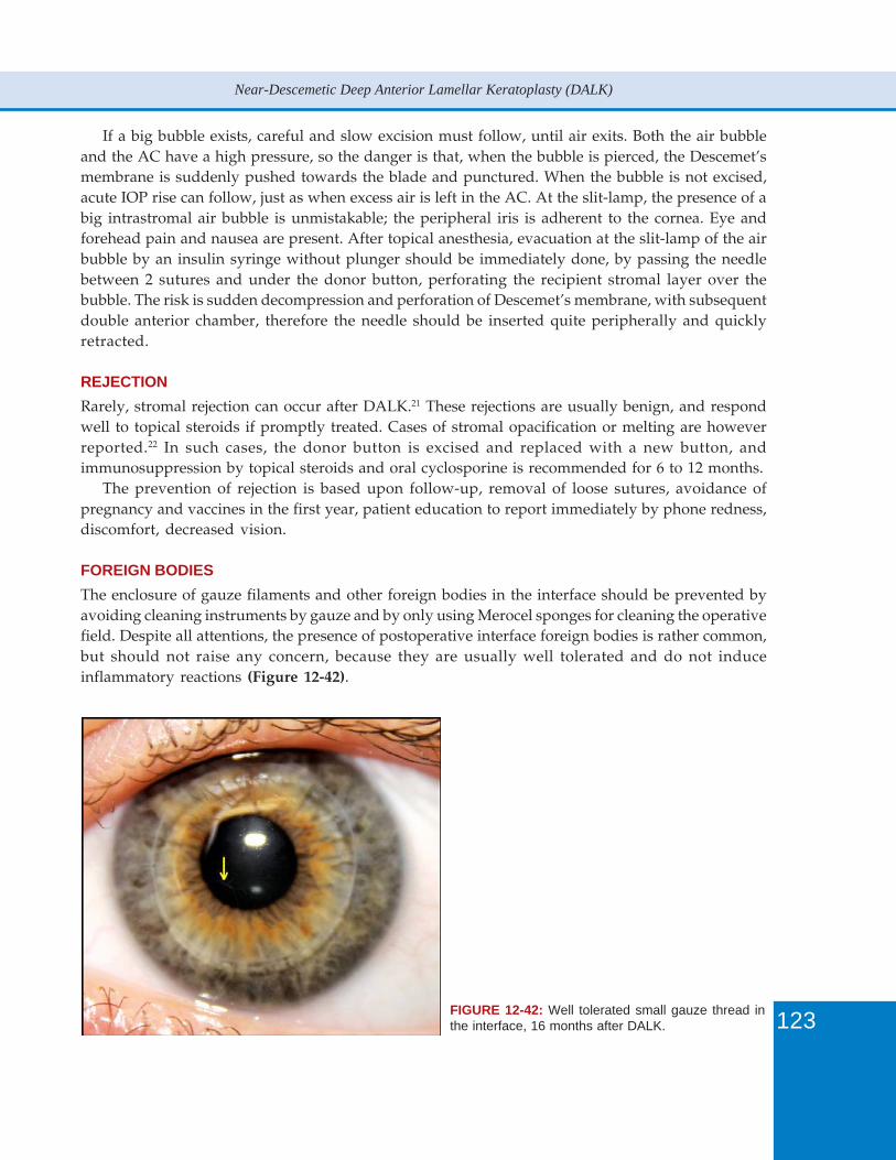
123
Near-Descemetic Deep Anterior Lamellar Keratoplasty (DALK)
If a big bubble exists, careful and slow excision must follow, until air exits. Both the air bubbleand the AC have a high pressure, so the danger is that, when the bubble is pierced, the Descemet’smembrane is suddenly pushed towards the blade and punctured. When the bubble is not excised,acute IOP rise can follow, just as when excess air is left in the AC. At the slit-lamp, the presence of abig intrastromal air bubble is unmistakable; the peripheral iris is adherent to the cornea. Eye andforehead pain and nausea are present. After topical anesthesia, evacuation at the slit-lamp of the airbubble by an insulin syringe without plunger should be immediately done, by passing the needlebetween 2 sutures and under the donor button, perforating the recipient stromal layer over thebubble. The risk is sudden decompression and perforation of Descemet’s membrane, with subsequentdouble anterior chamber, therefore the needle should be inserted quite peripherally and quicklyretracted.
REJECTION
Rarely, stromal rejection can occur after DALK.21 These rejections are usually benign, and respondwell to topical steroids if promptly treated. Cases of stromal opacification or melting are howeverreported.22 In such cases, the donor button is excised and replaced with a new button, andimmunosuppression by topical steroids and oral cyclosporine is recommended for 6 to 12 months.
The prevention of rejection is based upon follow-up, removal of loose sutures, avoidance ofpregnancy and vaccines in the first year, patient education to report immediately by phone redness,discomfort, decreased vision.
FOREIGN BODIES
The enclosure of gauze filaments and other foreign bodies in the interface should be prevented byavoiding cleaning instruments by gauze and by only using Merocel sponges for cleaning the operativefield. Despite all attentions, the presence of postoperative interface foreign bodies is rather common,but should not raise any concern, because they are usually well tolerated and do not induceinflammatory reactions (Figure 12-42).
FIGURE 12-42: Well tolerated small gauze thread inthe interface, 16 months after DALK.
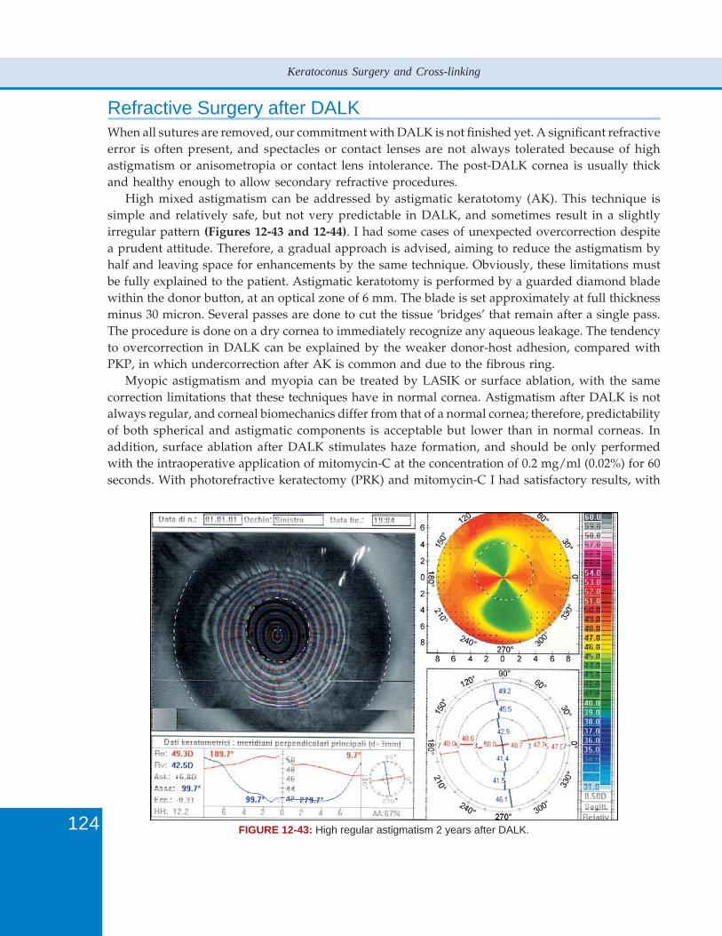
124
Keratoconus Surgery and Cross-linking
Refractive Surgery after DALKWhen all sutures are removed, our commitment with DALK is not finished yet. A significant refractiveerror is often present, and spectacles or contact lenses are not always tolerated because of highastigmatism or anisometropia or contact lens intolerance. The post-DALK cornea is usually thickand healthy enough to allow secondary refractive procedures.
High mixed astigmatism can be addressed by astigmatic keratotomy (AK). This technique issimple and relatively safe, but not very predictable in DALK, and sometimes result in a slightlyirregular pattern (Figures 12-43 and 12-44). I had some cases of unexpected overcorrection despitea prudent attitude. Therefore, a gradual approach is advised, aiming to reduce the astigmatism byhalf and leaving space for enhancements by the same technique. Obviously, these limitations mustbe fully explained to the patient. Astigmatic keratotomy is performed by a guarded diamond bladewithin the donor button, at an optical zone of 6 mm. The blade is set approximately at full thicknessminus 30 micron. Several passes are done to cut the tissue ‘bridges’ that remain after a single pass.The procedure is done on a dry cornea to immediately recognize any aqueous leakage. The tendencyto overcorrection in DALK can be explained by the weaker donor-host adhesion, compared withPKP, in which undercorrection after AK is common and due to the fibrous ring.
Myopic astigmatism and myopia can be treated by LASIK or surface ablation, with the samecorrection limitations that these techniques have in normal cornea. Astigmatism after DALK is notalways regular, and corneal biomechanics differ from that of a normal cornea; therefore, predictabilityof both spherical and astigmatic components is acceptable but lower than in normal corneas. Inaddition, surface ablation after DALK stimulates haze formation, and should be only performedwith the intraoperative application of mitomycin-C at the concentration of 0.2 mg/ml (0.02%) for 60seconds. With photorefractive keratectomy (PRK) and mitomycin-C I had satisfactory results, with
FIGURE 12-43: High regular astigmatism 2 years after DALK.
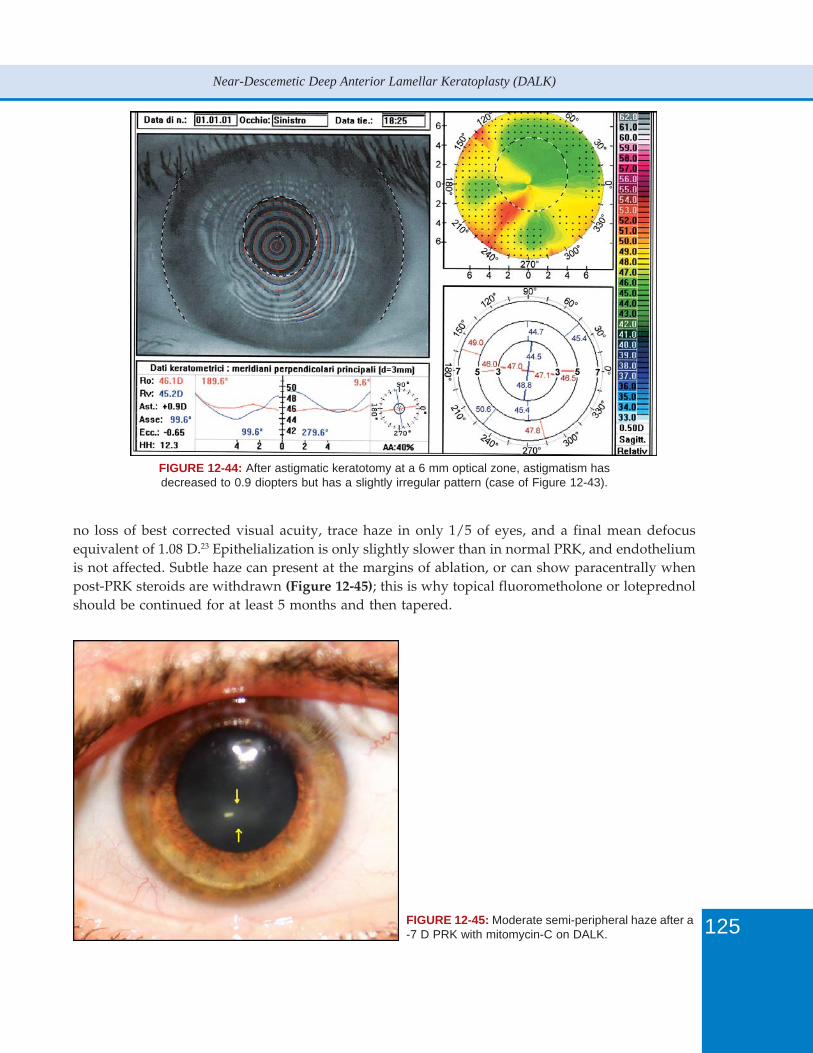
125
Near-Descemetic Deep Anterior Lamellar Keratoplasty (DALK)
FIGURE 12-44: After astigmatic keratotomy at a 6 mm optical zone, astigmatism hasdecreased to 0.9 diopters but has a slightly irregular pattern (case of Figure 12-43).
no loss of best corrected visual acuity, trace haze in only 1/5 of eyes, and a final mean defocusequivalent of 1.08 D.23 Epithelialization is only slightly slower than in normal PRK, and endotheliumis not affected. Subtle haze can present at the margins of ablation, or can show paracentrally whenpost-PRK steroids are withdrawn (Figure 12-45); this is why topical fluorometholone or loteprednolshould be continued for at least 5 months and then tapered.
FIGURE 12-45: Moderate semi-peripheral haze after a-7 D PRK with mitomycin-C on DALK.
126
Keratoconus Surgery and Cross-linking
Larger spherical errors require intraocular techniques, which will be explained in detail in otherchapters of this book. Refractive lens exchange is advised in presbyopic age or when a lens opacityis present or developing. Phakic IOL implantation is instead the preferred technique in youngereyes, provided that a good endothelial cell density is present.
Personal SeriesI have moved to near-Descemetic in May 2006, after noticing the difficulty of a consistent big bubbleformation. Since then, I applied the near-Descemetic technique (as described above) to all cases of KCundergoing DALK. The following series is a consecutive, single-surgeon retrospective analysis of allcases with a minimum follow-up of 1 year, and all refractive data were collected without sutures.
DALK was completed in 37 eyes of 37 patients with KC (mean age 32.6 years, SD 8.4, range 17 to49). Males were 18. In 1 case a microperforation occurred, but PKP was not needed. In 3 early cases,a big bubble formed, but dissection was only continued to evacuate air, but no attempt was done tobare Descemet’s membrane.
In 1 case, a peripheral abscess in the donor button developed in correspondence of a loose suture,6 months postoperatively, when the patient was abroad and could access to medical assistance onlyafter a week of pain and redness. Topical treatment with gatifloxacin was carried out, but the suturewas not removed. The patient returned to our institute, the suture removed, and the abscess healedwith no visual consequences.
Inferior unexplained melting of the donor button was noticed in another case, 2 weekspostoperatively. Sutures in the damaged area were instantly removed, and visual acuity remainedacceptable (0.7 unaided), but topography showed an inferior steepening. Central pachymetry was610 micron. At surgery, the donor cornea had appeared less rigid than usual donor corneas, butotherwise normal and transparent.
No cases of rejection occurred.Despite the near-Descemetic dissection, visual acuity was comparable to my previous cases of
DALK by the Big Bubble technique with Descemet’s membrane baring. Mean best-spectacle correctedvisual acuity (BSCVA) was 0.77 (SD 0.13; range 0.5 to 1). Mean refractive astigmatism was 3.88 D (SD2.06; range 2 to 8). Mean spherical equivalent was -3.27 D (SD 2.28; range 0 to -9).
Mean BSCVA in the 25-eye Big Bubble series was 0.78 (SD 0.15) (not statistically significantdifference with t-test):
ConclusionThe development of DALK and its diffusion have enormously improved the perspectives of patientswith advanced KC. Nowadays there is only justification for PKP in KC in very thin or scarredcorneas. Surgical difficulty of most proposed DALK technique is essentially derived from the attemptto bare Descemet’s membrane at all costs, which is a risky and unnecessary goal. Near-DescemeticDALK yields comparable visual results and is accessible to all corneal surgeons, with a short learningcurve and minimal equipment. Since most postoperative complications arise from sutures (and visualacuity can be improved by early removal of tight sutures), follow-up is based upon their timelymanagement, minimal but prolonged topical therapy, patient information, and, eventually, possiblemanagement of the final refractive error.
127
Near-Descemetic Deep Anterior Lamellar Keratoplasty (DALK)
References1. Borderie VM, Werthel AL, Touzeau O, Allouch C, Boutboul S, Laroche L. Comparison of techniques used for
removing the recipient stroma in anterior lamellar keratoplasty. Arch Ophthalmol 2008;126:31-7.2. Marchini G, Mastropasqua L, Pedrotti E, Nubile M, Ciancaglini M, Sbabo A. Deep lamellar keratoplasty by
intracorneal dissection: a prospective clinical and confocal microscopic study. Ophthalmology 2006;113:1289-1300.3. Ardjomand N, Hau S, McAlister JC, et al. Quality of vision and graft thickness in deep anterior lamellar and
penetrating keratoplasty. Am J Ophthalmol 2007;143:228-35.4. Archila EA. Deep lamellar keratoplasty dissection of host tissue with intrastromal air injection. Cornea 1984/
85;3:217-8.5. Sugita J, Kondo J. Deep lamellar keratoplasty with complete removal of pathological stroma for vision improvement.
Br J Ophthalmol 1997;81:184-8.6. Melles GRJ, Lander F, Rietveld FJR, Remeijer L, Beekhuis WH, Binder PS. A new surgical technique for deep
stromal, anterior lamellar keratoplasty. Br J Ophthalmol 1999;83:327-33.7. Duong MH, Thimel S, Hoang Xuan T. Kératoplastie lamellaire avec aero-visco dissection. J Fr Ophthalmol
2001;24:930-6.8. Anwar M, Teichmann KD. Big-bubble technique to bare Descemet’s membrane in anterior lamellar keratoplasty.
J Cataract Refract Surg 2002;28:398-403.9. Anwar M, Teichmann KD. Deep lamellar keratoplasty. Surgical techniques for anterior lamellar keratoplasty with
and without baring of Descemet’s membrane. Cornea 2002;21:374-83.10. Anwar M, Teichmann KD. Planned near-Descemet’s dissection in deep lamellar keratoplasty, using air or fluid. In
John T (Ed). Surgical Techniques in Anterior and Posterior Lamellar Corneal Surgery. New Delhi: Jaypee, 2006;125-39.
11. Leccisotti A, Fields VS. Angle-supported phakic intraocular lenses in eyes with keratoconus and myopia. J CataractRefract Surg 2003;29:1530-6.
12. Leccisotti A. Refractive lens exchange in keratoconus. J Cataract Refract Surg 2006;32:742-6.13. Wollensak G, Spoerl E, Seiler T. Riboflavin/ultraviolet-A-induced collagen crosslinking for the treatment of
keratoconus. Am J Ophthalmol 2003;135:620-7.14. Colin J, Malet FJ. Intacs for the correction of keratoconus: two-year follow-up. J Cataract Refract Surg 2007;33:
69-74.15. Jurkunas U, Azar DT. Potential complications of ocular surgery in patients with coexistent keratoconus and Fuchs’
endothelial dystrophy. Ophthalmology 2006;113:2187-97.16. Leccisotti A. Descemet’s membrane perforation during deep anterior lamellar keratoplasty: Prognosis. J Cataract
Refract Surg 2007;33:825-9.17. Pakrou N, Fung S, Selva D, Chedade M, Leibovitch I. Deep lamellar keratoplasty in the treatment of keratoconus.
Ophthalmologica 2006;220:164-9.18. Teichmann KD, Anwar M. Intrastromal air, Big-Bubble technique. In John T (Ed) Surgical Techniques in Anterior
and Posterior Lamellar Corneal Surgery. New Dehli: Jaypee, 2006;102-16.19. Maurino V, Allan BD, Stevens JD, Tuft SJ. Fixed dilated pupil (Urrets-Zavalia syndrome) after air/gas injection after
deep lamellar keratoplasty for keratoconus. Am J Ophthalmol 2002;133:266-8.20. Parthasarathy A, Por YM, Tan DT. Use of a “small-bubble technique” to increase the success of Anwar’s “big-
bubble technique” for deep lamellar keratoplasty with complete baring of Descemet’s membrane. Br J Ophthalmol2007;91:1369-73.
21. Fontana L, Parente G, Tassinari G. Clinical outcomes after deep anterior lamellar keratoplasty using the Big-bubbletechnique in patients with keratoconus. Am J Ophthalmol 2007;143:117-24.
22. Wylegala E, Tarnawska D, Dobrowolski D. Deep lamellar keratoplasty for various corneal lesions. Eur J Ophthalmol2004;14:467-72.
23. Leccisotti A. Photorefractive keratectomy with mitomycin C after deep anterior lamellar keratoplasty forkeratoconus. Cornea 2008;27:417-20.

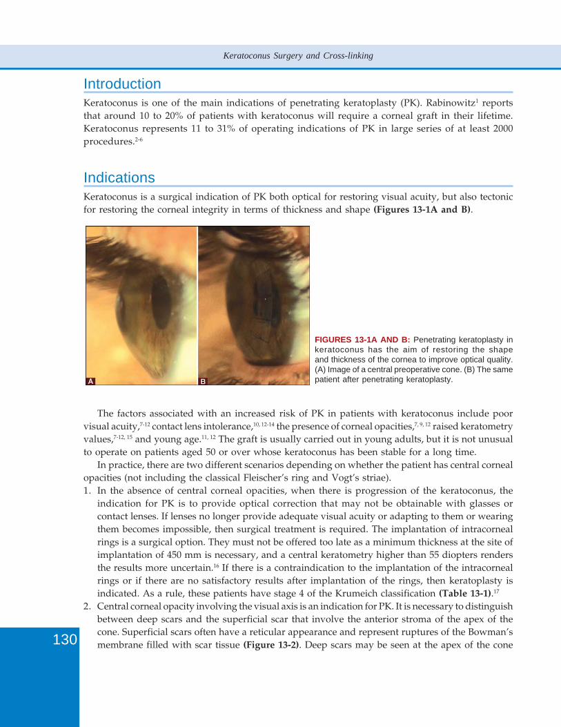
130
Keratoconus Surgery and Cross-linking
IntroductionKeratoconus is one of the main indications of penetrating keratoplasty (PK). Rabinowitz1 reportsthat around 10 to 20% of patients with keratoconus will require a corneal graft in their lifetime.Keratoconus represents 11 to 31% of operating indications of PK in large series of at least 2000procedures.2-6
IndicationsKeratoconus is a surgical indication of PK both optical for restoring visual acuity, but also tectonicfor restoring the corneal integrity in terms of thickness and shape (Figures 13-1A and B).
FIGURES 13-1A AND B: Penetrating keratoplasty inkeratoconus has the aim of restoring the shapeand thickness of the cornea to improve optical quality.(A) Image of a central preoperative cone. (B) The samepatient after penetrating keratoplasty.
The factors associated with an increased risk of PK in patients with keratoconus include poorvisual acuity,7-12 contact lens intolerance,10, 12-14 the presence of corneal opacities,7, 9, 12 raised keratometryvalues,7-12, 15 and young age.11, 12 The graft is usually carried out in young adults, but it is not unusualto operate on patients aged 50 or over whose keratoconus has been stable for a long time.
In practice, there are two different scenarios depending on whether the patient has central cornealopacities (not including the classical Fleischer’s ring and Vogt’s striae).1. In the absence of central corneal opacities, when there is progression of the keratoconus, the
indication for PK is to provide optical correction that may not be obtainable with glasses orcontact lenses. If lenses no longer provide adequate visual acuity or adapting to them or wearingthem becomes impossible, then surgical treatment is required. The implantation of intracornealrings is a surgical option. They must not be offered too late as a minimum thickness at the site ofimplantation of 450 mm is necessary, and a central keratometry higher than 55 diopters rendersthe results more uncertain.16 If there is a contraindication to the implantation of the intracornealrings or if there are no satisfactory results after implantation of the rings, then keratoplasty isindicated. As a rule, these patients have stage 4 of the Krumeich classification (Table 13-1).17
2. Central corneal opacity involving the visual axis is an indication for PK. It is necessary to distinguishbetween deep scars and the superficial scar that involve the anterior stroma of the apex of thecone. Superficial scars often have a reticular appearance and represent ruptures of the Bowman’smembrane filled with scar tissue (Figure 13-2). Deep scars may be seen at the apex of the cone
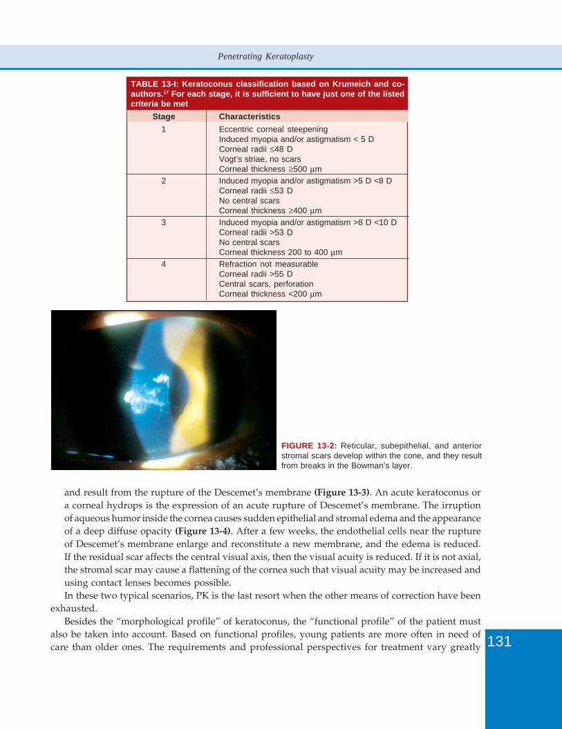
131
Penetrating Keratoplasty
TABLE 13-I: Keratoconus classification based on Krumeich and co-authors.17 For each stage, it is sufficient to have just one of the listedcriteria be met
Stage Characteristics1 Eccentric corneal steepening
Induced myopia and/or astigmatism < 5 DCorneal radii ≤48 DVogt’s striae, no scarsCorneal thickness ≥500 μm
2 Induced myopia and/or astigmatism >5 D <8 DCorneal radii ≤53 DNo central scarsCorneal thickness ≥400 μm
3 Induced myopia and/or astigmatism >8 D <10 DCorneal radii >53 DNo central scarsCorneal thickness 200 to 400 μm
4 Refraction not measurableCorneal radii >55 DCentral scars, perforationCorneal thickness <200 μm
FIGURE 13-2: Reticular, subepithelial, and anteriorstromal scars develop within the cone, and they resultfrom breaks in the Bowman’s layer.
and result from the rupture of the Descemet’s membrane (Figure 13-3). An acute keratoconus ora corneal hydrops is the expression of an acute rupture of Descemet’s membrane. The irruptionof aqueous humor inside the cornea causes sudden epithelial and stromal edema and the appearanceof a deep diffuse opacity (Figure 13-4). After a few weeks, the endothelial cells near the ruptureof Descemet’s membrane enlarge and reconstitute a new membrane, and the edema is reduced.If the residual scar affects the central visual axis, then the visual acuity is reduced. If it is not axial,the stromal scar may cause a flattening of the cornea such that visual acuity may be increased andusing contact lenses becomes possible.In these two typical scenarios, PK is the last resort when the other means of correction have been
exhausted.Besides the “morphological profile” of keratoconus, the “functional profile” of the patient must
also be taken into account. Based on functional profiles, young patients are more often in need ofcare than older ones. The requirements and professional perspectives for treatment vary greatly
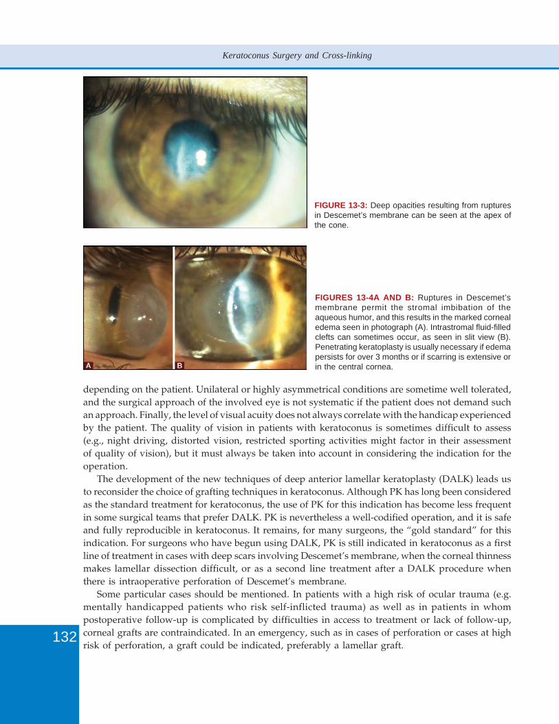
132
Keratoconus Surgery and Cross-linking
FIGURE 13-3: Deep opacities resulting from rupturesin Descemet’s membrane can be seen at the apex ofthe cone.
FIGURES 13-4A AND B: Ruptures in Descemet’smembrane permit the stromal imbibation of theaqueous humor, and this results in the marked cornealedema seen in photograph (A). Intrastromal fluid-filledclefts can sometimes occur, as seen in slit view (B).Penetrating keratoplasty is usually necessary if edemapersists for over 3 months or if scarring is extensive orin the central cornea.
depending on the patient. Unilateral or highly asymmetrical conditions are sometime well tolerated,and the surgical approach of the involved eye is not systematic if the patient does not demand suchan approach. Finally, the level of visual acuity does not always correlate with the handicap experiencedby the patient. The quality of vision in patients with keratoconus is sometimes difficult to assess(e.g., night driving, distorted vision, restricted sporting activities might factor in their assessmentof quality of vision), but it must always be taken into account in considering the indication for theoperation.
The development of the new techniques of deep anterior lamellar keratoplasty (DALK) leads usto reconsider the choice of grafting techniques in keratoconus. Although PK has long been consideredas the standard treatment for keratoconus, the use of PK for this indication has become less frequentin some surgical teams that prefer DALK. PK is nevertheless a well-codified operation, and it is safeand fully reproducible in keratoconus. It remains, for many surgeons, the “gold standard” for thisindication. For surgeons who have begun using DALK, PK is still indicated in keratoconus as a firstline of treatment in cases with deep scars involving Descemet’s membrane, when the corneal thinnessmakes lamellar dissection difficult, or as a second line treatment after a DALK procedure whenthere is intraoperative perforation of Descemet’s membrane.
Some particular cases should be mentioned. In patients with a high risk of ocular trauma (e.g.mentally handicapped patients who risk self-inflicted trauma) as well as in patients in whompostoperative follow-up is complicated by difficulties in access to treatment or lack of follow-up,corneal grafts are contraindicated. In an emergency, such as in cases of perforation or cases at highrisk of perforation, a graft could be indicated, preferably a lamellar graft.
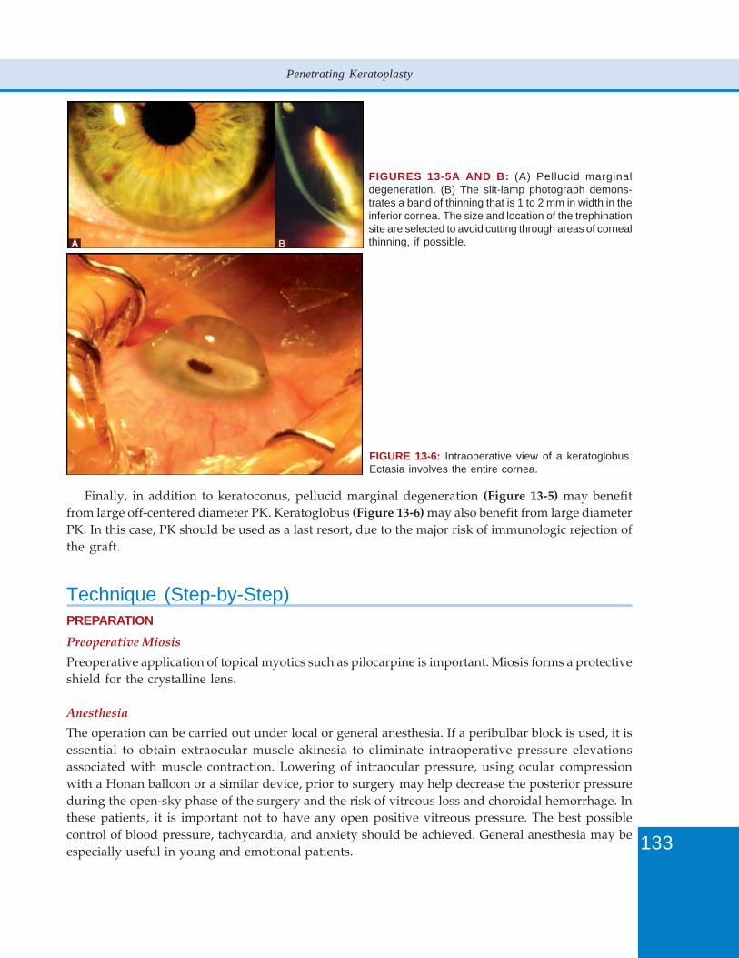
133
Penetrating Keratoplasty
FIGURES 13-5A AND B: (A) Pellucid marginaldegeneration. (B) The slit-lamp photograph demons-trates a band of thinning that is 1 to 2 mm in width in theinferior cornea. The size and location of the trephinationsite are selected to avoid cutting through areas of cornealthinning, if possible.
Finally, in addition to keratoconus, pellucid marginal degeneration (Figure 13-5) may benefitfrom large off-centered diameter PK. Keratoglobus (Figure 13-6) may also benefit from large diameterPK. In this case, PK should be used as a last resort, due to the major risk of immunologic rejection ofthe graft.
Technique (Step-by-Step)PREPARATIONPreoperative Miosis
Preoperative application of topical myotics such as pilocarpine is important. Miosis forms a protectiveshield for the crystalline lens.
Anesthesia
The operation can be carried out under local or general anesthesia. If a peribulbar block is used, it isessential to obtain extraocular muscle akinesia to eliminate intraoperative pressure elevationsassociated with muscle contraction. Lowering of intraocular pressure, using ocular compressionwith a Honan balloon or a similar device, prior to surgery may help decrease the posterior pressureduring the open-sky phase of the surgery and the risk of vitreous loss and choroidal hemorrhage. Inthese patients, it is important not to have any open positive vitreous pressure. The best possiblecontrol of blood pressure, tachycardia, and anxiety should be achieved. General anesthesia may beespecially useful in young and emotional patients.
FIGURE 13-6: Intraoperative view of a keratoglobus.Ectasia involves the entire cornea.
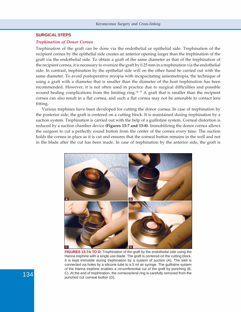
134
Keratoconus Surgery and Cross-linking
SURGICAL STEPSTrephination of Donor Cornea
Trephination of the graft can be done via the endothelial or epithelial side. Trephination of therecipient cornea by the epithelial side creates an anterior opening larger than the trephination of thegraft via the endothelial side. To obtain a graft of the same diameter as that of the trephination ofthe recipient cornea, it is necessary to oversize the graft by 0.25 mm in a trephination via the endothelialside. In contrast, trephination by the epithelial side will on the other hand be carried out with thesame diameter. To avoid postoperative myopia with incapacitating anisometropia, the technique ofusing a graft with a diameter that is smaller than the diameter of the host trephination has beenrecommended. However, it is not often used in practice due to surgical difficulties and possiblewound healing complications from the limiting ring.18, 19 A graft that is smaller than the recipientcornea can also result in a flat cornea, and such a flat cornea may not be amenable to contact lensfitting.
Various trephines have been developed for cutting the donor cornea. In case of trephination bythe posterior side, the graft is centered on a cutting block. It is maintained during trephination by asuction system. Trephination is carried out with the help of a guillotine system. Corneal distortion isreduced by a suction chamber device (Figures 13-7 and 13-8). Immobilizing the donor cornea allowsthe surgeon to cut a perfectly round button from the center of the cornea every time. The suctionholds the cornea in place as it is cut and ensures that the corneal button remains in the well and notin the blade after the cut has been made. In case of trephination by the anterior side, the graft is
FIGURES 13-7A TO D: Trephination of the graft by the endothelial side using theHanna trephine with a single use blade. The graft is centered on the cutting block.It is kept immobile during trephination by a system of suction (A). The well isconnected via holes by a silicone tube to a 5 ml air syringe. The guillotine systemof the Hanna trephine enables a circumferential cut of the graft by punching (B,C). At the end of trephination, the corneoscleral ring is carefully removed from thepunched cut corneal button (D).
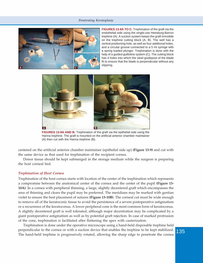
135
Penetrating Keratoplasty
FIGURES 13-8A TO C: Trephination of the graft via theendothelial side using the single-use Heesburg-Barrontrephine (A). A suction system keeps the graft immobileon the trephine cutting block (A, B). The well has acentral positioning hole, as well as four additional holes,and a circular groove connected to a 5 ml syringe witha spring loaded plunger. Trephination is done with thehelp of a guided guillotine system (C). The cutting blockhas 4 holes into which the steel guidepost of the bladefit to ensure that the blade is perpendicular without anyslipping.
FIGURES 13-9A AND B: Trephination of the graft via the epithelial side using theHanna trephine. The graft is mounted on the artificial anterior chamber maintainer(A) then cut with the Hanna trephine (B).
centered on the artificial anterior chamber maintainer (epithelial side up) (Figure 13-9) and cut withthe same device as that used for trephination of the recipient cornea.
Donor tissue should be kept submerged in the storage medium while the surgeon is preparingthe host corneal bed.
Trephination of Host Cornea
Trephination of the host cornea starts with location of the center of the trephination which representsa compromise between the anatomical center of the cornea and the center of the pupil (Figure 13-10A). In a cornea with peripheral thinning, a large, slightly decentered graft which encompasses thearea of thinning and clears the pupil may be preferred. The meridians may be marked with gentianviolet to ensure the best placement of sutures (Figure 13-10B). The corneal cut must be wide enoughto remove all of the keratoconic tissue to avoid the persistence of a severe postoperative astigmatismor a recurrence of the keratoconus. A lower peripheral cone is the most common form of keratoconus.A slightly decentered graft is well tolerated, although major decentration may be complicated by agiant postoperative astigmatism as well as by potential graft rejection. In case of marked protrusionof the cone, trephination is facilitated after flattening the apex with cauterization.
Trephination is done under the operative microscope using a hand-held disposable trephine heldperpendicular to the cornea or with a suction device that enables the trephine to be kept stabilized.The hand-held trephine is progressively rotated, allowing the sharp edge to penetrate the cornea
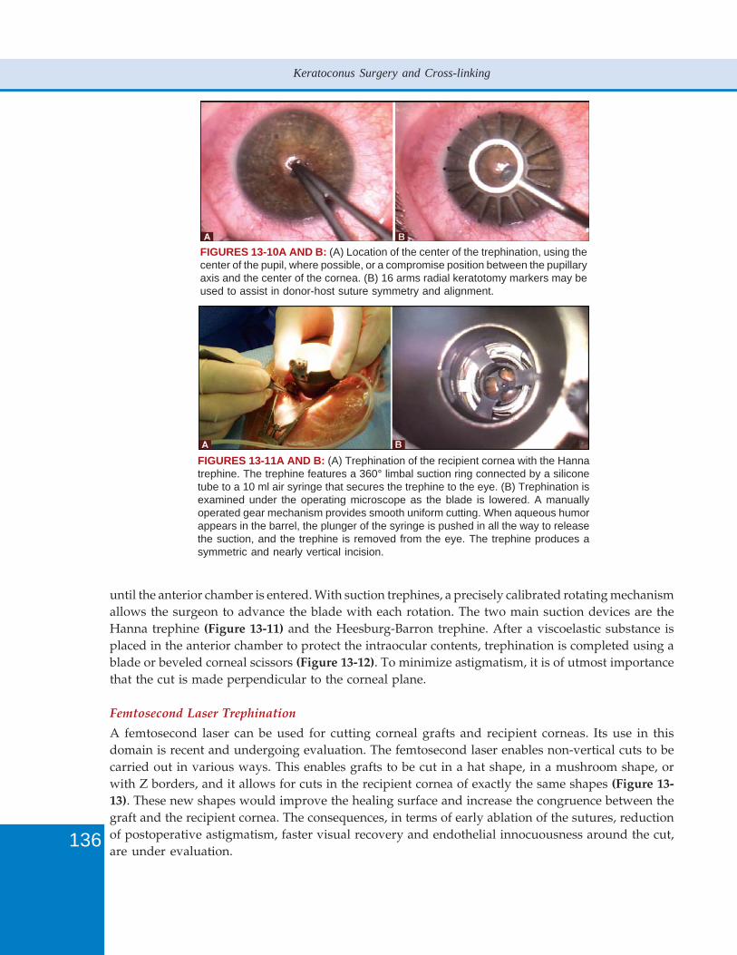
136
Keratoconus Surgery and Cross-linking
FIGURES 13-10A AND B: (A) Location of the center of the trephination, using thecenter of the pupil, where possible, or a compromise position between the pupillaryaxis and the center of the cornea. (B) 16 arms radial keratotomy markers may beused to assist in donor-host suture symmetry and alignment.
FIGURES 13-11A AND B: (A) Trephination of the recipient cornea with the Hannatrephine. The trephine features a 360° limbal suction ring connected by a siliconetube to a 10 ml air syringe that secures the trephine to the eye. (B) Trephination isexamined under the operating microscope as the blade is lowered. A manuallyoperated gear mechanism provides smooth uniform cutting. When aqueous humorappears in the barrel, the plunger of the syringe is pushed in all the way to releasethe suction, and the trephine is removed from the eye. The trephine produces asymmetric and nearly vertical incision.
until the anterior chamber is entered. With suction trephines, a precisely calibrated rotating mechanismallows the surgeon to advance the blade with each rotation. The two main suction devices are theHanna trephine (Figure 13-11) and the Heesburg-Barron trephine. After a viscoelastic substance isplaced in the anterior chamber to protect the intraocular contents, trephination is completed using ablade or beveled corneal scissors (Figure 13-12). To minimize astigmatism, it is of utmost importancethat the cut is made perpendicular to the corneal plane.
Femtosecond Laser Trephination
A femtosecond laser can be used for cutting corneal grafts and recipient corneas. Its use in thisdomain is recent and undergoing evaluation. The femtosecond laser enables non-vertical cuts to becarried out in various ways. This enables grafts to be cut in a hat shape, in a mushroom shape, orwith Z borders, and it allows for cuts in the recipient cornea of exactly the same shapes (Figure 13-13). These new shapes would improve the healing surface and increase the congruence between thegraft and the recipient cornea. The consequences, in terms of early ablation of the sutures, reductionof postoperative astigmatism, faster visual recovery and endothelial innocuousness around the cut,are under evaluation.
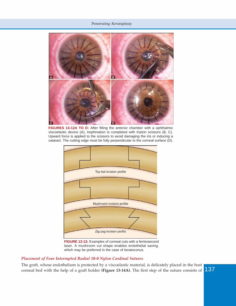
137
Penetrating Keratoplasty
FIGURES 13-12A TO D: After filling the anterior chamber with a ophthalmicviscoelastic device (A), trephination is completed with Katzin scissors (B, C).Upward force is applied to the scissors to avoid damaging the iris or inducing acataract. The cutting edge must be fully perpendicular to the corneal surface (D).
FIGURE 13-13: Examples of corneal cuts with a femtosecondlaser. A mushroom cut shape enables endothelial saving,which may be preferred in the case of keratoconus.
Placement of Four Interrupted Radial 10-0 Nylon Cardinal Sutures
The graft, whose endothelium is protected by a viscoelastic material, is delicately placed in the hostcorneal bed with the help of a graft holder (Figure 13-14A). The first step of the suture consists of
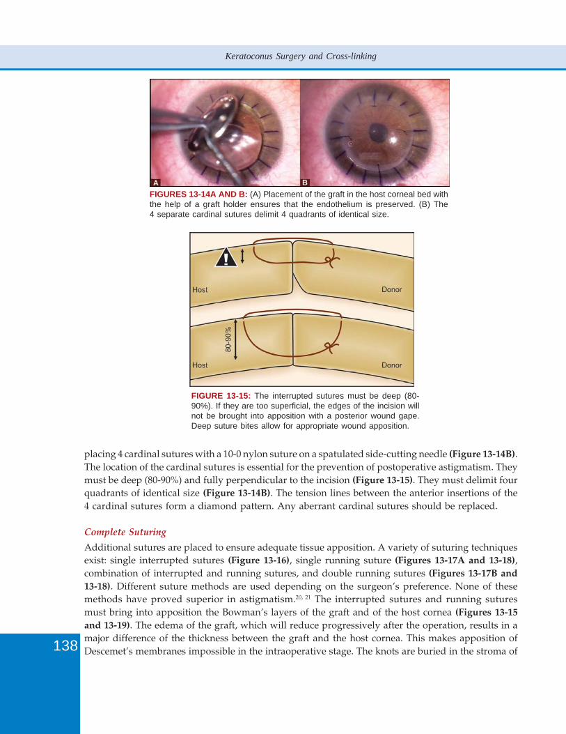
138
Keratoconus Surgery and Cross-linking
FIGURES 13-14A AND B: (A) Placement of the graft in the host corneal bed withthe help of a graft holder ensures that the endothelium is preserved. (B) The4 separate cardinal sutures delimit 4 quadrants of identical size.
FIGURE 13-15: The interrupted sutures must be deep (80-90%). If they are too superficial, the edges of the incision willnot be brought into apposition with a posterior wound gape.Deep suture bites allow for appropriate wound apposition.
placing 4 cardinal sutures with a 10-0 nylon suture on a spatulated side-cutting needle (Figure 13-14B).The location of the cardinal sutures is essential for the prevention of postoperative astigmatism. Theymust be deep (80-90%) and fully perpendicular to the incision (Figure 13-15). They must delimit fourquadrants of identical size (Figure 13-14B). The tension lines between the anterior insertions of the4 cardinal sutures form a diamond pattern. Any aberrant cardinal sutures should be replaced.
Complete Suturing
Additional sutures are placed to ensure adequate tissue apposition. A variety of suturing techniquesexist: single interrupted sutures (Figure 13-16), single running suture (Figures 13-17A and 13-18),combination of interrupted and running sutures, and double running sutures (Figures 13-17B and13-18). Different suture methods are used depending on the surgeon’s preference. None of thesemethods have proved superior in astigmatism.20, 21 The interrupted sutures and running suturesmust bring into apposition the Bowman’s layers of the graft and of the host cornea (Figures 13-15and 13-19). The edema of the graft, which will reduce progressively after the operation, results in amajor difference of the thickness between the graft and the host cornea. This makes apposition ofDescemet’s membranes impossible in the intraoperative stage. The knots are buried in the stroma of

139
Penetrating Keratoplasty
FIGURES 13-16A AND B: (A) At the end of the operation, the viscoelastic materialis exchanged for a balanced salt solution before tying the last suture. (B) Shownis a set of 16-interrupted sutures after the suture knots are buried.
FIGURES 13-17A AND B: Shown is a single continuous running suture (A) anda double continuous running suture (B).
FIGURE 13-18: Schematic suture technique for a single continuous runningsuture. Some surgeons believe that an anti-torque running suture is helpfulin reducing astigmatism, whereas others have not observed any significanteffects with such a suture.
Antitorque pattern of single running suture. On the left, the overlyingradial sutures (full line) would result in a minimal suture torque and a reducedrisk of induced astigmatism. Intrastromal suture bites (dotted line) areantitorque.
Torque pattern of single running suture. On the right, the intrastromalsuture is radial (dotted line), and the overlying torquing suture bites (fullline) could rotate the graft in the arrow direction and induce astigmatism.
the graft rather than in the recipient side to minimize vascularization. The total number of interruptedsutures and passages of the running suture is usually between 16 and 24.
Readjustment of Sutures to Minimize Astigmatism
When a running suture is carried out, it is necessary to harmoniously distribute its tension beforeburying the knot. The tension of the sutures may be checked at the end of the operation afterreplacing the viscoelastic material in the anterior chamber with balanced salt solution (Figure 13-16).
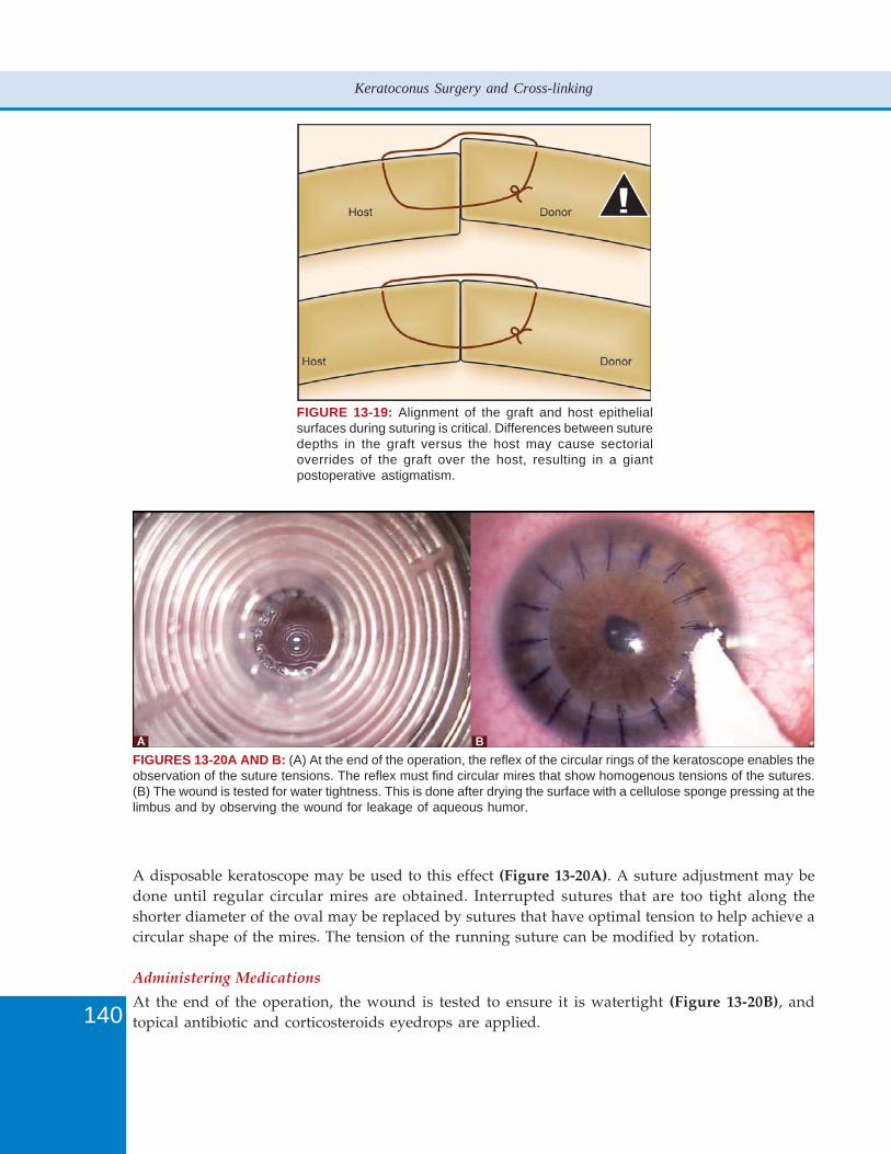
140
Keratoconus Surgery and Cross-linking
A disposable keratoscope may be used to this effect (Figure 13-20A). A suture adjustment may bedone until regular circular mires are obtained. Interrupted sutures that are too tight along theshorter diameter of the oval may be replaced by sutures that have optimal tension to help achieve acircular shape of the mires. The tension of the running suture can be modified by rotation.
Administering Medications
At the end of the operation, the wound is tested to ensure it is watertight (Figure 13-20B), andtopical antibiotic and corticosteroids eyedrops are applied.
FIGURE 13-19: Alignment of the graft and host epithelialsurfaces during suturing is critical. Differences between suturedepths in the graft versus the host may cause sectorialoverrides of the graft over the host, resulting in a giantpostoperative astigmatism.
FIGURES 13-20A AND B: (A) At the end of the operation, the reflex of the circular rings of the keratoscope enables theobservation of the suture tensions. The reflex must find circular mires that show homogenous tensions of the sutures.(B) The wound is tested for water tightness. This is done after drying the surface with a cellulose sponge pressing at thelimbus and by observing the wound for leakage of aqueous humor.
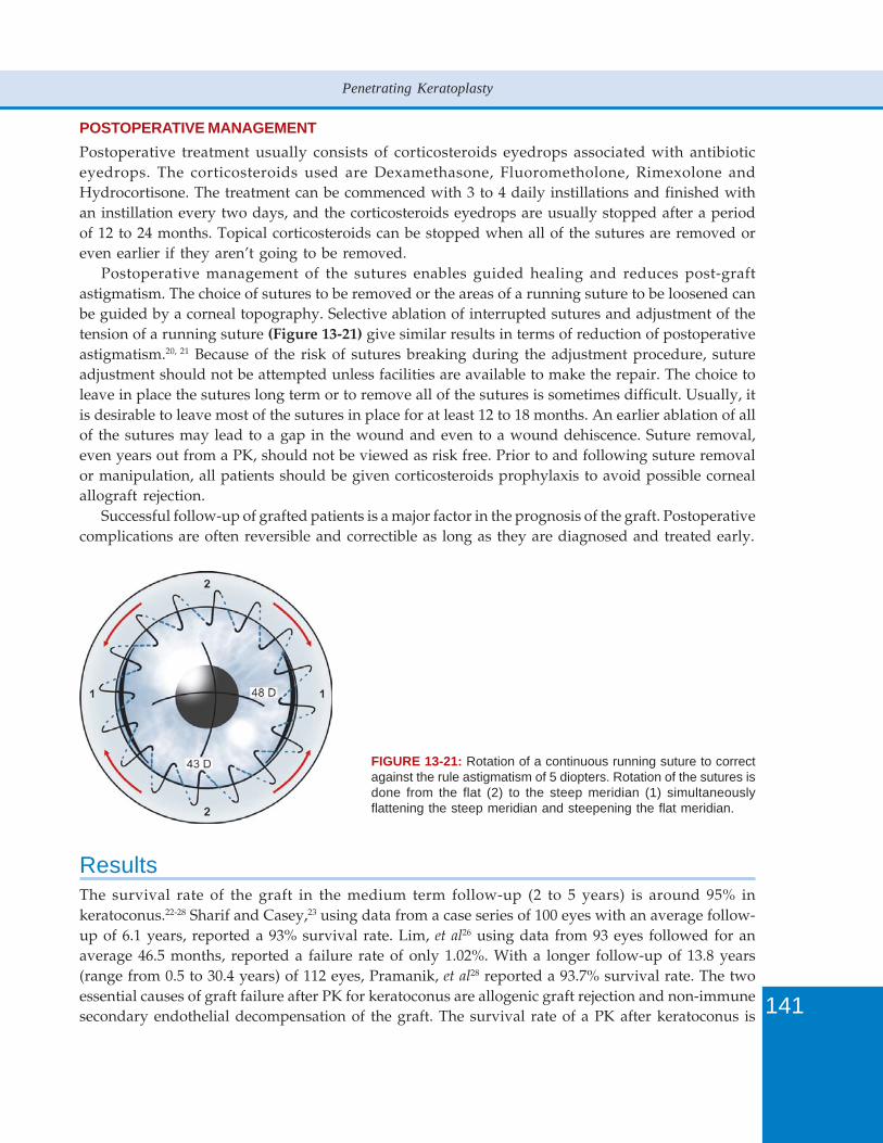
141
Penetrating Keratoplasty
POSTOPERATIVE MANAGEMENTPostoperative treatment usually consists of corticosteroids eyedrops associated with antibioticeyedrops. The corticosteroids used are Dexamethasone, Fluorometholone, Rimexolone andHydrocortisone. The treatment can be commenced with 3 to 4 daily instillations and finished withan instillation every two days, and the corticosteroids eyedrops are usually stopped after a periodof 12 to 24 months. Topical corticosteroids can be stopped when all of the sutures are removed oreven earlier if they aren’t going to be removed.
Postoperative management of the sutures enables guided healing and reduces post-graftastigmatism. The choice of sutures to be removed or the areas of a running suture to be loosened canbe guided by a corneal topography. Selective ablation of interrupted sutures and adjustment of thetension of a running suture (Figure 13-21) give similar results in terms of reduction of postoperativeastigmatism.20, 21 Because of the risk of sutures breaking during the adjustment procedure, sutureadjustment should not be attempted unless facilities are available to make the repair. The choice toleave in place the sutures long term or to remove all of the sutures is sometimes difficult. Usually, itis desirable to leave most of the sutures in place for at least 12 to 18 months. An earlier ablation of allof the sutures may lead to a gap in the wound and even to a wound dehiscence. Suture removal,even years out from a PK, should not be viewed as risk free. Prior to and following suture removalor manipulation, all patients should be given corticosteroids prophylaxis to avoid possible cornealallograft rejection.
Successful follow-up of grafted patients is a major factor in the prognosis of the graft. Postoperativecomplications are often reversible and correctible as long as they are diagnosed and treated early.
FIGURE 13-21: Rotation of a continuous running suture to correctagainst the rule astigmatism of 5 diopters. Rotation of the sutures isdone from the flat (2) to the steep meridian (1) simultaneouslyflattening the steep meridian and steepening the flat meridian.
ResultsThe survival rate of the graft in the medium term follow-up (2 to 5 years) is around 95% inkeratoconus.22-28 Sharif and Casey,23 using data from a case series of 100 eyes with an average follow-up of 6.1 years, reported a 93% survival rate. Lim, et al26 using data from 93 eyes followed for anaverage 46.5 months, reported a failure rate of only 1.02%. With a longer follow-up of 13.8 years(range from 0.5 to 30.4 years) of 112 eyes, Pramanik, et al28 reported a 93.7% survival rate. The twoessential causes of graft failure after PK for keratoconus are allogenic graft rejection and non-immunesecondary endothelial decompensation of the graft. The survival rate of a PK after keratoconus is
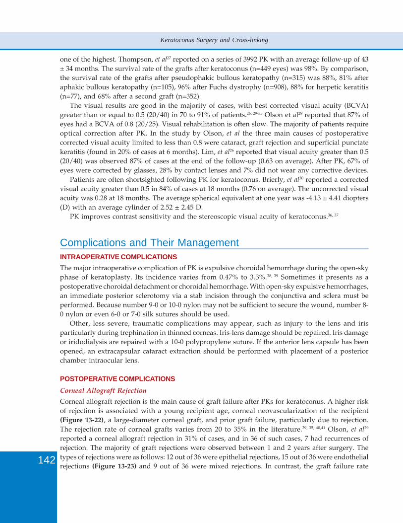
142
Keratoconus Surgery and Cross-linking
one of the highest. Thompson, et al27 reported on a series of 3992 PK with an average follow-up of 43± 34 months. The survival rate of the grafts after keratoconus (n=449 eyes) was 98%. By comparison,the survival rate of the grafts after pseudophakic bullous keratopathy (n=315) was 88%, 81% afteraphakic bullous keratopathy (n=105), 96% after Fuchs dystrophy (n=908), 88% for herpetic keratitis(n=77), and 68% after a second graft (n=352).
The visual results are good in the majority of cases, with best corrected visual acuity (BCVA)greater than or equal to 0.5 (20/40) in 70 to 91% of patients.26, 29-35 Olson et al29 reported that 87% ofeyes had a BCVA of 0.8 (20/25). Visual rehabilitation is often slow. The majority of patients requireoptical correction after PK. In the study by Olson, et al the three main causes of postoperativecorrected visual acuity limited to less than 0.8 were cataract, graft rejection and superficial punctatekeratitis (found in 20% of cases at 6 months). Lim, et al26 reported that visual acuity greater than 0.5(20/40) was observed 87% of cases at the end of the follow-up (0.63 on average). After PK, 67% ofeyes were corrected by glasses, 28% by contact lenses and 7% did not wear any corrective devices.
Patients are often shortsighted following PK for keratoconus. Brierly, et al30 reported a correctedvisual acuity greater than 0.5 in 84% of cases at 18 months (0.76 on average). The uncorrected visualacuity was 0.28 at 18 months. The average spherical equivalent at one year was -4.13 ± 4.41 diopters(D) with an average cylinder of 2.52 ± 2.45 D.
PK improves contrast sensitivity and the stereoscopic visual acuity of keratoconus.36, 37
Complications and Their ManagementINTRAOPERATIVE COMPLICATIONSThe major intraoperative complication of PK is expulsive choroidal hemorrhage during the open-skyphase of keratoplasty. Its incidence varies from 0.47% to 3.3%.38, 39 Sometimes it presents as apostoperative choroidal detachment or choroidal hemorrhage. With open-sky expulsive hemorrhages,an immediate posterior sclerotomy via a stab incision through the conjunctiva and sclera must beperformed. Because number 9-0 or 10-0 nylon may not be sufficient to secure the wound, number 8-0 nylon or even 6-0 or 7-0 silk sutures should be used.
Other, less severe, traumatic complications may appear, such as injury to the lens and irisparticularly during trephination in thinned corneas. Iris-lens damage should be repaired. Iris damageor iridodialysis are repaired with a 10-0 polypropylene suture. If the anterior lens capsule has beenopened, an extracapsular cataract extraction should be performed with placement of a posteriorchamber intraocular lens.
POSTOPERATIVE COMPLICATIONSCorneal Allograft Rejection
Corneal allograft rejection is the main cause of graft failure after PKs for keratoconus. A higher riskof rejection is associated with a young recipient age, corneal neovascularization of the recipient(Figure 13-22), a large-diameter corneal graft, and prior graft failure, particularly due to rejection.The rejection rate of corneal grafts varies from 20 to 35% in the literature.29, 35, 40,41 Olson, et al29
reported a corneal allograft rejection in 31% of cases, and in 36 of such cases, 7 had recurrences ofrejection. The majority of graft rejections were observed between 1 and 2 years after surgery. Thetypes of rejections were as follows: 12 out of 36 were epithelial rejections, 15 out of 36 were endothelialrejections (Figure 13-23) and 9 out of 36 were mixed rejections. In contrast, the graft failure rate
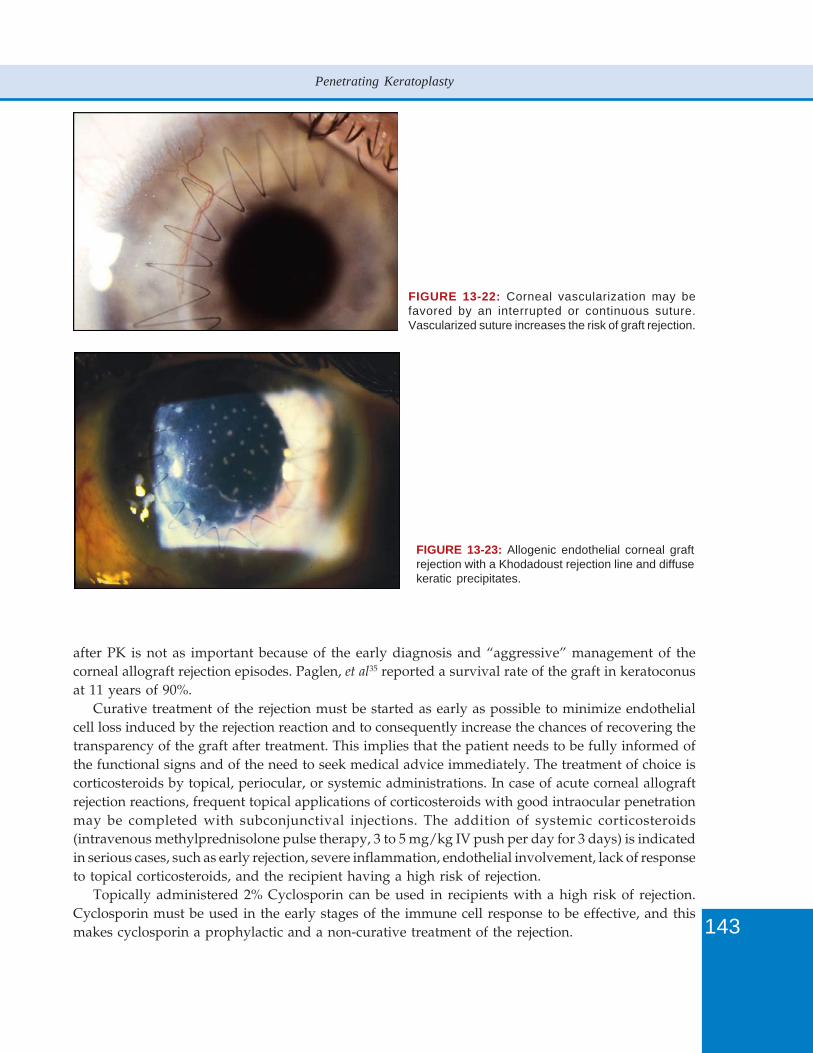
143
Penetrating Keratoplasty
FIGURE 13-22: Corneal vascularization may befavored by an interrupted or continuous suture.Vascularized suture increases the risk of graft rejection.
FIGURE 13-23: Allogenic endothelial corneal graftrejection with a Khodadoust rejection line and diffusekeratic precipitates.
after PK is not as important because of the early diagnosis and “aggressive” management of thecorneal allograft rejection episodes. Paglen, et al35 reported a survival rate of the graft in keratoconusat 11 years of 90%.
Curative treatment of the rejection must be started as early as possible to minimize endothelialcell loss induced by the rejection reaction and to consequently increase the chances of recovering thetransparency of the graft after treatment. This implies that the patient needs to be fully informed ofthe functional signs and of the need to seek medical advice immediately. The treatment of choice iscorticosteroids by topical, periocular, or systemic administrations. In case of acute corneal allograftrejection reactions, frequent topical applications of corticosteroids with good intraocular penetrationmay be completed with subconjunctival injections. The addition of systemic corticosteroids(intravenous methylprednisolone pulse therapy, 3 to 5 mg/kg IV push per day for 3 days) is indicatedin serious cases, such as early rejection, severe inflammation, endothelial involvement, lack of responseto topical corticosteroids, and the recipient having a high risk of rejection.
Topically administered 2% Cyclosporin can be used in recipients with a high risk of rejection.Cyclosporin must be used in the early stages of the immune cell response to be effective, and thismakes cyclosporin a prophylactic and a non-curative treatment of the rejection.
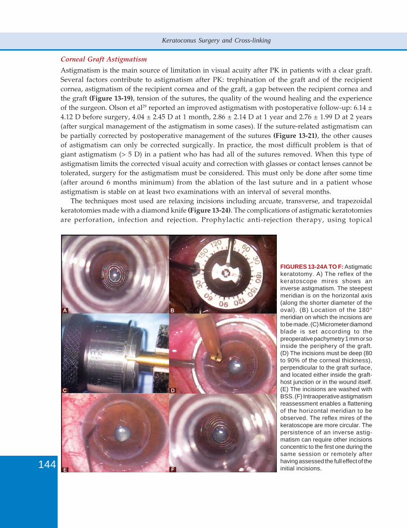
144
Keratoconus Surgery and Cross-linking
Corneal Graft Astigmatism
Astigmatism is the main source of limitation in visual acuity after PK in patients with a clear graft.Several factors contribute to astigmatism after PK: trephination of the graft and of the recipientcornea, astigmatism of the recipient cornea and of the graft, a gap between the recipient cornea andthe graft (Figure 13-19), tension of the sutures, the quality of the wound healing and the experienceof the surgeon. Olson et al29 reported an improved astigmatism with postoperative follow-up: 6.14 ±4.12 D before surgery, 4.04 ± 2.45 D at 1 month, 2.86 ± 2.14 D at 1 year and 2.76 ± 1.99 D at 2 years(after surgical management of the astigmatism in some cases). If the suture-related astigmatism canbe partially corrected by postoperative management of the sutures (Figure 13-21), the other causesof astigmatism can only be corrected surgically. In practice, the most difficult problem is that ofgiant astigmatism (> 5 D) in a patient who has had all of the sutures removed. When this type ofastigmatism limits the corrected visual acuity and correction with glasses or contact lenses cannot betolerated, surgery for the astigmatism must be considered. This must only be done after some time(after around 6 months minimum) from the ablation of the last suture and in a patient whoseastigmatism is stable on at least two examinations with an interval of several months.
The techniques most used are relaxing incisions including arcuate, transverse, and trapezoidalkeratotomies made with a diamond knife (Figure 13-24). The complications of astigmatic keratotomiesare perforation, infection and rejection. Prophylactic anti-rejection therapy, using topical
FIGURES 13-24A TO F: Astigmatickeratotomy. A) The reflex of thekeratoscope mires shows aninverse astigmatism. The steepestmeridian is on the horizontal axis(along the shorter diameter of theoval). (B) Location of the 180°meridian on which the incisions areto be made. (C) Micrometer diamondblade is set according to thepreoperative pachymetry 1 mm or soinside the periphery of the graft.(D) The incisions must be deep (80to 90% of the corneal thickness),perpendicular to the graft surface,and located either inside the graft-host junction or in the wound itself.(E) The incisions are washed withBSS. (F) Intraoperative astigmatismreassessment enables a flatteningof the horizontal meridian to beobserved. The reflex mires of thekeratoscope are more circular. Thepersistence of an inverse astig-matism can require other incisionsconcentric to the first one during thesame session or remotely afterhaving assessed the full effect of theinitial incisions.
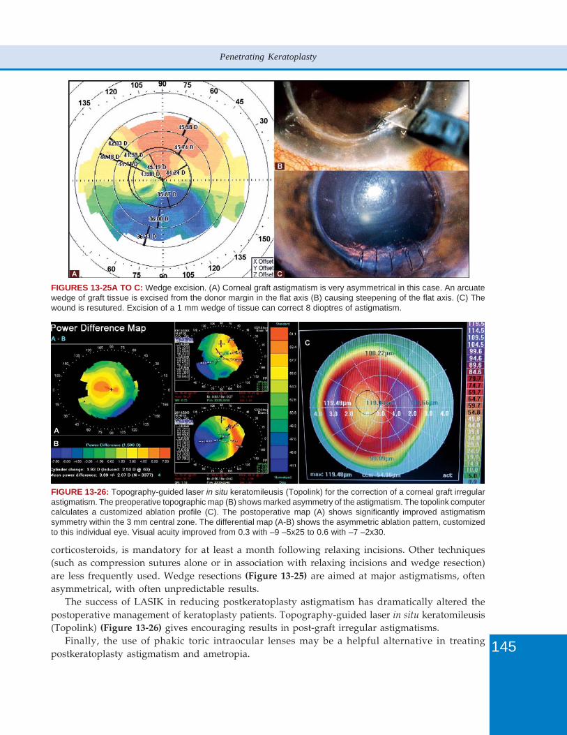
145
Penetrating Keratoplasty
FIGURES 13-25A TO C: Wedge excision. (A) Corneal graft astigmatism is very asymmetrical in this case. An arcuatewedge of graft tissue is excised from the donor margin in the flat axis (B) causing steepening of the flat axis. (C) Thewound is resutured. Excision of a 1 mm wedge of tissue can correct 8 dioptres of astigmatism.
corticosteroids, is mandatory for at least a month following relaxing incisions. Other techniques(such as compression sutures alone or in association with relaxing incisions and wedge resection)are less frequently used. Wedge resections (Figure 13-25) are aimed at major astigmatisms, oftenasymmetrical, with often unpredictable results.
The success of LASIK in reducing postkeratoplasty astigmatism has dramatically altered thepostoperative management of keratoplasty patients. Topography-guided laser in situ keratomileusis(Topolink) (Figure 13-26) gives encouraging results in post-graft irregular astigmatisms.
Finally, the use of phakic toric intraocular lenses may be a helpful alternative in treatingpostkeratoplasty astigmatism and ametropia.
FIGURE 13-26: Topography-guided laser in situ keratomileusis (Topolink) for the correction of a corneal graft irregularastigmatism. The preoperative topographic map (B) shows marked asymmetry of the astigmatism. The topolink computercalculates a customized ablation profile (C). The postoperative map (A) shows significantly improved astigmatismsymmetry within the 3 mm central zone. The differential map (A-B) shows the asymmetric ablation pattern, customizedto this individual eye. Visual acuity improved from 0.3 with –9 –5x25 to 0.6 with –7 –2x30.
146
Keratoconus Surgery and Cross-linking
Side Effects of Topical Corticosteroids
Glaucoma and cataract may occur.Intraocular pressure elevation is the second leading cause of graft failure after PK. In patients
with keratoconus, it is then often secondary to corticosteroids. Sustained elevation of intraocularpressure not only damages the optic nerve head, but it also has a deleterious effect on the cornealendothelium. Correction of elevated intraocular pressure refers to a reduction in corticosteroidseyedrops (e.g. replacement of Dexamethasone with Fluorometholone), the use of topical or systemicantiglaucoma medications, and even traditional filtering surgery on rare occasions (when either theoptic nerve or the graft is threatened by persistent elevation of intraocular pressure).
Cataract can be extracted through a separate incision after the wound is totally healed. Theendothelium must be protected by a good viscoelastic because of the risk of endothelialdecompensation. Cataract extraction in a young subject results in a constant loss of accommodation.
Other Complications
Shallow anterior chamber and wound leak: Lack of water tightness of the wound in the early postoperativeperiod can be due to a full thickness suture or an insufficient number of sutures. Additional suturesmay be applied in the operating room under topical anesthesia.
Epithelial defects: Epithelial defects are the source of visual loss, infection, rejection, stromal melting,perforations, and even graft failure. Survival of a corneal graft is critically dependent on an intactepithelial barrier. The quality of the postoperative epithelial wound healing necessitates frequentuse of lubricants, preferably preservative-free, to promote epithelial growth, as well as the use of abandage soft contact lens with high oxygen permeability in certain occasions.
Urrets-Zavalia syndrome: The first description of Urrets-Zavalia syndrome or fixed dilated pupil wasmade by Urrets-Zavalia after a corneal graft for keratoconus.42 However, this syndrome is notspecific to PK in keratoconus, and it was reported following lamellar grafts and in other indications.The mechanism is poorly understood, but it may involve iris ischemia. The role of mydriatics hasbeen brought up as well as that of early and transitory acute postoperative raised intraocular pressure.Although rare, this complication prompts the use of mydriatics to be limited unless it is absolutelynecessary. A careful ablation of viscoelastic at the end of the operation seems just as suitable, in ourclinical experience.
Infections after PK: Postoperative endophthalmitis is a rare but sight-threatening complication of PK.Its incidence is around 0.5%.43 Often, the infectious agents come from the conjunctival and palpebralflora of the patient. The clinical signs, diagnosis and treatment of postoperative endophthalmitisafter PK are the same as those for endophthalmitis after cataract surgery.
Bacterial and fungal keratitis are complications that usually involve the graft or the wound. Theincidence of microbial keratitis ranges from 1 to 12% in patients operated for PK. Microbial keratitismay be more frequent with topical steroids, persistent epithelial defect and suture related problems.It presents in the form of a stromal abscess with a high risk of graft melting. A particular form is aninfectious crystalline keratopathy which takes the appearance of crystalline branching opacities (Figure13-27). The most common organism causing crystalline keratopathy is Streptococcus viridans. Theidentification of the etiology must be done with corneal scrapings for smear and culture in appropriatemedia, as fungal species have also been implicated. Treatment for bacterial and fungal keratitis isbased on intensive regimen of broad-spectrum, fortified antibiotic eyedrops often with systemic
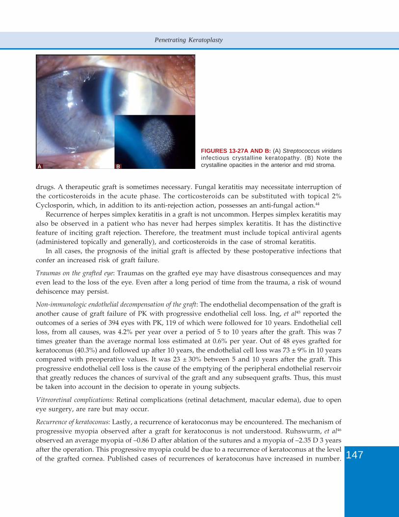
147
Penetrating Keratoplasty
FIGURES 13-27A AND B: (A) Streptococcus viridansinfectious crystalline keratopathy. (B) Note thecrystalline opacities in the anterior and mid stroma.
drugs. A therapeutic graft is sometimes necessary. Fungal keratitis may necessitate interruption ofthe corticosteroids in the acute phase. The corticosteroids can be substituted with topical 2%Cyclosporin, which, in addition to its anti-rejection action, possesses an anti-fungal action.44
Recurrence of herpes simplex keratitis in a graft is not uncommon. Herpes simplex keratitis mayalso be observed in a patient who has never had herpes simplex keratitis. It has the distinctivefeature of inciting graft rejection. Therefore, the treatment must include topical antiviral agents(administered topically and generally), and corticosteroids in the case of stromal keratitis.
In all cases, the prognosis of the initial graft is affected by these postoperative infections thatconfer an increased risk of graft failure.
Traumas on the grafted eye: Traumas on the grafted eye may have disastrous consequences and mayeven lead to the loss of the eye. Even after a long period of time from the trauma, a risk of wounddehiscence may persist.
Non-immunologic endothelial decompensation of the graft: The endothelial decompensation of the graft isanother cause of graft failure of PK with progressive endothelial cell loss. Ing, et al45 reported theoutcomes of a series of 394 eyes with PK, 119 of which were followed for 10 years. Endothelial cellloss, from all causes, was 4.2% per year over a period of 5 to 10 years after the graft. This was 7times greater than the average normal loss estimated at 0.6% per year. Out of 48 eyes grafted forkeratoconus (40.3%) and followed up after 10 years, the endothelial cell loss was 73 ± 9% in 10 yearscompared with preoperative values. It was 23 ± 30% between 5 and 10 years after the graft. Thisprogressive endothelial cell loss is the cause of the emptying of the peripheral endothelial reservoirthat greatly reduces the chances of survival of the graft and any subsequent grafts. Thus, this mustbe taken into account in the decision to operate in young subjects.
Vitreoretinal complications: Retinal complications (retinal detachment, macular edema), due to openeye surgery, are rare but may occur.
Recurrence of keratoconus: Lastly, a recurrence of keratoconus may be encountered. The mechanism ofprogressive myopia observed after a graft for keratoconus is not understood. Ruhswurm, et al46
observed an average myopia of –0.86 D after ablation of the sutures and a myopia of –2.35 D 3 yearsafter the operation. This progressive myopia could be due to a recurrence of keratoconus at the levelof the grafted cornea. Published cases of recurrences of keratoconus have increased in number.
148
Keratoconus Surgery and Cross-linking
Pramanik, et al28 reported on 112 eyes of 84 patients operated with PK for keratoconus with anaverage follow-up of 13.8 years (range from 0.5 to 30.4 years). A recurrence of keratoconus wasconfirmed in 6 cases (5.4%) after an average period of 17.9 years (range of 11 to 27 years). Theprobability of having a recurrence of keratoconus after PK over 25 years was estimated at 11.7%.
It has not yet been clearly established whether a keratoconus recurs or a mild keratoconus fromthe donor that has gone unnoticed becomes manifest. However, the average time of 17 years untilthe onset of a recurrence of keratoconus is quite comparable with the time to natural developmentof keratoconus in a teenager.
Discussion/ConclusionsWill the number of indications of corneal graft for keratoconus be reduced? Would the advances incontact lenses, suppressive (corneal collagen cross-linking with Riboflavin: CXL) and additive(intracorneal rings) treatments be able to avoid or delay a corneal graft?
CXL is aimed at progressive keratoconus.47 Its purpose is to “rigidify” a cornea which isbiomechanically unstable. This effect that was observed in vitro48, 49 is more difficult to quantify invivo. However, the first clinical results are encouraging,50-52 and the efficacy will continue to beassessed over a longer period.
Of course, contact lenses, which are essentially rigid, remain the basic treatment for eyes withkeratoconus. Unfortunately, lenses are not tolerated in some patients who are often those withallergies and whose corneas remain transparent. In these cases, the implantation of intracornealrings is a worthwhile alternative treatment to a corneal graft.53, 54 A longer period of time is alsonecessary to assess what will become of these corneas. A combination of these two treatments(CXL/rings) or even of these three treatments (CXL/lenses/rings) may also be used.
When the progression of keratoconus leads to central corneal scarring or major thinning, a cornealgraft is required. In this case, will the proportion of PK be reduced in favor of lamellar grafts?
The growing number of publications of lamellar graft techniques already marks this trend. Severalstudies comparing the results of PK and lamellar grafts in keratoconus have shown identical visualresults.55-57 The operating technique in lamellar grafts is more difficult than that of PK but they offernotable advantages in terms of conservation of the endothelium, graft rejection, faster corneal woundhealing, and a shorter duration of the use of corticosteroids.
In certain cases, when the endothelium is involved, PK remains the only surgical alternative. Ifnot, the choice between PK and lamellar grafts depends essentially on the preference and experienceof the surgeon.
Long considered as the reference surgical treatment for keratoconus, today, PK is part of atherapeutic arsenal that has been considerably enriched in the past few years. PK in keratoconusrequires a perfect surgical technique. The benefits of the femtosecond laser in the surgical techniqueremains to be demonstrated, and the technique may still benefit from future improvements. Thevisual results of PK in keratoconus are good in the majority of cases. This surgery, carried out mostoften in young patients, may lead to complications which should be early detected, in order todetect them early and provide appropriate solutions. Thus, the surgical indication of PK in a patientwith keratoconus must always take into account this benefits/risks ratio. Although PK may beincreasingly replaced with lamellar grafts, nevertheless, PK is an established surgical technique inkeratoconus that has been greatly studied for its efficacy.
149
Penetrating Keratoplasty
References1. Rabinowitz YS. Keratoconus. Surv Ophthalmol 1998;42:297-319.2. The Australian Corneal Graft Registry. 1990 to 1992 report. Aust N Z J Ophthalmol 1993;21:1-48.3. Legeais JM, Parc C, d’Hermies F, Pouliquen Y, Renard G. Nineteen years of penetrating keratoplasty in the Hotel-
Dieu Hospital in Paris. Cornea 2001;20:603-6.4. Lois N, Kowal VO, Cohen EJ, Rapuano CJ, Gault JA, Raber IM, Laibson PR. Indications for penetrating keratoplasty
and associated procedures, 1989-1995. Cornea 1997;16:623-9.5. Maeno A, Naor J, Lee HM, Hunter WS, Rootman DS. Three decades of corneal transplantation: indications and
patient characteristics. Cornea 2000;19:7-11.6. Dobbins KR, Price FWJ, Whitson WE. Trends in the indications for penetrating keratoplasty in the midwestern
United States. Cornea 2000;19:813-6.7. Crews MJ, Driebe WT Jr, Stern GA. The clinical management of keratoconus: A 6 year retrospective study. CLAO
J 1994;20:194-7.8. Lass JH, Lembach RG, Park SB, Hom DL, Fritz ME, Svilar GM, Nuamah IF, Reinhart WJ, Stocker EG, Keates RH, et
al. Clinical management of keratoconus. A multicenter analysis. Ophthalmology 1990;97:433-45.9. Sray WA, Cohen EJ, Rapuano CJ, Laibson PR. Factors associated with the need for penetrating keratoplasty in
keratoconus. Cornea 2002;21:784-6.10. Dana MR, Putz JL, Viana MA, Sugar J, McMahon TT. Contact lens failure in keratoconus management.
Ophthalmology 1992;99:1187-92.11. Reeves SW, Stinnett S, Adelman RA, Afshari NA. Risk factors for progression to penetrating keratoplasty in
patients with keratoconus. Am J Ophthalmol 2005;140:607-11.12. Gordon MO, Steger-May K, Szczotka-Flynn L, Riley C, Joslin CE, Weissman BA, Fink BA, Edrington TB, Olafsson
HE, Zadnik K. CLEK Study Group. Baseline factors predictive of incident penetrating keratoplasty in keratoconus.Am J Ophthalmol 2006;142:923-30.
13. Smiddy WE, Hamburg TR, Kracher GP, Stark WJ. Keratoconus. Contact lens or keratoplasty? Ophthalmology1988;95:487-92.
14. Belin MW, Fowler WG, Chambers WA. Keratoconus. Evaluation of recent trends in the surgical and nonsurgicalcorrection of keratoconus. Ophthalmology 1988;95:335-9..
15. Tuft SJ, Moodaley LC, Gregory WM, Davison CR, Buckley RJ. Prognostic factors for the progression of keratoconus.Ophthalmology 1994;101:439-47.
16. Colin J. European clinical evaluation: use of Intacs for the treatment of keratoconus. J Cataract Refract Surg2006;32:747-55.
17. Krumeich JH, Daniel J, Knulle A. Live-epikeratophakia for keratoconus. J Cataract Refract Surg 1998;24:456-63.18. Wilson EE, Bourne WM. Effect of recipient-donor trephine size disparity on refractive error in keratoconus.
Ophthalmology 1989;96:299-305.19. Girald LJ, Esnaola N, Rao R, Barnett L, Rand WJ. Use of grafts smaller than the opening for keratoconic myopia and
astigmatism. J Cataract Refract Surg 1992;18:380-4.20. Karabatsas CH, Cook SD, Figueiredo FC, Diamond JP, Easty DL. Combined interrupted and continuous versus
single continuous adjustable suturing in penetrating keratoplasty: a prospective, randomized study of inducedastigmatism during the first postoperative year. Ophthalmology 1998;105:1991-8.
21. Spadea L, Cifariello F, Bianco G, Balestrazzi E. Long-term results of penetrating keratoplasty using a single ordouble running suture technique. Graefes Arch Clin Exp Ophthalmol 2002;240:415-9.
22. Ehlers N, Olsen T. Long term results of corneal grafting in keratoconus. Acta Ophthalmol 1983;61:918-26.23. Sharif KW, Casey TA. Penetrating keratoplasty for keratoconus: complications and long-term success. Br J
Ophthalmol 1991;75:142-6.24. Price FW Jr, Whitson WE, John S, Gonzales JS. Risk factors for corneal graft failure. J Refract Surg 1996;12:134-47.25. Dandona L, Naduvilath TJ, Janarthanan M, Ragu K, Rao GN. Survival analysis and visual outcome in a large series
of corneal transplants in India. Br J Ophthalmol 1997;81:726-31.26. Lim L, Pesudovs K, Coster DJ. Penetrating keratoplasty for keratoconus: visual outcome and success. Ophthalmology
2000;107:1125-31.27. Thompson RW, Price MO, Bowers PJ, Price FW. Long-term graft survival after penetrating keratoplasty.
Ophthalmology 2003;110:1396-402.28. Pramanik S, Musch DC, Sutphin JE, Farjo AA. Extended long-term outcomes of penetrating keratoplasty for
keratoconus. Ophthalmology 2006;113:1633-8.29. Olson RJ, Pingree M, Ridges R, Lundergan M, Alldredge C, Clinch TE. Penetrating keratoplasty for keratoconus: a
long-term review of results and complications. J Cataract Ref Surg 2000;26:987-91.

150
Keratoconus Surgery and Cross-linking
30. Brierly SC, Isquierdo L, Mannis MJ. Penetrating keratoplasty for keratoconus. Cornea 2000;19:329-32.31. Silbiger JS, Cohen EJ, Laibson PR. The rate of visual recovery after penetrating keratoplasty for keratoconus.
CLAO J 1996;22:266-9.32. Italon C, Pieh S, Hansemayer G, Kahraman G, Kaminski S, Skorpik C, Dejaco-Ruhswurm I. Changes of axial length
and keratometry after keratoplasty for keratoconus using the guided trephine system. Am J Ophthalmol2002;134:696-700.
33. Tuft S, Gregory W. Long-term refraction and keratometry after penetrating keratoplasty for keratoconus. Cornea1995;14:614-7.
34. Tuft S, Fritzke FW, Buckley RJ. Myopia following penetrating keratoplasty for keratoconus. Br J Ophthalmol1992;76:642-5.
35. Paglen PG, Fine M, Abbott RL, Webster RG. The prognosis for keratoplasty in keratoconus. Ophthalmology1982;89:651-4.
36. Brahma A, Ennis F, Harper R, Ridgway A, Tullo A. Visual function after penetrating keratoplasty for keratoconus:a prospective longitudinal evaluation. Br J Ophthalmol 2000;84:60-6.
37. Pesudovs K, Schoneveld P, Seto RJ, Coster DJ. Contrast and glare testing in keratoconus and after penetratingkeratoplasty. Br J Ophthalmol 2004;88:653-7.
38. Purcell JJ Jr. Expulsive hemorrhage. In Brightbill F(Ed): Corneal Surgery, Theory Technique and Tissue (2nd edn).St. Louis, Mosby, 1993;221-4.
39. Taylor DM, Stern AL, McDonald P. The triple procedure: 1 to 10 year follow-up. Trans Am Ophthalmol Soc1986;84:221-49.
40. Kirkness CM, Ficker LA, Steele ADMcG, Rice NSC. The success of penetrating keratoplasty for keratoconus. Eye1990;4:673-88.
41. Chandler JW, Kaufmann HE. Graft rejections after keratoplasty for keratoconus. Am J Ophthalmol 1973;77:543-7.42. Urrets-Zavalia A. A fixed, dilated pupil iris atrophy and secondary glaucoma. A distinct clinical entity following
penetrating keratoplasty in keratoconus. Am J Ophthalmol 1963;56:257-65.43. Eifrig CW, Flynn HW Jr, Scott IU, Newton J. Acute-onset postoperative endophthalmitis: review of incidence and
visual outcomes (1995-2001). Ophthalmic Surg Lasers. 2002;33:378-8.44. Bell NP, Karp CL, Alfonso EC, Schiffman J, Miller D. Effects of methylprednisolone and cyclosporine A on fungal
growth in vitro. Cornea 1999;18:306-13.45. Ing JI, Ing HH, Nelson LR, Hodge DO, Bourne WM. Ten-year postoperative results of penetrating keratoplasty.
Ophthalmology 1998;105:1855-65.46. Ruhswurm I, Scholz U, Pfleger T, Zehetmayer M, Hanselmayer G, Skorpik C. Three-year clinical outcome after
penetrating keratoplasty for keratoconus with the guided trephine system. Am J Ophthalmol 1999;127:666-73.47. Spoerl E, Huhle M, Seiler T. Induction of cross-links in corneal tissue. Exp Eye Res 1998;66:97-103.48. Wollensak G, Spoerl E, Seiler T. Stress-strain measurements of human and porcine corneas after riboflavin-
ultraviolet-A-induced cross-linking. J Cataract Refract Surg 2003;29:1780-5.49. Spoerl E, Wollensak G, Seiler T. Increased resistance of crosslinked cornea against enzymatic digestion. Curr Eye
Res 2004;29:35-40.50. Wollensak G, Spoerl E, Seiler T. Riboflavin/Ultraviolet-A-induced collagen crosslinking for the treatment of
keratoconus. Am J Ophthalmol 2003;135:620-7.51. Caporossi A, Baiocchi S, Mazzotta C, Traversi C, Caporossi T. Parasurgical therapy for keratoconus by riboflavin-
ultraviolet type A rays induced cross-linking of corneal collagen: preliminary refractive results in an Italian study.J Cataract Refract Surg 2006;32:837-45.
52. Raiskup-Wolf F, Hoyer A, Spoerl E, Pillunat LE. Collagen crosslinking with riboflavin and ultraviolet-A light inkeratoconus: long-term results. J Cataract Refract Surg 2008;34:796-801.
53. Colin J, Cochener B, Savary G, Malet F. Correcting keratoconus with intracorneal rings. J Cataract Refract Surg2000;26:1117-22.
54. Colin J, Cochener B, Savary G, Malet F, Holmes-Higgin D. Intacs inserts for treating keratoconus: one year results.Ophthalmology 2001;108:1409-14.
55. Shimazaki J, Shimmura S, Ishioka M, Tsubota K. Randomized clinical trial of deep lamellar keratoplasty vs.penetrating keratoplasty. Am J Ophthalmol 2002;134:159-65.
56. Watson SL, Ramsay A, Dart JKG, Bunce K, Craig E. Comparison of deep lamellar keratoplasty and penetratingkeratoplasty in patients with keratoconus. Ophthalmology 2004;111:1676-82.
57. Borderie VM, Werthel AL, Touzeau O, Allouch C, Boutboul S, Laroche L. Comparison of techniques used forremoving the recipient stroma in anterior lamellar keratoplasty. Arch Ophthalmol 2008; 126:31-7.
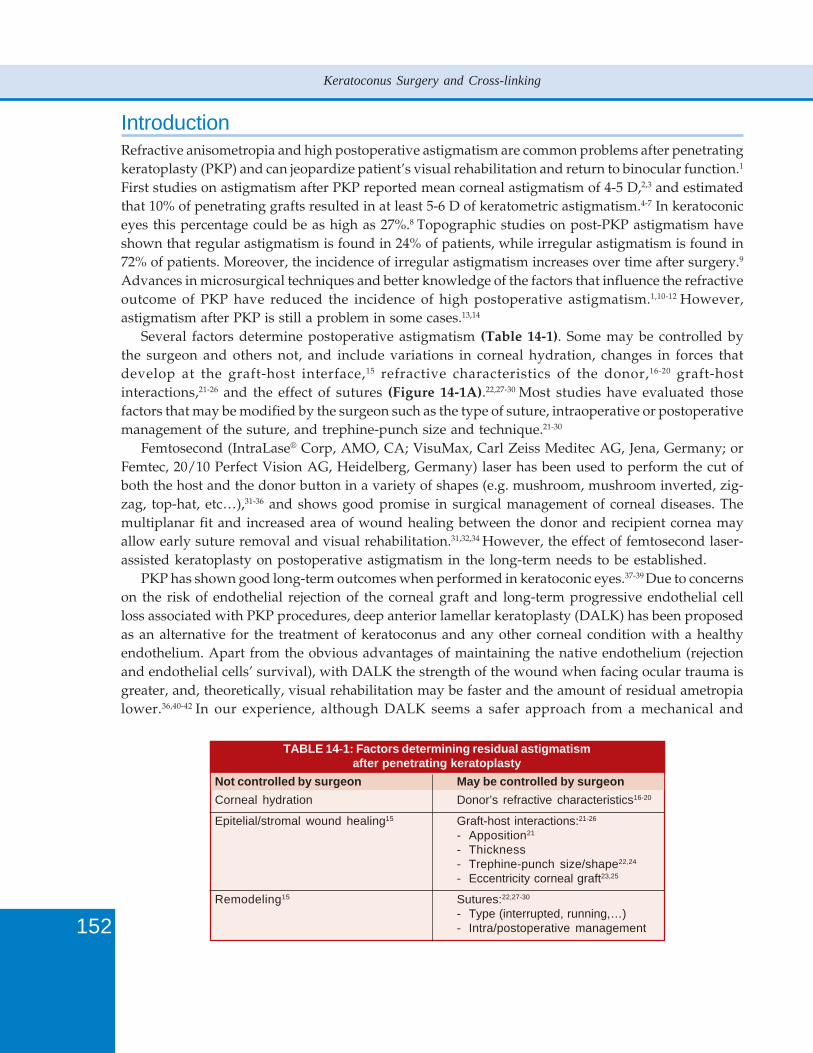
152
Keratoconus Surgery and Cross-linking
IntroductionRefractive anisometropia and high postoperative astigmatism are common problems after penetratingkeratoplasty (PKP) and can jeopardize patient’s visual rehabilitation and return to binocular function.1
First studies on astigmatism after PKP reported mean corneal astigmatism of 4-5 D,2,3 and estimatedthat 10% of penetrating grafts resulted in at least 5-6 D of keratometric astigmatism.4-7 In keratoconiceyes this percentage could be as high as 27%.8 Topographic studies on post-PKP astigmatism haveshown that regular astigmatism is found in 24% of patients, while irregular astigmatism is found in72% of patients. Moreover, the incidence of irregular astigmatism increases over time after surgery.9
Advances in microsurgical techniques and better knowledge of the factors that influence the refractiveoutcome of PKP have reduced the incidence of high postoperative astigmatism.1,10-12 However,astigmatism after PKP is still a problem in some cases.13,14
Several factors determine postoperative astigmatism (Table 14-1). Some may be controlled bythe surgeon and others not, and include variations in corneal hydration, changes in forces thatdevelop at the graft-host interface,15 refractive characteristics of the donor,16-20 graft-hostinteractions,21-26 and the effect of sutures (Figure 14-1A).22,27-30 Most studies have evaluated thosefactors that may be modified by the surgeon such as the type of suture, intraoperative or postoperativemanagement of the suture, and trephine-punch size and technique.21-30
Femtosecond (IntraLase® Corp, AMO, CA; VisuMax, Carl Zeiss Meditec AG, Jena, Germany; orFemtec, 20/10 Perfect Vision AG, Heidelberg, Germany) laser has been used to perform the cut ofboth the host and the donor button in a variety of shapes (e.g. mushroom, mushroom inverted, zig-zag, top-hat, etc…),31-36 and shows good promise in surgical management of corneal diseases. Themultiplanar fit and increased area of wound healing between the donor and recipient cornea mayallow early suture removal and visual rehabilitation.31,32,34 However, the effect of femtosecond laser-assisted keratoplasty on postoperative astigmatism in the long-term needs to be established.
PKP has shown good long-term outcomes when performed in keratoconic eyes.37-39 Due to concernson the risk of endothelial rejection of the corneal graft and long-term progressive endothelial cellloss associated with PKP procedures, deep anterior lamellar keratoplasty (DALK) has been proposedas an alternative for the treatment of keratoconus and any other corneal condition with a healthyendothelium. Apart from the obvious advantages of maintaining the native endothelium (rejectionand endothelial cells’ survival), with DALK the strength of the wound when facing ocular trauma isgreater, and, theoretically, visual rehabilitation may be faster and the amount of residual ametropialower.36,40-42 In our experience, although DALK seems a safer approach from a mechanical and
TABLE 14-1: Factors determining residual astigmatismafter penetrating keratoplasty
Not controlled by surgeon May be controlled by surgeonCorneal hydration Donor’s refractive characteristics16-20
Epitelial/stromal wound healing15 Graft-host interactions:21-26
- Apposition21
- Thickness- Trephine-punch size/shape22,24
- Eccentricity corneal graft23,25
Remodeling15 Sutures:22,27-30
- Type (interrupted, running,…)- Intra/postoperative management
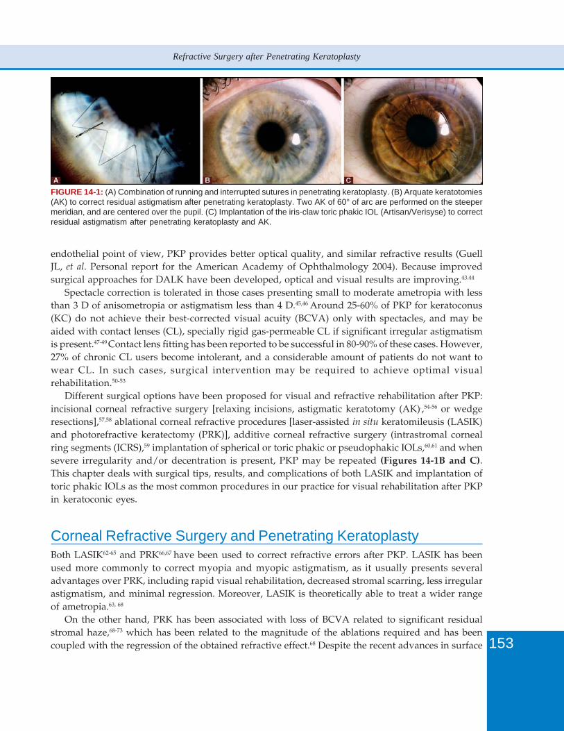
153
Refractive Surgery after Penetrating Keratoplasty
FIGURE 14-1: (A) Combination of running and interrupted sutures in penetrating keratoplasty. (B) Arquate keratotomies(AK) to correct residual astigmatism after penetrating keratoplasty. Two AK of 60° of arc are performed on the steepermeridian, and are centered over the pupil. (C) Implantation of the iris-claw toric phakic IOL (Artisan/Verisyse) to correctresidual astigmatism after penetrating keratoplasty and AK.
endothelial point of view, PKP provides better optical quality, and similar refractive results (GuellJL, et al. Personal report for the American Academy of Ophthalmology 2004). Because improvedsurgical approaches for DALK have been developed, optical and visual results are improving.43.44
Spectacle correction is tolerated in those cases presenting small to moderate ametropia with lessthan 3 D of anisometropia or astigmatism less than 4 D.45,46 Around 25-60% of PKP for keratoconus(KC) do not achieve their best-corrected visual acuity (BCVA) only with spectacles, and may beaided with contact lenses (CL), specially rigid gas-permeable CL if significant irregular astigmatismis present.47-49 Contact lens fitting has been reported to be successful in 80-90% of these cases. However,27% of chronic CL users become intolerant, and a considerable amount of patients do not want towear CL. In such cases, surgical intervention may be required to achieve optimal visualrehabilitation.50-53
Different surgical options have been proposed for visual and refractive rehabilitation after PKP:incisional corneal refractive surgery [relaxing incisions, astigmatic keratotomy (AK).,54-56 or wedgeresections],57,58 ablational corneal refractive procedures [laser-assisted in situ keratomileusis (LASIK)and photorefractive keratectomy (PRK)], additive corneal refractive surgery (intrastromal cornealring segments (ICRS),59 implantation of spherical or toric phakic or pseudophakic IOLs,60,61 and whensevere irregularity and/or decentration is present, PKP may be repeated (Figures 14-1B and C).This chapter deals with surgical tips, results, and complications of both LASIK and implantation oftoric phakic IOLs as the most common procedures in our practice for visual rehabilitation after PKPin keratoconic eyes.
Corneal Refractive Surgery and Penetrating KeratoplastyBoth LASIK62-65 and PRK66,67 have been used to correct refractive errors after PKP. LASIK has beenused more commonly to correct myopia and myopic astigmatism, as it usually presents severaladvantages over PRK, including rapid visual rehabilitation, decreased stromal scarring, less irregularastigmatism, and minimal regression. Moreover, LASIK is theoretically able to treat a wider rangeof ametropia.63, 68
On the other hand, PRK has been associated with loss of BCVA related to significant residualstromal haze,68-73 which has been related to the magnitude of the ablations required and has beencoupled with the regression of the obtained refractive effect.68 Despite the recent advances in surface
154
Keratoconus Surgery and Cross-linking
ablation techniques which might change our standard in the near future, LASIK is currently thetechnique of choice to correct low and moderate post-PKP refractive errors.
In those cases with high levels of astigmatism, AK may be performed first to reduce astigmatismto a level at which a more predictable refractive surgery such as LASIK or implantation of a phakicIOL can be used to correct residual ametropia.54,65,74-77
INDICATIONSExcimer refractive surgery should be considered to correct refractive errors after PKP whenanisometropia is not successfully corrected with spectacles, and in cases of CL intolerance. Theprimary goal of LASIK after PKP is to reduce the degree of ametropia, which allows spectaclecorrection and return to binocularity. The inclusion criteria for LASIK after PKP are the same as forLASIK in non-PKP patients. However, some additional cautions must be taken into account (Table14-2).78-80
TABLE 14-2: Indications and contraindications for laser-assisted in-situkeratomileusis for residual ametropia after penetrating keratoplasty
Indications Contraindications• Refractive stability at least 6 months • Graft rejection, recurrence of herpetic
after extraction of sutures ocular disease, or inflammatory• Minimal maintenance or no topical condition
corticosteroids • Marked peripheral corneal• CCT > 500 µm79 vascularization• Residual stromal bed > 250 µm • Significant corneal anesthesia• Sim K 38 – 55 D80 • Thin host tissue
• Wound ectasia• Significant graft override/Wound
malapposition:78,80
- misalignment graft-host junction > 10%- internal gaping > 20%
Although refractive stability after PKP is not easily determined, it has been generally accepted toperform corneal refractive surgery at least 6 months after extraction of all sutures, which means, atleast about 18 months after PKP. Apart from the time elapsed after PKP, the stability of refractionand corneal topography suggest a low level of active wound remodeling. The amount of whiteningand scarring, as well as its thickness profile, of the wound should also be verified to avoid wounddehiscence or problems when cutting the flap.81
Dry eye or corneal hypo/anesthesia are more common in post-PKP patients, especially the elderlyones, and increase the risk of ocular surface problems after PKP. Therefore, management of ocularsurface problems is mandatory before LASIK. Endothelial cell count should also be performed. Theeffect of LASIK on endothelial cell counts on PKP eyes remains unclear. Although some studiesreport an endothelial cell loss of 8.6%-10.56%,62,82 other series have found no significant alterations.63,83
STEP-BY-STEP SURGICAL TECHNIQUELASIK technique in PKP eyes is essentially the same as our standard LASIK procedure. Diazepam(5 to 10 mg) is administered orally about 30 minutes before surgery. Topical anesthesia (proparacainehydrochloride 1%) is instilled 1 drop every two minutes starting 6 minutes before surgery. In cases
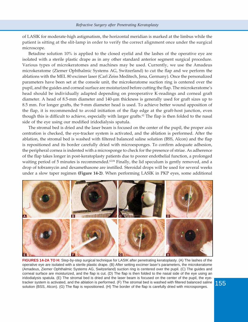
155
Refractive Surgery after Penetrating Keratoplasty
of LASIK for moderate-high astigmatism, the horizontal meridian is marked at the limbus while thepatient is sitting at the slit-lamp in order to verify the correct alignment once under the surgicalmicroscope.
Betadine solution 10% is applied to the closed eyelid and the lashes of the operative eye areisolated with a sterile plastic drape as in any other standard anterior segment surgical procedure.Various types of microkeratomes and machines may be used. Currently, we use the Amadeusmicrokeratome (Ziemer Ophthalmic Systems AG, Switzerland) to cut the flap and we perform theablations with the MEL 80 excimer laser (Carl Zeiss Meditech, Jena, Germany). Once the personalizedparameters have been set at the console unit, the microkeratome suction ring is centered over thepupil, and the guides and corneal surface are moisturized before cutting the flap. The microkeratome’shead should be individually adapted depending on preoperative K-readings and corneal graftdiameter. A head of 8.5-mm diameter and 140-µm thickness is generally used for graft sizes up to8.5 mm. For longer grafts, the 9-mm diameter head is used. To achieve better wound apposition ofthe flap, it is recommended to avoid initiation of the flap edge at the graft-host junction, eventhough this is difficult to achieve, especially with larger grafts.62 The flap is then folded to the nasalside of the eye using our modified iridodialysis spatula.
The stromal bed is dried and the laser beam is focused on the center of the pupil, the proper axiscentration is checked, the eye-tracker system is activated, and the ablation is performed. After theablation, the stromal bed is washed with filtered balanced saline solution (BSS, Alcon) and the flapis repositioned and its border carefully dried with microesponges. To confirm adequate adhesion,the peripheral cornea is indented with a microsponge to check for the presence of striae. As adherenceof the flap takes longer in post-keratoplasty patients due to poorer endothelial function, a prolongedwaiting period of 5 minutes is recommended.62,84 Finally, the lid speculum is gently removed, and adrop of tobramycin and dexamethasone are instilled. Steroidal drops will be used for several weeksunder a slow taper regimen (Figure 14-2). When performing LASIK in PKP eyes, some additional
FIGURES 14-2A TO H: Step-by-step surgical technique for LASIK after penetrating keratoplasty. (A) The lashes of theoperative eye are isolated with a sterile plastic drape. (B) After setting excimer laser’s parameters, the microkeratome(Amadeus, Ziemer Ophthalmic Systems AG, Switzerland) suction ring is centered over the pupil. (C) The guides andcorneal surface are moisturized, and the flap is cut. (D) The flap is then folded to the nasal side of the eye using aniridodialysis spatula. (E) The stromal bed is dried and the laser beam is focused on the center of the pupil, the eye-tracker system is activated, and the ablation is performed. (F) The stromal bed is washed with filtered balanced salinesolution (BSS, Alcon). (G) The flap is repositioned. (H) The border of the flap is carefully dried with microsponges.
156
Keratoconus Surgery and Cross-linking
difficulties may be encountered. It is of utmost importance to check the correct apposition andscarring of the graft-host interface. As there is a long-term instability of PKP wounds, partial wounddehiscence may occur while cutting the flap. If this is the case, LASIK may be performed uneventfullyand, in some cases, the wound may require suture.
High keratometry readings are relatively common in PKP patients. Therefore, flap complicationstypical of steep corneas such as thin flaps or buttonholes may occur. Moreover, it has also beenpostulated an increased risk of recurrence of keratoconus in the graft after excimer laser ablations.
Topography-guided ablations constitute adequate options to correct or reduce irregular astigmatism.However, the efficacy of guided ablations compared to the standard approach is still controversial.85-87
In those cases with high levels of astigmatism, AK may be performed first to reduce astigmatism toa level at which LASIK can be used to correct residual ametropia, while less tissue is ablated.54,65,88,89
It is generally recommended to perform LASIK at least 6 months after incisional surgery.62
Simply performing a hinged lamellar keratotomy induces a biomechanical response of the corneathat results in changes of its shape.90,91 Also, cutting a 160-µm corneal flap in eyes after PKP results insignificant changes of both spherical equivalent refraction and astigmatism that may result in inaccuraterefractive corrections if pre-flap refraction is applied.92,93 For this reason, several authors advocate toperform LASIK after PKP as a 2-stage procedure. Excimer ablation by relifting the flap is performedonce the corneal shape is stabilized (1 to 3 months after keratotomy).93-99 Nevertheless, as we havepreviously mentioned with topo-guided or wavefront-guided ablations, a superior efficacy of the2-step approach is still controversial.
Toric Phakic Intraocular Lenses and PKPINDICATIONSInclusion and exclusion criteria for implantation of phakic IOLs are summarized in Table 14-3. Thetoric Artisan-Verisyse is our phakic IOL of choice for the correction of residual moderate-highmyopic or hyperopic astigmatism after PKP. The toric Artisan/Verisyse pIOL is a one piece, 5-mmoptic, PMMA lens which is available in powers ranging from +12 D to -23.5 D in 0.5-D increments,
TABLE 14-3: Generally recommended inclusion and exclusion criteria forphakic IOL implantation
Inclusion criteria Exclusion criteria• Age > 21 years • Background of active disease in the• Stability of refraction at least 1 year anterior segment• Ametropia not correctable with • Recurrent or chronic uveitis
excimer laser surgery • Any form of clinically significant• Unsatisfactory vision with/intolerance cataract
of contact lenses or spectacles • Previous corneal or intraocular• Irido-corneal angle ≥ 30° surgery (to be evaluated)• cECC > 2300 cells/mm2*: • IOP > 21mmHg or glaucoma
(> 2500 cells/mm2 if >21 years old, • Preexisting macular degeneration or> 2000 if >40 years old) macular pathology
• No anomaly of the iris or pupil function • Abnormal retinal condition• Scotopic pupil size < 5-6 mm • Systemic diseases (e.g.• Toric PIOL if astigmatism > 2 D autoimmune disorder, connective
tissue disease, atopia, diabetesmellitus)
* Lower cECC are accepted in selected cases
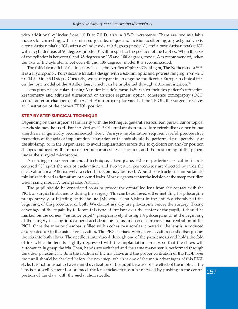
157
Refractive Surgery after Penetrating Keratoplasty
with additional cylinder from 1.0 D to 7.0 D, also in 0.5-D increments. There are two availablemodels for correcting, with a similar surgical technique and incision positioning, any astigmatic axis:a toric Artisan phakic IOL with a cylinder axis at 0 degrees (model A) and a toric Artisan phakic IOLwith a cylinder axis at 90 degrees (model B) with respect to the position of the haptics. When the axisof the cylinder is between 0 and 45 degrees or 135 and 180 degrees, model A is recommended; whenthe axis of the cylinder is between 45 and 135 degrees, model B is recommended.
The foldable model of the iris-claw lens is the Artiflex (Ophtec, Groningen, The Netherlands).100,101
It is a Hydrophobic Polysiloxane foldable design with a 6.0-mm optic and powers ranging from –2 Dto –14.5 D in 0.5 D steps. Currently, we participate in an ongoing multicenter European clinical trialon the toric model of the Artiflex lens, which can be implanted through a 3.1-mm incision.102
Lens power is calculated using Van der Heijde’s formula,103 which includes patient’s refraction,keratometry and adjusted ultrasound or anterior segment optical coherence tomography (OCT)central anterior chamber depth (ACD). For a proper placement of the TPIOL, the surgeon receivesan illustration of the correct TPIOL position.
STEP-BY-STEP SURGICAL TECHNIQUEDepending on the surgeon’s familiarity with the technique, general, retrobulbar, peribulbar or topicalanesthesia may be used. For the Verisyse® PIOL implantation procedure retrobulbar or peribulbaranesthesia is generally recommended. Toric Verisyse implantation requires careful preoperativemarcation of the axis of implantation. Marcation of the axis should be performed preoperatively atthe slit-lamp, or in the Argon laser, to avoid implantation errors due to cyclotorsion and/or positionchanges induced by the retro or peribulbar anesthesia injection, and the positioning of the patientunder the surgical microscope.
According to our recommended technique, a two-plane, 5.2-mm posterior corneal incision iscentered 90° apart the axis of enclavation, and two vertical paracenteses are directed towards theenclavation area. Alternatively, a scleral incision may be used. Wound construction is important tominimize induced astigmatism or wound leaks. Most surgeons center the incision at the steep meridianwhen using model A toric phakic Artisan.
The pupil should be constricted so as to protect the crystalline lens from the contact with thePIOL or surgical instruments during the surgery. This can be achieved either instilling 1% pilocarpinepreoperatively or injecting acetylcholine (Myochol, Ciba Vision) in the anterior chamber at thebeginning of the procedure, or both. We do not usually use pilocarpine before the surgery. Takingadvantage of the capability to locate this type of implant over the center of the pupil, it should bemarked on the cornea (“entrance pupil”) preoperatively if using 1% pilocarpine, or at the beginningof the surgery if using intracameral acetylcholine, so as to enable a proper, final centration of thePIOL. Once the anterior chamber is filled with a cohesive viscoelastic material, the lens is introducedand rotated up to the axis of enclavation. The PIOL is fixed with an enclavation needle that pushesthe iris into both claws. The needle is introduced through one of the paracentesis and holds the foldof iris while the lens is slightly depressed with the implantation forceps so that the claws willautomatically grasp the iris. Then, hands are switched and the same maneuver is performed throughthe other paracentesis. Both the fixation of the iris claws and the proper centration of the PIOL overthe pupil should be checked before the next step, which is one of the main advantages of this PIOLstyle. It is not unusual to have a mild ovalization of the pupil because of the effect of the miotic. If thelens is not well centered or oriented, the lens enclavation can be released by pushing in the centralportion of the claw with the enclavation needle.
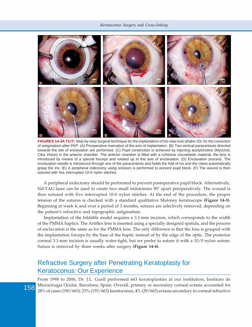
158
Keratoconus Surgery and Cross-linking
A peripheral iridectomy should be performed to prevent postoperative pupil block. Alternatively,Nd:YAG laser can be used to create two small iridotomies 90° apart preoperatively. The wound isthen sutured with five interrupted 10-0 nylon stitches. At the end of the procedure, the propertension of the sutures is checked with a standard qualitative Maloney keratoscope (Figure 14-3).Beginning at week 4, and over a period of 3 months, sutures are selectively removed, depending onthe patient’s refractive and topographic astigmatism.
Implantation of the foldable model requires a 3.1-mm incision, which corresponds to the widthof the PMMA haptics. The Artiflex lens is inserted using a specially designed spatula, and the processof enclavation is the same as for the PMMA lens. The only difference is that the lens is grasped withthe implantation forceps by the base of the haptic instead of by the edge of the optic. The posteriorcorneal 3.1-mm incision is usually water-tight, but we prefer to suture it with a 10/0 nylon suture.Suture is removed by three weeks after surgery (Figure 14-4).
Refractive Surgery after Penetrating Keratoplasty forKeratoconus: Our ExperienceFrom 1998 to 2006, Dr. J.L. Guell performed 663 keratoplasties at our institution, Instituto deMicrocirugia Ocular, Barcelona, Spain. Overall, primary or secondary corneal ectasia accounted for28% of cases (190/663): 23% (155/663) keratoconus, 4% (29/663) ectasia secondary to corneal refractive
FIGURES 14-3A TO F: Step-by-step surgical technique for the implantation of iris-claw toric phakic IOL for the correctionof astigmatism after PKP. (A) Preoperative marcation of the axis of implantation. (B) Two vertical paracentesis directedtowards the site of enclavation are performed. (C) Pupil constriction is achieved by injecting acetylcholine (Myochol,Ciba Vision) in the anterior chamber. The anterior chamber is filled with a cohesive viscoelastic material, the lens isintroduced by means of a special forceps and rotated up to the axis of enclavation. (D) Enclavation process. Theenclavation needle is introduced through one of the paracentesis and holds the fold of iris and the claws automaticallygrasp the iris. (E) A peripheral iridectomy using scissors is performed to prevent pupil block. (F) The wound is thensutured with five interrupted 10-0 nylon stitches.
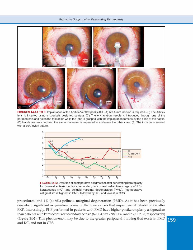
159
Refractive Surgery after Penetrating Keratoplasty
FIGURES 14-4A TO F: Implantation of the Artiflex/Veriflex phakic IOL (A) A 3.1-mm incision is required. (B) The Artiflexlens is inserted using a specially designed spatula. (C) The enclavation needle is introduced through one of theparacentesis and holds the fold of iris while the lens is grasped with the implantation forceps by the base of the haptic.(D) Hands are switched and the same maneuver is repeated to enclavate the other claw. (E) The incision is suturedwith a 10/0 nylon suture.
FIGURE 14-5: Evolution of postoperative astigmatism after penetrating keratoplastyfor corneal ectasia: ectasia secondary to corneal refractive surgery (CRS),keratoconus (KC), and pellucid marginal degeneration (PMD). Postoperativeastigmatism is highest in PMD, followed by KC, and lowest in CRS.
procedures, and 1% (6/663) pellucid marginal degeneration (PMD). As it has been previouslydescribed, significant astigmatism is one of the main causes that impair visual rehabilitation afterPKP. Interestingly, PKP performed in patients with PMD have higher postkeratoplasty astigmatismthan patients with keratoconus or secondary ectasia (6.8 ± 4.6 vs 2.98 ± 1.63 and 2.25 ± 2.38, respectively)(Figure 14-5). This phenomenon may be due to the greater peripheral thinning that exists in PMDand KC, and not in CRS.
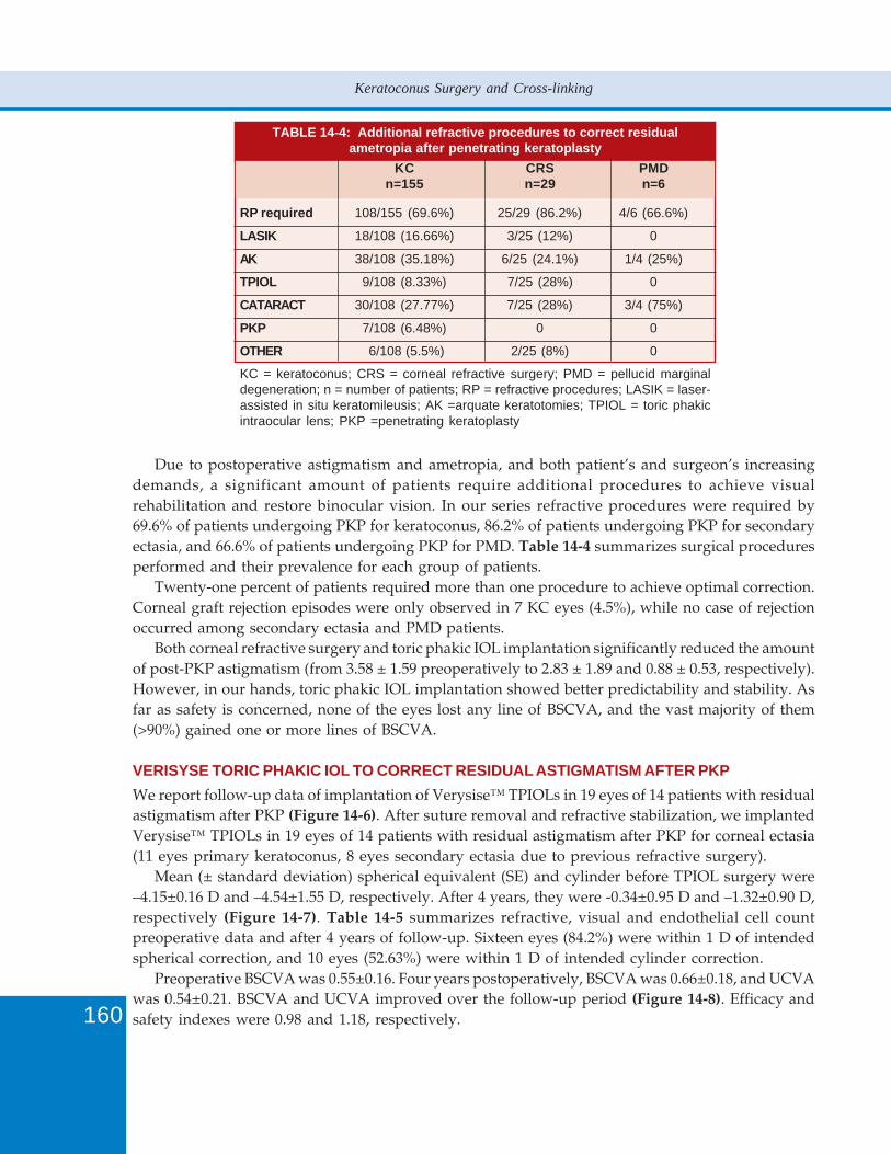
160
Keratoconus Surgery and Cross-linking
Due to postoperative astigmatism and ametropia, and both patient’s and surgeon’s increasingdemands, a significant amount of patients require additional procedures to achieve visualrehabilitation and restore binocular vision. In our series refractive procedures were required by69.6% of patients undergoing PKP for keratoconus, 86.2% of patients undergoing PKP for secondaryectasia, and 66.6% of patients undergoing PKP for PMD. Table 14-4 summarizes surgical proceduresperformed and their prevalence for each group of patients.
Twenty-one percent of patients required more than one procedure to achieve optimal correction.Corneal graft rejection episodes were only observed in 7 KC eyes (4.5%), while no case of rejectionoccurred among secondary ectasia and PMD patients.
Both corneal refractive surgery and toric phakic IOL implantation significantly reduced the amountof post-PKP astigmatism (from 3.58 ± 1.59 preoperatively to 2.83 ± 1.89 and 0.88 ± 0.53, respectively).However, in our hands, toric phakic IOL implantation showed better predictability and stability. Asfar as safety is concerned, none of the eyes lost any line of BSCVA, and the vast majority of them(>90%) gained one or more lines of BSCVA.
VERISYSE TORIC PHAKIC IOL TO CORRECT RESIDUAL ASTIGMATISM AFTER PKPWe report follow-up data of implantation of Verysise™ TPIOLs in 19 eyes of 14 patients with residualastigmatism after PKP (Figure 14-6). After suture removal and refractive stabilization, we implantedVerysise™ TPIOLs in 19 eyes of 14 patients with residual astigmatism after PKP for corneal ectasia(11 eyes primary keratoconus, 8 eyes secondary ectasia due to previous refractive surgery).
Mean (± standard deviation) spherical equivalent (SE) and cylinder before TPIOL surgery were–4.15±0.16 D and –4.54±1.55 D, respectively. After 4 years, they were -0.34±0.95 D and –1.32±0.90 D,respectively (Figure 14-7). Table 14-5 summarizes refractive, visual and endothelial cell countpreoperative data and after 4 years of follow-up. Sixteen eyes (84.2%) were within 1 D of intendedspherical correction, and 10 eyes (52.63%) were within 1 D of intended cylinder correction.
Preoperative BSCVA was 0.55±0.16. Four years postoperatively, BSCVA was 0.66±0.18, and UCVAwas 0.54±0.21. BSCVA and UCVA improved over the follow-up period (Figure 14-8). Efficacy andsafety indexes were 0.98 and 1.18, respectively.
TABLE 14-4: Additional refractive procedures to correct residualametropia after penetrating keratoplasty
KC CRS PMDn=155 n=29 n=6
RP required 108/155 (69.6%) 25/29 (86.2%) 4/6 (66.6%)
LASIK 18/108 (16.66%) 3/25 (12%) 0
AK 38/108 (35.18%) 6/25 (24.1%) 1/4 (25%)
TPIOL 9/108 (8.33%) 7/25 (28%) 0
CATARACT 30/108 (27.77%) 7/25 (28%) 3/4 (75%)
PKP 7/108 (6.48%) 0 0
OTHER 6/108 (5.5%) 2/25 (8%) 0
KC = keratoconus; CRS = corneal refractive surgery; PMD = pellucid marginaldegeneration; n = number of patients; RP = refractive procedures; LASIK = laser-assisted in situ keratomileusis; AK =arquate keratotomies; TPIOL = toric phakicintraocular lens; PKP =penetrating keratoplasty
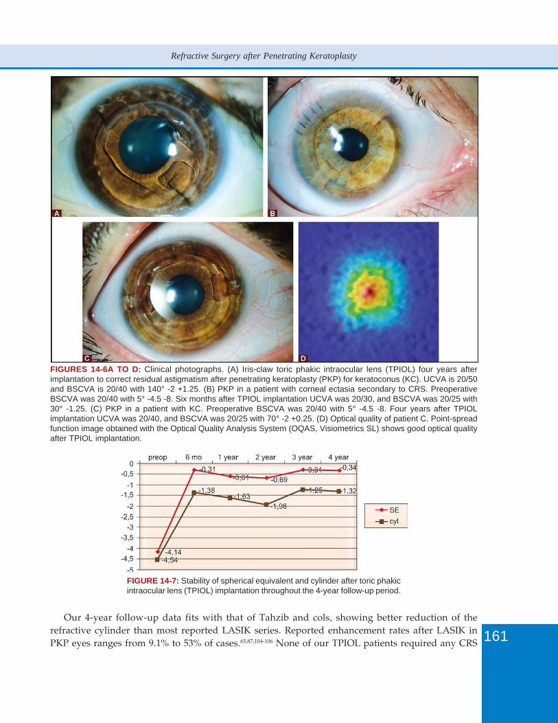
161
Refractive Surgery after Penetrating Keratoplasty
FIGURES 14-6A TO D: Clinical photographs. (A) Iris-claw toric phakic intraocular lens (TPIOL) four years afterimplantation to correct residual astigmatism after penetrating keratoplasty (PKP) for keratoconus (KC). UCVA is 20/50and BSCVA is 20/40 with 140° -2 +1.25. (B) PKP in a patient with corneal ectasia secondary to CRS. PreoperativeBSCVA was 20/40 with 5° -4.5 -8. Six months after TPIOL implantation UCVA was 20/30, and BSCVA was 20/25 with30° -1.25. (C) PKP in a patient with KC. Preoperative BSCVA was 20/40 with 5° -4.5 -8. Four years after TPIOLimplantation UCVA was 20/40, and BSCVA was 20/25 with 70° -2 +0.25. (D) Optical quality of patient C. Point-spreadfunction image obtained with the Optical Quality Analysis System (OQAS, Visiometrics SL) shows good optical qualityafter TPIOL implantation.
FIGURE 14-7: Stability of spherical equivalent and cylinder after toric phakicintraocular lens (TPIOL) implantation throughout the 4-year follow-up period.
Our 4-year follow-up data fits with that of Tahzib and cols, showing better reduction of therefractive cylinder than most reported LASIK series. Reported enhancement rates after LASIK inPKP eyes ranges from 9.1% to 53% of cases.65,87,104-106 None of our TPIOL patients required any CRS
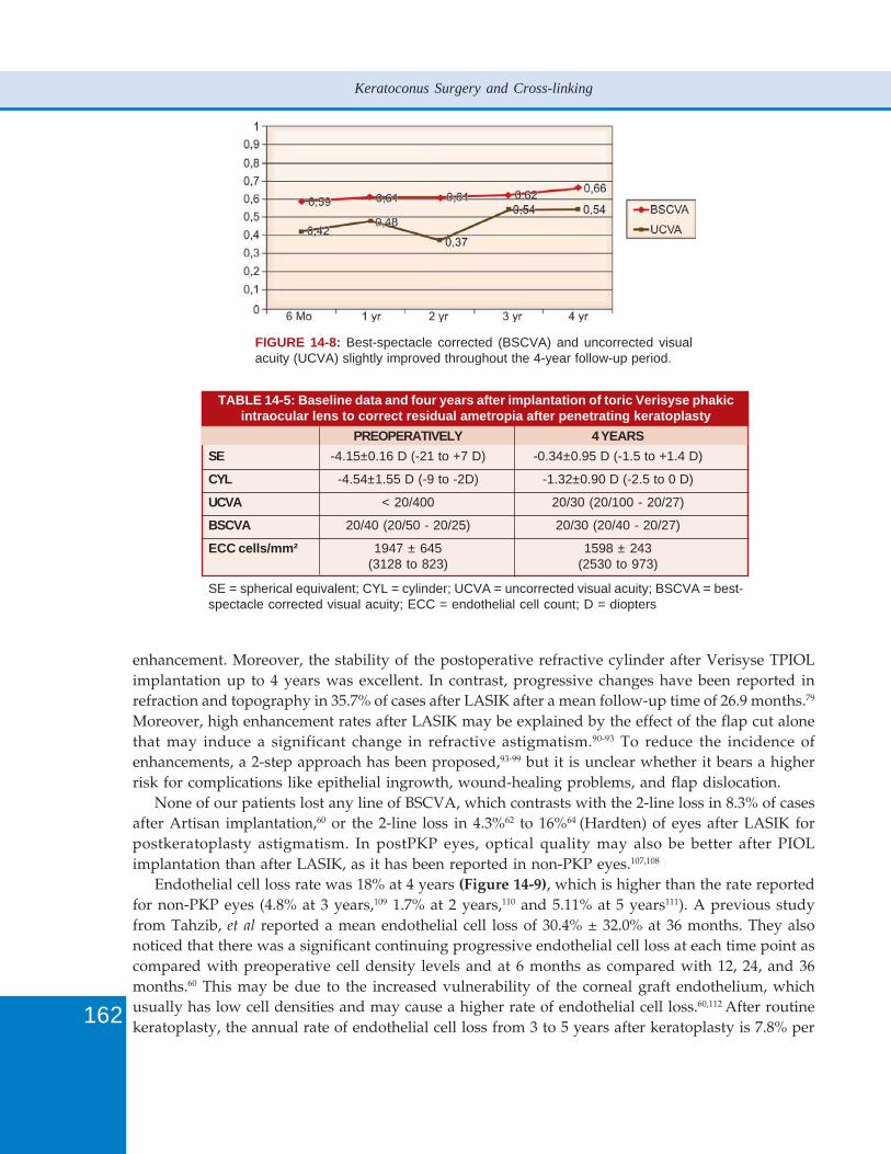
162
Keratoconus Surgery and Cross-linking
FIGURE 14-8: Best-spectacle corrected (BSCVA) and uncorrected visualacuity (UCVA) slightly improved throughout the 4-year follow-up period.
TABLE 14-5: Baseline data and four years after implantation of toric Verisyse phakicintraocular lens to correct residual ametropia after penetrating keratoplasty
PREOPERATIVELY 4 YEARSSE -4.15±0.16 D (-21 to +7 D) -0.34±0.95 D (-1.5 to +1.4 D)
CYL -4.54±1.55 D (-9 to -2D) -1.32±0.90 D (-2.5 to 0 D)
UCVA < 20/400 20/30 (20/100 - 20/27)
BSCVA 20/40 (20/50 - 20/25) 20/30 (20/40 - 20/27)
ECC cells/mm² 1947 ± 645 1598 ± 243(3128 to 823) (2530 to 973)
SE = spherical equivalent; CYL = cylinder; UCVA = uncorrected visual acuity; BSCVA = best-spectacle corrected visual acuity; ECC = endothelial cell count; D = diopters
enhancement. Moreover, the stability of the postoperative refractive cylinder after Verisyse TPIOLimplantation up to 4 years was excellent. In contrast, progressive changes have been reported inrefraction and topography in 35.7% of cases after LASIK after a mean follow-up time of 26.9 months.79
Moreover, high enhancement rates after LASIK may be explained by the effect of the flap cut alonethat may induce a significant change in refractive astigmatism.90-93 To reduce the incidence ofenhancements, a 2-step approach has been proposed,93-99 but it is unclear whether it bears a higherrisk for complications like epithelial ingrowth, wound-healing problems, and flap dislocation.
None of our patients lost any line of BSCVA, which contrasts with the 2-line loss in 8.3% of casesafter Artisan implantation,60 or the 2-line loss in 4.3%62 to 16%64 (Hardten) of eyes after LASIK forpostkeratoplasty astigmatism. In postPKP eyes, optical quality may also be better after PIOLimplantation than after LASIK, as it has been reported in non-PKP eyes.107,108
Endothelial cell loss rate was 18% at 4 years (Figure 14-9), which is higher than the rate reportedfor non-PKP eyes (4.8% at 3 years,109 1.7% at 2 years,110 and 5.11% at 5 years111). A previous studyfrom Tahzib, et al reported a mean endothelial cell loss of 30.4% ± 32.0% at 36 months. They alsonoticed that there was a significant continuing progressive endothelial cell loss at each time point ascompared with preoperative cell density levels and at 6 months as compared with 12, 24, and 36months.60 This may be due to the increased vulnerability of the corneal graft endothelium, whichusually has low cell densities and may cause a higher rate of endothelial cell loss.60,112 After routinekeratoplasty, the annual rate of endothelial cell loss from 3 to 5 years after keratoplasty is 7.8% per
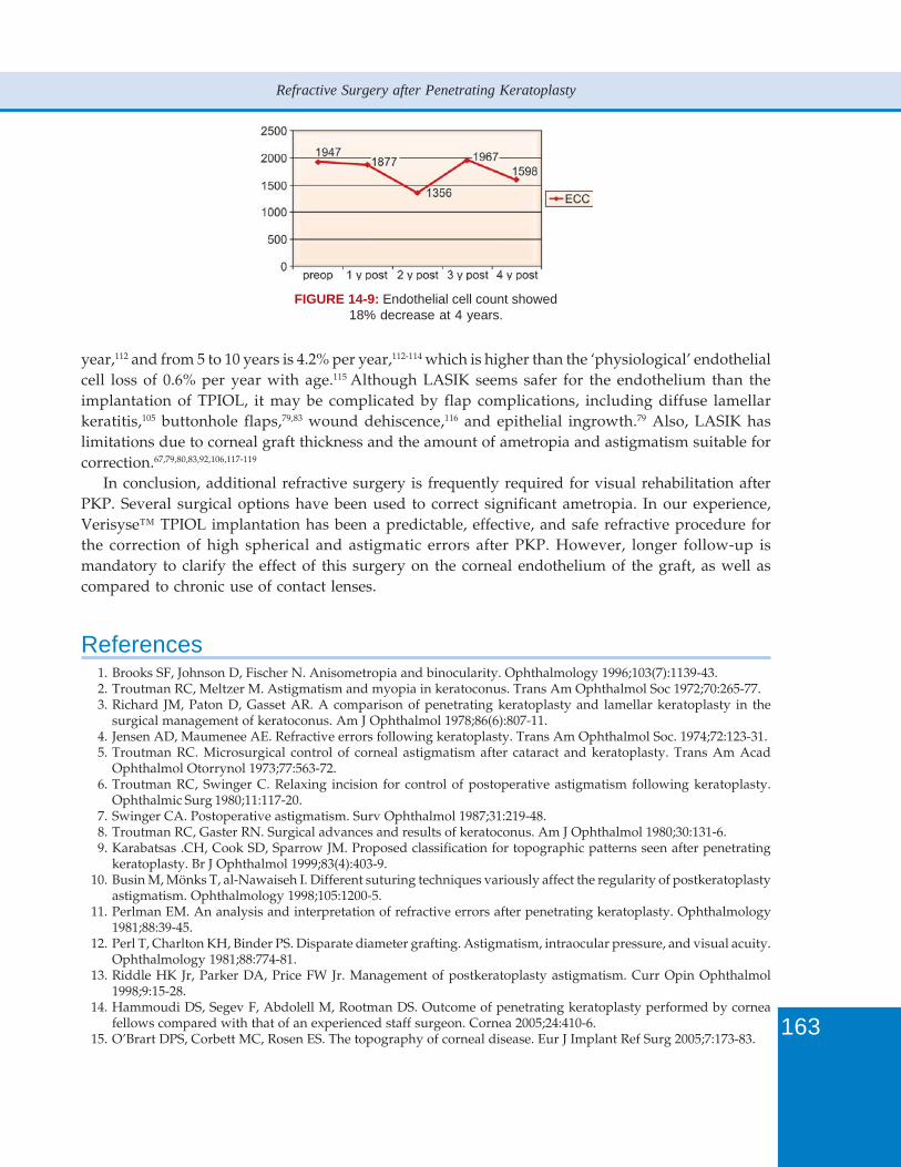
163
Refractive Surgery after Penetrating Keratoplasty
FIGURE 14-9: Endothelial cell count showed18% decrease at 4 years.
year,112 and from 5 to 10 years is 4.2% per year,112-114 which is higher than the ‘physiological’ endothelialcell loss of 0.6% per year with age.115 Although LASIK seems safer for the endothelium than theimplantation of TPIOL, it may be complicated by flap complications, including diffuse lamellarkeratitis,105 buttonhole flaps,79,83 wound dehiscence,116 and epithelial ingrowth.79 Also, LASIK haslimitations due to corneal graft thickness and the amount of ametropia and astigmatism suitable forcorrection.67,79,80,83,92,106,117-119
In conclusion, additional refractive surgery is frequently required for visual rehabilitation afterPKP. Several surgical options have been used to correct significant ametropia. In our experience,Verisyse™ TPIOL implantation has been a predictable, effective, and safe refractive procedure forthe correction of high spherical and astigmatic errors after PKP. However, longer follow-up ismandatory to clarify the effect of this surgery on the corneal endothelium of the graft, as well ascompared to chronic use of contact lenses.
References1. Brooks SF, Johnson D, Fischer N. Anisometropia and binocularity. Ophthalmology 1996;103(7):1139-43.2. Troutman RC, Meltzer M. Astigmatism and myopia in keratoconus. Trans Am Ophthalmol Soc 1972;70:265-77.3. Richard JM, Paton D, Gasset AR. A comparison of penetrating keratoplasty and lamellar keratoplasty in the
surgical management of keratoconus. Am J Ophthalmol 1978;86(6):807-11.4. Jensen AD, Maumenee AE. Refractive errors following keratoplasty. Trans Am Ophthalmol Soc. 1974;72:123-31.5. Troutman RC. Microsurgical control of corneal astigmatism after cataract and keratoplasty. Trans Am Acad
Ophthalmol Otorrynol 1973;77:563-72.6. Troutman RC, Swinger C. Relaxing incision for control of postoperative astigmatism following keratoplasty.
Ophthalmic Surg 1980;11:117-20.7. Swinger CA. Postoperative astigmatism. Surv Ophthalmol 1987;31:219-48.8. Troutman RC, Gaster RN. Surgical advances and results of keratoconus. Am J Ophthalmol 1980;30:131-6.9. Karabatsas .CH, Cook SD, Sparrow JM. Proposed classification for topographic patterns seen after penetrating
keratoplasty. Br J Ophthalmol 1999;83(4):403-9.10. Busin M, Mönks T, al-Nawaiseh I. Different suturing techniques variously affect the regularity of postkeratoplasty
astigmatism. Ophthalmology 1998;105:1200-5.11. Perlman EM. An analysis and interpretation of refractive errors after penetrating keratoplasty. Ophthalmology
1981;88:39-45.12. Perl T, Charlton KH, Binder PS. Disparate diameter grafting. Astigmatism, intraocular pressure, and visual acuity.
Ophthalmology 1981;88:774-81.13. Riddle HK Jr, Parker DA, Price FW Jr. Management of postkeratoplasty astigmatism. Curr Opin Ophthalmol
1998;9:15-28.14. Hammoudi DS, Segev F, Abdolell M, Rootman DS. Outcome of penetrating keratoplasty performed by cornea
fellows compared with that of an experienced staff surgeon. Cornea 2005;24:410-6.15. O’Brart DPS, Corbett MC, Rosen ES. The topography of corneal disease. Eur J Implant Ref Surg 2005;7:173-83.

164
Keratoconus Surgery and Cross-linking
16. Liu Y, Seitz B, Langenbucher A, et al. Impact of preoperative corneal curvature on the outcome of penetratingkeratoplasty in keratoconus. Cornea 2003;22:409-12.
17. Ousley PJ, Terry MA. Use of a portable topography machine for screening donor tissue for prior refractivesurgery. Cornea 2002;21:745-50.
18. Lin RC, Li Y, Tang M, et al. Screening for previous refractive surgery in eye bank corneas by using opticalcoherence tomography. Cornea 2007;26:594-9.
19. Hick S, Laliberte JF, Meunier J, et al. Topographic screening of donor eyes for previous refractive surgery. JCataract Refract Surg 2006;32:309-17.
20. Krivoy D, McCormick S, Zaidman GW. Postkeratoplasty keratoconus in a nonkeratoconus patient. Am J Ophthalmol2001;131:653-4.
21. Kaiserman I, Bahar I, Rootman DS. Corneal wound malapposition after penetrating keratoplasty: an opticalcoherence tomography study. Br J Ophthalmol 2008;92(8):1103-7.
22. Seitz B, Langenbucher A, Küchle M, Naumann GO. Impact of graft diameter on corneal power and the regularityof postkeratoplasty astigmatism before and after suture removal. Ophthalmology 2003;110:2162-7.
23. Kerenyi A, Süveges I. Corneal topographic results after eccentric, biconvex penetrating keratoplasty. J CataractRefract Surg 2003;29:752-6.
24. Jaycock PD, Jones MN, Males J, et al. Outcomes of same-sizing versus oversizing donor trephines in keratoconicpatients undergoing first penetrating keratoplasty. Ophthalmology. 2008;115(2):268-75.
25. Chern KC, Meisler DM, Wilson SE, et al. Small-diameter, round, eccentric penetrating keratoplasties and cornealtopographic correlation. Ophthalmology 1997;104:643-7.
26. Troutman RC. Astigmatic considerations in corneal graft. Ophthalmic Surg. 1979;10(5):21-6.27. Van Rij G, Waring GO. Changes in corneal curvature induced by sutures and incisions. Am J Ophthalmol
1984;98:773-83.28. Javadi MA, Naderi M, Zare M, et al. Comparison of the effect of three suturing techniques on postkeratoplasty
astigmatism in keratoconus. Cornea 2006;25(9):1029-33.29. Serdarevic ON, Renard GJ, Pouliquen Y. Randomized clinical trial of penetrating keratoplasty. Before and after
suture removal comparison of intraoperative and postoperative suture adjustment. Ophthalmology1995;102(10):1497-503.
30. Mader TH, Yuan R, Lynn MJ, Stulting RD, Wilson LA, Waring GO 3rd. Changes in keratometric astigmatism aftersuture removal more than one year after penetrating keratoplasty. Ophthalmology. 1993;100(1):119-26;discussion-127.
31. Cheng YY, Tahzib NG, van Rij G, et al. Femtosecond laser-assisted inverted mushroom keratoplasty. Cornea2008;27:679-85.
32. Price FW, Price MO. Femtosecond laser shaped penetrating keratoplasty: one-year results utilizing a top-hatconfiguration. Am J Ophthalmol 2008;145:210-4.
33. Por YM, Cheng JY, Parthasarathy A, et al. Outcomes of femtosecond laser-assisted penetrating keratoplasty. AmJ Ophthalmol 2008;145:772-4.
34. Buratto L, Böhm E. The use of the femtosecond laser in penetrating keratoplasty. Am J Ophthalmol 2007;143:737-42.
35. Farid M, Kim M, Steinert RF. Results of penetrating keratoplasty performed with a femtosecond laser zigzagincision initial report. Ophthalmology 2007;114:2208-12.
36. Bahar I, Kaiserman I, Srinivasan S, Ya-Ping J, Slomovic AR, Rootman DS. Comparison of Three Different Techniquesof Corneal Transplantation for Keratoconus. Am J Ophthalmol 2008;22:e-pub ahead of print.
37. Pramanik S, Musch DC, Sutphin JE, Fario AA. Extended long-term outcomes of penetrating keratoplasty forkeratoconus. Ophthalmology. 2006;113(9):1633-8.
38. Gracia Garcia GP, Martinez JB. Outcomes of penetrating keratoplasty in mentally retarded patients with keratoconus.Cornea 2008;27(9):980-7.
39. Beckingsale P, Mavrikakis I, Al-Yousuf N, et al. Penetrating keratoplasty: outcomes from a corneal unit comparedto national data. Br J Ophthalmol 2006;90(6):728-31.
40. Watson SL, Ramsay A, Dart JK. Comparison of deep lamellar keratoplasty and penetrating keratoplasty in patientswith keratoconus. Ophthalmology 2004;111:1676-82.
41. Vabres B, Bosnjakowski M, Bekri L, et al. Deep lamellar keratoplasty versus penetrating keratoplasty for keratoconus.J Fr Ophthalmol 2006;29:361-71.
42. Shimazaki J, Shimmura S, Ishioka M, Tsubota K. Randomized clinical trial of deep lamellar keratoplasty vs penetratingkeratoplasty. Am J Ophthalmol 2002;134:159-65.
43. Borderie VM, Werthel AL, Touzeau O, et al. Comparison of techniques used for removing the recipient stroma inanterior lamellar keratoplasty. Arch Ophthalmol 2008;126:31-7.
44. Anwar M, Teichmann KD. Big-bubble technique to bare Descemet’s membrane in anterior lamellar keratoplasty.J Cataract Refract Surg 2002;28:398-403.
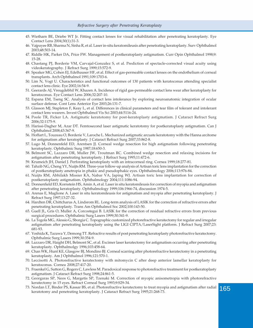
165
Refractive Surgery after Penetrating Keratoplasty
45. Wietharn BE, Driebe WT Jr. Fitting contact lenses for visual rehabilitation after penetrating keratoplasty. EyeContact Lens 2004;30(1):31-3.
46. Vajpayee RB, Sharma N, Sinha R, et al. Laser in-situ keratomileusis after penetrating keratoplasty. Surv Ophthalmol2003;48:503-14.
47. Riddle HK, Parker DA, Price FW. Management of postkeratoplasty astigmatism. Curr Opin Ophthalmol 1998;9:15-28.
48. Chastang PJ, Borderie VM, Carvajal-Gonzalez S, et al. Prediction of spectacle-corrected visual acuity usingvideokeratography. J Refract Surg 1999;15:572-9.
49. Speaker MG, Cohen EJ, Edelhauser HF, et al. Effect of gas-permeable contact lenses on the endothelium of cornealtransplants. Arch Ophthalmol 1991;109:1703-6.
50. Lim N, Vogt U. Characteristics and functional outcomes of 130 patients with keratoconus attending specialistcontact lens clinic. Eye 2002;16:54-9.
51. Geerards AJ, Vreugdebhil W, Khazen A. Incidence of rigid gas-permeable contact lens wear after keratoplasty forkeratoconus. Eye Contact Lens 2006;32:207-10.
52. Espana EM, Tseng SC. Analysis of contact lens intolerance by exploring neuroanatomic integration of ocularsurface defense. Cont Lens Anterior Eye 2003;26:131-7.
53. Glasson MJ, Stapleton F, Keay L, et al. Differences in clinical parameters and tear film of tolerant and intolerantcontact lens wearers. Invest Ophthalmol Vis Sci 2003;44:5116-24.
54. Poole TR, Ficker LA. Astigmatic keratotomy for post-keratoplasty astigmatism. J Cataract Refract Surg2006;32:1175-9.
55. Harissi-Dagher M, Azar DT. Femtosecond laser astigmatic keratotomy for postkeratoplasty astigmatism. Can JOphthalmol 2008;43:367-9.
56. Hoffart L, Touzeau O, Borderie V, Laroche L. Mechanized astigmatic arcuate keratotomy with the Hanna arcitomefor astigmatism after keratoplasty. J Cataract Refract Surg 2007;33:862-8.
57. Lugo M, Donnenfeld ED, Arentsen JJ. Corneal wedge resection for high astigmatism following penetratingkeratoplasty. Ophthalmic Surg 1987;18:650-3.
58. Belmont SC, Lazzaro DR, Muller JW, Troutman RC. Combined wedge resection and relaxing incisions forastigmatism after penetrating keratoplasty. J Refract Surg 1995;11:472-6.
59. Krumeich JH, Daniel J. Perforating keratoplasty with an intracorneal ring. Cornea 1999;18:277-81.60. Tahzib NG, Cheng YY, Nuijts RM. Three-year follow-up analysis of Artisan toric lens implantation for the correction
of postkeratoplasty ametropia in phakic and pseudophakic eyes. Ophthalmology 2006;113:976-84.61. Nuijts RM, Abhilakh Missier KA, Nabar VA, Japing WJ. Artisan toric lens implantation for correction of
postkeratoplasty astigmatism. Ophthalmology 2004;111:1086-94.62. Donnenfield ED, Kornstein HS, Amin A, et al. Laser in situ keratomileusis for correction of myopia and astigmatism
after penetrating keratoplasty. Ophthalmology 1999;106:1966-74, discussion 1974-5.63. Arenas E, Maglione A. Laser in situ keratomileusis for astigmatism and myopia after penetrating keratoplasty. J
Refract Surg 1997;13:27-32.64. Hardten DR, Chittcharus A, Lindsrom RL. Long-term analysis of LASIK for the correction of refractive errors after
penetrating keratoplasty. Trans Am Ophthalmol Soc 2002;100:143-50.65. Guell JL, Gris O, Muller A, Corcostegui B. LASIK for the correction of residual refractive errors from previous
surgical procedures. Ophthalmic Surg Lasers 1999;30:341-9.66. La Tegola MG, Alessio G, Sborgia C. Topographic customized photorefractive keratectomy for regular and irregular
astigmatism after penetrating keratoplasty using the LIGI CIPTA/LaserSight platform. J Refract Surg 2007;23:681-93.
67. Yoshida K, Tazawa Y, Demong TT. Refractive results of post penetrating keratoplasty photorefractive keratectomy.Ophthalmic Surg Lasers 1999;30:354-9.
68. Lazzaro DR, Haight DH, Belmont SC, et al. Excimer laser keratectomy for astigmatism occurring after penetratingkeratoplasty. Ophthalmolgy 1996;103:458-64.
69. Chan WK, Hunt KE, Glasgow BJ, Mondino BJ. Corneal scarring after photorefractive keratectomy in a penetratingkeratoplasty. Am J Ophthalmol 1996;121:570-1.
70. Leccisotti A. Photorefractive keratectomy with mitomycin C after deep anterior lamellar keratoplasty forkeratoconus. Cornea 2008;27:417-20.
71. Fraenkel G, Sutton G, Rogers C, Lawless M. Paradoxical response to photorefractive treatment for postkeratoplastyastigmatism. J Cataract Refract Surg 1998;24:861-5.
72. Georgaras SP, Neos G, Margetis SP, Tzenaki M. Correction of myopic anisometropia with photorefractivekeratectomy in 15 eyes. Refract Corneal Surg 1993;9:S29-34.
73. Nordan LT, Binder PS, Kassar BS, et al. Photorefractive keratectomy to treat myopia and astigmatism after radialkeratotomy and penetrating keratoplasty. J Cataract Refract Surg 1995;21:268-73.

166
Keratoconus Surgery and Cross-linking
74. Afshari NA, Schirra F, Rapoza PA, et al. Laser in situ keratomileusis outcomes following radial keratotomy,astigmatic keratotomy, photorefractive keratectomy, and penetrating keratoplasty. J Cataract Refract Surg2005;31:2093-2100.
75. Guell Jl, Vazquez M. Correction of high astigmatism with astigmatic keratotomy combined with laser in situkeratomileusis. J Cataract Refract Surg 2000;26:960-6.
76. Bochmann F, Schipper I. Correction of post-keratoplasty astigmatism with keratotomies in the host cornea.J Cataract Refract Surg 2006;32(6):923-8.
77. Harissi-Dagher M, Azar DT. Femtosecond laser astigmatic keratotomy for postkeratoplasty astigmatism. Can JOphthalmol 2008;43(3):367-9.
78. Serdarevic ON, Renard GJ, Pouliquen Y. Randomized clinical trial comparing astigmatism and visual rehabilitationafter penetrating keratoplasty with and without intraoperative suture adjustment. Ophthalmology 1994;101:990-9.
79. Kwitko S, Marinho DR, Rymer S, Ramos Filho S. Laser in situ keratomileusis for ametropia after penetratingkeratoplasty. J Cataract Refract Surg 2001;27:374-9.
80. Koay PY, McGhee CN, Weed KH, Craig JP. Laser in situ keratomileusis after penetrating keratoplasty. J RefractSurg 2000;16:140-7.
81. Lam DS, Leung A, Wu JT, et al. How long should one wait to perform LASIK after PKP? J Cataract Refract Surg1998;24:6-7.
82. Nassaralla BR, Nassaralla JJ, Horst J. Laser in situ keratomileusis after penetrating keratoplasty. J Refract Surg2000;16:431-7.
83. Forseto AS, Francesconi CM, Nose RA, Nose W. Laser in situ keratomileusis to correct refractive errors afterkeratoplasty. J Cataract Refract Surg 1999;25:479-85.
84. Preschel N, Hardten DR, Lindstrom RL. LASIK after penetrating keratoplasty. Int Ophthalmol Clin 2000;40:111-23.85. Alessio G, Boscia F, La Tegola MG, Shorgia C. Corneal Interactive Programmed Topographic Ablation Customized
photorefractive keratectomy for correction of postkeratoplasty astigmatism. Ophthalmology 2001;108:2029-37.86. Knorz MC, Jendritza B. Topographically-guided laser in situ keratomileusis to treat corneal irregularities.
Ophthalmology 2000;107:1138-43.87. Guell JL, Velasco F. Topographically guided ablations for the correction of irregular astigmatism after corneal
surgery. Int Ophthalmol Clin 2003;43:111-28.88. Afshari NA, Schirra F, Rapoza PA, et al. Laser in situ keratomileusis outcomes following radial keratotomy,
astigmatic keratotomy, photorefractive keratectomy, and penetrating keratoplasty. J Cataract Refract Surg 2005;31:2093-2100.
89. Guell Jl, Vazquez M. Correction of high astigmatism with astigmatic keratotomy combined with laser in situkeratomileusis. J Cataract Refract Surg 2000;26:960-6.
90. Roberts C. The cornea is not a piece of plastic. J Refract Surg 2000;16:407-13.91. Guell JL, Velasco F, Roberts C, et al. Corneal flap thickness and topography changes induced by flap creation
during laser in situ keratomileusis. J Cataract Refract Surg 2005;31:115-9.92. Busin M, Arffa RC, Zambianchi L, et al. Effect of hinged lamellar keratotomy on postkeratoplasty eyes.
Ophthalmology 2001;108:1845-51.93. Dada T, Vajpayee RB, Gupta V, et al. Microkeratome-induced reduction of astigmatism after penetrating
keratoplasty. Am J Ophthalmol 2001;131:507-8.94. Busin M, Zambianchi L, Garzione F, et al. Two-stage laser in situ keratomileusis to correct refractive errors after
penetrating keratoplasty. J Refract Surg 2003;19:301-8.95. Mularoni A, Laffi GL, Bassein L, Tassinari G. Two-step LASIK with topography-guided ablation to correct
astigmatism after penetrating keratoplasty. J Refract Surg 2006;22:67-74.96. Dada T, Vajpayee RB. Laser in-situ keratomileusis after PKP. J Cataract Refract Surg 2002;28:7-8.97. Tuunanen TH, Ruusuvaara PJ, Uusitalo RJ, Tervo TM. Photoastigmatic keratectomy for correction of astigmatism
in corneal grafts. Cornea 1997;16:48-53.98. Vajpayee RB, Dada T. LASIK after penetrating keratoplasty. Ophthalmology 2000;107:1801-2.99. Kohnen T, Bühren J. Corneal first-surface aberration analysis of the biomechanical effects of astigmatic keratotomy
and a microkeratome cut after penetrating keratoplasty. J Cataract Refract Surg 2005;31:185-9.100. Güell JL, Morral M, Gris O, et al. Evaluation of Verisyse and Artiflex phakic intraocular lenses during accommodation
using Visante optical coherence tomography. J Cataract Refract Surg 2007;33(8):1398-1404.101. Guell JL, Velasco F. Phakic intraocular lens implantation. Int Ophthalmol Clin 2002;42(4):119-30.102. Dick HB, Tehrani M. Short-term follow-up after implantation of a foldable iris-fixated intraocular lens in phakic
eyes. Ophthalmology 2005;112:2189-95.103. Van der Heijde GL. Some optical aspects of implantation of an intraocular lens in a myopia eye. Eur J Implant
Refract Surg 1989;1:245-8.
167
Refractive Surgery after Penetrating Keratoplasty
104. Donnenfeld ED, Solomon R, Biser S. Laser in situ keratomileusis after penetrating keratoplasty. Int OphthalmolClin 2002;42(4):67-87.
105. Malecha MA, Holland EJ. Correction of myopia and astigmatism after penetrating keratoplasty with laser in situkeratomileusis. Cornea 2002;21:564-9.
106. Rashad KM. Laser in situ keratomileusis for correction of high astigmatism after penetrating keratoplasty. JRefract Surg 2000;16:701-10. 1291-6, discussion 1297.
107. Malecaze FJ, Hulin H, Bierer P, et al. A randomized paired eye comparison of two techniques for treating moderatelyhigh myopia: LASIK and artisan phakic lens. Ophthalmology 2002;109:1622-30.
108. El Danasoury MA, El Maghraby A, Gamali TO. Comparison of iris-fixed Artisan lens implantation with excimerlaser in situ keratomileusis in correcting myopia between 9.00 and 19.50 diopters: a randomized study.Ophthalmology 2002; 109:955-64.
109. Stulting RD, John ME, Maloney RK. US Verisyse Study Group. Three-Year results of Artisan/Verisyse phakicintraocular lens implantation results of the United States Food and Drug Administration Clinical Trial.Ophthalmology 2008;115(3):464-72.
110. Budo C, Hessloehl JC, Izak M, et al. Multicenter study of the Artisan phakic intraocular lens. J Cataract Refract Surg2000;26(8):1163-71.
111. Güell JL, Morral M, Gris O, et al. Five-year follow-up of 399 phakic Artisan®-Verisyse® implantation for myopia,hyperopia and/or astigmatism. Ophthalmology 115(6):1002-12.
112. Bourne WM. Corneal endothelium five years after transplantation. Am J Ophthalmol 1994;118:185-96.113. Bourne WM, Nelson LR, Hodge DO. Continued endothelial cell loss ten years after lens implantation. Ophthalmology
1994;101:1014-22, discussion 1022-3.114. Patel SV, Hodge DO, Bourne WM. Corneal endothelium and postoperative outcomes 15 years after penetrating
keratoplasty. Am J Ophthalmol 2005;139:311-9.115. Bourne WM, Nelson LR, Hodge DO. Central corneal endothelial cell changes over a ten-year period. Invest
Ophthalmol Vis Sci 1997;38(3):779-82.116. Ranchod TM, McLeod SD. Wound dehiscence in a patient with keratoconus after penetrating keratoplasty and
LASIK. Arch Ophthalmol 2004;122:920-1.117. Nassaralla BR, Nassaralla JJ. Laser in situ keratomileusis after penetrating keratoplasty. J Refract Surg 2000;16:
431-7.118. Buzard K, Febbraro JL, Fundingsland BR. Laser in situ keratomileusis for the correction of residual ametropia after
penetrating keratoplasty. J Cataract Refract Surg 2004;30:1006-13.119. Barraquer CC, Rodriguez-Barraquer T. Five-year results of laser in-situ keratomileusis (LASIK) after penetrating
keratoplasty. Cornea 2004;23:243-8.

170
Keratoconus Surgery and Cross-linking
IntroductionHigh astigmatism is one of the most common and difficult problems after grafting, representing oneof the major vision-limiting factors after penetrating keratoplasty (PK). Some known causes forpost-keratoplasty astigmatism are size thickness and shape differences between donor and hosteccentric placement, irregular scarring at host junction and asymmetric forces in the cornea resultingin shape alterations.1, 2 Despite substantial improvement in visual acuity after PK and a clear graft,significant postoperative astigmatism can remain and limit visual performance and decrease patientsatisfaction after successful PK. A thorough study that classified astigmatism patterns after PK foundthat 24% showed regular astigmatic patterns and 72% showed irregular astigmatic patterns.3 Weknow that patients generally do not tolerate more than 3-4 diopters (D) of anisometropia and/orastigmatism.4
Currently, there is no standard approach for the management of astigmatism after PK. Treatingthis type of astigmatism remains a challenge and many techniques can be applied. Various methodshave been tried to treat post-keratoplasty astigmatism and include wedge resections, relaxing incisions,customized laser ablation such as photorefractive keratectomy (PRK), laser in situ keratomileusis(LASIK) and laser-assisted subepithelial keratectomy (LASEK). Until now, LASIK appears to be thepreferred technique for correction of anisometropia and astigmatism after keratoplasty.5-13 Thereduction of refractive astigmatism after LASIK varies from 48% to 88%. However, enhancementswere reported in 9.1%,14 15%,15 42.9%,6 45%16 and 53%8 of cases and in one study, LASIK was combinedwith arcuate incisions in the stromal bed in 54% of eyes (Table 15-1).17
Another treatment option is the surgical implantation of a toric phakic intraocular lens (IOL). Theadvantage of this surgical technique is the fact that the implantation requires no direct manipulationor ablation of the donor cornea. The iris-fixated toric Artisan IOL can provide a wide field forcorrection of post-keratoplasty astigmatism and ametropia. A study by Nuijts, et al18 demonstratedthe safety and efficacy of this procedure, showing a 91.0% reduction of the refractive cylinder,which is higher than LASIK post-PK studies have shown. None of the treated eyes lost best-correctedvisual acuity (BSCVA) lines and 50% gained at least 2 lines of BSCVA. They also showed that thepostoperative refractive error remained stable up to 12 months after the Artisan lens implantation.They reported an endothelial cell loss of 7.6% at 3 months, 21.7% at 6 months, and 16.6% at the lastfollow-up (mean of 8.4 months).18 Moshirfar, et al have described 2 successful cases of implantationof the iris-fixated phakic IOL for the correction of myopic refractive error after keratoplasty,demonstrating no significant endothelial cell loss.19 A more recent study by Moshirfar, et al showed2 cases of Artisan lens implantation post-PK, demonstrating an endothelial cell loss of 25.6% in onecase and 42% in the other.20 The Artisan lens also has shown to be rotationally stable,21-23 which is ofgreat value when attempting to correct higher degrees of astigmatism, since minimal differencesbetween achieved and intended lens axis alignment can lead to a reduced corrective value of thelens. Approximately one-third of the cylindrical correction is lost if the IOL is rotated 10° off theaxis. Axial misplacement of the Artisan toric IOL is probably related to incorrect alignment duringthe surgical procedure, since the lens is fixed firmly to the midperipheral iris stroma.
Recently, new imaging devices became available, which enable us to visualize the phakic IOL inthe anterior chamber. The position of the phakic IOL is important as it provides information aboutthe distance of the IOL to its surrounding structures. To ensure their long-term safety, the distancefrom the phakic IOL to the corneal endothelium and to the crystalline lens should be carefullymonitored. Anterior segment optical coherence tomography (AS-OCT) is a non-contact device, which
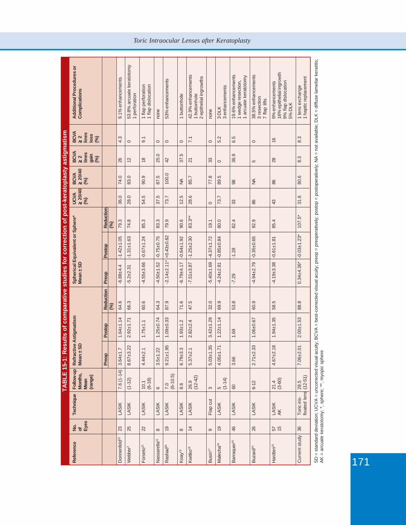
171
Toric Intraocular Lenses after Keratoplasty
TAB
LE 1
5-1:
Res
ults
of c
ompa
rativ
e st
udie
s fo
r co
rrec
tion
of p
ost-
kera
topl
asty
ast
igm
atis
mR
efer
ence
No.
Tech
niqu
eFo
llow
-up
Ref
ract
ive
Ast
igm
atis
mS
pher
ical
Equ
ival
ent o
r Sph
ere*
UCVA
BCVA
BCVA
BCVA
Add
ition
al P
roce
dure
s or
ofM
onth
s,M
ean
± SD
Mea
n ±
SD≥≥≥≥ ≥
20/4
0≥≥≥≥ ≥
20/4
0≥≥≥≥ ≥
2≥≥≥≥ ≥
2C
ompl
icat
ions
Eyes
Mea
n(%
)(%
)lin
eslin
es(r
ange
)ga
inlo
ss(%
)(%
)
Preo
pPo
stop
Red
uctio
nPr
eop
Post
opR
educ
tion
(%)
(%)
Don
nenf
eld33
23LA
SIK
7.6
(1-1
4)3.
64±1
.71.
64±1
.14
64.6
-6.8
8±4.
4-1
.42±
1.05
79.3
36.0
74.0
264.
39.
1% e
nhan
cem
ents
Web
ber2
25LA
SIK
(1-1
2)8.
67±3
.22
2.92
±1.7
166
.3-5
.2±2
.31
-1.3
1±1.
6374
.828
.083
.012
053
.8%
arc
uate
ker
atot
omy
1 pe
rfora
tion
Fors
eto22
22LA
SIK
10.1
4.44
±2.1
1.75
±1.1
60.6
-4.5
5±3.
66-0
.67±
1.24
85.3
54.5
90.9
189.
11
flap
perfo
ratio
n(6
-18)
1 fla
p di
sloc
atio
n
Nas
sare
lla21
8LA
SIK
63.
5±1.
221.
25±0
.74
64.3
-4.5
0±1.
52-0
.75±
0.75
83.3
37.5
87.5
25.0
0no
ne
Ras
had20
19LA
SIK
7.0
9.21
±1.9
51.
09±0
.33
87.9
-2.1
4±2.
11*
+0.4
3±0.
8279
.973
.710
0.0
420
53%
enh
ance
men
ts(6
-10.
5)
Koa
y198
LAS
IK8.
66.
79±3
.31.
93±1
.271
.6-6
.79±
4.17
-0.6
4±1.
9290
.612
.5N
A37
.50
1 bu
ttonh
ole
Kw
itko18
14LA
SIK
26.9
5.37
±2.1
2.82
±2.4
47.5
-7.5
1±3.
87-1
.25±
2.30
83.3
**28
.685
.721
7.1
42.9
% e
nhan
cem
ents
(12-
42)
1 bu
ttonh
ole
2 ep
ithel
ial i
ngro
wth
s
Bus
in17
9Fl
ap c
ut3
5.03
±1.3
53.
42±1
.29
32.0
-5.4
0±1.
69-4
.37±
1.72
19.1
077
.833
0no
ne
Mal
echa
3419
LAS
IK5
4.05
±1.7
11.
22±1
.14
69.9
-4.2
4±2.
81-0
.85±
0.84
80.0
73.7
89.5
05.
23
DLK
(1-1
4)3
enha
ncem
ents
Bar
raqu
er25
46LA
SIK
603.
661.
6953
.8-7
.29
-1.2
882
.433
9836
.96.
519
.6%
enh
ance
men
ts1
wed
ge re
sect
ion,
1 ar
cuat
e ke
rato
tom
y
Buz
ard24
26LA
SIK
6-12
2.71
±2.3
31.
06±0
.67
60.9
-4.9
4±2.
79-0
.35±
0.65
92.9
86N
A5
038
.5%
enh
ance
men
ts3
rese
ctio
n7
flap
lifts
Har
dten
2357
LAS
IK21
.44.
67±2
.18
1.94
±1.3
558
.5-4
.19±
3.38
-0.6
1±1.
8185
.443
8628
169%
enh
ance
men
ts15
AK
(3-6
0)16
% e
pith
elia
l ing
row
th9%
flap
dis
loca
tion
5% D
LK
Cur
rent
stu
dy36
Toric
iris
-28
.57.
06±2
.01
2.00
±1.5
388
.80.
34±4
.36*
-0.0
3±1.
23*
107.
5*31
.680
.68.
38.
31
lens
exc
hang
efix
ated
lens
(12-
51)
1 ha
ptic
repl
acem
ent
SD
= s
tand
ard
devi
atio
n; U
CV
A =
unc
orre
cted
vis
ual a
cuity
; BC
VA
= b
est-c
orre
cted
vis
ual a
cuity
; pre
op =
pre
oper
ativ
ely;
pos
top
= po
stop
erat
ivel
y; N
A =
not
ava
ilabl
e; D
LK =
diff
use
lam
ella
r ker
atiti
s;A
K =
arc
uate
ker
atot
omy;
*, s
pher
e; *
*, m
yopi
c sp
here
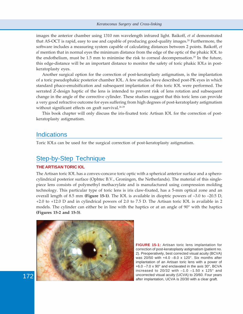
172
Keratoconus Surgery and Cross-linking
images the anterior chamber using 1310 nm wavelength infrared light. Baikoff, et al demonstratedthat AS-OCT is rapid, easy to use and capable of producing good-quality images.24 Furthermore, thesoftware includes a measuring system capable of calculating distances between 2 points. Baikoff, et
al mention that in normal eyes the minimum distance from the edge of the optic of the phakic IOL tothe endothelium, must be 1.5 mm to minimise the risk to corneal decompensation.25 In the future,this edge-distance will be an important distance to monitor the safety of toric phakic IOLs in post-keratoplasty eyes.
Another surgical option for the correction of post-keratoplasty astigmatism, is the implantationof a toric pseudophakic posterior chamber IOL. A few studies have described post-PK eyes in whichstandard phaco-emulsification and subsequent implantation of this toric IOL were performed. Theserrated Z-design haptic of the lens is intended to prevent risk of lens rotation and subsequentchange in the angle of the corrective cylinder. These studies suggest that this toric lens can providea very good refractive outcome for eyes suffering from high degrees of post-keratoplasty astigmatismwithout significant effects on graft survival.26-29
This book chapter will only discuss the iris-fixated toric Artisan IOL for the correction of post-keratoplasty astigmatism.
IndicationsToric IOLs can be used for the surgical correction of post-keratoplasty astigmatism.
Step-by-Step TechniqueTHE ARTISAN TORIC IOLThe Artisan toric IOL has a convex-concave toric optic with a spherical anterior surface and a sphero-cylindrical posterior surface (Ophtec B.V., Groningen, the Netherlands). The material of this single-piece lens consists of polymethyl methacrylate and is manufactured using compression moldingtechnology. This particular type of toric lens is iris claw-fixated, has a 5-mm optical zone and anoverall length of 8.5 mm (Figure 15-1). The IOL is available in dioptric powers of –3.0 to –20.5 D,+2.0 to +12.0 D and in cylindrical powers of 2.0 to 7.5 D. The Artisan toric IOL is available in 2models. The cylinder can either be in line with the haptics or at an angle of 90° with the haptics(Figures 15-2 and 15-3).
FIGURE 15-1: Artisan toric lens implantation forcorrection of post-keratoplasty astigmatism (patient no.2). Preoperatively, best corrected visual acuity (BCVA)was 20/50 with +4.0 –8.0 x 120°. Six months afterimplantation of an Artisan toric lens with a power of+6.0 –7.0 x 90° and enclavated in the axis 30°, BCVAincreased to 20/32 with –1.0 –1.50 x 125° anduncorrected visual acuity (UCVA) to 20/60. Four yearsafter implantation, UCVA is 20/30 with a clear graft.
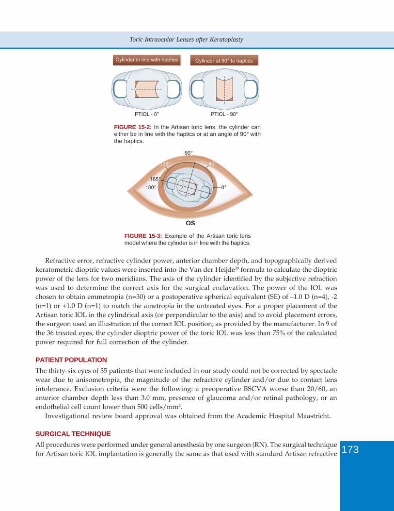
173
Toric Intraocular Lenses after Keratoplasty
FIGURE 15-2: In the Artisan toric lens, the cylinder caneither be in line with the haptics or at an angle of 90° withthe haptics.
FIGURE 15-3: Example of the Artisan toric lensmodel where the cylinder is in line with the haptics.
Refractive error, refractive cylinder power, anterior chamber depth, and topographically derivedkeratometric dioptric values were inserted into the Van der Heijde30 formula to calculate the dioptricpower of the lens for two meridians. The axis of the cylinder identified by the subjective refractionwas used to determine the correct axis for the surgical enclavation. The power of the IOL waschosen to obtain emmetropia (n=30) or a postoperative spherical equivalent (SE) of –1.0 D (n=4), -2(n=1) or +1.0 D (n=1) to match the ametropia in the untreated eyes. For a proper placement of theArtisan toric IOL in the cylindrical axis (or perpendicular to the axis) and to avoid placement errors,the surgeon used an illustration of the correct IOL position, as provided by the manufacturer. In 9 ofthe 36 treated eyes, the cylinder dioptric power of the toric IOL was less than 75% of the calculatedpower required for full correction of the cylinder.
PATIENT POPULATIONThe thirty-six eyes of 35 patients that were included in our study could not be corrected by spectaclewear due to anisometropia, the magnitude of the refractive cylinder and/or due to contact lensintolerance. Exclusion criteria were the following: a preoperative BSCVA worse than 20/60, ananterior chamber depth less than 3.0 mm, presence of glaucoma and/or retinal pathology, or anendothelial cell count lower than 500 cells/mm2.
Investigational review board approval was obtained from the Academic Hospital Maastricht.
SURGICAL TECHNIQUEAll procedures were performed under general anesthesia by one surgeon (RN). The surgical techniquefor Artisan toric IOL implantation is generally the same as that used with standard Artisan refractive
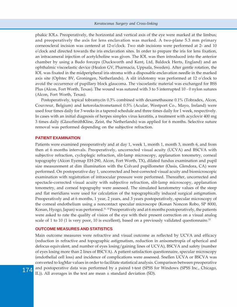
174
Keratoconus Surgery and Cross-linking
phakic IOLs. Preoperatively, the horizontal and vertical axis of the eye were marked at the limbus;and preoperatively the axis for lens enclavation was marked. A two-plane 5.3 mm primarycorneoscleral incision was centered at 12-o’clock. Two stab incisions were performed at 2- and 10o’clock and directed towards the iris enclavation sites. In order to prepare the iris for lens fixation,an intracameral injection of acetylcholine was given. The IOL was then introduced into the anteriorchamber by using a Budo forceps (Duckworth and Kent, Ltd, Baldock Herts, England) and anophthalmic viscoelastic device (Healon GV, Pharmacia, Uppsala, Sweden). After gentle rotation, theIOL was fixated in the midperipheral iris stroma with a disposable enclavation needle in the markedaxis site (Ophtec BV, Groningen, Netherlands). A slit iridotomy was performed at 12 o’clock toavoid the occurrence of pupillary block glaucoma. The viscoelastic material was exchanged for BSSPlus (Alcon, Fort Worth, Texas). The wound was sutured with 3 to 5 interrupted 10 - 0 nylon sutures(Alcon, Fort Worth, Texas).
Postoperatively, topical tobramycin 0.3% combined with dexamethasone 0.1% (Tobradex, Alcon,Couvreur, Belgium) and ketorolactrometamol 0.5% (Acular, Westport Co., Mayo, Ireland) wereused four times daily for 3 weeks in a tapered schedule and three times daily for 1 week, respectively.In cases with an initial diagnosis of herpes simplex virus keratitis, a treatment with acyclovir 400 mg3 times daily (GlaxoSmithKline, Zeist, the Netherlands) was applied for 6 months. Selective sutureremoval was performed depending on the subjective refraction.
PATIENT EXAMINATIONPatients were examined preoperatively and at day 1, week 1, month 1, month 3, month 6, and fromthen at 6 months intervals. Preoperatively, uncorrected visual acuity (UCVA) and BSCVA withsubjective refraction, cycloplegic refraction, slit-lamp microscopy, applanation tonometry, cornealtopography (Alcon Eyemap EH-290, Alcon, Fort Worth, TX), dilated fundus examination and pupilsize measurement at dim illumination with the Colvard pupillometer (Oasis, Glendora, CA) wereperformed. On postoperative day 1, uncorrected and best-corrected visual acuity and biomicroscopicexamination with registration of intraocular pressure were performed. Thereafter, uncorrected andspectacle-corrected visual acuity with subjective refraction, slit-lamp microscopy, applanationtonometry, and corneal topography were assessed. The simulated keratometry values of the steepand flat meridians were used for calculation of the topographically induced surgical astigmatism.Preoperatively and at 6 months, 1 year, 2 years, and 3 years postoperatively, specular microscopy ofthe corneal endothelium using a noncontact specular microscope (Konan Noncon Robo, SP 8000,Konan, Hyogo, Japan) was performed.31, 32 Preoperatively and at 6 months postoperatively, the patientswere asked to rate the quality of vision of the eye with their present correction on a visual analogscale of 1 to 10 (1 is very poor, 10 is excellent), based on a previously validated questionnaire.33
OUTCOME MEASURES AND STATISTICSMain outcome measures were refractive and visual outcome as reflected by UCVA and efficacy(reduction in refractive and topographic astigmatism, reduction in anisometropia of spherical anddefocus equivalent, and number of eyes losing/gaining lines of UCVA), BSCVA and safety (numberof eyes losing more than 2 lines of BSCVA). A patient satisfaction questionnaire, specular microscopy(endothelial cell loss) and incidence of complications were assessed. Snellen UCVA or BSCVA wasconverted to logMar values in order to facilitate statistical analysis. Comparison between preoperativeand postoperative data was performed by a paired t-test (SPSS for Windows (SPSS Inc., Chicago,IL)). All averages in the text are mean ± standard deviation (SD).
175
Toric Intraocular Lenses after Keratoplasty
ANALYSIS OF ASTIGMATISMBoth vector analysis and non-vector analysis of the cylinder was performed. The efficacy of theprocedure (i.e., the proportion of astigmatism correction which was achieved) was quantified usingthe correction index expressed as a percentage of the surgically induced astigmatism (SIA) dividedby the target induced astigmatism (TIA) for each individual eye and aggregated.34 The Holladaymethod to convert polar values (cylinder and axis) to a Cartesian (X and Y) coordinate system wasused to determine the mean ± SD value of the refractive and topographical keratometric astigmatism.35
The coordinates were plotted in a doubled-angle plot and the centroid was determined.
ResultsPATIENT POPULATIONTwenty-seven patients were female and eight were male. The mean age was 63.8 ± 17.0 (range,23-82 years). The mean time interval between PK and toric lens implantation was 57.1 ± 30.2 months(range, 26 to 142 months) and the interval between suture removal and lens implantation was 30.8± 28.2 months (range, 3 to 144 months). Twenty-five eyes were pseudophakic after previousimplantation of a posterior chamber IOL and one eye was aphakic. The initial diagnosis requiringcorneal transplantation was Fuchs endothelial dystrophy (33.3%), (pseudophakic) bullous keratopathy(13.9%), herpes simplex keratitis (16.7%), keratoconus (13.9%), corneal scarring (11.1%) re-grafting(8.3%) and high astigmatism (2.8%).
The baseline parameters were a mean sphere of 0.34 ± 4.36 D (range, + 9.0 to –10.0 D), a mean SErefraction of –3.19 ± 4.31 D (range, +5.5 to –14.25 D), a mean baseline refractive cylinder power of-7.06 ± 2.01 (range, -3.0 to –11.0 D), a mean defocus equivalent of 6.99 ± 2.59 D (range, 3.25 to 14.25D) and a mean baseline topographically derived simulated keratometric cylinder of 7.00 ± 2.27 D(range, +3.51 to +11.65 D). There were no significant differences for the baseline parameters betweenthe pseudophakic eye group and the phakic eye group. The mean follow-up was 28.5 ± 12.5 months(range, 12 to 51 months).
VISUAL ACUITY OUTCOMEThe mean preoperative logMar UCVA was 1.39 ± 0.44 and increased to 0.55 ± 0.35 at the last follow-up (p < 0.001, paired t-test). Postoperatively, 31.6% of eyes had a UCVA better than 20/40, 63.9% ofeyes better than 20/80 as compared to 0% preoperatively. In the phakic and pseudophakic eyegroups, 27.8% and 35.3% of eyes had a UCVA better than 20/40, respectively (p = 0.71, Pearson chi-square). The mean number of gained lines of UCVA in all eyes was 8.50 ± 5.51 (range, -2 to 18 lines).The mean preoperative logMar BSCVA was 0.26 ± 0.17 and postoperatively it was 0.26 ± 0.24(p = 0.802, paired t-test). Postoperatively, 80.6% of eyes had a BSCVA better than 20/40 as comparedto 69.4% preoperatively and 25.0% had a BSCVA better than 20/25 (Figure 15-4). In the phakic andpseudophakic eye groups, 77.8% and 82.4% of eyes had a BSCVA better than 20/40, respectively(p = 0.83, Pearson chi-square). The mean number of gained lines of BSCVA in all eyes was -0.06 ±2.14 (range, -6 to 4 lines). There was a loss of BSCVA of greater than 2 lines in 8.3% of eyes and again of at least 2 lines in 8.3% of eyes (Figure 15-5). The predictability of intended versus achievedcylinder correction showed 26 of 36 eyes (72.3%) within 2 D and 18 of 36 eyes (50.0%) within 1 D ofthe intended correction. In the phakic eye group, 66.7% and 50.0% of eyes were within 2 D and 1 Dof the intended correction, and in the pseudophakic eye group 76.5% and 52.9% of eyes were within
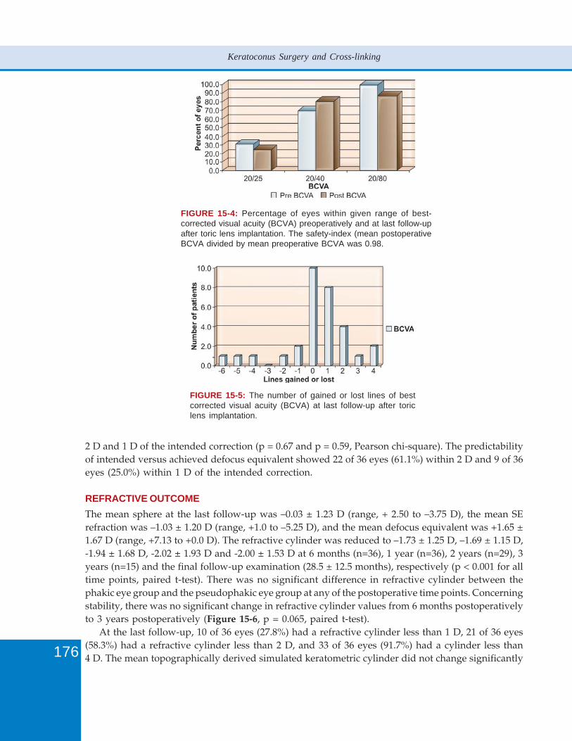
176
Keratoconus Surgery and Cross-linking
FIGURE 15-4: Percentage of eyes within given range of best-corrected visual acuity (BCVA) preoperatively and at last follow-upafter toric lens implantation. The safety-index (mean postoperativeBCVA divided by mean preoperative BCVA was 0.98.
FIGURE 15-5: The number of gained or lost lines of bestcorrected visual acuity (BCVA) at last follow-up after toriclens implantation.
2 D and 1 D of the intended correction (p = 0.67 and p = 0.59, Pearson chi-square). The predictabilityof intended versus achieved defocus equivalent showed 22 of 36 eyes (61.1%) within 2 D and 9 of 36eyes (25.0%) within 1 D of the intended correction.
REFRACTIVE OUTCOMEThe mean sphere at the last follow-up was –0.03 ± 1.23 D (range, + 2.50 to –3.75 D), the mean SErefraction was –1.03 ± 1.20 D (range, +1.0 to –5.25 D), and the mean defocus equivalent was +1.65 ±1.67 D (range, +7.13 to +0.0 D). The refractive cylinder was reduced to –1.73 ± 1.25 D, –1.69 ± 1.15 D,-1.94 ± 1.68 D, -2.02 ± 1.93 D and -2.00 ± 1.53 D at 6 months (n=36), 1 year (n=36), 2 years (n=29), 3years (n=15) and the final follow-up examination (28.5 ± 12.5 months), respectively (p < 0.001 for alltime points, paired t-test). There was no significant difference in refractive cylinder between thephakic eye group and the pseudophakic eye group at any of the postoperative time points. Concerningstability, there was no significant change in refractive cylinder values from 6 months postoperativelyto 3 years postoperatively (Figure 15-6, p = 0.065, paired t-test).
At the last follow-up, 10 of 36 eyes (27.8%) had a refractive cylinder less than 1 D, 21 of 36 eyes(58.3%) had a refractive cylinder less than 2 D, and 33 of 36 eyes (91.7%) had a cylinder less than4 D. The mean topographically derived simulated keratometric cylinder did not change significantly
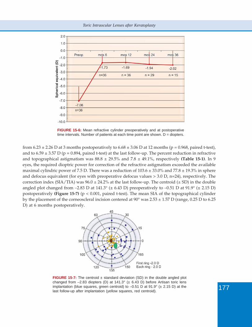
177
Toric Intraocular Lenses after Keratoplasty
FIGURE 15-6: Mean refractive cylinder preoperatively and at postoperativetime intervals. Number of patients at each time point are shown. D = diopters.
from 6.23 ± 2.26 D at 3 months postoperatively to 6.68 ± 3.06 D at 12 months (p = 0.968, paired t-test),and to 6.59 ± 3.57 D (p = 0.894, paired t-test) at the last follow-up. The percent reduction in refractiveand topographical astigmatism was 88.8 ± 29.5% and 7.8 ± 49.1%, respectively (Table 15-1). In 9eyes, the required dioptric power for correction of the refractive astigmatism exceeded the availablemaximal cylindric power of 7.5 D. There was a reduction of 103.6 ± 33.0% and 77.8 ± 19.3% in sphereand defocus equivalent (for eyes with preoperative defocus values > 3.0 D, n=24), respectively. Thecorrection index (SIA/TIA) was 96.0 ± 24.2% at the last follow-up. The centroid (± SD) in the doubleangled plot changed from –2.83 D at 141.3° (± 6.43 D) preoperatively to –0.51 D at 91.9° (± 2.15 D)postoperatively (Figure 15-7) (p < 0.001, paired t-test). The mean SIA of the topographical cylinderby the placement of the corneoscleral incision centered at 90° was 2.53 ± 1.57 D (range, 0.25 D to 6.25D) at 6 months postoperatively.
FIGURE 15-7: The centroid ± standard deviation (SD) in the double angled plotchanged from –2.83 diopters (D) at 141.3° (± 6.43 D) before Artisan toric lensimplantation (blue squares, green centroid) to –0.51 D at 91.9° (± 2.15 D) at thelast follow-up after implantation (yellow squares, red centroid).
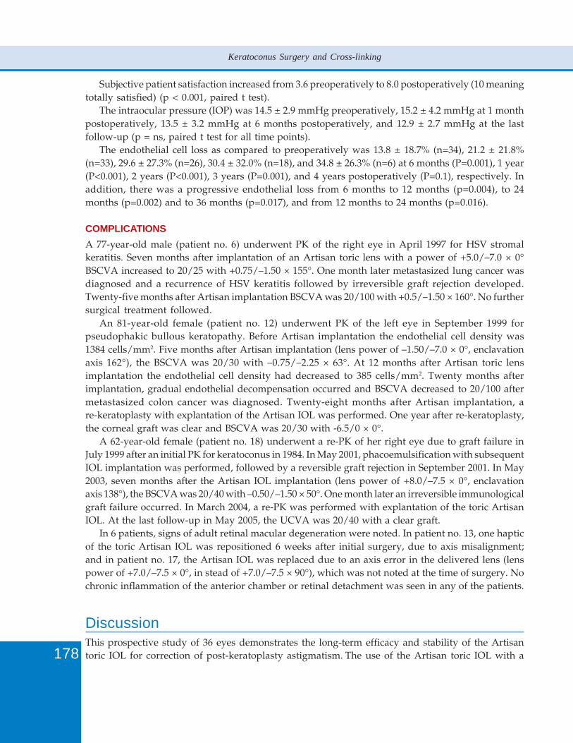
178
Keratoconus Surgery and Cross-linking
Subjective patient satisfaction increased from 3.6 preoperatively to 8.0 postoperatively (10 meaningtotally satisfied) (p < 0.001, paired t test).
The intraocular pressure (IOP) was 14.5 ± 2.9 mmHg preoperatively, 15.2 ± 4.2 mmHg at 1 monthpostoperatively, 13.5 ± 3.2 mmHg at 6 months postoperatively, and 12.9 ± 2.7 mmHg at the lastfollow-up (p = ns, paired t test for all time points).
The endothelial cell loss as compared to preoperatively was 13.8 ± 18.7% (n=34), 21.2 ± 21.8%(n=33), 29.6 ± 27.3% (n=26), 30.4 ± 32.0% (n=18), and 34.8 ± 26.3% (n=6) at 6 months (P=0.001), 1 year(P<0.001), 2 years (P<0.001), 3 years (P=0.001), and 4 years postoperatively (P=0.1), respectively. Inaddition, there was a progressive endothelial loss from 6 months to 12 months (p=0.004), to 24months (p=0.002) and to 36 months (p=0.017), and from 12 months to 24 months (p=0.016).
COMPLICATIONSA 77-year-old male (patient no. 6) underwent PK of the right eye in April 1997 for HSV stromalkeratitis. Seven months after implantation of an Artisan toric lens with a power of +5.0/–7.0 × 0°BSCVA increased to 20/25 with +0.75/–1.50 × 155°. One month later metastasized lung cancer wasdiagnosed and a recurrence of HSV keratitis followed by irreversible graft rejection developed.Twenty-five months after Artisan implantation BSCVA was 20/100 with +0.5/–1.50 × 160°. No furthersurgical treatment followed.
An 81-year-old female (patient no. 12) underwent PK of the left eye in September 1999 forpseudophakic bullous keratopathy. Before Artisan implantation the endothelial cell density was1384 cells/mm2. Five months after Artisan implantation (lens power of –1.50/–7.0 × 0°, enclavationaxis 162°), the BSCVA was 20/30 with –0.75/–2.25 × 63°. At 12 months after Artisan toric lensimplantation the endothelial cell density had decreased to 385 cells/mm2. Twenty months afterimplantation, gradual endothelial decompensation occurred and BSCVA decreased to 20/100 aftermetastasized colon cancer was diagnosed. Twenty-eight months after Artisan implantation, are-keratoplasty with explantation of the Artisan IOL was performed. One year after re-keratoplasty,the corneal graft was clear and BSCVA was 20/30 with -6.5/0 × 0°.
A 62-year-old female (patient no. 18) underwent a re-PK of her right eye due to graft failure inJuly 1999 after an initial PK for keratoconus in 1984. In May 2001, phacoemulsification with subsequentIOL implantation was performed, followed by a reversible graft rejection in September 2001. In May2003, seven months after the Artisan IOL implantation (lens power of +8.0/–7.5 × 0°, enclavationaxis 138°), the BSCVA was 20/40 with –0.50/–1.50 × 50°. One month later an irreversible immunologicalgraft failure occurred. In March 2004, a re-PK was performed with explantation of the toric ArtisanIOL. At the last follow-up in May 2005, the UCVA was 20/40 with a clear graft.
In 6 patients, signs of adult retinal macular degeneration were noted. In patient no. 13, one hapticof the toric Artisan IOL was repositioned 6 weeks after initial surgery, due to axis misalignment;and in patient no. 17, the Artisan IOL was replaced due to an axis error in the delivered lens (lenspower of +7.0/–7.5 × 0°, in stead of +7.0/–7.5 × 90°), which was not noted at the time of surgery. Nochronic inflammation of the anterior chamber or retinal detachment was seen in any of the patients.
DiscussionThis prospective study of 36 eyes demonstrates the long-term efficacy and stability of the Artisantoric IOL for correction of post-keratoplasty astigmatism. The use of the Artisan toric IOL with a

179
Toric Intraocular Lenses after Keratoplasty
power range of 7.5 D of cylinder and -20.5 D of myopia to +12.0 D of hyperopia, provides a widefield for correction of post-keratoplasty astigmatism and ametropia. In our series this is reflected bythe magnitude of baseline spherical error (range + 9.0 to –10.0 D) and cylindrical error (range -3.0 to–11.0 D), which is much higher than in most post-keratoplasty LASIK series. To our knowledge, thereduction of the refractive cylinder by 88.8 ± 29.5% (without any enhancements) is better than inmost reported LASIK series.5-15
Improving the percentage of eyes with an UCVA of 20/40 or better from 0% preoperatively to31.6% at the last follow-up illustrates the efficacy of the Artisan toric IOL procedure in this patientgroup with highly ametropic eyes. In most LASIK series with lower preoperative ametropia UCVAbetter than 20/40 varied from 28% to 74%.6, 8, 10, 14, 15, 17 With respect to safety, there was a loss ofBSCVA of greater than 2 lines in 8.3% of eyes and a gain of at least 2 lines in 8.3% of eyes. This is inaccordance with two recent randomized studies in routine refractive surgery for the correction ofhigh myopia that showed a greater gain of BSCVA with Artisan phakic IOL implantation as comparedto a greater loss of BSCVA with LASIK and a better quality of vision with the Artisan lens inmoderate to high myopia.36, 37 The loss of greater than 2 lines of BSCVA in 8.3% in our series iscomparable to series of LASIK for postkeratoplasty astigmatism that show a greater than 2 lines lossof BSCVA in 4.3%,38 7.1%,6 9.1%10 and 16%.11However, the pattern of complications induced by thetwo techniques is very different. LASIK surgery may be complicated by flap complications in steepcorneas and has limitations due to corneal graft thickness and amount of ametropia and astigmatismsuitable for correction.5-13 LASIK related complications like diffuse lamellar keratitis,15 buttonholeflaps,6, 10 wound dehiscence39 and epithelial ingrowth6 have been reported. Because the majority ofeyes in the reported LASIK series were grafted in young patients for keratoconus with a rapidwound healing, wound dehiscence problems were less likely to occur than in a group of olderpatients grafted for Fuchs endothelial dystrophy or bullous keratopathy.40 Since the effect of the flapcut alone may induce a significant reduction of refractive astigmatism in up to 50% in some patientsand because of the high enhancement rate a two-stage LASIK procedure has been proposed.5, 41-43
However, it is unclear whether a two-stage procedure bears a higher risk for complications likeepithelial ingrowth, wound healing problems and flap dislocation. In our present Artisan series,irreversible corneal decompensation occurred in two patients after metastasized cancer wasdiagnosed. Before the diagnosis of malignant disease BSCVA was 20/25 in both patients and nosigns of immunological rejection had been noted. We believe that changes in the immune system dueto the concomitant development of malignant systemic disease might have initiated the graft failures.In a third patient with a re-keratoplasty for a graft failure after an initial diagnosis of keratoconus animmunological irreversible graft failure occurred. This was the second rejection period after there-keratoplasty following a previous reversible rejection period 4 months after cataract surgery. Wecannot exclude that the last rejection, although 8 months after surgery, may have been related to theArtisan toric lens implantation. Two of the three cases with corneal decompensation underwentsuccessful regrafting with explantation of the toric Artisan intraocular lens. In 6 patients signs ofadult retinal macular degeneration that decreased BSCVA were seen as can be expected in this agegroup. No other complications like chronic inflammation of the anterior chamber or retinal detachmentin any of the patients were noted.
The stability of the postoperative refractive cylinder after Artisan toric lens implantation up to 36months was excellent. After LASIK however, progressive changes were seen in refraction andtopography in 35.7% of cases after a mean follow-up time of 26.9 months.6 A potential limitation ofthe Artisan toric IOL for the correction of post-keratoplasty astigmatism is SIA by implantation of

180
Keratoconus Surgery and Cross-linking
the rigid polymethylmethacrylate IOL through a 5.3 mm incision. In a recent series of implantationof the Artisan toric IOL for correction of myopia or hyperopia with astigmatism the SIA was 0.53D.44 However, after keratoplasty the biomechanical response of the corneoscleral tissue to the incisionmay be somewhat unpredictable and a greater variability in SIA may be seen. Indeed, in our seriesthe mean SIA was 2.53 D six months postoperatively and varied from 0.25 D to 6.25 D. Due to thisvariability we believe that the SIA cannot be incorporated into the power calculation of the lens.Since the goal of correcting post-keratoplasty astigmatism is mainly to reduce the refractive astigmatismand ametropia to enable patients to wear spectacles, we feel that a lesser predictability of astigmatismreduction may be acceptable.
Concerns have been raised especially with respect to the development of complications likeendothelial cell loss, chronic inflammation and cystoid macular edema after Artisan toric lensimplantation. A study using fluorometry showed inflammation comparable with cataract surgery at6 months postoperatively45 whereas a study using a flare-cell meter found chronic inflammation 1 to2 years after implantation of the older Worst-Fechner intraocular lens.46 We found no chronicinflammation by slit-lamp examination in the present study and cystoid macular edema withconcomitant loss of best corrected visual acuity was not seen in the immediate postoperative phase.
After routine PK, the mean annual rate of endothelial cell loss from 3 to 5 years seems to be 7.8%per year and from 5 to 10 years it seems to be 4.2% per year.47-50 The corneal endothelium seems toremain relatively stable between 10 and 15 years after PK and it has also been suggested that therate of endothelial cell loss from 10 to 15 years after surgery may be similar to that of normalcorneas.47 It was hypothesized that late endothelial cell loss may be caused by an aspecific, non-rejection-like inflammation, or perhaps a chronic breakdown of the blood aqueous barrier, but notby an allograft rejection mechanism.
The mean endothelial cell loss in our study group was 13.8 ± 18.7%, 21.2 ± 21.8%, 29.6 ± 27.3%,and 30.4 ± 32.0% at 6, 12, 24 and 36 months. There was a significant continuing progressive endothelialcell loss at each time point as compared to preoperative cell density levels and between 6 months ascompared to 12, 24, and 36 months. For an experienced surgeon, the implantation of the Artisantoric lens in post-keratoplasty eyes does not appear to be more traumatic as compared to Artisanlens implantation in phakic eyes for correction of myopia. However, the endothelial cell loss in thepresent series is much higher than the reported endothelial cell loss in studies of Artisan IOLimplantation for correction of high myopia in virgin phakic eyes, which varies from 0.7% to 11.7%over 3 years time.36, 37, 51-55 Risk factors that may lead to higher levels of endothelial cell loss after PKare low donor endothelial cell count, older donor age, aphakia, pseudophakia, and older recipientage. In addition, the implantation of an Artisan lens in post-keratoplasty eyes could be more traumaticfor the corneal endothelium, relating to decreased visualization, as compared to virgin eyes. Thiscould lead to higher endothelial cell loss in the post-keratoplasty group. At the time of toric Artisanlens implantation, 50% of our patients were between 3 to 5 years, 27.7% between 5 to 10 years and5.6% longer than 10 years after PK. For our 24 months cohort, the cell loss rate was 14.8% per yearand for the 36 months cohort it was 10.1% per year. If we correct these values for the expected cellloss induced by the PK, there appears to be an additional cell loss of 6% to 7% per year. Therefore,we cannot exclude that the Artisan IOL in the presence of a corneal graft with low cell densities maycause a higher rate of endothelial cell loss due to the compromised endothelium. Cell counts as lowas 370 cells/mm2 and 515 cells/mm2 have been measured before decompensation.56-59 Nevertheless,we feel that an endothelial cell density of at least 500 cells/mm2 as exclusion criterion is permittedsince no other treatment modalities exist but corneal regrafting, and the Artisan lens is perfectly
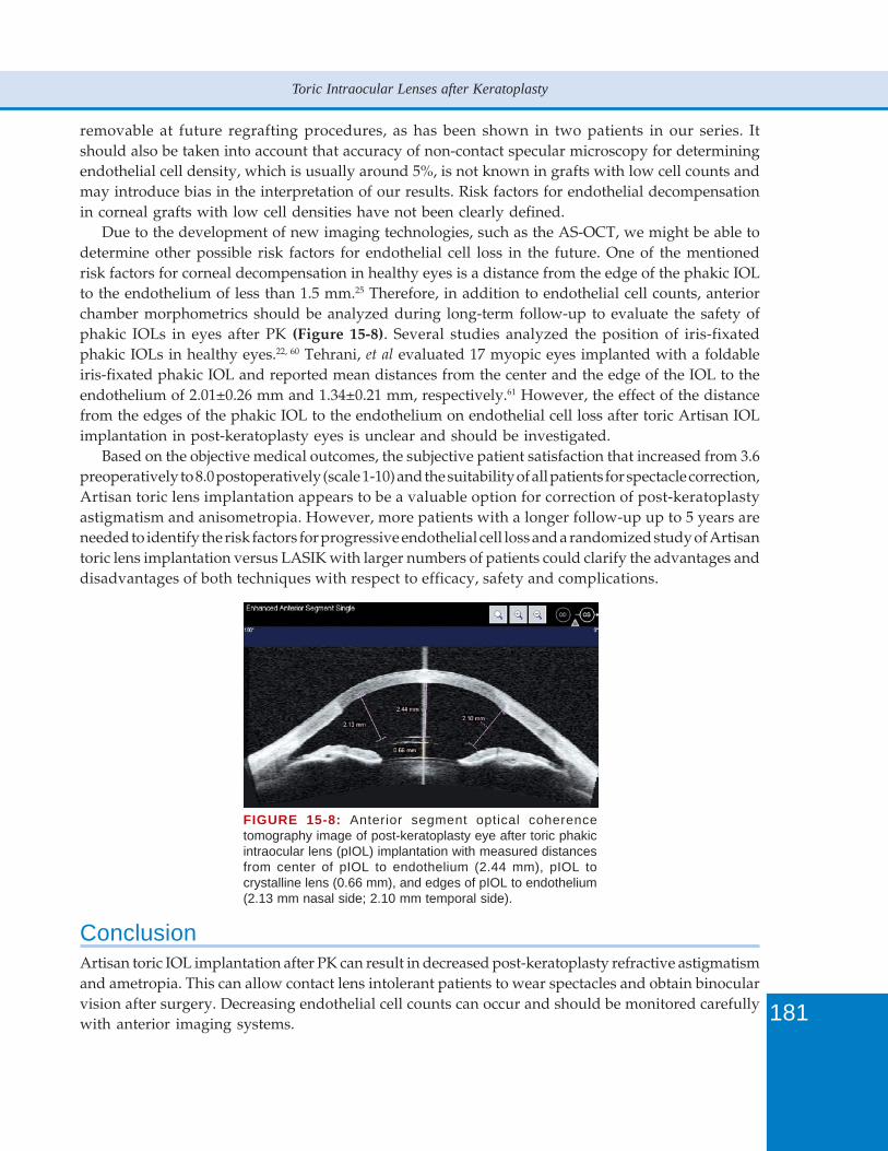
181
Toric Intraocular Lenses after Keratoplasty
removable at future regrafting procedures, as has been shown in two patients in our series. Itshould also be taken into account that accuracy of non-contact specular microscopy for determiningendothelial cell density, which is usually around 5%, is not known in grafts with low cell counts andmay introduce bias in the interpretation of our results. Risk factors for endothelial decompensationin corneal grafts with low cell densities have not been clearly defined.
Due to the development of new imaging technologies, such as the AS-OCT, we might be able todetermine other possible risk factors for endothelial cell loss in the future. One of the mentionedrisk factors for corneal decompensation in healthy eyes is a distance from the edge of the phakic IOLto the endothelium of less than 1.5 mm.25 Therefore, in addition to endothelial cell counts, anteriorchamber morphometrics should be analyzed during long-term follow-up to evaluate the safety ofphakic IOLs in eyes after PK (Figure 15-8). Several studies analyzed the position of iris-fixatedphakic IOLs in healthy eyes.22, 60 Tehrani, et al evaluated 17 myopic eyes implanted with a foldableiris-fixated phakic IOL and reported mean distances from the center and the edge of the IOL to theendothelium of 2.01±0.26 mm and 1.34±0.21 mm, respectively.61 However, the effect of the distancefrom the edges of the phakic IOL to the endothelium on endothelial cell loss after toric Artisan IOLimplantation in post-keratoplasty eyes is unclear and should be investigated.
Based on the objective medical outcomes, the subjective patient satisfaction that increased from 3.6preoperatively to 8.0 postoperatively (scale 1-10) and the suitability of all patients for spectacle correction,Artisan toric lens implantation appears to be a valuable option for correction of post-keratoplastyastigmatism and anisometropia. However, more patients with a longer follow-up up to 5 years areneeded to identify the risk factors for progressive endothelial cell loss and a randomized study of Artisantoric lens implantation versus LASIK with larger numbers of patients could clarify the advantages anddisadvantages of both techniques with respect to efficacy, safety and complications.
FIGURE 15-8: Anterior segment optical coherencetomography image of post-keratoplasty eye after toric phakicintraocular lens (pIOL) implantation with measured distancesfrom center of pIOL to endothelium (2.44 mm), pIOL tocrystalline lens (0.66 mm), and edges of pIOL to endothelium(2.13 mm nasal side; 2.10 mm temporal side).
ConclusionArtisan toric IOL implantation after PK can result in decreased post-keratoplasty refractive astigmatismand ametropia. This can allow contact lens intolerant patients to wear spectacles and obtain binocularvision after surgery. Decreasing endothelial cell counts can occur and should be monitored carefullywith anterior imaging systems.
182
Keratoconus Surgery and Cross-linking
A potential drawback of implanting an Artisan toric IOL for treatment of post-keratoplastyastigmatism is SIA, related to the required 5.3 mm incision size. The biomechanical response of thecorneoscleral tissue to the incision may be somewhat unpredictable after PK, and a greater variabilityin SIA may be seen. A recent study on implantation of the Artisan toric lens for correction of myopiaor hyperopia with astigmatism in virgin non-operated eyes, showed a SIA of 0.53 D.44 However, ina post-keratoplasty astigmatism treatment group a lesser predictability of astigmatism reductionmay be acceptable, since the primary goal of treating of astigmatism after PK is the correction ofsufficient myopia and astigmatism in order to enable patients to wear spectacles or contact lenses.
References1. van Rij G, Cornell FM, Waring GO, 3rd, et al. Postoperative astigmatism after central vs eccentric penetrating
keratoplasties. Am J Ophthalmol 1985;99:317-20.2. Kerenyi A, Suveges I. Corneal topographic results after eccentric, biconvex penetrating keratoplasty. J Cataract
Refract Surg 2003;29:752-6.3. Karabatsas CH, Cook SD, Sparrow JM. Proposed classification for topographic patterns seen after penetrating
keratoplasty. Br J Ophthalmol 1999;83:403-9.4. Brooks SE, Johnson D, Fischer N. Anisometropia and binocularity. Ophthalmology 1996;103:1139-43.5. Busin M, Arffa RC, Zambianchi L, et al. Effect of hinged lamellar keratotomy on postkeratoplasty eyes.
Ophthalmology 2001;108:1845-51; discussion 1851-52.6. Kwitko S, Marinho DR, Rymer S, Ramos Filho S. Laser in situ keratomileusis after penetrating keratoplasty. J
Cataract Refract Surg 2001;27:374-9.7. Koay PY, McGhee CN, Weed KH, Craig JP. Laser in situ keratomileusis for ametropia after penetrating keratoplasty.
J Refract Surg 2000;16:140-7.8. Rashad KM. Laser in situ keratomileusis for correction of high astigmatism after penetrating keratoplasty. J
Refract Surg 2000;16:701-10.9. Nassaralla BR, Nassaralla JJ. Laser in situ keratomileusis after penetrating keratoplasty. J Refract Surg 2000;16:
431-7.10. Forseto AS, Francesconi CM, Nose RA, Nose W. Laser in situ keratomileusis to correct refractive errors after
keratoplasty. J Cataract Refract Surg 1999;25:479-85.11. Hardten DR, Chittcharus A, Lindstrom RL. Long term analysis of LASIK for the correction of refractive errors after
penetrating keratoplasty. Cornea 2004;23:479-489.12. Buzard K, Febbraro JL, Fundingsland BR. Laser in situ keratomileusis for the correction of residual ametropia after
penetrating keratoplasty. J Cataract Refract Surg 2004;30:1006-13.13. Barraquer CC, Rodriguez-Barraquer T. Five-year results of laser in-situ keratomileusis (LASIK) after penetrating
keratoplasty. Cornea 2004;23:243-8.14. Donnenfeld ED, Solomon R, Biser S. Laser in situ keratomileusis after penetrating keratoplasty. Int Ophthalmol
Clin 2002;42:67-87.15. Malecha MA, Holland EJ. Correction of myopia and astigmatism after penetrating keratoplasty with laser in situ
keratomileusis. Cornea 2002;21:564-9.16. Guell JL, Gris O, de Muller A, Corcostegui B. LASIK for the correction of residual refractive errors from previous
surgical procedures. Ophthalmic Surg Lasers 1999;30:341-9.17. Webber SK, Lawless MA, Sutton GL, Rogers CM. LASIK for post penetrating keratoplasty astigmatism and
myopia. Br J Ophthalmol 1999;83:1013-8.18. Nuijts RM, Abhilakh Missier KA, Nabar VA, Japing WJ. Artisan toric lens implantation for correction of
postkeratoplasty astigmatism. Ophthalmology 2004;111:1086-94.19. Moshirfar M, Barsam CA, Parker JW. Implantation of an Artisan phakic intraocular lens for the correction of high
myopia after penetrating keratoplasty. J Cataract Refract Surg 2004;30:1578-81.20. Moshirfar M, Feilmeier MR, Kang PC. Implantation of Verisyse phakic intraocular lens to correct myopic refractive
error after penetrating keratoplasty in pseudophakic eyes. Cornea 2006;25:107-11.21. Tehrani M, Dick HB, Schwenn O, et al. Postoperative astigmatism and rotational stability after artisan toric phakic
intraocular lens implantation. J Cataract Refract Surg 2003;29:1761-6.22. Baumeister M, Buhren J, Kohnen T. Position of angle-supported, iris-fixated, and ciliary sulcus-implanted myopic
phakic intraocular lenses evaluated by Scheimpflug photography. Am J Ophthalmol 2004;138:723-31.

183
Toric Intraocular Lenses after Keratoplasty
23. Bartels MC, Saxena R, van den Berg TJ, et al. The influence of incision-induced astigmatism and axial lens positionon the correction of myopic astigmatism with the Artisan toric phakic intraocular lens. Ophthalmology 2006;113:1110-7.
24. Baikoff G, Lutun E, Ferraz C, Wei J. Static and dynamic analysis of the anterior segment with optical coherencetomography. J Cataract Refract Surg 2004;30:1843-50.
25. Baikoff G. Anterior segment OCT and phakic intraocular lenses: a perspective. J Cataract Refract Surg 2006;32:1827-35.
26. Viestenz A, Kuchle M, Seitz B, Langenbucher A. Toric intraocular lenses for correction of persistent cornealastigmatism after penetrating keratoplasty. Ophthalmologe 2005;102:148-52.
27. McMullan TF, Goldsmith C, Illingworth CD. Toric posterior chamber (in-the-bag) intraocular lens implantation tocorrect postpenetrating keratoplasty astigmatism. Eye 2007;21:150-2.
28. Kersey JP, O’Donnell A, Illingworth CD. Cataract surgery with toric intraocular lenses can optimize uncorrectedpostoperative visual acuity in patients with marked corneal astigmatism. Cornea 2007;26:133-5.
29. Buchwald HJ, Lang GK. Cataract surgery with implantation of toric silicone lenses for severe astigmatism afterkeratoplasty. Klin Monatsbl Augenheilkd 2004;221:489-94.
30. van der Heijde GL, Fechner PU, Worst JG. [Optical consequences of implantation of a negative intraocular lens inmyopic patients]. Klin Monatsbl Augenheilkd 1988;193:99-102.
31. Waring GO, 3rd. Standard graphs for reporting refractive surgery. J Refract Surg 2000;16:459-66.32. Koch DD, Kohnen T, Obstbaum SA, Rosen ES. Format for reporting refractive surgical data. J Cataract Refract Surg
1998;24:285-7.33. Nijkamp MD, Dolders MG, de Brabander J, et al. Effectiveness of multifocal intraocular lenses to correct presbyopia
after cataract surgery: a randomized controlled trial. Ophthalmology 2004;111:1832-9.34. Alpins N. Astigmatism analysis by the Alpins method. J Cataract Refract Surg 2001;27:31-49.35. Holladay JT, Cravy TV, Koch DD. Calculating the surgically induced refractive change following ocular surgery. J
Cataract Refract Surg 1992;18:429-43.36. Malecaze FJ, Hulin H, Bierer P, et al. A randomized paired eye comparison of two techniques for treating moderately
high myopia: LASIK and artisan phakic lens. Ophthalmology 2002;109:1622-30.37. El Danasoury MA, El Maghraby A, Gamali TO. Comparison of iris-fixed Artisan lens implantation with excimer
laser in situ keratomileusis in correcting myopia between -9.00 and -19.50 diopters: a randomized study.Ophthalmology 2002;109:955-64.
38. Donnenfeld ED, Kornstein HS, Amin A, et al. Laser in situ keratomileusis for correction of myopia and astigmatismafter penetrating keratoplasty. Ophthalmology 1999;106:1966-74; discussion 1974-75.
39. Ranchod TM, McLeod SD. Wound dehiscence in a patient with keratoconus after penetrating keratoplasty andLASIK. Arch Ophthalmol 2004;122:920-1.
40. Abou-Jaoude ES, Brooks M, Katz DG, Van Meter WS. Spontaneous wound dehiscence after removal of singlecontinuous penetrating keratoplasty suture. Ophthalmology 2002;109:1291-96; discussion 1297.
41. Busin M, Zambianchi L, Garzione F, et al. Two-stage laser in situ keratomileusis to correct refractive errors afterpenetrating keratoplasty. J Refract Surg 2003;19:301-8.
42. Alio JL, Javaloy J, Osman AA, et al. Laser in situ keratomileusis to correct post-keratoplasty astigmatism; 1-stepversus 2-step procedure. J Cataract Refract Surg 2004;30:2303-10.
43. Dada T, Vajpayee RB, Gupta V, et al. Microkeratome-induced reduction of astigmatism after penetratingkeratoplasty. Am J Ophthalmol 2001;131:507-8.
44. Dick HB, Alio J, Bianchetti M, et al. Toric phakic intraocular lens: European multicenter study. Ophthalmology2003;110:150-62.
45. Perez-Santonja JJ, Bueno JL, Zato MA. Surgical correction of high myopia in phakic eyes with Worst-Fechnermyopia intraocular lenses. J Refract Surg 1997;13:268-281; discussion 281-4.
46. Perez-Santonja JJ, Iradier MT, Benitez del Castillo JM, et al. Chronic subclinical inflammation in phakic eyes withintraocular lenses to correct myopia. J Cataract Refract Surg 1996;22:183-7.
47. Patel SV, Hodge DO, Bourne WM. Corneal endothelium and postoperative outcomes 15 years after penetratingkeratoplasty. Am J Ophthalmol 2005;139:311-9.
48. Ing JJ, Ing HH, Nelson LR, et al. Ten-year postoperative results of penetrating keratoplasty. Ophthalmology1998;105:1855-65.
49. Bourne WM, McLaren JW. Clinical responses of the corneal endothelium. Exp Eye Res 2004;78:561-72.50. Bourne WM, Nelson LR, Hodge DO. Continued endothelial cell loss ten years after lens implantation. Ophthalmology
1994;101:1014-22; discussion 1022-23.51. Maloney RK, Nguyen LH, John ME. Artisan phakic intraocular lens for myopia: short-term results of a prospective,
multicenter study. Ophthalmology 2002;109:1631-41.
184
Keratoconus Surgery and Cross-linking
52. Menezo JL, Cisneros AL, Rodriguez-Salvador V. Endothelial study of iris-claw phakic lens: four year follow-up. JCataract Refract Surg 1998;24:1039-49.
53. Menezo JL, Peris-Martinez C, Cisneros AL, Martinez-Costa R. Phakic intraocular lenses to correct high myopia:Adatomed, Staar, and Artisan. J Cataract Refract Surg 2004;30:33-44.
54. Güell JL, Vazquez M, Malecaze F, et al. Artisan toric phakic intraocular lens for the correction of high astigmatism.Am J Ophthalmol 2003;136:442-7.
55. Lifshitz T, Levy J, Aizenman I, et al. Artisan phakic intraocular lens for correcting high myopia. Int Ophthalmol2004;25:233-8.
56. Hoffer KJ. Corneal decomposition after corneal endothelium cell count. Am J Ophthalmol 1979;87:252-3.57. Nuyts RM, Boot N, van Best JA, et al. Long term changes in human corneal endothelium following toxic endothelial
cell destruction: a specular microscopic and fluorophotometric study. Br J Ophthalmol 1996;80:15-20.58. Breebaart AC, Nuyts RM, Pels E, et al. Toxic endothelial cell destruction of the cornea after routine extracapsular
cataract surgery. Arch Ophthalmol 1990;108:1121-5.59. Armitage WJ, Dick AD, Bourne WM. Predicting endothelial cell loss and long-term corneal graft survival. Invest
Ophthalmol Vis Sci 2003;44:3326-31.60. Guell JL, Morral M, Gris O, et al. Evaluation of Verisyse and Artiflex phakic intraocular lenses during accommodation
using Visante optical coherence tomography. J Cataract Refract Surg 2007;33:1398-1404.61. Tehrani M, Dick HB. Scheimpflug biometry of the anterior segment after implantation of foldable iris-fixated
lenses. J Refract Surg 2006;22:243-6.

Index
AAnterior lamellar keratoplasty 37Applanation tonometry 174Artisan implantation 162Ascorbic acid 24Astigmatic keratotomy 79Automated lamellar therapeutic
keratoplasty 85advantages 93instrumentation and procedure 87preparation of recipient bed 87preparation of the donor button 90suturing of graft 90
Axial map 5
BBest spectacle-corrected visual acuity 29Bio-microscope evaluation 29Budo forceps 174Busin’s technique 93
CChronic iridocyclitis 36Cohesive viscoelastic material 64Collagen triple helicoidal chain 23Corneal astigmatism 37Corneal collagen networks 23Corneal computerized topographic
examination 29Corneal distortion 134Corneal edema 23Corneal endothelium 24Corneal epithelium 25Corneal hydration 23Corneal hysteresis 9Corneal refractive surgery and penetrating
keratoplasty 153indications 154step-by-step surgical technique 154
Corneal resistance factor 9Cross-linking plus toric phakic IOL 52
DDeep anterior lamellar keratoplasty 100
air-induced acute IOP rise 121
anesthesia 104complications 115donor tissue 104double anterior chamber 120foreign bodies 123indications 100informed consent 102instrumentation 103management 115perforation 117
during air injection 119during stromal dissection 119during trephination 117
postoperative care 114preoperative work-up 101rejection 123surgical technique 104unnoticed big bubble 121
Deep anterior lamellar keratoplasty 60Descemet’s membrane 38, 96, 132Detecting corneal ectasia 1Diopter of myopia 37Dysphotopsia 37
EEffect and evidence of cross-linking
decrease of swelling 15enhanced resistance against
proteolytic enzymes 16increase in elasticity modulus 15increase in stiffness and bending
stiffness 15increased thickness of the collagen
lamellas 16raising of shrinking temperature 15
Epikeratophakia 2, 94complications 95preparation of patient 94technique 94
Epikeratoplasty 22Epithelium removal 26Extracapsular cataract extraction 2
FFemtosecond laser 95Forme fruste 8
HHydrogen links 23
IIatrogenic ectasia 2Implantation of foldable model 158Insertion of the intacs by femtosecond
laser dissection 53choice of sizes 54complications 54
extrusion 55migration of an intacs™ segment
54neovascularization 54overcorrection 54undercorrection 54
nomograms 53predictability 55prior to intralase dissection of intacs
tracks 53stability 55surgical outcomes 54
Interlenticular opacification 81Intraocular pressure 29Intraoperative autorefraction 37, 79Intrastromal air injection 107Intrastromal ring segments 42
clinical results 48complications 47indications 42management 47manual implantation technique 42
step-by-step technique 42postoperative management regime 47
Intrastromal rings 37IOL power for emmetropia 79Ionic links 23
KKeratansulphate type I 23Keratoconus 2
LLambert-Beer’s law 14Lamellar keratoplasty 52
186
Keratoconus Surgery and Cross-linking
MManifest refraction spherical equivalent
29Mucopolysaccharide 23Myopic spherical equivalent 60
NNidek Magellan 10
OOrbscan topography 53, 69
PPellucid marginal degeneration 2Penetrating keratoplasty 129
complications 142intraoperative 142postoperative 142
contraindications 130indications 130management 142other complications 146
epithelial defects 146infections after PK 146shallow anterior chamber and
wound leak 146Urrets-Zavalia syndrome 146
postoperative management 141results 141surgical steps 134
administering medications 140complete suturing 138femtosecond laser trephination 136readjustment of sutures to
minimize astigmatism 139trephination of donor cornea 134trephination of host cornea 135
technique (step-by-step) 133preparation 133
Penetrating keratoplasty 2, 38, 60Penetration of riboflavin under the
epithelium 29Peribulbar anesthesia 63Peripheral cornea 42Phakic IOLs 36Photochemical cross-linking 23Photooxidative collagen cross-linking 14Photorefractive keratotectomy 2Prevention of endophthalmitis 80
QQualitative Maloney keratoscope 64Quantitative projection keratometer
64
RRadial keratotomy 2Refractive lens exchange in keratoconus
and after keratoplasty 77indications 78preoperative work-up 79results 81surgical technique 80
Refractive surgery after DALK 124Refractive lens exchange 36Retinal detachment 36Riboflavin 14Riboflavin-UVA treatment 24
SSafety for endothelium and lens 15Scheimpflug imaging 7Scheimpflug map data 11Slit iridotomy 174Slit scanning 7Slit-lamp microscopy 174
step-by-step surgical technique 157Stromal emphysema 107Stronger cross-linking in anterior stroma
16treatment procedure 16
Surgical treatment of keratoconus 36principles 36techniques 36
TTopical anesthesia 80Toric intraocular lenses after
keratoplasty 169complications 178indications 172results 175
patient population 175step-by-step techinque 172
analysis of astigmatism 175artisan toric IOL 172
outcome measures and statistics174
patient examination 174patient population 173surgical technique 173
visual acuity outcome 175refractive outcome 176
Toric phakic intraocular lens 52, 59choosing diameter of lens 62indications 60lens power calculation 62results 66
efficacy index 68endothelial cell counts 68refractive outcome 67safety index 68visual acuity 68
step-by-step surgical technique 63foldable iris-claw TPIOLS 64implantable collamer lens 65PMMA iris-claw TPIOLS 63
Toric phakic intraocular lenses and PKPindications 156
Toric phakic IOLs in keratoconus 73case report 74limitations 75
Transepithelial corneal cross-linkingresults 27technique 27
Transepithelial technique 26Tropocollagen molecules 23
UUltraviolet light 14Uncorrected visual acuity 29UVA source 24
VVerisyse implantation 75Verisyse toric phakic IOL 74Vitrector 66
WWavefront sensing 8
ZZiemer galilei 8Zonal pachymetry 11