Embed Size (px)
Citation preview

The Integumentary System
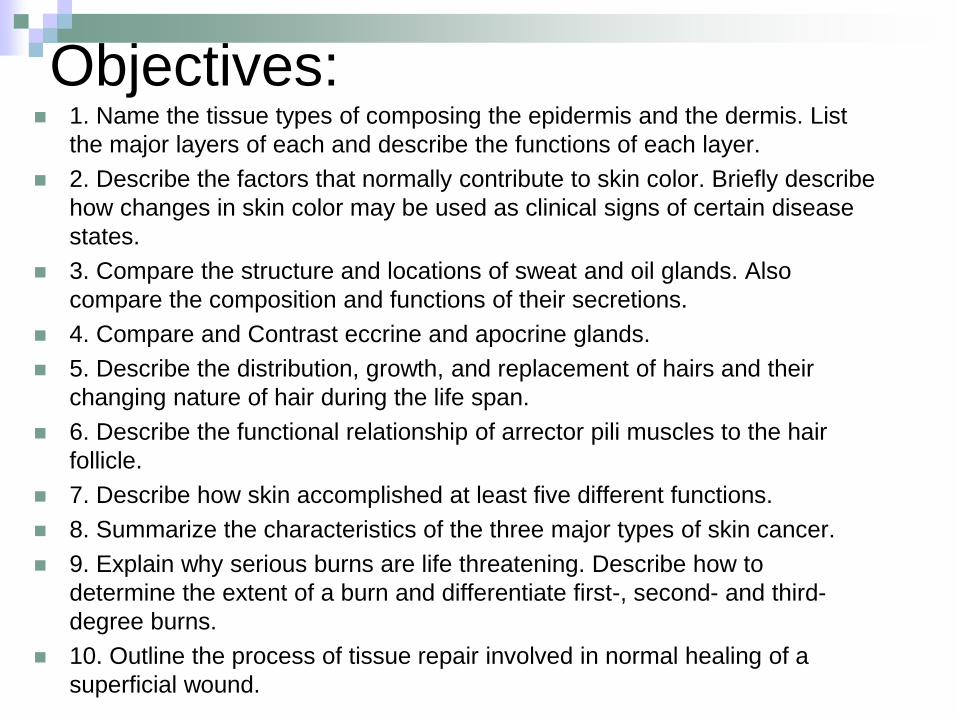
Objectives: 1. Name the tissue types of composing the epidermis and the dermis. List
the major layers of each and describe the functions of each layer.
2. Describe the factors that normally contribute to skin color. Briefly describe
how changes in skin color may be used as clinical signs of certain disease
states.
3. Compare the structure and locations of sweat and oil glands. Also
compare the composition and functions of their secretions.
4. Compare and Contrast eccrine and apocrine glands.
5. Describe the distribution, growth, and replacement of hairs and their
changing nature of hair during the life span.
6. Describe the functional relationship of arrector pili muscles to the hair
follicle.
7. Describe how skin accomplished at least five different functions.
8. Summarize the characteristics of the three major types of skin cancer.
9. Explain why serious burns are life threatening. Describe how to
determine the extent of a burn and differentiate first-, second- and third-
degree burns.
10. Outline the process of tissue repair involved in normal healing of a
superficial wound.
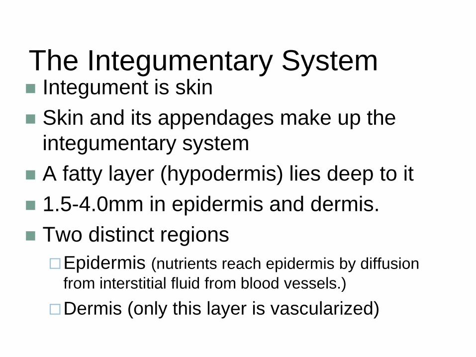
The Integumentary System Integument is skin
Skin and its appendages make up the
integumentary system
A fatty layer (hypodermis) lies deep to it
1.5-4.0mm in epidermis and dermis.
Two distinct regions
Epidermis (nutrients reach epidermis by diffusion
from interstitial fluid from blood vessels.)
Dermis (only this layer is vascularized)
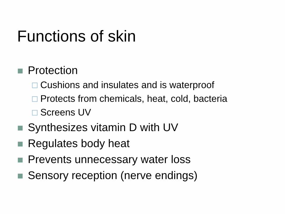
Functions of skin
Protection
Cushions and insulates and is waterproof
Protects from chemicals, heat, cold, bacteria
Screens UV
Synthesizes vitamin D with UV
Regulates body heat
Prevents unnecessary water loss
Sensory reception (nerve endings)
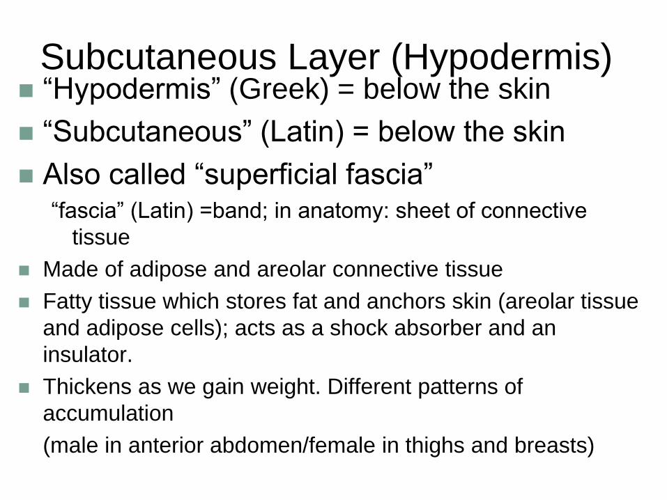
Subcutaneous Layer (Hypodermis) “Hypodermis” (Greek) = below the skin
“Subcutaneous” (Latin) = below the skin
Also called “superficial fascia” “fascia” (Latin) =band; in anatomy: sheet of connective
tissue
Made of adipose and areolar connective tissue
Fatty tissue which stores fat and anchors skin (areolar tissue
and adipose cells); acts as a shock absorber and an
insulator.
Thickens as we gain weight. Different patterns of
accumulation
(male in anterior abdomen/female in thighs and breasts)
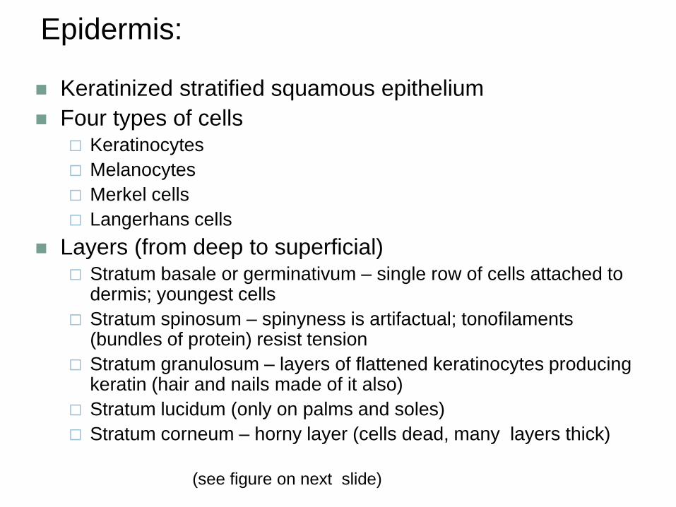
Epidermis:
Keratinized stratified squamous epithelium
Four types of cells Keratinocytes
Melanocytes
Merkel cells
Langerhans cells
Layers (from deep to superficial) Stratum basale or germinativum – single row of cells attached to
dermis; youngest cells
Stratum spinosum – spinyness is artifactual; tonofilaments (bundles of protein) resist tension
Stratum granulosum – layers of flattened keratinocytes producing keratin (hair and nails made of it also)
Stratum lucidum (only on palms and soles)
Stratum corneum – horny layer (cells dead, many layers thick)
(see figure on next slide)
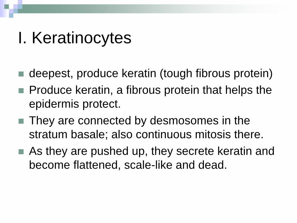
I. Keratinocytes
deepest, produce keratin (tough fibrous protein)
Produce keratin, a fibrous protein that helps the
epidermis protect.
They are connected by desmosomes in the
stratum basale; also continuous mitosis there.
As they are pushed up, they secrete keratin and
become flattened, scale-like and dead.
II. Melanocytes, Merkel cells
and Langerhans cells: Melanocytes -
make dark skin pigment melanin
Spider-shaped cells that make melanin in stratum basale.
Melanin is moved to end of arms of cell and transferred to keratinocytes.
The melanin then collects on the superficial side of the nucleus.
Merkel cells – associated with sensory nerve endings
Langerhans cells – macrophage-like dendritic cells
Cells come from the bone marrow to become macrophages.
Layers of Epidermis: A. Thick vs. Thin skin: palms of hands and soles of feet contain an additional layer of skin, giving them five. The rest of the skin has four distinct layers.
B. Stratum basale or germinativum – The deepest layer of the dermis. Made of a single row of cells attached to dermis; youngest cells (keratinocytes); rapid mitosis occurs here; melanocytes present here.
C. Stratum spinosum – spinyness is artifactual; tonofilaments (bundles of protein) resist tension; several layers thick; consists primarily of tension-resisting bundles of prekeratin filaments; the cells flatten and become spiny because the cells shrink in death but desmosomes hold tight.
Layers of Epidermis continued… D. Stratum granulosum – layers of flattened keratinocytes producing keratin (hair and nails made of it also); Consists of 3-5 layers, cells continue to flatten, internal structures disintegrate, granules develop that help form keratin in upper layers. Cells are now too far from blood vessels and are dead.
E. Stratum lucidum (only on palms and soles---making thick skin); Forms a thin, translucent band of clear, dead keratinocytes.
F. Stratum corneum – horny layer (cells dead, many layers thick); 20-30 cell layers, keratin and plasma membrane protect skin against abrasion, penetration from pathogens and now is waterproof.
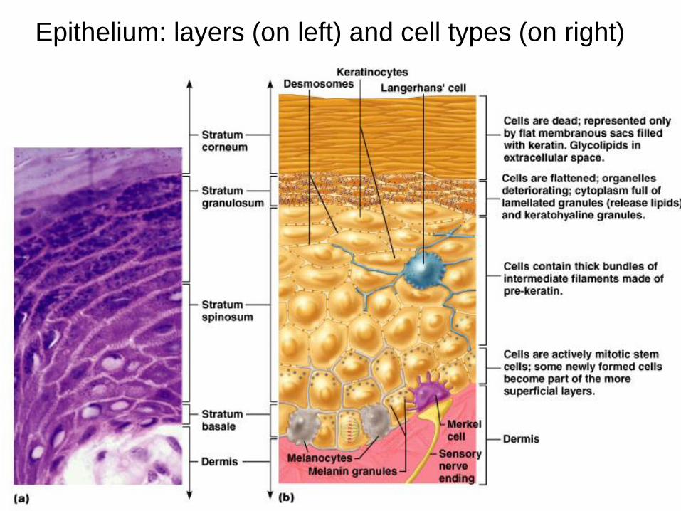
Epithelium: layers (on left) and cell types (on right)
Remember…
Four basic types of tissue
Epithelium – epidermis just discussed
Connective tissue - dermis
Muscle tissue
Nervous tissue
Dermis
Strong, flexible connective tissue: your “hide”
Cells: fibroblasts, macrophages, mast cells, WBCs (white blood cells)
Fiber types: collagen, elastic, reticular
Rich supply of nerves and vessels
Critical role in temperature regulation (the vessels)
Two layers (see next slides) Papillary – areolar connective tissue; includes dermal
papillae
Reticular – “reticulum” (network) of collagen and reticular fibers
Layers of the Dermis 1. Papillary Layer–
areolar connective tissue; includes dermal papillae
The upper layer of the dermis where areolar connective tissue with collagen and elastic fibers and blood vessels form a mat.
Its superior surface or borders forms peg like projections called dermal papillae into the epidermis.
These are filled with blood vessels and nerve endings (pain and touch receptors).
Palms and soles have dermal ridges to increase friction (fingerprints.)
2. Reticular Layer – “reticulum” (network) of collagen and reticular fibers
Makes up 80% of dermis; made of dense irregular connective tissue (aka DICT).
Collagen fibers are found in bundles parallel to the surface.
Produces lines of cleavage in between.
Stretch marks develop from this layer.
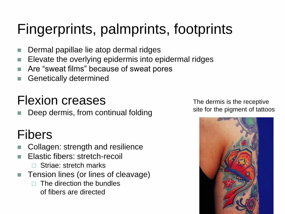
Fingerprints, palmprints, footprints
Dermal papillae lie atop dermal ridges
Elevate the overlying epidermis into epidermal ridges
Are “sweat films” because of sweat pores
Genetically determined
Flexion creases Deep dermis, from continual folding
Fibers Collagen: strength and resilience
Elastic fibers: stretch-recoil Striae: stretch marks
Tension lines (or lines of cleavage) The direction the bundles
of fibers are directed
The dermis is the receptive
site for the pigment of tattoos
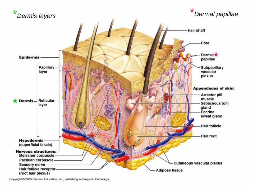
*Dermis layers
*
*
*Dermal papillae
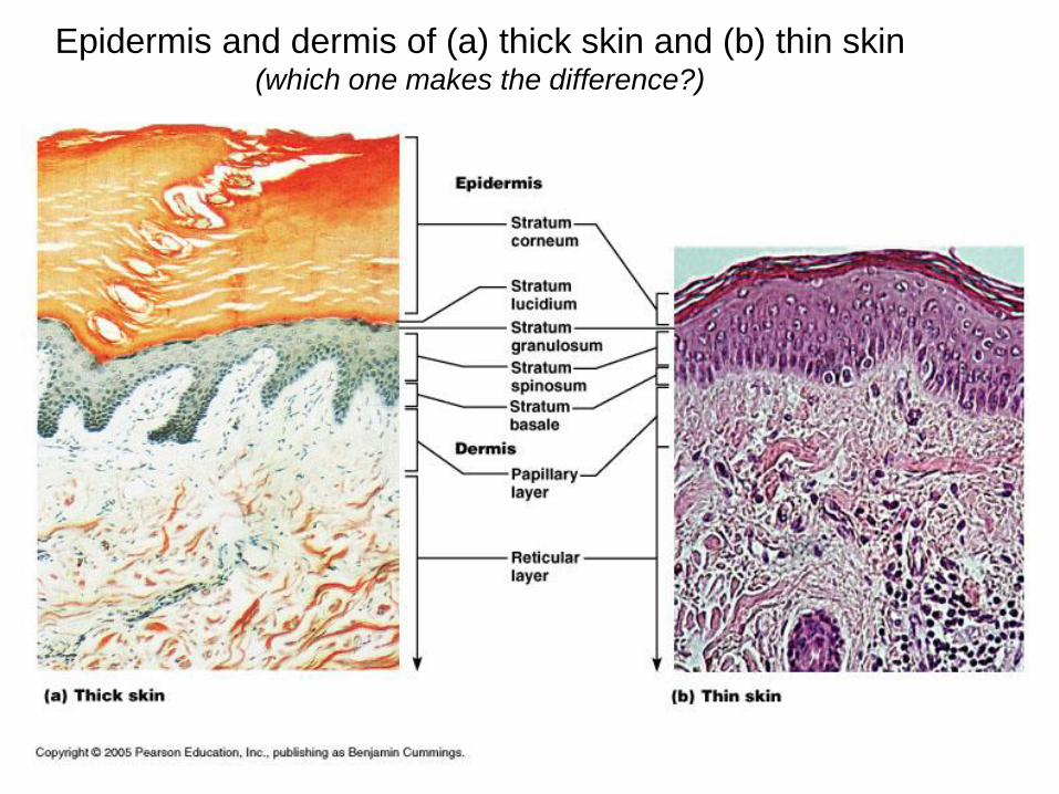
Epidermis and dermis of (a) thick skin and (b) thin skin (which one makes the difference?)