Embed Size (px)
Citation preview
London Cancer
Guidelines for Cutaneous Malignant Melanoma Management
August 2013 Review: August 2014
Version: 1.0
Lead author: David Chao
2
Contents
1. Introduction .............................................................................................................. 3
2. Screening and surveillance ......................................................................................... 3
3. Management in primary care ..................................................................................... 3
4. Management in secondary care ................................................................................. 4
4.1. Management of adult patients ................................................................................... 4
4.2. Management of TYA patients ..................................................................................... 4
5. Staging ...................................................................................................................... 6
6. Surgical Management ................................................................................................ 9
6.1. Primary excision .......................................................................................................... 9
6.2. Sentinel lymph node biopsy ........................................................................................ 9
6.3. Lymphadenopathy ...................................................................................................... 9
6.4. Lymph Node Dissection (LND) ................................................................................... 10
7. Histopathology and molecular profiling ................................................................... 11
7.1. Minimum dataset ...................................................................................................... 11
7.2. Molecular Profiling .................................................................................................... 11
8. Adjuvant Treatments ............................................................................................... 11
9. Follow up management ........................................................................................... 13
9.1. Definition of patients at high risk of relapse ............................................................. 13
9.2. Follow up for In Situ disease ..................................................................................... 13
9.3. Follow up for Stages I & IIA ....................................................................................... 13
9.4. Follow up for Stages IIB, IIC, IIIA, IIIB & IIIC ............................................................... 14
9.5. Follow up for occult primary ..................................................................................... 14
9.6. PET scans ................................................................................................................... 14
9.7. Patient preference .................................................................................................... 14
10. Imaging ................................................................................................................ 15
11. Management of advanced or metastatic disease .................................................. 15
3
1. Introduction
The London Cancer guidelines for the management of malignant melanoma are the result of the merger of the previous guidelines of the Skin Cancer Tumour Boards for North Central and North East London. The guidelines have been updated to incorporate the
2010 British Association of Dermatology guidelines (http://www.bad.org.uk/Portals/_Bad/Guidelines/Clinical%20Guidelines/Melanoma%20guidelines%202010.pdf), and the
2010 NICE IOG for Skin Cancers (http://guidance.nice.org.uk/CSGSTIM).
2. Screening and surveillance
The system will adopt the 2010 BAD guidelines for both screening and surveillance.
3. Management in primary care
Patients may present to their general practitioner with a changing pigmented lesion. The weighted 7-point checklist may be helpful in assessing pigmented lesions. Major features 2 points
Change in size
Irregular shape
Irregular colour Minor features 1 point
Largest diameter 7mm or more
Inflammation
Oozing
Change in sensation Suspicion is greater for lesions scoring 3 points. However, any one feature is adequate to prompt an urgent referral if the concerns about cancer are strong. Biopsy or attempted excision should not be carried out in primary care. Any melanoma excision in primary care is a breach of the NICE IOG but the clinical governance arrangements are in transition currently. In the past breaches were collated by the network who then reported these to the PCTs for further action. Since the PCTs have been dissolved it is unclear who will accept responsibility for such breaches and the Pathway Board is working with the Clinical Commissioning Groups on an appropriate alert system. At this stage, the GP can do one of the following:
Reassure the patient and discharge
Reassure the patient but point out signs to look for which may represent malignant change and request the patient to return
Reassure the patient but take a photograph. Once again advise if change is noted, to return
4
Review the patient within 2 weeks in the event that the lesion is infected/traumatised. The lesion must be covered by a dressing for the 2 week period
Refer to local Plastic Surgery/Dermatology Unit via the urgent suspected cancer route/two week rule (TWR). London Cancer has a Suspected Skin Cancer fax form with all dermatology units listed.
4. Management in secondary care
4.1. Management of adult patients
Patients are referred in via the TWR and can be seen by Dermatologists or Plastic Surgeons. In the clinic the following model is used,
A full history is taken
The patient is asked to undress and examined thoroughly in bright light with a magnifying glass or dermatascope.
The lesion may be photographed
The lesion is documented and if deemed suspicious excised
Where possible the clinic will offer immediate biopsy service. If not possible, the biopsy will be preformed as soon as possible
Biopsy is an excision biopsy (excising the lesion with a 2mm margin). If excision is not possible, then incision biopsy is acceptable
When biopsy is undertaken, care is given to orientation of scar with view to future excisions
The pathology request form will give the following details - name, age, gender, DOB, hospital number, site, brief history, differential diagnosis and orientation stitch if necessary.
Patients are requested to return in 2-3 weeks for the results. Patients should be encouraged to request their results if they do not hear within 4 weeks.
If the pathology records melanoma, then patients are requested to return to the appropriate clinic
If the patient may be managed by the LSMDT (in situ or stage IA melanoma) then definitive treatment may be carried out at that hospital and surgical margins are given in section 5.1. However, for patients under 18y or 18-24y additional guidance is available (see section 3.2). For patients with stage IB melanoma or above the option of Sentinel Lymph Node Biopsy should be considered. This staging procedure is offered by the two main SSMDT at the Royal Free Hospital and the Royal London Hospital. It is stressed that co-morbidities and informed patient choice are vital determinants for this procedure.
4.2. Management of TYA patients
UCLH is the designated Principal Treatment Centre for The North Thames Teenage and Young Adult (TYA) Network, which includes organisations affiliated with London Cancer. The
5
Royal Free Hospital (RFH) has been awarded TYA Designated Hospital status for patients with skin cancer acknowledging the specific expertise for managing skin cancer at this Trust. There are 3 distinct groups of young patients with skin cancer to be considered under this guidance.
1) All patients 18y and under: These patients must be referred to Adolescent Dermatology at UCLH and discussed at the Skin Cancer SSMDT at the Royal Free Hospital. If it is deemed appropriate then patients should undergo their further surgical treatment at the RFH. Follow up should take place back at UCLH.
2) Patients aged 19 to 24y with melanoma under breslow thickness 1mm and no other poor prognostic markers, such as ulceration or mitotic activity: There is a conflict here between the TYA and Skin Cancer IOG. Under TYA IOG such patients should be offered treatment at the Principal Treatment Centre (in this case UCLH) or the designated TYA centre (in this case RFH) only, but it must be recognised that this is generic guidance. Under Skin Cancer IOG such patients are dealt with locally by the LSMDT and do not require referral onwards to the SSMDT. Discussion at the Skin Cancer Pathway Board has highlighted that for this good prognosis group of patients mandatory referral to the SSMDT would only lengthen treatment and travelling times, potentially compromising outcome. Therefore the pragmatic compromise will be that these patients will be offered the choice between further surgery at the site of the LSMDT or to be referred to the SSMDT. Those patients who stay with the LSMDT for further treatment will be provided with a TYA information pack detailing appropriate support services for them and the LSMDT will be required to complete a form on each patient which will be forwarded directly to the TYA MDT. Those requiring further support will be contacted by the designated TYA CNS.
3) Patients aged 19 to 24y with melanoma of breslow thickness 1mm or greater, with or without other poor prognostic markers: This patient group is currently obliged to be referred to the SSMDT for further treatment under Skin Cancer IOG. These patients will be provided with an information pack detailing appropriate support services for them and the Skin Cancer CNS will dial into the TYA MDT on Wednesday afternoons on alternate weeks to discuss these patients. Those requiring further support will be contacted by the designated TYA CNS.
These guidelines have been ratified by the TYA Pathway Board.
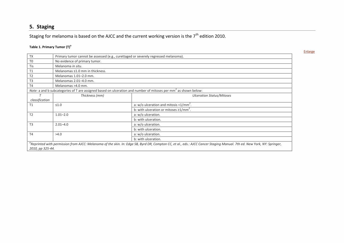
5. Staging
Staging for melanoma is based on the AJCC and the current working version is the 7th edition 2010. Table 1. Primary Tumor (T)
a
Enlarge
TX Primary tumor cannot be assessed (e.g., curettaged or severely regressed melanoma).
T0 No evidence of primary tumor.
Tis Melanoma in situ.
T1 Melanomas ≤1.0 mm in thickness.
T2 Melanomas 1.01–2.0 mm.
T3 Melanomas 2.01–4.0 mm.
T4 Melanomas >4.0 mm.
Note: a and b subcategories of T are assigned based on ulceration and number of mitoses per mm2 as shown below:
T classification
Thickness (mm) Ulceration Status/Mitoses
T1 ≤1.0 a: w/o ulceration and mitosis <1/mm2.
b: with ulceration or mitoses ≥1/mm2.
T2 1.01–2.0 a: w/o ulceration.
b: with ulceration.
T3 2.01–4.0 a: w/o ulceration.
b: with ulceration.
T4 >4.0 a: w/o ulceration.
b: with ulceration. aReprinted with permission from AJCC: Melanoma of the skin. In: Edge SB, Byrd DR, Compton CC, et al., eds.: AJCC Cancer Staging Manual. 7th ed. New York, NY: Springer,
2010, pp 325-44.
7
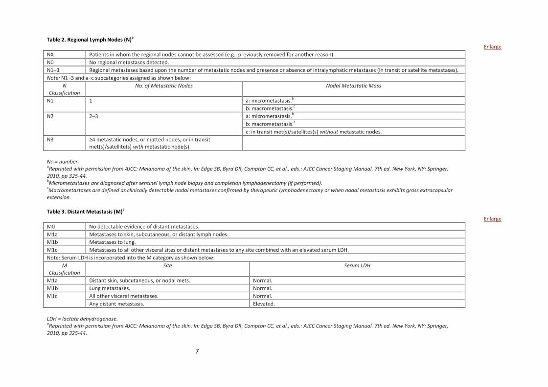
Table 2. Regional Lymph Nodes (N)a
Enlarge
NX Patients in whom the regional nodes cannot be assessed (e.g., previously removed for another reason).
N0 No regional metastases detected.
N1–3 Regional metastases based upon the number of metastatic nodes and presence or absence of intralymphatic metastases (in transit or satellite metastases).
Note: N1–3 and a–c subcategories assigned as shown below:
N Classification
No. of Metastatic Nodes Nodal Metastatic Mass
N1 1 a: micrometastasis.b
b: macrometastasis.c
N2 2–3 a: micrometastasis.b
b: macrometastasis.c
c: in transit met(s)/satellites(s) without metastatic nodes.
N3 ≥4 metastatic nodes, or matted nodes, or in transit met(s)/satellite(s) with metastatic node(s).
No = number. aReprinted with permission from AJCC: Melanoma of the skin. In: Edge SB, Byrd DR, Compton CC, et al., eds.: AJCC Cancer Staging Manual. 7th ed. New York, NY: Springer,
2010, pp 325-44. bMicrometastases are diagnosed after sentinel lymph node biopsy and completion lymphadenectomy (if performed).
cMacrometastases are defined as clinically detectable nodal metastases confirmed by therapeutic lymphadenectomy or when nodal metastasis exhibits gross extracapsular
extension. Table 3. Distant Metastasis (M)
a
Enlarge
M0 No detectable evidence of distant metastases.
M1a Metastases to skin, subcutaneous, or distant lymph nodes.
M1b Metastases to lung.
M1c Metastases to all other visceral sites or distant metastases to any site combined with an elevated serum LDH.
Note: Serum LDH is incorporated into the M category as shown below:
M Classification
Site Serum LDH
M1a Distant skin, subcutaneous, or nodal mets. Normal.
M1b Lung metastases. Normal.
M1c All other visceral metastases. Normal.
Any distant metastasis. Elevated.
LDH = lactate dehydrogenase. aReprinted with permission from AJCC: Melanoma of the skin. In: Edge SB, Byrd DR, Compton CC, et al., eds.: AJCC Cancer Staging Manual. 7th ed. New York, NY: Springer,
2010, pp 325-44.
8
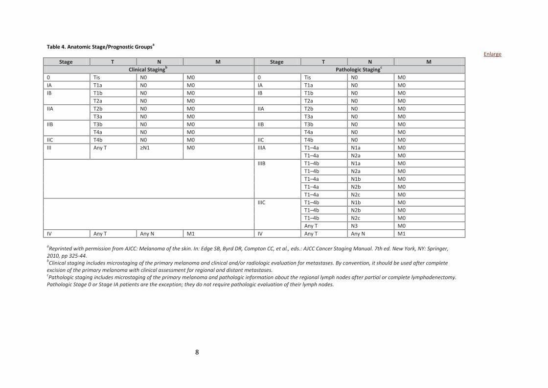
Table 4. Anatomic Stage/Prognostic Groups
a
Enlarge
Stage T N M Stage T N M
Clinical Stagingb Pathologic Staging
c
0 Tis N0 M0 0 Tis N0 M0
IA T1a N0 M0 IA T1a N0 M0
IB T1b N0 M0 IB T1b N0 M0
T2a N0 M0 T2a N0 M0
IIA T2b N0 M0 IIA T2b N0 M0
T3a N0 M0 T3a N0 M0
IIB T3b N0 M0 IIB T3b N0 M0
T4a N0 M0 T4a N0 M0
IIC T4b N0 M0 IIC T4b N0 M0
III Any T ≥N1 M0 IIIA T1–4a N1a M0
T1–4a N2a M0
IIIB T1–4b N1a M0
T1–4b N2a M0
T1–4a N1b M0
T1–4a N2b M0
T1–4a N2c M0
IIIC T1–4b N1b M0
T1–4b N2b M0
T1–4b N2c M0
Any T N3 M0
IV Any T Any N M1 IV Any T Any N M1
aReprinted with permission from AJCC: Melanoma of the skin. In: Edge SB, Byrd DR, Compton CC, et al., eds.: AJCC Cancer Staging Manual. 7th ed. New York, NY: Springer,
2010, pp 325-44. bClinical staging includes microstaging of the primary melanoma and clinical and/or radiologic evaluation for metastases. By convention, it should be used after complete
excision of the primary melanoma with clinical assessment for regional and distant metastases. cPathologic staging includes microstaging of the primary melanoma and pathologic information about the regional lymph nodes after partial or complete lymphadenectomy.
Pathologic Stage 0 or Stage IA patients are the exception; they do not require pathologic evaluation of their lymph nodes.
9
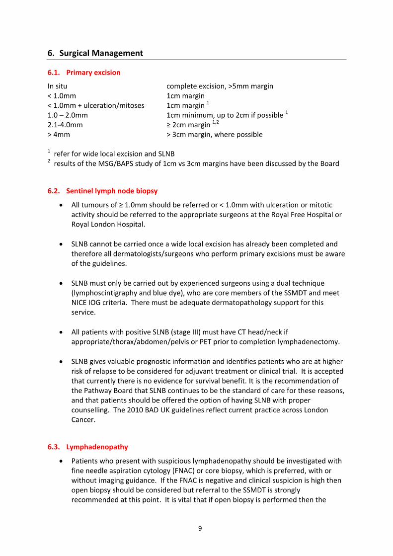
6. Surgical Management
6.1. Primary excision
In situ complete excision, >5mm margin < 1.0mm 1cm margin < 1.0mm + ulceration/mitoses 1cm margin 1
1.0 – 2.0mm 1cm minimum, up to 2cm if possible 1
2.1-4.0mm ≥ 2cm margin 1,2
> 4mm > 3cm margin, where possible 1 refer for wide local excision and SLNB 2 results of the MSG/BAPS study of 1cm vs 3cm margins have been discussed by the Board
6.2. Sentinel lymph node biopsy
All tumours of ≥ 1.0mm should be referred or < 1.0mm with ulceration or mitotic activity should be referred to the appropriate surgeons at the Royal Free Hospital or Royal London Hospital.
SLNB cannot be carried once a wide local excision has already been completed and therefore all dermatologists/surgeons who perform primary excisions must be aware of the guidelines.
SLNB must only be carried out by experienced surgeons using a dual technique (lymphoscintigraphy and blue dye), who are core members of the SSMDT and meet NICE IOG criteria. There must be adequate dermatopathology support for this service.
All patients with positive SLNB (stage III) must have CT head/neck if appropriate/thorax/abdomen/pelvis or PET prior to completion lymphadenectomy.
SLNB gives valuable prognostic information and identifies patients who are at higher risk of relapse to be considered for adjuvant treatment or clinical trial. It is accepted that currently there is no evidence for survival benefit. It is the recommendation of the Pathway Board that SLNB continues to be the standard of care for these reasons, and that patients should be offered the option of having SLNB with proper counselling. The 2010 BAD UK guidelines reflect current practice across London Cancer.
6.3. Lymphadenopathy
Patients who present with suspicious lymphadenopathy should be investigated with fine needle aspiration cytology (FNAC) or core biopsy, which is preferred, with or without imaging guidance. If the FNAC is negative and clinical suspicion is high then open biopsy should be considered but referral to the SSMDT is strongly recommended at this point. It is vital that if open biopsy is performed then the
10
incision must be such as to allow subsequent complete block dissection of the regional nodes without compromise.
All patients should have blood tests and CT head/neck if appropriate/ thorax/abdomen/pelvis or PET prior to lymphadenectomy.
6.4. Lymph Node Dissection (LND)
Radical lymph node dissections (LND) should only be performed by a designated core member of the SSMDT at the Royal Free Hospital or Royal London Hospital. Pre-operative staging investigations should be carried out as listed previously. The decision as to whether or not surgery should proceed prior to scanning should be made after SSMDT discussion with an informed patient. The block dissection specimen should be marked and orientated for the pathologist.
Axilla It is recommended that axillary LND should include all nodes in levels I – III, and this may require either resection or division of pectoralis minor. Inguinal The management of inguinal lymph node metastases is controversial. A superficial inguinal lymph node dissection should be considered in the presence of:
A single clinically involved node in the femoral triangle
Medical co-morbidities which would increase the risk associated with more extensive surgery
A positive superficial inguinal sentinel node A pelvic lymph node dissection ileo-obturator should be considered:
If there is >1 clinically palpable subinguinal node,
If there is U/S and / or CT evidence of pelvic node involvement
If pathological review of the superficial specimen shows multiple microscopically/ macroscopically involved nodes
Cervical Cervical nodal disease should be reviewed and treated by either surgeons in the SSMDT with expertise in head and neck skin cancer including melanoma, or by a Head and Neck MDT with a special interest in melanoma. Some LSMDT have core members who are Head and Neck surgeons and dissections may take place locally if in the best interests of the patient and only after discussion with the SSMDT. A comprehensive, and not a selective neck dissection should be performed. The term comprehensive allows either:
A radical dissection of levels 1-5
Modified radical – the above, sparing spinal accessory nerve, internal jugular vein and sternocleidomastoid muscle.
11
Extended radical – Radical dissection including parotid and/or posterior occipital chain.
The risk of recurrence is high (up to 28%) despite comprehensive surgery and so surgery may be combined with adjuvant radiotherapy. This is the only nodal group shown to have improved local recurrence rate with post-operative radiotherapy [ Guadagno BA et al, Lancet Oncol 2009; 10: 409–16]. If extra-capsular spread is noted, then the management should be discussed at the SSMDT.
7. Histopathology and molecular profiling
7.1. Minimum dataset
All pathology departments should be reporting the minimum dataset as stipulated by the Royal College of Pathologists. Staging should be according to the 7th edition AJCC staging 2010; the full text can be round on http://jco.ascopubs.org/content/27/36/6199.full.pdf+html and there is an excellent summary of the differences between the 6 & 7th versions on http://www.cancerstaging.org/staging/PDFs/gershenwald-melanoma.pdf
7.2. Molecular Profiling
This will be of increasing importance in determining the treatment options for all cancers. For melanoma the only target mutation with a licensed drug is in the BRaf oncogene as of May 2013 but there is no doubt that this list will grow. It may be helpful to know other target mutations for clinical trial purposes as well. For all melanomas stage IIB and above it is proposed to test for the BRaf mutation at the time of initial diagnosis where there is sufficient tissue. This will guide subsequent therapy, be it for clinical trials or standard therapy. Currently all sites in London Cancer use the Roche-funded central laboratories for this purpose.
8. Adjuvant Treatments
In the UK there are no formally recognised adjuvant therapies for any stage of malignant melanoma. Clinical trials remain the most important consideration and all LSMDT should be reviewing melanoma patients for their suitability for adjuvant clinical trials and referring them on to the SSMDT in good time. Radiotherapy remains a contentious adjuvant therapy. There is evidence for some benefit in reducing local relapse following lymphadenectomy for Head and Neck melanomas but there is as yet no evidence for an overall survival benefit [Guadagno BA et al, Lancet Oncol 2009; 10: 409–16]. Similarly following axillary or groin dissection adjuvant radiotherapy may improve progression-free survival but not overall survival and there may be reduction in quality of life due to lymphoedema [Henderson MA et al, J Clin Oncol 2013; 31 suppl; abstr 9001]. Such patients should be discussed in the SSMDT on a case by case basis and
12
referred for radiotherapy as appropriate taking into account comorbidities, toxicity from the radiotherapy and patient preference.
13
9. Follow up management
The purpose of follow up is to try to catch any relapse as early as possible. Arguably without effective therapies for advanced melanoma close follow up would have had little impact on survival. However, since 2011 with the introduction of the BRaf mutation inhibitors and immunotherapy as effective new treatments for advanced melanoma the therapeutic landscape has changed. Finding patients with minimal volume disease relapse, particularly in the brain, and avoiding high dose steroid usage, makes clinical sense especially for immunotherapeutic strategies which have a slower onset of action. Therefore there is now a case for closer surveillance. The following guidelines have been based on a consensus document signed by UK melanoma oncologists and due to be published on the Melanoma Focus website in 2013.
9.1. Definition of patients at high risk of relapse
There is no precise definition of high risk but most adjuvant clinical trials have taken stage IIC, IIIB & IIIC disease where the 5y survival is <60%. The Pathway Board feels that this may be too restrictive and prefers to broaden the net. Since the highest risk period is within the first 3y this should be the focus of the most intense surveillance.
9.2. Follow up for In Situ disease
Clinical Review Patients may be discharged and GP alerted or Yearly follow up for 5y before discharge Blood Tests No blood tests are recommended Imaging No routine imaging is recommended
9.3. Follow up for Stages I & IIA
Clinical Review Years 1-3, 3-4 monthly review Years 4-5, 6 monthly review Years 6-10, annual review or discharge depending upon patient preference with GP alerted Blood Tests No blood tests are recommended Imaging No routine surveillance CT scans are recommended
14
9.4. Follow up for Stages IIB, IIC, IIIA, IIIB & IIIC
Clinical Review Years 1-3, 3-4 monthly review Years 4-5, 4-6 monthly review Years 6-10, annual review or discharge depending upon patient preference with GP alerted Blood Tests No blood tests are recommended but routine kidney function tests may be needed prior to CT scanning Imaging CT Brain/Chest/Abdomen/Pelvis at baseline CT Brain thereafter only if symptomatic or upon systemic relapse CT Chest/Abdomen/Pelvis every 6 months for first 3 years only
9.5. Follow up for occult primary
This group of patients should be followed up as per Stages IIB, IIC, IIIA-B
9.6. PET scans
While CT imaging is considered the standard of care PET scans may be used instead depending upon availability and physician preference. PET scans may also be used if there is concern on CT imaging where the PET scan would influence clinical management and it may also be considered for occult primaries.
9.7. Patient preference
CT scans remain the gold standard investigation, particularly for some areas of the body such as the lungs, but entail ionizing radiation. While the additional risk to patients from exposure to CT scans remains controversial this is at the very least a theoretical concern. Patients should be counselled appropriately about the intention of CT scan follow up and ultimately it is very much patient preference whether to have follow up scans.
15
10. Imaging
Imaging modality Indications and notes
Staging CT, MRI
PET Ultrasonography +/FNA Bone scans
Required for surgical planning of locally invasive lesion and/or clinical suspicion of metastases May replace CT or MRI depending upon physician preference For investigation of local lymph node basin or other masses if clinically suspicious For suspected bony metastases
Surveillance CT,MRI, PET Detection of asymptomatic disease according to risk profile.
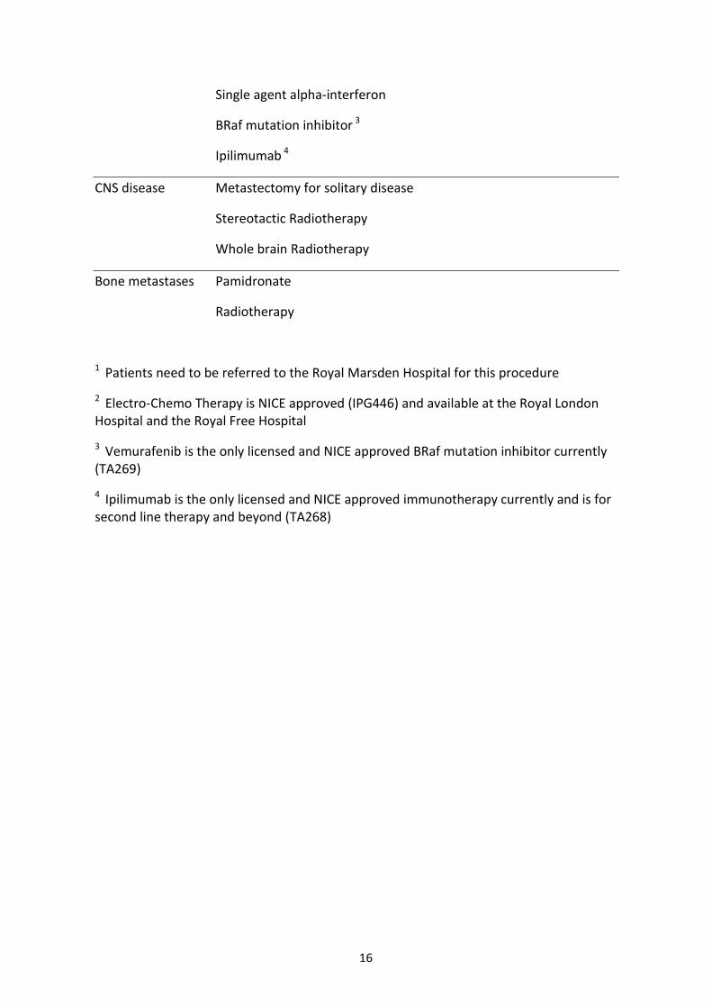
11. Management of advanced or metastatic disease
The treatment algorithms for advanced melanoma are evolving at a tremendous pace. The algorithms presented below are for licensed and/or NICE approved therapies but in most cases clinical trials afford the best prospects for patients by offering them the possibility of cutting edge therapies. All such patients must be discussed with the appropriate SSMDT and both SSMDTs operating in London Cancer must be fully aware of the clinical trial portfolio available within London Cancer which should include Phase I trials. Where there is a gap in the portfolio the Lead for Research will see if there are relevant trials available at other institutions in and around London. The clinical trial portfolio will be updated regularly and will be available on the London Cancer website.
Stage Off study
Skin metastases Excision
Isolated limb perfusion 1
Laser ablation
Radiotherapy
ECT 2
Visceral disease Metastectomy for solitary disease if stable
Single agent Dacarbazine
16
Single agent alpha-interferon
BRaf mutation inhibitor 3
Ipilimumab 4
CNS disease Metastectomy for solitary disease
Stereotactic Radiotherapy
Whole brain Radiotherapy
Bone metastases Pamidronate
Radiotherapy
1 Patients need to be referred to the Royal Marsden Hospital for this procedure
2 Electro-Chemo Therapy is NICE approved (IPG446) and available at the Royal London Hospital and the Royal Free Hospital
3 Vemurafenib is the only licensed and NICE approved BRaf mutation inhibitor currently (TA269)
4 Ipilimumab is the only licensed and NICE approved immunotherapy currently and is for second line therapy and beyond (TA268)