Embed Size (px)
Citation preview
THE MANY FACES OF MONOCLONAL GAMMOPATHIES
Marion S. Sternbach, MD, FRCP(C), FACP
Presented at Medicine Grand Rounds, St Joseph’s Hospital
Sept 19, 2007
Monoclonal GammopathiesObjectives.
• 1. Getting acquainted with varied aspects of clinical presentation of the Monoclonal Gammopathies (MG) and their association with different conditions.
• 2. Following the evolution and pathogenesis from MGUS to myelomas or lymphomas.

The immunoglobulin molecule
• The basic structure of immunoglobulins
Heavy Chain
CCFc Receptor Region
Hinge or J region
Light Chain
Fab. – Antibody binding site regions
N NN N
C C
(gamma, miu, alphaDelta)
( kappa, lambda)
S-S bridges
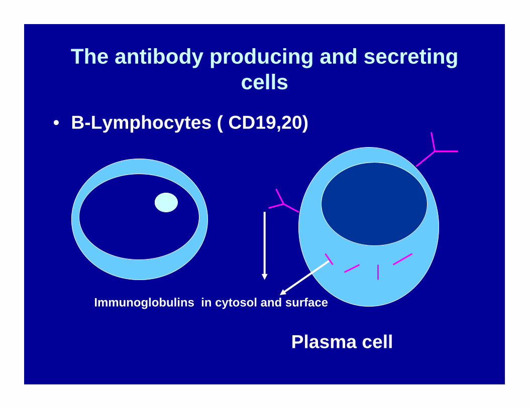
The antibody producing and secreting cells
• B-Lymphocytes ( CD19,20)
Plasma cell
Immunoglobulins in cytosol and surface
Monoclonal Gammopathies
Medicine involves a life time of learning, not only because treatments change and improve constantly, but because
NO TWO PATIENTS ARE IDENTICAL &THE SAME DIAGNOSIS PLAYS ITSELF OUT
DIFFERENTLY IN EVERY PATIENT.
Monoclonal Gammopathies,Definition
• A monoclonal expansion of a single NON FUNCTIONAL ANTIBODY, originating in a mutated MONOCLONAL B- LYMPHOCYTE or PLASMA CELL.
• Plasma cells are “terminal” non proliferating cells, secreting gamma globulins = functional antibodies.
• Once they mutated, they can proliferate producing the pathological antibody.
Monoclonal AntibodiesPatient presentations
1. FH, 46 year old female, dental assistant , never exposed to toxic chemicals, fumes or pollutants.Experienced fatigue, full physical check up by family MD and investigations revealed an abnormal protein electrophoresis.
Referred to Hematology. Physical Exam WNL,Past History non contributory, exc. For years
of smoking, quit 2 years ago.
Monoclonal Gammopathies,Patient presentations ( 1. cont.)
• Investigations: CBC, Renal, Hepatic functions N.
• Immune fixation: Monoclonal IgGK of 15 g/L.• Bone Marrow aspirate and biopsy – perfectly
Normal.• Skeletal survey, chest X ray, abdominal and
pelvic US Normal.• Pt. followed for 3 ½ years with no change.• Then sudden back pain in the T6-T7 area.
Monoclonal Gammopathies Patient presentation (1. cont.)
• All tests from before unchanged, exc. IgGK now up to 21 g/l., Skeletal survey showed a collapsed T6 vertebra.Bone marrow aspirate and biopsy Normal.
• MRI of spine showed lytic lesion in T6, nowhere else.
• Vertebral biopsy confirmed Plasmacytoma.• VAD chemotherapy administered for 6 cycles
with monthly Pamidronate, followed by• Autologous PBSC transplant. Successful.
Monoclonal GammopathiesPatient presentation 2.
• 2. 72 year old female from Poland, retired teacher. Suffered for years from Mixed Connective Tissue disorder. Got intermittent steroid therapy.
• Ref. to me for monoclonal gammopathy IgGL • Initial investigations, including BM Bx,
skeletal survey, CT-s –all Neg.• Two years later, followed regularly –
developed significant lymphadenopathy and splenomegaly.
Monoclonal GammopathiesPatient presentation (2. cont.)
• Lymph node and Bone marrow biopsy revealed low grade diffuse lymphocytic lymphoma.
• Treated with Cyclophosphamide, Vincristine, Prednisone – 8 cycles.
• Improved remarkably for over one year. Even the Mixed Conn. Tissue Dis. Symptoms abated.
• 18 months later – huge lymph nodes, aggressive large cell lymphoma and
• Hypercalcemia.• CHOP chemotherapy, short remission. Died in one
year.
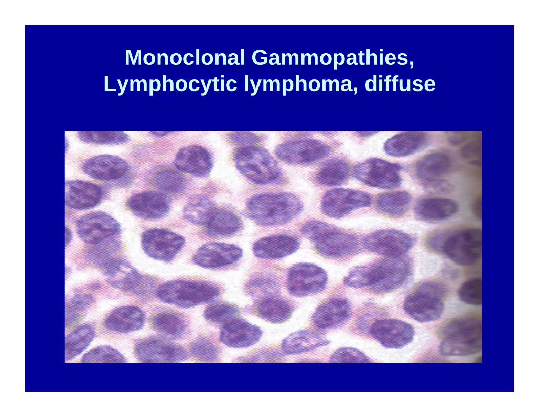
Monoclonal Gammopathies,Lymphocytic lymphoma, diffuse
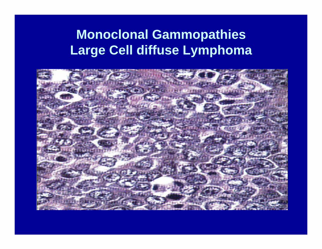
Monoclonal GammopathiesLarge Cell diffuse Lymphoma
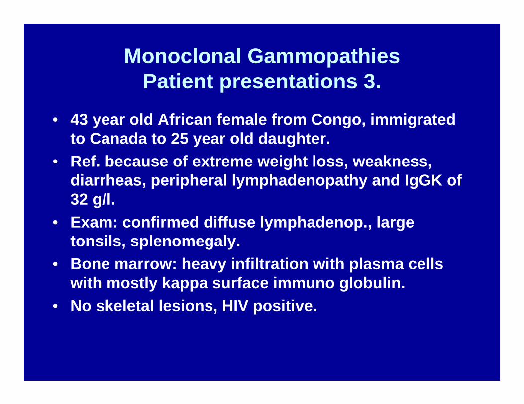
Monoclonal GammopathiesPatient presentations 3.
• 43 year old African female from Congo, immigrated to Canada to 25 year old daughter.
• Ref. because of extreme weight loss, weakness, diarrheas, peripheral lymphadenopathy and IgGK of 32 g/l.
• Exam: confirmed diffuse lymphadenop., large tonsils, splenomegaly.
• Bone marrow: heavy infiltration with plasma cells with mostly kappa surface immuno globulin.
• No skeletal lesions, HIV positive.
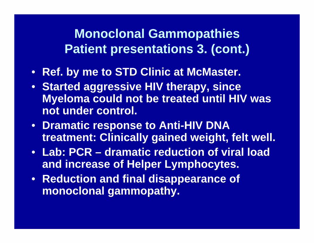
Monoclonal GammopathiesPatient presentations 3. (cont.)
• Ref. by me to STD Clinic at McMaster.• Started aggressive HIV therapy, since
Myeloma could not be treated until HIV was not under control.
• Dramatic response to Anti-HIV DNA treatment: Clinically gained weight, felt well.
• Lab: PCR – dramatic reduction of viral load and increase of Helper Lymphocytes.
• Reduction and final disappearance of monoclonal gammopathy.
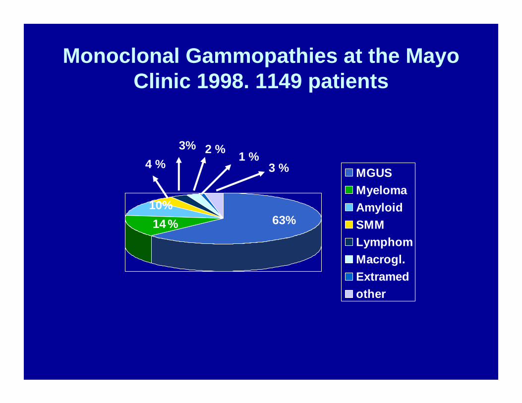
Monoclonal Gammopathies at the Mayo Clinic 1998. 1149 patients
MGUSMyelomaAmyloidSMMLymphomMacrogl.Extramedother
63%1410%
%
4 %3% 2 % 1 %
3 %
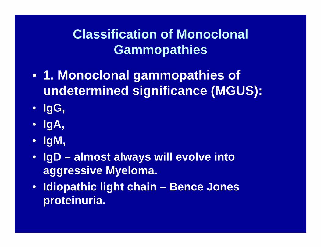
Classification of Monoclonal Gammopathies
• 1. Monoclonal gammopathies of undetermined significance (MGUS):
• IgG,• IgA,• IgM,• IgD – almost always will evolve into
aggressive Myeloma.• Idiopathic light chain – Bence Jones
proteinuria.
Classification of Monoclonal Gammopathies ( 2 cont. )
• Bi- & triclonal Gammopathies,• A. Malignant Monoclonal
Gammopathies:• 1. Myeloma,• 2. Smoldering myeloma,• 3. Plasma cell leukemia,• 4. Non secretory myeloma.• 5. Osteosclerotic myeloma (POEMS)
Classification of Monoclonal Gammopathies ( cont.3 )
• B. Plasmacytoma:• 1. Solitary Plasmacytoma of bone• 2. Extramedullary plasmacytoma.• C. Lymphoproliferative disorders :• 1. Waldenstrom’s Macroglobulinemia,• 2. Heavy chain diseases (HCD): • gamma, alpha, miu (u) • 3. Primary Amyloidosis.• 4. CLL and other B- cell Lymphomas, PTLD
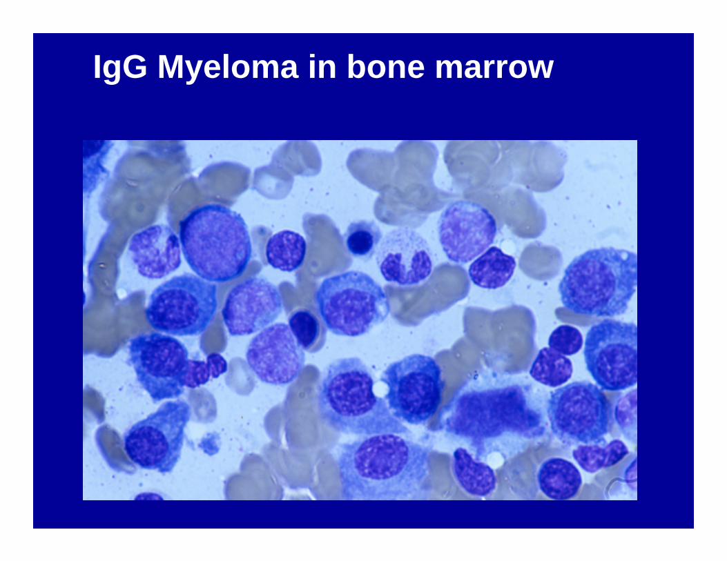
IgG Myeloma in bone marrow
Classification of Monoclonal Gammopathies ( cont. 4 )
• D. Connective Tissue disorders:• SLE,• Rheumatoid Arthritis,• Sjogren syndrome and Mixed conn. Tissue
Disorders,• Scleroderma,• Psoriatic Arthritis,• Polymyalgia rheumatica,• Cryoglobulinemia.
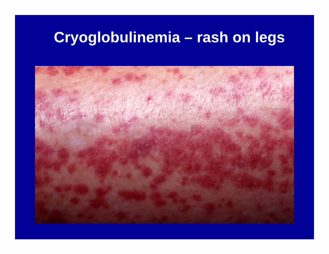
Cryoglobulinemia – rash on legs
Classification of Monoclonal Gammopathies ( cont. 5 )
• E. Associated with infections:• Hepatitis C virus,• HIV/AIDS• F. Dermatologic disorders:• Scleredema, Lichen myxoedematosus,• Diffuse plane xanthomatosis,• Urticaria and IgM ( Schnitzler’s syndrome )• Subcorneal pustular dermatosis,• Necrobiotic xantogranuloma.• Pyoderma gangrenosum
Classification of Monoclonal Gammopathies, ( cont. 6 )
• G. Miscellaneous:• Acquired von Willebrand’s syndrome with Anti –
Factor VIII Ag. Activity .• Acquired C 1 esterase deficiency ( angioedema )• Eosinophilic Fasciitis,• Myelodysplasia,• Chronic Neutrophilic Leukemia,• CIDP with MGUS,• Capillary Leak syndrome,• T-cell large granular lymphocyte leukemia.
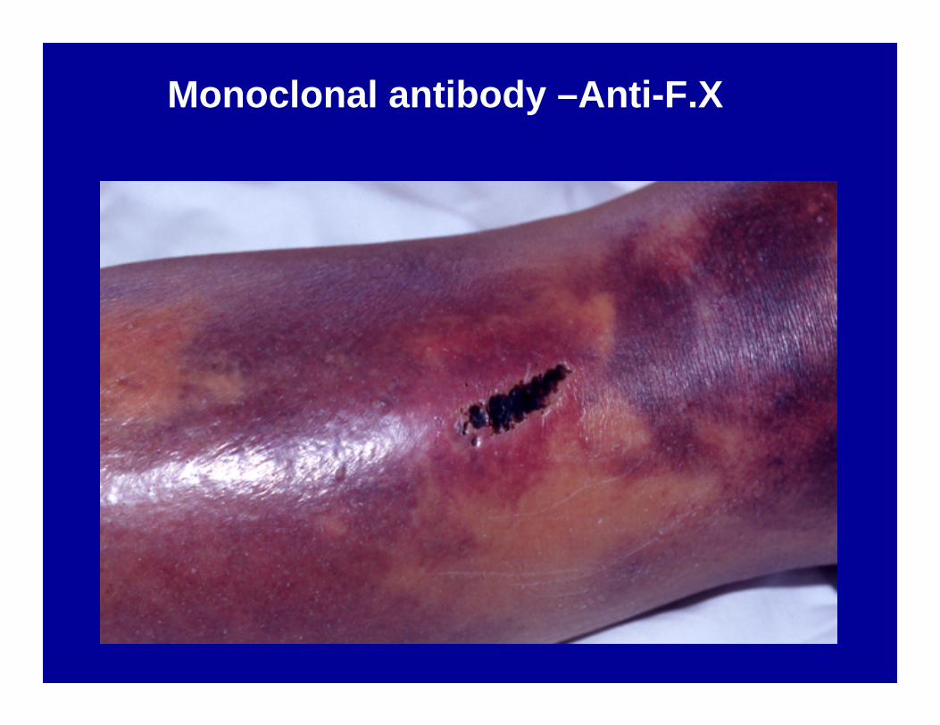
Monoclonal antibody –Anti-F.X
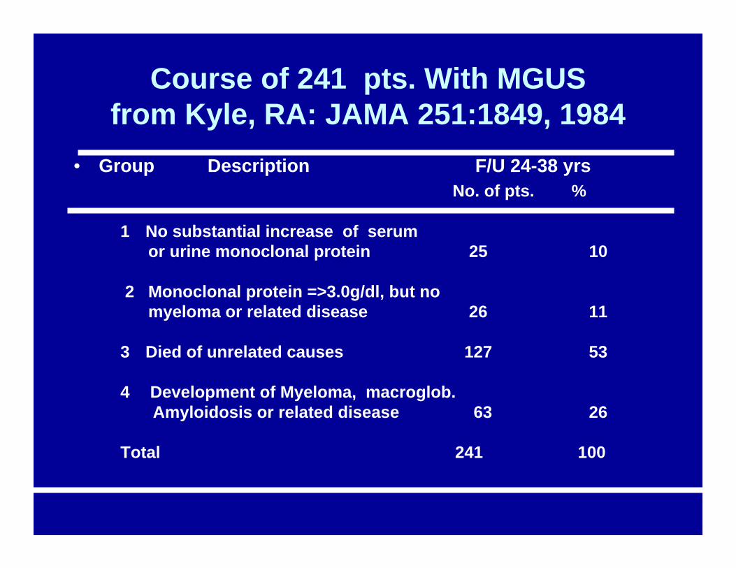
Course of 241 pts. With MGUSfrom Kyle, RA: JAMA 251:1849, 1984
• Group Description F/U 24-38 yrsNo. of pts. %
1 No substantial increase of serumor urine monoclonal protein 25 10
2 Monoclonal protein =>3.0g/dl, but nomyeloma or related disease 26 11
3 Died of unrelated causes 127 53
4 Development of Myeloma, macroglob.Amyloidosis or related disease 63 26
Total 241 100
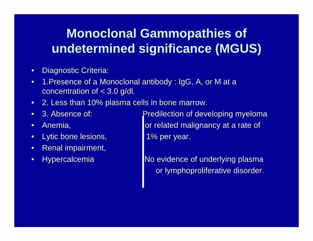
Monoclonal Gammopathies of undetermined significance (MGUS)
• Diagnostic Criteria:• 1.Presence of a Monoclonal antibody : IgG, A, or M at a
concentration of < 3.0 g/dl.• 2. Less than 10% plasma cells in bone marrow.• 3. Absence of: Predilection of developing myeloma• Anemia, or related malignancy at a rate of• Lytic bone lesions, 1% per year.• Renal impairment, • Hypercalcemia No evidence of underlying plasma
or lymphoproliferative disorder.
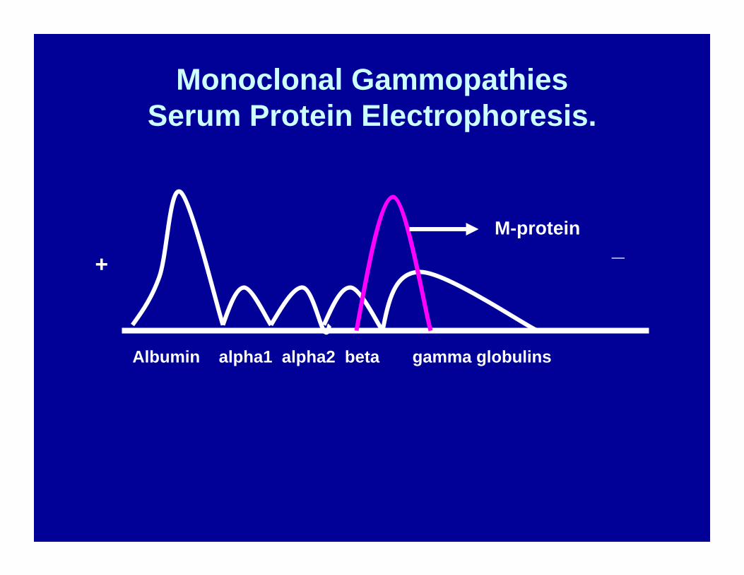
Monoclonal GammopathiesSerum Protein Electrophoresis.
+_
Albumin alpha1 alpha2 beta gamma globulins
M-protein
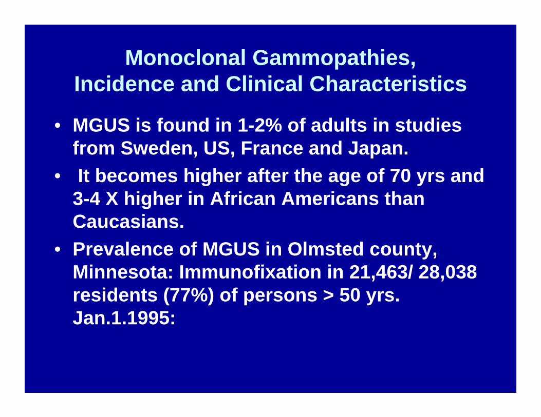
Monoclonal Gammopathies,Incidence and Clinical Characteristics
• MGUS is found in 1-2% of adults in studies from Sweden, US, France and Japan.
• It becomes higher after the age of 70 yrs and 3-4 X higher in African Americans than Caucasians.
• Prevalence of MGUS in Olmsted county, Minnesota: Immunofixation in 21,463/ 28,038 residents (77%) of persons > 50 yrs. Jan.1.1995:
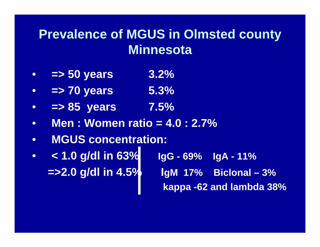
Prevalence of MGUS in Olmsted county Minnesota
• => 50 years 3.2%• => 70 years 5.3%• => 85 years 7.5%• Men : Women ratio = 4.0 : 2.7%• MGUS concentration:• < 1.0 g/dl in 63% IgG - 69% IgA - 11%
=>2.0 g/dl in 4.5% IgM 17% Biclonal – 3%kappa -62 and lambda 38%
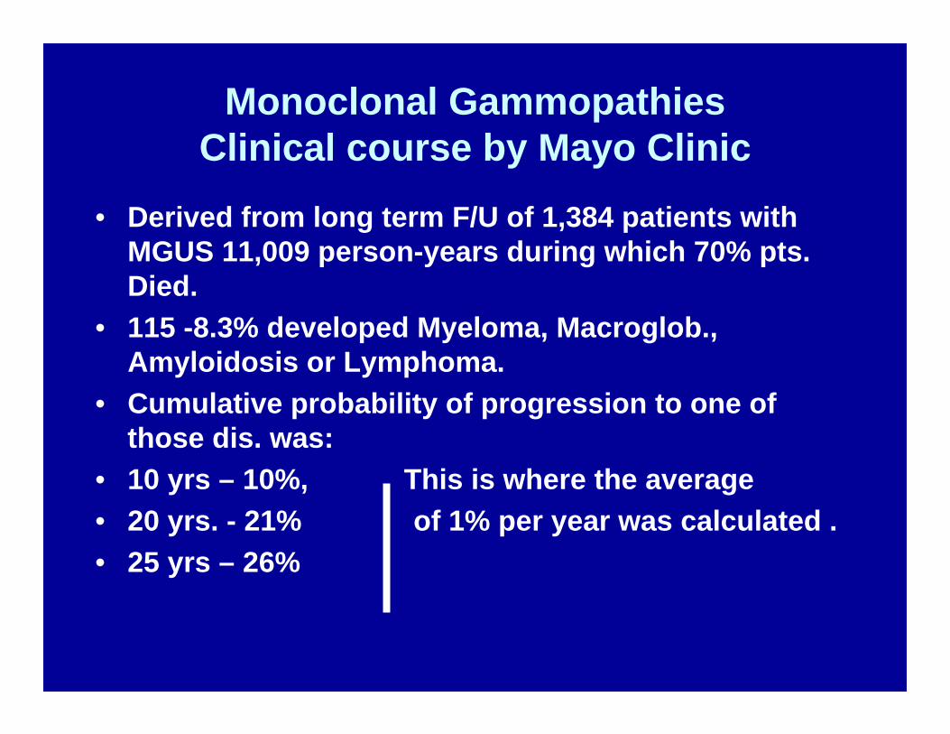
Monoclonal GammopathiesClinical course by Mayo Clinic
• Derived from long term F/U of 1,384 patients with MGUS 11,009 person-years during which 70% pts. Died.
• 115 -8.3% developed Myeloma, Macroglob., Amyloidosis or Lymphoma.
• Cumulative probability of progression to one of those dis. was:
• 10 yrs – 10%, This is where the average• 20 yrs. - 21% of 1% per year was calculated .• 25 yrs – 26%
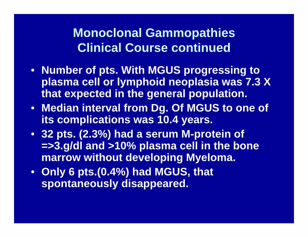
Monoclonal GammopathiesClinical Course continued
• Number of pts. With MGUS progressing to plasma cell or lymphoid neoplasia was 7.3 X that expected in the general population.
• Median interval from Dg. Of MGUS to one of its complications was 10.4 years.
• 32 pts. (2.3%) had a serum M-protein of =>3.g/dl and >10% plasma cell in the bone marrow without developing Myeloma.
• Only 6 pts.(0.4%) had MGUS, that spontaneously disappeared.
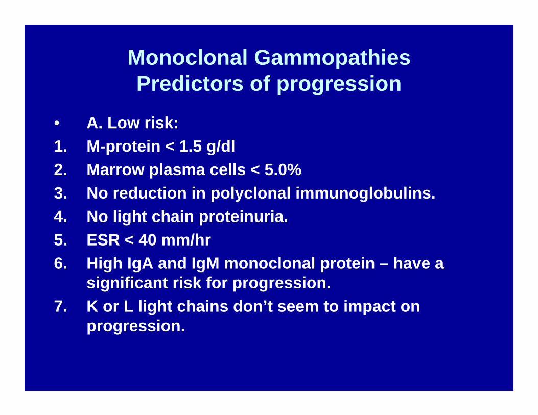
Monoclonal GammopathiesPredictors of progression
• A. Low risk:1. M-protein < 1.5 g/dl2. Marrow plasma cells < 5.0%3. No reduction in polyclonal immunoglobulins.4. No light chain proteinuria.5. ESR < 40 mm/hr6. High IgA and IgM monoclonal protein – have a
significant risk for progression.7. K or L light chains don’t seem to impact on
progression.
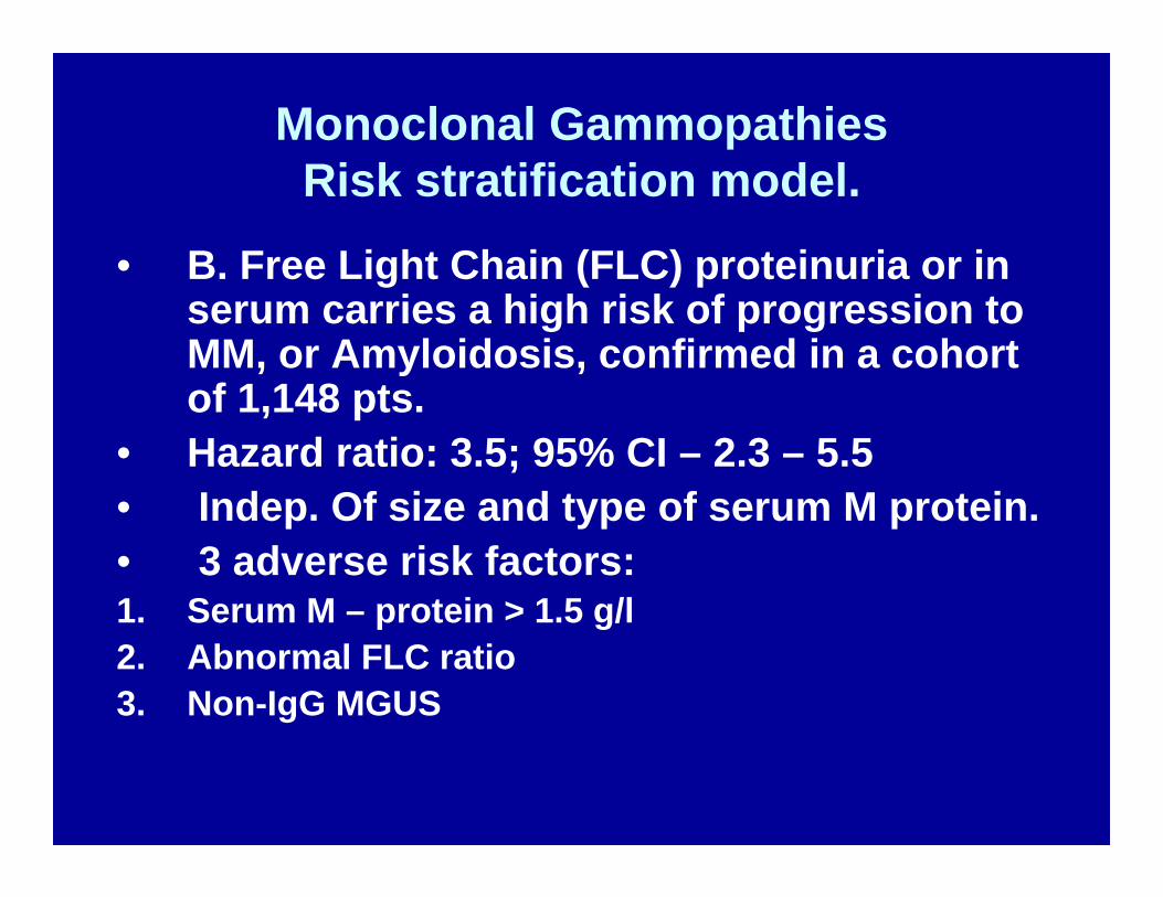
Monoclonal GammopathiesRisk stratification model.
• B. Free Light Chain (FLC) proteinuria or in serum carries a high risk of progression to MM, or Amyloidosis, confirmed in a cohort of 1,148 pts.
• Hazard ratio: 3.5; 95% CI – 2.3 – 5.5 • Indep. Of size and type of serum M protein. • 3 adverse risk factors:1. Serum M – protein > 1.5 g/l2. Abnormal FLC ratio3. Non-IgG MGUS
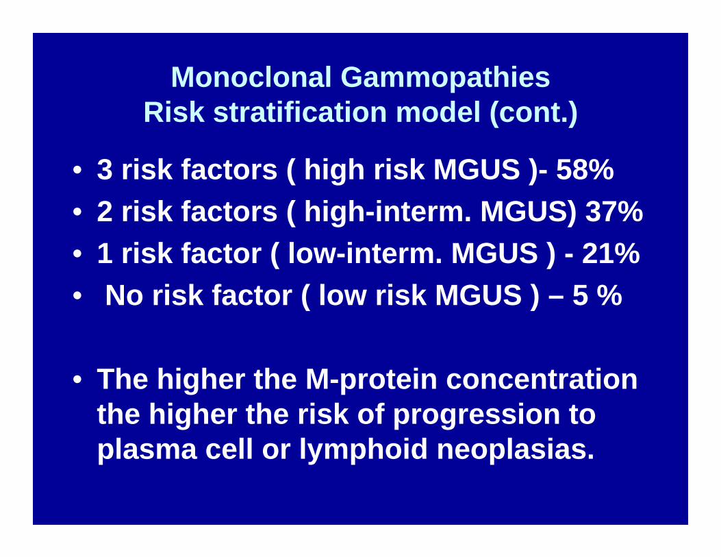
Monoclonal GammopathiesRisk stratification model (cont.)
• 3 risk factors ( high risk MGUS )- 58%• 2 risk factors ( high-interm. MGUS) 37%• 1 risk factor ( low-interm. MGUS ) - 21%• No risk factor ( low risk MGUS ) – 5 %
• The higher the M-protein concentration the higher the risk of progression to plasma cell or lymphoid neoplasias.
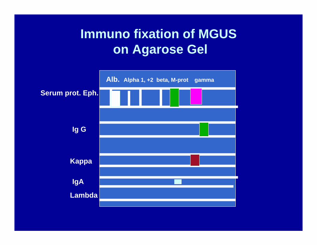
Immuno fixation of MGUSon Agarose Gel
Serum prot. Eph.
Alb. Alpha 1, +2 beta, M-prot gamma
Ig G
Kappa
IgA
Lambda
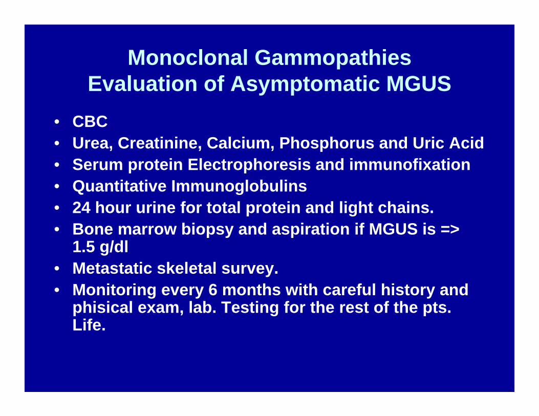
Monoclonal GammopathiesEvaluation of Asymptomatic MGUS
• CBC• Urea, Creatinine, Calcium, Phosphorus and Uric Acid• Serum protein Electrophoresis and immunofixation• Quantitative Immunoglobulins• 24 hour urine for total protein and light chains.• Bone marrow biopsy and aspiration if MGUS is =>
1.5 g/dl• Metastatic skeletal survey.• Monitoring every 6 months with careful history and
phisical exam, lab. Testing for the rest of the pts. Life.
Monoclonal GammopathiesSpeculation on their pathogenesis.
• 1. Chronic Antigenic stimulation as in HIV Hepatitis or other longstanding chronic infections and skin diseases.
• 2. Environmental Factors: Osserman had an inbred mouse highly susceptible to develop Plasmacytomas. If these mice were kept in a strictly sterile environment from birth, they did not develop the plasmacytoma.
Monoclonal GammopathiesSpeculation on their pathogenesis.2
• 3. Evolution from auto-antibodies in autoimmune diseases or from hyper-polyclonal gamma globulinemia to MGUS.
• 4. As age progresses immune surveillance diminishes.
• 1 in 1.0 X 10^6 cells is usually mutated, but gets eliminated by our cytotoxic lymphocytes and mono-macrophages.
• These defenses start failing with age and neoplasia is more frequent in elderly.
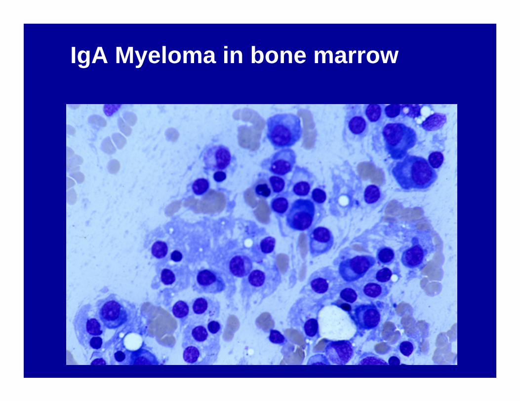
IgA Myeloma in bone marrow
Current Therapies for Myeloma
1. Induction therapy with Vincristine, Adriamycine and Dexamethasone,
2. Harvesting of peripheral blood stem cells (PBSC) through apheresis,
3. High dose chemotherapy with Melphalan +/-TBI only if necessary.
4. After relapse – Thalidomide and Prednisone, followed by
5. Bortezomib (Velcade) have been very successful.
Monoclonal GammopathiesSummary 1.
1. The prevalence of MG is high and increases with age after the 6-th decade.
2. Its potential and susceptibility to evolve at 1.0 to 1.5 % per year into a plasmacytoid or lymphoid neoplasia is high: 7.3 X from the base line in an unaffected population of the same age group.
Monoclonal Gammopathies (MG)Summary 2.
3. MG is often associated with underlying non-lympho-plasmacytoid disorders, which when treated successfully, lead also to the disappearance of the MG.
4. Diff. Dg. Of MGUS from Myeloma :• MG < 30 g/l• Plasma cells in bone marrow < 10 %• NO Anemia,• NO Hypogammaglobulinemia,• NO renal failure and urine light chain proteinuria.• NO hypercalcemia , NO lytic bone lesions.• NO CD 56 markers on plasma cells.
Monoclonal GammopathiesSummary 3.
Life long periodic monitoring of MG, as well as searching for an underlying or associated disorder is extremely important, since when they evolve into lymphomas or myelomas these diseases can be very successfully treated.