Embed Size (px)
Citation preview
The medicinal plant goldenseal is a natural LDL-lowering
agent with multiple bioactive components and new
action mechanisms
Parveen Abidi,1 Wei Chen,1 Fredric B. Kraemer, Hai Li, and Jingwen Liu2
Department of Veterans Affairs Palo Alto Health Care System, Palo Alto, CA 94304
Abstract Our previous studies have identified berberine(BBR), an alkaloid isolated from the Chinese herb huang-lian, as a unique cholesterol-lowering drug that upregulateshepatic low density lipoprotein receptor (LDLR) expressionthrough a mechanism of mRNA stabilization. Here, we dem-onstrate that the root extract of goldenseal, a BBR-containingmedicinal plant, is highly effective in upregulation of liverLDLR expression in HepG2 cells and in reducing plasmacholesterol and low density lipoprotein cholesterol (LDL-c)in hyperlipidemic hamsters, with greater activities than thepure compound BBR. By conducting bioassay-driven semi-purifications, we demonstrate that the higher potency ofgoldenseal is achieved through concerted actions of multi-ple bioactive compounds in addition to BBR. We identifycanadine (CND) and two other constituents of goldensealas new upregulators of LDLR expression. We further showthat the activity of BBR on LDLR expression is attenuatedby multiple drug resistance-1 (MDR1)-mediated efflux fromliver cells, whereas CND is resistant to MDR1. This find-ing defines a molecular mechanism for the higher activityof CND than BBR. We also provide substantial evidence toshow that goldenseal contains natural MDR1 antagonist(s)that accentuate the upregulatory effect of BBR on LDLRmRNA expression. These new findings identify golden-seal as a natural LDL-c-lowering agent, and our studies pro-vide a molecular basis for the mechanisms of action.—Abidi, P., W. Chen, F. B. Kraemer, H. Li, and J. Liu. Themedicinal plant goldenseal is a natural LDL-lowering agentwith multiple bioactive components and new action mecha-nisms. J. Lipid Res. 2006. 47: 2134–2147.
Supplementary key words low density lipoprotein cholesterol &canadine & berberine & mRNA stabilization & multiple drug resistance-1& extracellular signal-regulated kinase activation & hypercholesterolemia
Coronary heart disease is the major cause of morbidityand mortality in the Western population (1). Increasedplasma low density lipoprotein cholesterol (LDL-c) level ispostulated to be the primary risk factor for the develop-ment of coronary heart disease and atherosclerosis (2, 3).
In humans,.70% of LDL-c is removed from plasma by lowdensity lipoprotein receptor (LDLR)-mediated uptake inthe liver (4). Hence, the expression level of hepatic LDLRdirectly influences plasma cholesterol levels; therefore,regulation of liver LDLR represents a key mechanism bywhich therapeutic agents could intervene in the develop-ment of coronary heart disease and atherosclerosis.
Hepatic LDLR expression is predominantly regulatedat the transcriptional level through a negative feedbackmechanism by intracellular cholesterol pools. This regu-lation is controlled through specific interactions of thesterol-regulatory element of the LDLR promoter (5, 6)and a family of sterol-regulatory element binding proteins(7–9). The activation of LDLR transcription through thedepletion of intracellular cholesterol is the principal work-ing mechanism of the current cholesterol-lowering statindrugs (10). Statins are specific inhibitors of HMG-CoA re-ductase, the rate-limitingenzyme incellular cholesterol bio-synthesis. The depletion of the regulatory cholesterol poolin the liver results in an increased expression of LDLR andan enhanced uptake of LDL particles from the circulation.Since the development of lovastatin as the first HMGreductase inhibitor several decades ago, statin therapy hasbeen the primary treatment choice for hypercholesterol-emia (11–16), because of its high efficacy and improvedclinical outcomes. Nevertheless, there is still a need todevelop additional cholesterol-lowering agents to treathyperlipidemia (1, 17).
Our interest in the discovery of new LDLR modulatorsfrom natural resources has led to the identification ofberberine (BBR), an alkaloid isolated from the Chineseherb huanglian, as a novel upregulator of hepatic LDLR
Manuscript received 4 May 2006 and in revised form 16 June 2006 and inre-revised form 19 July 2006 and in re-re-revised form 2 August 2006.
Published, JLR Papers in Press, August 2, 2006.DOI 10.1194/jlr.M600195-JLR200
Abbreviations: BBR, berberine; CND, canadine; ELSD, evaporativelight-scattering detection; ERK, extracellular signal-regulated kinase;HC, high-cholesterol; HDT, hydrastine; HDTN, hydrastinine; LDL-c,low density lipoprotein cholesterol; LDLR, low density lipoprotein re-ceptor; MDR, multiple drug resistance; OM, oncostatin M; PMT, pal-matine; siRNA, small interfering RNA; TC, total cholesterol; TG,triglyceride; VRPM, verapamil.
1 P. Abidi and W. Chen contributed equally to this work.2 To whom correspondence should be addressed.e-mail: [email protected]
This article is available online at http://www.jlr.org2134 Journal of Lipid Research Volume 47, 2006
by guest, on Novem
ber 15, 2018w
ww
.jlr.orgD
ownloaded from
(18, 19). By conducting studies in human hepatoma-derived cell lines, we showed that BBR strongly increasesLDLR mRNA and protein expression. Our studies furtherrevealed that BBR upregulates LDLR expression by extend-ing the half-life of LDLR mRNA without affecting genetranscription. Thus, BBR uses a mechanism of action dif-ferent from statins. A placebo-controlled clinical study con-ducted in China showed that oral administration of BBR in32 hypercholesterolemic patients at a daily dose of 1 g for3 months reduced plasma total cholesterol (TC) by 29%,triglyceride (TG) by 35%, and LDL-c by 25% without sideeffects (18). These in vitro and clinical studies suggested apossible use of BBR in the treatment of hyperlipidemia.
BBR is not only present in the Chinese herb huanglian(Coptis chinesis) but is also an indigenous component ofother members of the plant family Ranunculaceae, suchas goldenseal (Hydrastis canadensis L.) (20). Goldenseal isnative to the eastern region of North America, and itsproducts are extracts from the dried root of the plant.Goldenseal is among the top five herbal products currentlyon the U.S. market and has been used to treat a variety ofillnesses, such as digestive disorders, urinary tract infec-tion, and upper respiratory inflammation (21). However,the effects of goldenseal in regulating plasma lipid andcholesterol levels have never been studied. In this investi-gation, we examined the effects of goldenseal in regulat-ing hepatic LDLR expression and LDL-c metabolism inthe human hepatoma-derived cell line HepG2 and inhypercholesterolemic hamsters.
MATERIALS AND METHODS
Analysis and quantitation of alkaloid componentsin goldenseal
BBR chloride, (2)-canadine (CND), b-hydrastine (HDT),hydrastinine (HDTN), and palmatine (PMT) were purchasedfrom Sigma, and stock solutions of 10 mg/ml in DMSO wereprepared and used as standards in HPLC, LC-MS, and evapo-rative light-scattering detection (ELSD). Goldenseal root extractLot 1 was manufactured by Solgar Vitamin and Herb (Leonia,NJ), Lots 2, 8, and 9 were manufactured by Country Sun (PaloAlto, CA), Lot 3 was manufactured by Now Foods (Bloomingdale,IL), Lot 4 was manufactured by The Vitamin Shoppe (NorthBergen, NJ), Lots 5 and 6 were manufactured by Nature’s WayProducts, Inc. (Springville, UT), and Lot 7 was manufactured byGala Herbs (Brevard, NC). Lots 1–6 were in powder form andwere extracted with ethanol. Lots 7–9 were in 60% grain alcohol.The ethanol extract of goldenseal was diluted in methanol andsubjected to HPLC, ELSD, and LC-MS to determine the alkaloidcontents. Chemical analyses were performed by Combinix, Inc.(Mountain View, CA).
Quantitation of LDLRmRNA expression by Northern blotanalysis and real-time PCR
Isolation of total RNA from HepG2 and from hamster liversand analysis of LDLR and GAPDH mRNA by Northern blot wereperformed as described previously (18, 22). Differences in hy-bridization signals of Northern blots were quantitated with aPhosphorImager. For comparative real-time PCR assays, the re-verse transcription was conducted with random primers using
Moloney murine leukemia virus (Promega) at 378C for 1 h in avolume of 25 ml containing 1 mg of total RNA. Real-time PCR wasperformed on the cDNA using the ABI Prism 7900-HT SequenceDetection System and Universal MasterMix. Human and hamsterLDLR and GAPDH PreDeveloped TaqMan Assay Reagents(Applied Biosystems) were used to assess mRNA expression inHepG2 cells and hamster livers. Multiple drug resistance-1(MDR1) mRNA expression in HepG2 cells was assayed similarlyusing the PreDeveloped probes from Applied Biosystems.
LDL uptake assay
HepG2 cells on six-well culture plates were treated with com-pounds for 18 h. Fluorescent DiI-LDL (Biomedical Technolo-gies, Stoughton, MA) at a concentration of 6 mg/ml was added tothe cells at the end of treatment for 4 h, and cells were tryp-sinized. The mean red fluorescence of 23 104 cells was measuredusing FACScan (filter 610/20 DF, BD LSRII; Becton Dickinson).
Transient transfection and dual luciferase reporter assays
HepG2 cells were transfected with plasmid DNA (100 ng/well)using FuGENE 6 transfection reagent. The DNA ratio ofpLDLR234Luc (22) to renilla luciferase reporter pRL-SV40 was90:10. Twenty hours after transfection, medium was changed to0.5% FBS and drugs were added for 8 h followed by cell lysis. Theluciferase activity in cell lysate was measured using the DualLuciferase Assay System from Promega. Triplicate wells were as-sayed for each transfection condition.
Semipurification of goldenseal alkaloid components
One milliliter of goldenseal liquid extract was subjected toflash chromatography over a silica gel column with a chloroform-methanol 90–50% gradient as an eluting solvent. Twenty-six15 ml fractions were collected. A total of 200 ml of each fractionwas used directly to measure the fluorescence intensity with afluorescent microplate reader (Spectra MaxGemini; MolecularDevices, Sunnyvale, CA) at 350 nm excitation and 545 nm emis-sion (23). The rest of the fraction was evaporated under N2, andresidues in each fraction were dissolved in 250 ml of DMSO. Tenmicroliters from each fraction was diluted with 90 ml of ethanoland applied to HPLC, ELSD, and LC-MS.
BBR uptake assay
HepG2 cells were seeded on six-well culture plates at a densityof 0.83 106 cells/well in medium containing 10% FBS. The nextday, cells were incubated with medium containing 0.5% FBS.BBR at a concentration of 15 mg/ml or goldenseal with an equiva-lent amount of BBR was added to the cells for the indicatedtimes. At the end of treatment, cells were washed with cold PBSand trypsinized. Cell suspensions in PBS were placed on ice tominimize efflux activity. The mean green fluorescence of 23 104
cells was measured using FACScan (filter 525/50HQ, BD LSRII;Becton Dickinson).
MDR direct dye efflux assay
The MDR Direct Dye Efflux Assay kit (No. ECM910; ChemiconInternational, Inc., Temecula, CA) was used tomeasureMDR1 ac-tivity. HepG2 cells seeded on six-well culture plates were incu-bated in efflux buffer (RPMI 1 2% BSA) and 0.2 mg/ml DiOC2
in the absence or presence of the tested compounds at 378C for2 h. Cells were washed with cold PBS and trypsinized. Cell sus-pensions in PBS were placed on ice to minimize efflux activity.Themean green fluorescence of 23 104 cells was measured using
Regulation of liver LDL receptor expression by goldenseal 2135
by guest, on Novem
ber 15, 2018w
ww
.jlr.orgD
ownloaded from

FACScan (filter 530/30DF, BD LSRII; Becton Dickinson). Theweak green fluorescence of goldenseal constituted ,1% of thefluorescent signal of DiOC2 and was ignored.
Small interfering RNA transfection
Predesigned small interfering RNAs (siRNAs) targeted to hu-man MDR1 (No. 51320) and a negative control with a scrambledsequence (No. 4618G) were obtained from Ambion. HepG2 cellswere seeded on six-well culture plates and were transfected withsiRNA using the SilencerTM siRNA Transfection II Kit (Ambion)according to the given instructions. After 3 days, transfected cellswere untreated or treated with BBR, CND, or goldenseal for 6 hbefore RNA isolation.
Goldenseal in vivo studies
Thirty-three male Golden Syrian hamsters at 6–8 weeks ofage were purchased from Charles River Laboratories and werehoused in cages (three animals per cage) in an air-conditionedroom with a 12 h light cycle. Animals had free access to auto-claved water and food. After 1 week on a regular rodent chowdiet, 27 hamsters were switched to a rodent high-cholesterol(HC) diet containing 1.25% cholesterol (product D12108, Re-search Diet, Inc., New Brunswick, NJ) and 6 hamsters were fed acontrol normal diet containing 0.37% fat and no cholesterol(product D12102; Research Diet, Inc.). After 21 days, hamsterson the HC diet were randomly divided into three groups (n 5 9per group) and were given goldenseal at 125 ml/day or BBR at1.8 mg/day intraperitoneally once per day at 9 AM. The controlgroup received an equal volume of vehicle (20% hydroxypropyl-b-cyclodextrin). Goldenseal grain alcohol extract Lot 9 was driedunder a nitrogen stream and resuspended in 23 volume of 20%hydroxypropyl-b-cyclodextrin to a final BBR concentration of3.6 mg/ml. BBR chloride was dissolved in the same vehicle solu-tion. Four hours after the last drug treatment, all animals wereeuthanized. At the time of dissection, body weight, liver weight,and gross morphology of the liver were recorded. Livers wereimmediately removed, cut into small pieces, and stored at 2808Cfor RNA isolation, protein isolation, and cholesterol contentmeasurement. Animal use and experimental procedures wereapproved by the Institutional Animal Care and Use Committee ofthe Department of Veterans Affairs Palo Alto Health Care System.
Histopathology assessment
For histological examination, pieces of liver tissues were eitherimmersed in OCT solution under liquid N2 and stored at 2808Cfor Oil Red O staining or fixed in 10% paraformaldehyde at roomtemperature for hematoxylin and eosin staining. Tissue sectionswere processed and stained at Stanford University’s HistologyResearch Core Laboratory using routine laboratory procedures.After staining, tissue sections were evaluated by a veterinary pa-thologist and an experienced scientist independently.
Serum isolation and cholesterol determination
Blood samples (0.2 ml) were collected from the retro-orbitalplexus using heparinized capillary tubes under anesthesia (2–3%isoflurane and 1–2 l/min oxygen) after an 8 h fast (7 AM to 3 PM)before and during the drug treatments. Serum was isolatedat room temperature and stored at 2808C. Standard enzymaticmethods were used to determine TC, TG, LDL-c, HDL-c, and FFAlevels with commercially available kits purchased from StanbioLaboratory and Wako Chemical GmbH (Neuss, Germany). Eachsample was assayed in duplicate.
Measurement of hepatic cholesterol
One hundred milligrams of frozen liver tissue was thawed andhomogenized in 2 ml of chloroform-methanol (2:1). After ho-mogenization, lipids were further extracted by rocking samplesfor 1 h at room temperature, followed by centrifugation at 5,000 gfor 10 min. One milliliter of lipid extract was dried under a ni-trogen stream and redissolved in 1 ml of ethanol. TC and TGwere measured using commercially available kits.
HPLC analysis of lipoprotein profiles
Twenty microliters of each serum sample from hamsters on anormal diet (n 5 6), an HC diet (n 5 9), and an HC diet treatedwith goldenseal (n 5 9) was pooled. Cholesterol and TG levelsof each of the major lipoprotein classes, including chylomicron,VLDL, LDL, and HDL, in the pool sera were analyzed by HPLC(24) at Skylight Biotech, Inc. (Tokyo, Japan).
Western blot analysis of phosphorylated extracellularsignal-regulated kinase in liver tissues and in HepG2 cells
Approximately 90–100 mg of hamster liver tissue from eachanimal was pooled from the same treatment group (n 5 9) andhomogenized in 5 ml of buffer containing 20 mM Tris-HCl, pH8.0, 0.1 M NaCl, 1 mM CaCl2, and cocktails of phosphatase in-hibitors (Sigma) and protease inhibitors (Complete Mini; RocheDiagnostic). Total homogenate was centrifuged at 800 g for 5 minto pellet nuclei, and the supernatant was filtered through muslincloth. The filtrate was subjected to 100,000 g centrifugation for1 h at 48C to obtain the cytosolic fraction. After protein quan-titation using the BCATM protein assay reagent (Pierce), 50 mg ofprotein from each pooled sample was subjected to SDS-PAGE,followed by Western blotting using anti-phosphorylated extra-cellular signal-regulated kinase (ERK; Cell Signaling) antibodyand antibody against total ERK (Santa Cruz Biotechnology). Toanalyze ERK activation in HepG2 cells, cells seeded on six-wellculture plates in 0.5% FBS Eagle’s Minimum Essential Medium(EMEM) were treated with 10 mg/ml of each alkaloid as well asgoldenseal (1.5 ml/ml; Lot 8) for 2 h, and cell lysates were col-lected as described previously (18).
Statistical analysis
Significant differences between control and treatment groupsor between BBR- and CND-treated samples were assessed by Stu-dent’s t-test. P , 0.05 was considered statistically significant.
RESULTS
Goldenseal strongly upregulates LDLR expression inHepG2 cells
Goldenseal contains three major isoquinoline alkaloids,BBR, CND, and HDT, as well as some minor alkaloid com-ponents such as HDTN (20, 21, 25, 26) (Fig. 1A). AlthoughPMT exists in Coptis, Oregon grape root, and several otherBBR-containing plants (20), CND and HDT are the onlynative components of goldenseal (26–28). Goldenseal rootextract typically contains 2.5–6% total alkaloids (27).
To determine the activity of goldenseal in the regulationof LDLR expression, we first performed HPLC analysison goldenseal ethanol extracts obtained from six differentcommercial suppliers. HPLC/ultraviolet photodiode arraydetector (UV-DAD) spectroscopic comparisons with stan-dard solutions were used to confirm the presence of BBR,
2136 Journal of Lipid Research Volume 47, 2006
by guest, on Novem
ber 15, 2018w
ww
.jlr.orgD
ownloaded from
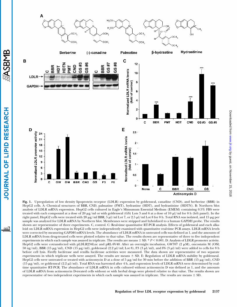
Fig. 1. Upregulation of low density lipoprotein receptor (LDLR) expression by goldenseal, canadine (CND), and berberine (BBR) inHepG2 cells. A: Chemical structures of BBR, CND, palmatine (PMT), hydrastine (HDT), and hydrastinine (HDTN). B: Northern blotanalysis of LDLR mRNA expression. HepG2 cells cultured in Eagle’s Mimumum Essential Medium (EMEM) containing 0.5% FBS weretreated with each compound at a dose of 20 mg/ml or with goldenseal (GS) Lots 3 and 6 at a dose of 10 ml/ml for 8 h (left panel). In theright panel, HepG2 cells were treated with 20 mg/ml BBR, 5 ml/ml Lot 7, or 2.5 ml/ml Lot 8 for 8 h. Total RNA was isolated, and 15 mg persample was analyzed for LDLR mRNA by Northern blot. Membranes were stripped and hybridized to a human GAPDH probe. The resultsshown are representative of three experiments. C, control. C: Real-time quantitative RT-PCR analysis. Effects of goldenseal and each alka-loid on LDLR mRNA expression in HepG2 cells were independently examined with quantitative real-time PCR assays. LDLR mRNA levelswere corrected by measuring GAPDHmRNA levels. The abundance of LDLRmRNA in untreated cells was defined as 1, and the amounts ofLDLR mRNA from drug-treated cells were plotted relative to that value. The results shown are representative of three to five independentexperiments in which each sample was assayed in triplicate. The results are means6 SD. * P, 0.001. D: Analysis of LDLR promoter activity.HepG2 cells were cotransfected with pLDLR234Luc and pRL-SV40. After an overnight incubation, GW707 (2 mM), oncostatin M (OM;50 ng/ml), BBR (15 mg/ml), CND (15 mg/ml), goldenseal (2.2 ml/ml; Lot 8), F3 (3 ml/ml), and F6 (3 ml/ml) were added to cells for 8 hbefore cell lysis. Firefly luciferase and renilla luciferase activities were measured. The data shown are representative of two separateexperiments in which triplicate wells were assayed. The results are means 6 SD. E: Regulation of LDLR mRNA stability by goldenseal.HepG2 cells were untreated or treated with actinomycin D at a dose of 5 mg/ml for 30 min before the addition of BBR (15 mg/ml), CND(15 mg/ml), or goldenseal (2.2 ml/ml). Total RNA was harvested after 4 h, and expression levels of LDLR mRNA were determined by real-time quantitative RT-PCR. The abundance of LDLR mRNA in cells cultured without actinomycin D was defined as 1, and the amountsof LDLR mRNA from actinomycin D-treated cells without or with herbal drugs were plotted relative to that value. The results shown arerepresentative of two independent experiments in which each sample was assayed in triplicate. The results are means 6 SD.
Regulation of liver LDL receptor expression by goldenseal 2137
by guest, on Novem
ber 15, 2018w
ww
.jlr.orgD
ownloaded from
CND, HDT, and HDTN as well as the absence of PMT.Concentrations of CND and HDT in sample extracts weredetermined using single-point calibration, and concentra-tions of BBR in sample extracts were calculated using astandard curve. We further verified the identities of BBR,CND, and HDT in extracts by LC-MS analysis. Table 1 liststhe concentrations of alkaloids in different lots of golden-seal extracts. After these comprehensive quantitative analy-ses, HepG2 cells were treated for 8 h with goldensealextract Lots 3 and 6 at a concentration of 10ml/ml (equiva-lent to a BBR concentration ofz15 mg/ml) and with eachalkaloid at a concentration of 20 mg/ml. Northern blotanalysis showed that HDT, HDTN, and PMT have no ef-fects, but CND and BBR are both strong inducers of LDLRmRNA expression (Fig. 1B, left panel). Interestingly, gold-enseal extract Lots 3 and 6 with lower BBR concentrationsproduced the greatest increase of LDLRmRNA levels. TheNorthern blot results were independently confirmed byreal-time quantitative RT-PCR (Fig. 1C). A 9.8-fold in-crease in the level of LDLR mRNA was achieved by gold-enseal extract Lot 3, which contains 15 mg/ml BBR and0.9 mg/ml CND, whereas the pure compound BBR at aconcentration of 20 mg/ml only produced a 3-fold in-crease in LDLR mRNA expression. Strong activities ofgoldenseal Lots 7 and 8 on LDLR expression were demon-strated by Northern blot (Fig. 1B, right panel). Similarexperiments were repeated three to four times using gold-enseal extracts from all six different suppliers. In allassays, goldenseal extracts outperformed the pure com-pound BBR in the upregulation of LDLR mRNA expres-sion. At comparable concentrations of BBR, the activity ofgoldenseal extract is typically two to three times higherthan that of pure BBR. To confirm the higher potency ofgoldenseal on LDLR expression, we measured the DiI-LDL uptake of HepG2 cells untreated or treated for 15 hwith BBR (10 mg/ml) or goldenseal containing an equiva-lent amount of BBR (1.5 ml/ml of Lot 8). The LDLR-mediated ligand uptake in HepG2 cells was increased2.5-fold by BBR and 4.9-fold by goldenseal compared withuntreated cells.
Our previous studies demonstrated that BBR doesnot activate LDLR gene transcription but has a stabilizingeffect on LDLR mRNA (18, 19). To determine whethermRNA half-life prolongation is the primary mechanismthrough which goldenseal increases LDLR expression,HepG2 cells were transfected with the LDLR promoter
luciferase construct pLDLR234Luc along with the nor-malizing reporter pRL-SV40Luc. After transfection, cellswere treated for 8 h with BBR or CND at a concentrationof 15 mg/ml or with 2.2 ml/ml goldenseal Lot 8 alongwith two known activators of the LDLR promoter, cytokineoncostatin M (OM; 50 ng/ml) and the compound GW707(2 mM). OM activates LDLR transcription through a sterol-independent regulatory element of the LDLR promoter(29), and GW707 is a sterol-like compound that increasesLDLR transcription through sterol-regulatory element-1(30, 31). Figure 1D shows that LDLR promoter activity wasstrongly increased by GW707 and OM but was not affectedat all by goldenseal, CND, or BBR. To further corroboratethis finding, HepG2 cells were untreated or treated withactinomycin D for 30 min before the addition of BBR,CND, or goldenseal, and total RNA was isolated after a 4 htreatment. Real-time quantitative RT-PCR showed thatinhibition of transcription by actinomycin D reduced theabundance of LDLR mRNA but did not prevent the up-regulatory effects of these agents on LDLR mRNA ex-pression. Under the same condition of transcriptionalsuppression, LDLR mRNA was increased 2.5-fold by BBRor CND and 3.4-fold by goldenseal compared with controls(Fig. 1E). Collectively, these results illustrate that golden-seal extract is highly effective in the upregulation of LDLRexpression through mRNA stabilization, with a greater ac-tivity than the pure compound BBR.
Goldenseal increases LDLR expression through theconcerted action of multiple bioactive components inaddition to BBR
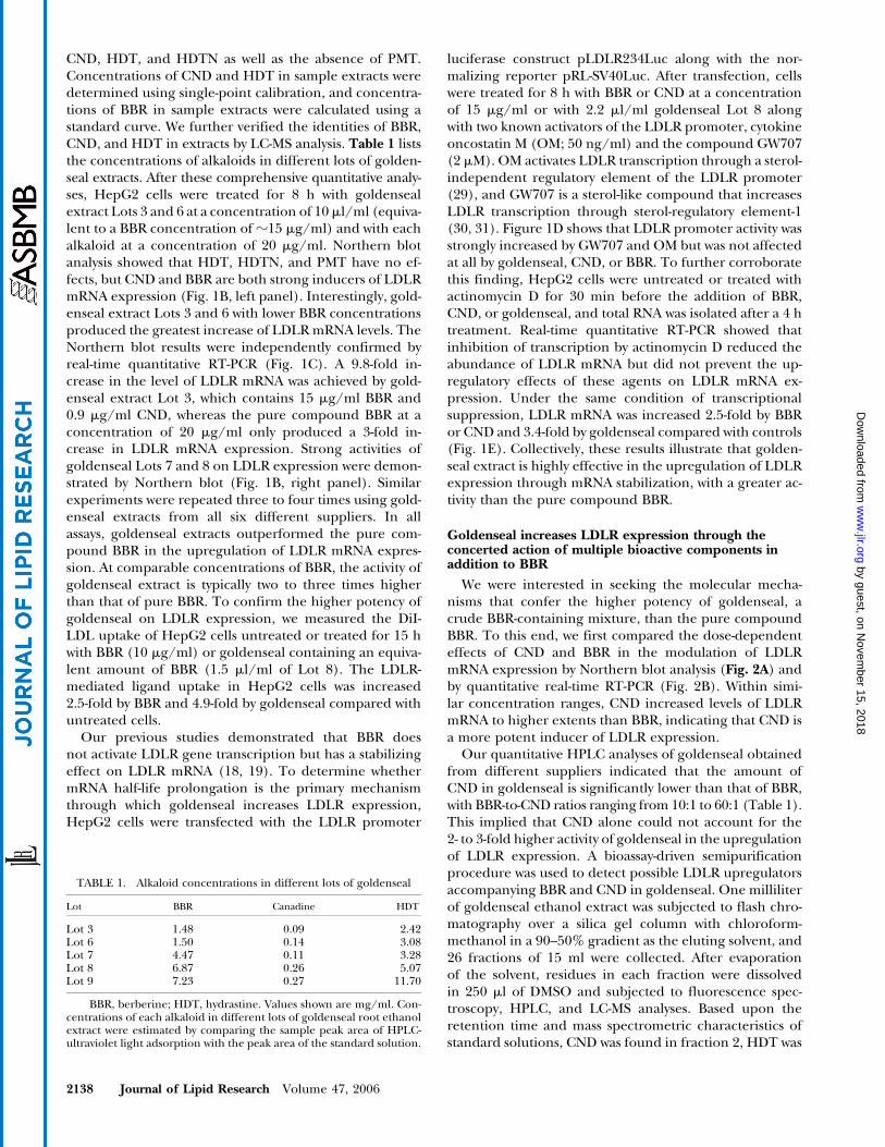
We were interested in seeking the molecular mecha-nisms that confer the higher potency of goldenseal, acrude BBR-containing mixture, than the pure compoundBBR. To this end, we first compared the dose-dependenteffects of CND and BBR in the modulation of LDLRmRNA expression by Northern blot analysis (Fig. 2A) andby quantitative real-time RT-PCR (Fig. 2B). Within simi-lar concentration ranges, CND increased levels of LDLRmRNA to higher extents than BBR, indicating that CND isa more potent inducer of LDLR expression.
Our quantitative HPLC analyses of goldenseal obtainedfrom different suppliers indicated that the amount ofCND in goldenseal is significantly lower than that of BBR,with BBR-to-CND ratios ranging from 10:1 to 60:1 (Table 1).This implied that CND alone could not account for the2- to 3-fold higher activity of goldenseal in the upregulationof LDLR expression. A bioassay-driven semipurificationprocedure was used to detect possible LDLR upregulatorsaccompanying BBR and CND in goldenseal. One milliliterof goldenseal ethanol extract was subjected to flash chro-matography over a silica gel column with chloroform-methanol in a 90–50% gradient as the eluting solvent, and26 fractions of 15 ml were collected. After evaporationof the solvent, residues in each fraction were dissolvedin 250 ml of DMSO and subjected to fluorescence spec-troscopy, HPLC, and LC-MS analyses. Based upon theretention time and mass spectrometric characteristics ofstandard solutions, CND was found in fraction 2, HDT was
TABLE 1. Alkaloid concentrations in different lots of goldenseal
Lot BBR Canadine HDT
Lot 3 1.48 0.09 2.42Lot 6 1.50 0.14 3.08Lot 7 4.47 0.11 3.28Lot 8 6.87 0.26 5.07Lot 9 7.23 0.27 11.70
BBR, berberine; HDT, hydrastine. Values shown are mg/ml. Con-centrations of each alkaloid in different lots of goldenseal root ethanolextract were estimated by comparing the sample peak area of HPLC-ultraviolet light adsorption with the peak area of the standard solution.
2138 Journal of Lipid Research Volume 47, 2006
by guest, on Novem
ber 15, 2018w
ww
.jlr.orgD
ownloaded from
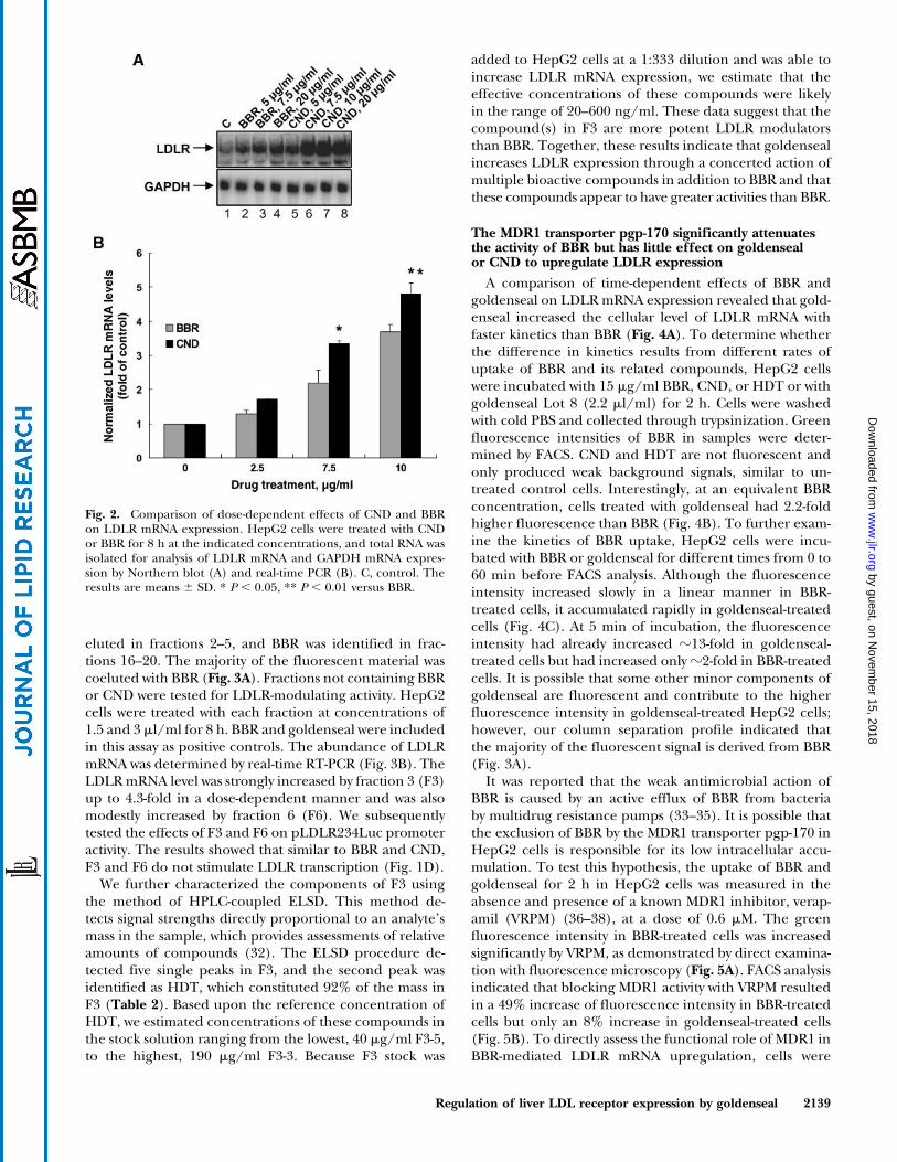
eluted in fractions 2–5, and BBR was identified in frac-tions 16–20. The majority of the fluorescent material wascoeluted with BBR (Fig. 3A). Fractions not containing BBRor CND were tested for LDLR-modulating activity. HepG2cells were treated with each fraction at concentrations of1.5 and 3 ml/ml for 8 h. BBR and goldenseal were includedin this assay as positive controls. The abundance of LDLRmRNA was determined by real-time RT-PCR (Fig. 3B). TheLDLRmRNA level was strongly increased by fraction 3 (F3)up to 4.3-fold in a dose-dependent manner and was alsomodestly increased by fraction 6 (F6). We subsequentlytested the effects of F3 and F6 on pLDLR234Luc promoteractivity. The results showed that similar to BBR and CND,F3 and F6 do not stimulate LDLR transcription (Fig. 1D).
We further characterized the components of F3 usingthe method of HPLC-coupled ELSD. This method de-tects signal strengths directly proportional to an analyte’smass in the sample, which provides assessments of relativeamounts of compounds (32). The ELSD procedure de-tected five single peaks in F3, and the second peak wasidentified as HDT, which constituted 92% of the mass inF3 (Table 2). Based upon the reference concentration ofHDT, we estimated concentrations of these compounds inthe stock solution ranging from the lowest, 40 mg/ml F3-5,to the highest, 190 mg/ml F3-3. Because F3 stock was
added to HepG2 cells at a 1:333 dilution and was able toincrease LDLR mRNA expression, we estimate that theeffective concentrations of these compounds were likelyin the range of 20–600 ng/ml. These data suggest that thecompound(s) in F3 are more potent LDLR modulatorsthan BBR. Together, these results indicate that goldensealincreases LDLR expression through a concerted action ofmultiple bioactive compounds in addition to BBR and thatthese compounds appear to have greater activities than BBR.
The MDR1 transporter pgp-170 significantly attenuatesthe activity of BBR but has little effect on goldensealor CND to upregulate LDLR expression
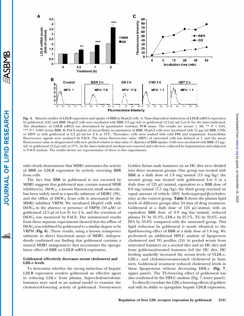
A comparison of time-dependent effects of BBR andgoldenseal on LDLRmRNA expression revealed that gold-enseal increased the cellular level of LDLR mRNA withfaster kinetics than BBR (Fig. 4A). To determine whetherthe difference in kinetics results from different rates ofuptake of BBR and its related compounds, HepG2 cellswere incubated with 15 mg/ml BBR, CND, or HDT or withgoldenseal Lot 8 (2.2 ml/ml) for 2 h. Cells were washedwith cold PBS and collected through trypsinization. Greenfluorescence intensities of BBR in samples were deter-mined by FACS. CND and HDT are not fluorescent andonly produced weak background signals, similar to un-treated control cells. Interestingly, at an equivalent BBRconcentration, cells treated with goldenseal had 2.2-foldhigher fluorescence than BBR (Fig. 4B). To further exam-ine the kinetics of BBR uptake, HepG2 cells were incu-bated with BBR or goldenseal for different times from 0 to60 min before FACS analysis. Although the fluorescenceintensity increased slowly in a linear manner in BBR-treated cells, it accumulated rapidly in goldenseal-treatedcells (Fig. 4C). At 5 min of incubation, the fluorescenceintensity had already increased z13-fold in goldenseal-treated cells but had increased onlyz2-fold in BBR-treatedcells. It is possible that some other minor components ofgoldenseal are fluorescent and contribute to the higherfluorescence intensity in goldenseal-treated HepG2 cells;however, our column separation profile indicated thatthe majority of the fluorescent signal is derived from BBR(Fig. 3A).
It was reported that the weak antimicrobial action ofBBR is caused by an active efflux of BBR from bacteriaby multidrug resistance pumps (33–35). It is possible thatthe exclusion of BBR by the MDR1 transporter pgp-170 inHepG2 cells is responsible for its low intracellular accu-mulation. To test this hypothesis, the uptake of BBR andgoldenseal for 2 h in HepG2 cells was measured in theabsence and presence of a known MDR1 inhibitor, verap-amil (VRPM) (36–38), at a dose of 0.6 mM. The greenfluorescence intensity in BBR-treated cells was increasedsignificantly by VRPM, as demonstrated by direct examina-tion with fluorescence microscopy (Fig. 5A). FACS analysisindicated that blocking MDR1 activity with VRPM resultedin a 49% increase of fluorescence intensity in BBR-treatedcells but only an 8% increase in goldenseal-treated cells(Fig. 5B). To directly assess the functional role of MDR1 inBBR-mediated LDLR mRNA upregulation, cells were
Fig. 2. Comparison of dose-dependent effects of CND and BBRon LDLR mRNA expression. HepG2 cells were treated with CNDor BBR for 8 h at the indicated concentrations, and total RNA wasisolated for analysis of LDLR mRNA and GAPDH mRNA expres-sion by Northern blot (A) and real-time PCR (B). C, control. Theresults are means 6 SD. * P , 0.05, ** P , 0.01 versus BBR.
Regulation of liver LDL receptor expression by goldenseal 2139
by guest, on Novem
ber 15, 2018w
ww
.jlr.orgD
ownloaded from
treated with BBR, CND, or goldenseal in the absence orpresence of VRPM and levels of LDLR mRNA were deter-mined. The results showed that VRPM did not increase theactivity of goldenseal or CND but enhanced the activityof BBR on LDLR mRNA expression in a dose-dependentmanner (Fig. 5C, D). The fact that the activity of CND was
not affected at all by VRPM suggests that CND is not asubstrate of MDR1.
To further examine the inhibitory role of pgp-170 onBBR activity, HepG2 cells were transfected with siRNA ofMDR1 or a control siRNA for 3 days. Western blot analysisof MDR1 abundance showed a significant reduction ofMDR1 protein level by the transfection of MDR1 siRNA(Fig. 5F, inset). Thus, the siRNA-transfected cells weretreated with BBR for 2 h to measure BBR uptake or for 6 hfor RNA isolation. FACS analysis showed that the cellularretention of BBR in MDR1 siRNA-transfected cells was in-creased by 47% (P , 0.01) compared with that in mock-transfected cells (Fig. 5E). Quantitative RT-PCR showedthat the mRNA level of MDR1 was decreased by 69% incontrol cells and by 71% in BBR-treated cells comparedwith nonspecific siRNA-transfected cells (mock). Reduc-tion of MDR1 expression by siRNA did not affect LDLRmRNA level in control cells; however, it caused a 45% in-crease (P , 0.001) in the activity of BBR to increase LDLRmRNA level (Fig. 5F). As expected, the activity of CND orgoldenseal on LDLR expression was not affected by MDR1siRNA transfection (data not shown). Together, these re-
TABLE 2. Analysis of components of F3 by HPLC coupled withevaporative light-scattering detection
CompoundRetention
Time Peak AreaPercentage
Area
HDT (10 mg/ml) 1.7 1,621,945 100.0F3-1 (unknown, 0.15 mg/ml) 1.58 24,813 2.5F3-2 (HDT, 5.7 mg/ml) 1.78 926,022 91.5F3-3 (unknown, 0.19 mg/ml) 11.09 32,185 3.2F3-4 (unknown, 0.12 mg/ml) 12.78 20,780 2.1F3-5 (unknown, 0.04 mg/ml) 13.62 7,805 0.8
HPLC-coupled evaporative light-scattering detection was used toanalyze the components of F3. The standard solution of HDT was usedas a reference. The amount of mass in each peak was estimated bycomparing the peak area of each peak with the peak area of HDT.
Fig. 3. Separation of goldenseal extract by silica gel column chromatography and detection of LDLRmodulation activity in column eluates. A: One milliliter of goldenseal extract was separated into 26 fractionsby silica gel column chromatography using chloroform-methanol as the elution solvent. The fluorescenceintensity of 200 ml from each fraction was measured by a fluorescent microplate reader at 350 nm excitationand 545 nm emission. The presence of CND, HDT, or BBR in eluates was determined by HPLC and LC-MSwith standard solutions of each compound as the reference. B: HepG2 cells were treated for 8 h with 1.5 or3 ml of each fraction after evaporation of the solvent and redissolving in DMSO. BBR (15 mg/ml) andgoldenseal (GS; 2.2 ml/ml; Lot 8) were used in these experiments as positive controls. The inducing effectsof F3 and F6 on LDLR mRNA expression were consistently observed in four separate experiments. C,control. The results are means 6 SD. *** P , 0.001 versus control.
2140 Journal of Lipid Research Volume 47, 2006
by guest, on Novem
ber 15, 2018w
ww
.jlr.orgD
ownloaded from
sults clearly demonstrate that MDR1 attenuates the activityof BBR on LDLR expression by actively excreting BBRfrom cells.
The fact that BBR in goldenseal is not excreted byMDR1 suggests that goldenseal may contain natural MDRinhibitor(s). DiOC2, a known fluorescent small molecule,has been widely used as a specific substrate of MDR1 (39),and the efflux of DiOC2 from cells is attenuated by theMDR1 inhibitor VRPM. We incubated HepG2 cells withDiOC2 in the absence or presence of VRPM (50 mM) orgoldenseal (2.5 ml of Lot 8) for 2 h, and the retention ofDiOC2 was measured by FACS. The summarized resultsfrom three separate experiments showed that the efflux ofDiOC2 was inhibited by goldenseal to a similar degree as byVRPM (Fig. 6). These results, using a known transportersubstrate in direct functional assays of MDR1, indepen-dently confirmed our finding that goldenseal contains anatural MDR1 antagonist(s) that accentuates the upregu-latory effect of BBR on LDLR mRNA expression.
Goldenseal effectively decreases serum cholesterol andLDL-c levels
To determine whether the strong induction of hepaticLDLR expression renders goldenseal an effective agentin reducing LDL-c from plasma, hypercholesterolemichamsters were used as an animal model to examine thecholesterol-lowering activity of goldenseal. Twenty-seven
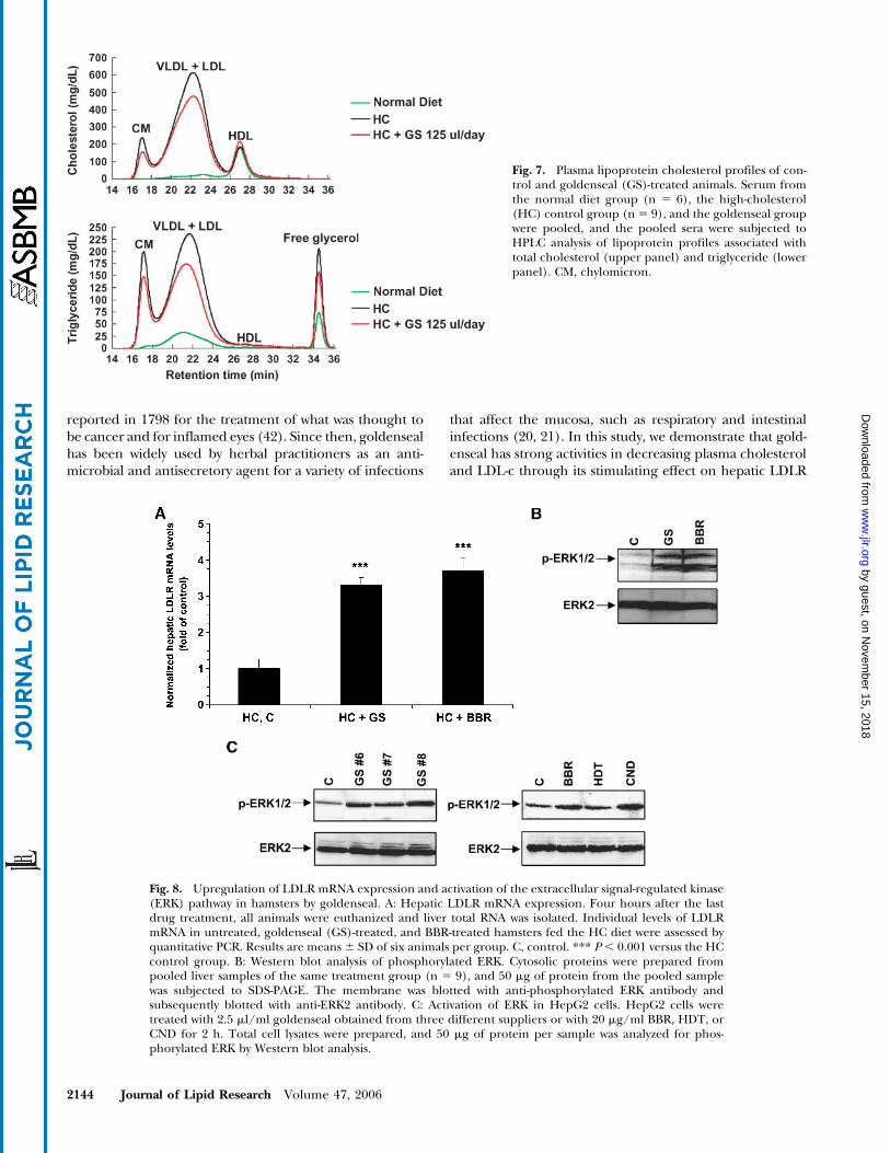
Golden Syrian male hamsters on an HC diet were dividedinto three treatment groups. One group was treated withBBR at a daily dose of 1.8 mg/animal (15 mg/kg); thesecond group was treated with goldenseal Lot 9 at adaily dose of 125 ml/animal, equivalent to a BBR dose of0.9 mg/animal (7.5 mg/kg); the third group received anequal amount of vehicle (20% hydroxypropyl-b-cyclodex-trin) as the control group. Table 3 shows the plasma lipidlevels of different groups after 24 days of drug treatment.Goldenseal at a daily dose of 125 ml/animal, with anequivalent BBR dose of 0.9 mg/day/animal, reducedplasma TC by 31.3%, LDL-c by 25.1%, TG by 32.6%, andFFA by 33.8% compared with the untreated group. Thislipid reduction by goldenseal is nearly identical to thelipid-lowering effect of BBR at a daily dose of 1.8 mg. Weperformed an additional HPLC analysis of lipoproteincholesterol and TG profiles (24) in pooled serum fromuntreated hamsters on a normal diet and an HC diet andfrom goldenseal-treated hamsters fed the HC diet. HCfeeding markedly increased the serum levels of VLDL-c,LDL-c, and chylomicron-associated cholesterol in ham-sters. Goldenseal treatment reduced cholesterol levels inthese lipoproteins without decreasing HDL-c (Fig. 7,upper panel). The TG-lowering effect of goldenseal wasalso confirmed by the HPLC analysis (Fig. 7, lower panel).
To directly correlate the LDL-c-lowering effects of golden-seal with its ability to upregulate hepatic LDLR expression,
Fig. 4. Kinetics studies of LDLR expression and uptake of BBR in HepG2 cells. A: Time-dependent inductions of LDLRmRNA expressionby goldenseal (GS) and BBR. HepG2 cells were incubated with BBR (15 mg/ml) or goldenseal (2.2 ml/ml; Lot 8) for the times indicated.The abundance of LDLR mRNA was determined by quantitative real-time PCR assays. The results are means 6 SD. ** P , 0.01,*** P , 0.001 versus BBR. B: FACS analysis of intracellular accumulation of BBR. HepG2 cells were incubated with 15 mg/ml BBR, CND,or HDT or with goldenseal at 2.2 ml/ml for 2 h at 378C. Thereafter, cells were washed with cold PBS and trypsinized. Intracellularfluorescence signals were analyzed by FACS. The mean fluorescence value (MFV) of untreated cells was defined as 1, and the meanfluorescence value in drug-treated cells were plotted relative to that value. C: Kinetics of BBR uptake. Cells were incubated with BBR (15 mg/ml) or goldenseal (2.2 ml/ml) at 378C. At the times indicated, medium was removed and cells were collected by trypsinization and subjectedto FACS analysis. The results shown are representative of three to five experiments.
Regulation of liver LDL receptor expression by goldenseal 2141
by guest, on Novem
ber 15, 2018w
ww
.jlr.orgD
ownloaded from
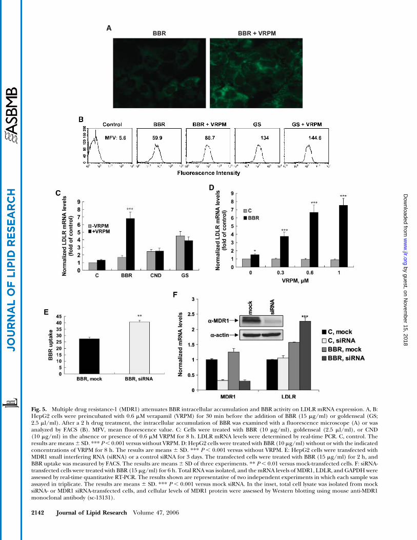
Fig. 5. Multiple drug resistance-1 (MDR1) attenuates BBR intracellular accumulation and BBR activity on LDLR mRNA expression. A, B:HepG2 cells were preincubated with 0.6 mM verapamil (VRPM) for 30 min before the addition of BBR (15 mg/ml) or goldenseal (GS;2.5 ml/ml). After a 2 h drug treatment, the intracellular accumulation of BBR was examined with a fluorescence microscope (A) or wasanalyzed by FACS (B). MFV, mean fluorescence value. C: Cells were treated with BBR (10 mg/ml), goldenseal (2.5 ml/ml), or CND(10 mg/ml) in the absence or presence of 0.6 mM VRPM for 8 h. LDLR mRNA levels were determined by real-time PCR. C, control. Theresults are means6 SD. *** P, 0.001 versus without VRPM. D: HepG2 cells were treated with BBR (10 mg/ml) without or with the indicatedconcentrations of VRPM for 8 h. The results are means 6 SD. *** P , 0.001 versus without VRPM. E: HepG2 cells were transfected withMDR1 small interfering RNA (siRNA) or a control siRNA for 3 days. The transfected cells were treated with BBR (15 mg/ml) for 2 h, andBBR uptake was measured by FACS. The results are means 6 SD of three experiments. ** P , 0.01 versus mock-transfected cells. F: siRNA-transfected cells were treated with BBR (15 mg/ml) for 6 h. Total RNA was isolated, and themRNA levels of MDR1, LDLR, and GAPDHwereassessed by real-time quantitative RT-PCR. The results shown are representative of two independent experiments in which each sample wasassayed in triplicate. The results are means 6 SD. *** P , 0.001 versus mock siRNA. In the inset, total cell lysate was isolated from mocksiRNA- or MDR1 siRNA-transfected cells, and cellular levels of MDR1 protein were assessed by Western blotting using mouse anti-MDR1monoclonal antibody (sc-13131).
2142 Journal of Lipid Research Volume 47, 2006
by guest, on Novem
ber 15, 2018w
ww
.jlr.orgD
ownloaded from
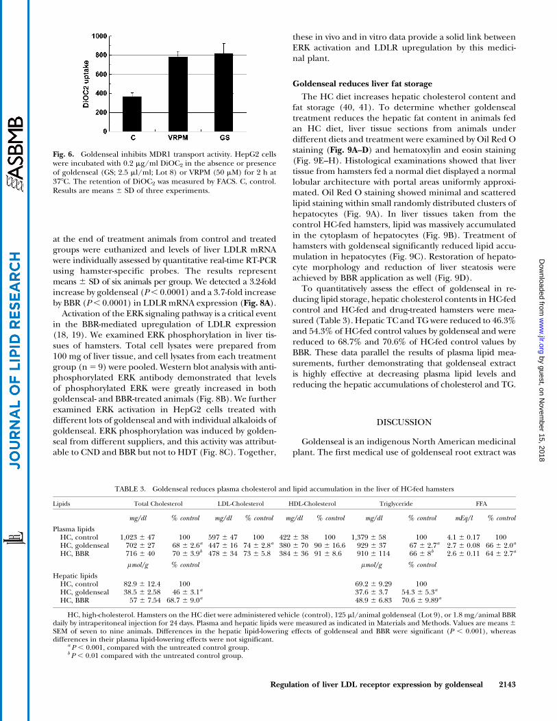
at the end of treatment animals from control and treatedgroups were euthanized and levels of liver LDLR mRNAwere individually assessed by quantitative real-time RT-PCRusing hamster-specific probes. The results representmeans 6 SD of six animals per group. We detected a 3.2-foldincrease by goldenseal (P, 0.0001) and a 3.7-fold increaseby BBR (P, 0.0001) in LDLRmRNA expression (Fig. 8A).
Activation of the ERK signaling pathway is a critical eventin the BBR-mediated upregulation of LDLR expression(18, 19). We examined ERK phosphorylation in liver tis-sues of hamsters. Total cell lysates were prepared from100 mg of liver tissue, and cell lysates from each treatmentgroup (n5 9) were pooled. Western blot analysis with anti-phosphorylated ERK antibody demonstrated that levelsof phosphorylated ERK were greatly increased in bothgoldenseal- and BBR-treated animals (Fig. 8B). We furtherexamined ERK activation in HepG2 cells treated withdifferent lots of goldenseal and with individual alkaloids ofgoldenseal. ERK phosphorylation was induced by golden-seal from different suppliers, and this activity was attribut-able to CND and BBR but not to HDT (Fig. 8C). Together,
these in vivo and in vitro data provide a solid link betweenERK activation and LDLR upregulation by this medici-nal plant.
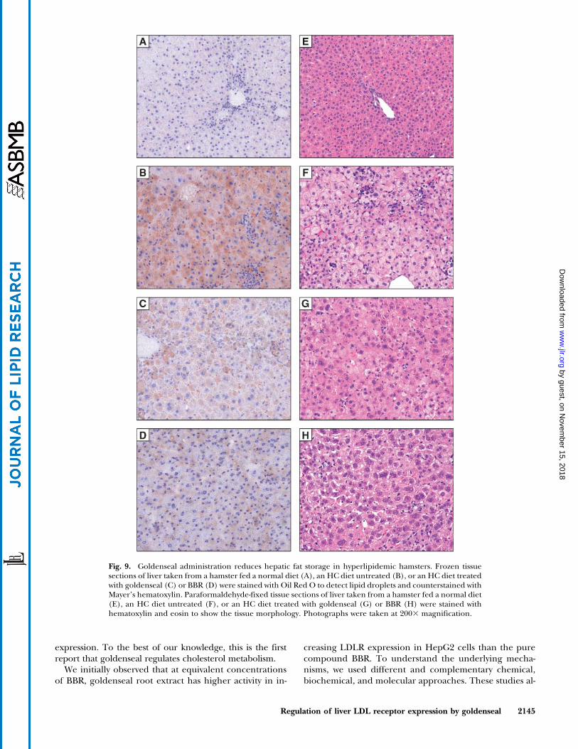
Goldenseal reduces liver fat storage
The HC diet increases hepatic cholesterol content andfat storage (40, 41). To determine whether goldensealtreatment reduces the hepatic fat content in animals fedan HC diet, liver tissue sections from animals underdifferent diets and treatment were examined by Oil Red Ostaining (Fig. 9A–D) and hematoxylin and eosin staining(Fig. 9E–H). Histological examinations showed that livertissue from hamsters fed a normal diet displayed a normallobular architecture with portal areas uniformly approxi-mated. Oil Red O staining showed minimal and scatteredlipid staining within small randomly distributed clusters ofhepatocytes (Fig. 9A). In liver tissues taken from thecontrol HC-fed hamsters, lipid was massively accumulatedin the cytoplasm of hepatocytes (Fig. 9B). Treatment ofhamsters with goldenseal significantly reduced lipid accu-mulation in hepatocytes (Fig. 9C). Restoration of hepato-cyte morphology and reduction of liver steatosis wereachieved by BBR application as well (Fig. 9D).
To quantitatively assess the effect of goldenseal in re-ducing lipid storage, hepatic cholesterol contents in HC-fedcontrol and HC-fed and drug-treated hamsters were mea-sured (Table 3). Hepatic TC and TG were reduced to 46.3%and 54.3% of HC-fed control values by goldenseal and werereduced to 68.7% and 70.6% of HC-fed control values byBBR. These data parallel the results of plasma lipid mea-surements, further demonstrating that goldenseal extractis highly effective at decreasing plasma lipid levels andreducing the hepatic accumulations of cholesterol and TG.
DISCUSSION
Goldenseal is an indigenous North American medicinalplant. The first medical use of goldenseal root extract was
Fig. 6. Goldenseal inhibits MDR1 transport activity. HepG2 cellswere incubated with 0.2 mg/ml DiOC2 in the absence or presenceof goldenseal (GS; 2.5 ml/ml; Lot 8) or VRPM (50 mM) for 2 h at378C. The retention of DiOC2 was measured by FACS. C, control.Results are means 6 SD of three experiments.
TABLE 3. Goldenseal reduces plasma cholesterol and lipid accumulation in the liver of HC-fed hamsters
Lipids Total Cholesterol LDL-Cholesterol HDL-Cholesterol Triglyceride FFA
mg/dl % control mg/dl % control mg/dl % control mg/dl % control mEq/l % control
Plasma lipidsHC, control 1,023 6 47 100 597 6 47 100 422 6 38 100 1,379 6 58 100 4.1 6 0.17 100HC, goldenseal 702 6 27 68 6 2.6a 447 6 16 74 6 2.8a 380 6 70 90 6 16.6 929 6 37 67 6 2.7a 2.7 6 0.08 66 6 2.0a
HC, BBR 716 6 40 70 6 3.9b 478 6 34 73 6 5.8 384 6 36 91 6 8.6 910 6 114 66 6 8b 2.6 6 0.11 64 6 2.7a
lmol/g % control lmol/g % control
Hepatic lipidsHC, control 82.9 6 12.4 100 69.2 6 9.29 100HC, goldenseal 38.5 6 2.58 46 6 3.1a 37.6 6 3.7 54.3 6 5.3a
HC, BBR 57 6 7.54 68.7 6 9.0a 48.9 6 6.83 70.6 6 9.89a
HC, high-cholesterol. Hamsters on the HC diet were administered vehicle (control), 125 ml/animal goldenseal (Lot 9), or 1.8 mg/animal BBRdaily by intraperitoneal injection for 24 days. Plasma and hepatic lipids were measured as indicated in Materials and Methods. Values are means 6SEM of seven to nine animals. Differences in the hepatic lipid-lowering effects of goldenseal and BBR were significant (P , 0.001), whereasdifferences in their plasma lipid-lowering effects were not significant.
a P , 0.001, compared with the untreated control group.b P , 0.01 compared with the untreated control group.
Regulation of liver LDL receptor expression by goldenseal 2143
by guest, on Novem
ber 15, 2018w
ww
.jlr.orgD
ownloaded from
reported in 1798 for the treatment of what was thought tobe cancer and for inflamed eyes (42). Since then, goldensealhas been widely used by herbal practitioners as an anti-microbial and antisecretory agent for a variety of infections
that affect the mucosa, such as respiratory and intestinalinfections (20, 21). In this study, we demonstrate that gold-enseal has strong activities in decreasing plasma cholesteroland LDL-c through its stimulating effect on hepatic LDLR
Fig. 7. Plasma lipoprotein cholesterol profiles of con-trol and goldenseal (GS)-treated animals. Serum fromthe normal diet group (n 5 6), the high-cholesterol(HC) control group (n5 9), and the goldenseal groupwere pooled, and the pooled sera were subjected toHPLC analysis of lipoprotein profiles associated withtotal cholesterol (upper panel) and triglyceride (lowerpanel). CM, chylomicron.
Fig. 8. Upregulation of LDLR mRNA expression and activation of the extracellular signal-regulated kinase(ERK) pathway in hamsters by goldenseal. A: Hepatic LDLR mRNA expression. Four hours after the lastdrug treatment, all animals were euthanized and liver total RNA was isolated. Individual levels of LDLRmRNA in untreated, goldenseal (GS)-treated, and BBR-treated hamsters fed the HC diet were assessed byquantitative PCR. Results are means 6 SD of six animals per group. C, control. *** P , 0.001 versus the HCcontrol group. B: Western blot analysis of phosphorylated ERK. Cytosolic proteins were prepared frompooled liver samples of the same treatment group (n 5 9), and 50 mg of protein from the pooled samplewas subjected to SDS-PAGE. The membrane was blotted with anti-phosphorylated ERK antibody andsubsequently blotted with anti-ERK2 antibody. C: Activation of ERK in HepG2 cells. HepG2 cells weretreated with 2.5 ml/ml goldenseal obtained from three different suppliers or with 20 mg/ml BBR, HDT, orCND for 2 h. Total cell lysates were prepared, and 50 mg of protein per sample was analyzed for phos-phorylated ERK by Western blot analysis.
2144 Journal of Lipid Research Volume 47, 2006
by guest, on Novem
ber 15, 2018w
ww
.jlr.orgD
ownloaded from
expression. To the best of our knowledge, this is the firstreport that goldenseal regulates cholesterol metabolism.
We initially observed that at equivalent concentrationsof BBR, goldenseal root extract has higher activity in in-
creasing LDLR expression in HepG2 cells than the purecompound BBR. To understand the underlying mecha-nisms, we used different and complementary chemical,biochemical, and molecular approaches. These studies al-
Fig. 9. Goldenseal administration reduces hepatic fat storage in hyperlipidemic hamsters. Frozen tissuesections of liver taken from a hamster fed a normal diet (A), an HC diet untreated (B), or an HC diet treatedwith goldenseal (C) or BBR (D) were stained with Oil Red O to detect lipid droplets and counterstained withMayer’s hematoxylin. Paraformaldehyde-fixed tissue sections of liver taken from a hamster fed a normal diet(E), an HC diet untreated (F), or an HC diet treated with goldenseal (G) or BBR (H) were stained withhematoxylin and eosin to show the tissue morphology. Photographs were taken at 2003 magnification.
Regulation of liver LDL receptor expression by goldenseal 2145
by guest, on Novem
ber 15, 2018w
ww
.jlr.orgD
ownloaded from
lowed us to discover several important factors that contrib-ute to the higher activity of goldenseal in the modulationof LDLR expression.
First, we have identified CND, another major isoquino-line compound of goldenseal, as a new modulator ofLDLR expression with greater activity than BBR. It is note-worthy that CND and PMT are structurally closely relatedto BBR, yet PMT has no regulatory activity on LDLRexpression. On the other hand, both BBR and PMT havestrong DNA binding affinities, whereas CND, a hydroge-nated product of BBR, does not bind to DNA (43). It hasbeen proposed that the quaternary ammonium and planarstructure play critical roles in the DNA binding of BBR andPMT. The fact that CND lacks both critical features forDNA binding but shares the common activity with BBR instabilizing LDLR mRNA provides the first evidence thatseparates the DNA binding property from the activity ofmRNA stabilization in these isoquinoline compounds.
Second, we have demonstrated the presence of addi-tional LDLR regulators in goldenseal extract. We showedthat eluates F3 and F6 of silica gel columns loaded withgoldenseal have LDLR-inducing activities that cannotbe attributed to BBR or CND. At present, it is not clearwhether the increased LDLR expression is caused by asingle compound in F3 or F6 or results from a combinedaction of the mixture. Because neither F3 nor F6 increasedLDLR promoter activity (Fig. 1D), the unknown com-pound(s) likely acts on the stability of LDLR mRNA. Thus,our studies demonstrate that goldenseal contains a groupof natural compounds that have unique properties instabilizing LDLR mRNA. Experiments to isolate and struc-turally characterize these unknown compounds are cur-rently under way in our laboratory.
The third factor that contributes to the strong activity ofgoldenseal in increasing LDLR expression is the resistanceto MDR1-mediated drug excretion. Using two differentapproaches, MDR1 inhibitors that inhibit the transportactivity of MDR1 and siRNA that blocks the expression ofMDR1, we demonstrated that pgp-170 actively excludesBBR from HepG2 cells, resulting in a lower efficacy of BBRin LDLR regulation. BBR and PMT, which are strong am-phipathic cations, have been identified as natural sub-strates of theMDRNorA pump ofmicroorganisms (33–35),and our data are consistent with these literature reports.The fact that BBR in goldenseal has a longer intracellularretention time, with greater influx and lesser efflux thanBBR alone, suggested the existence of an MDR inhibi-tor(s) in goldenseal. Using DiOC2, a well-characterizedMDR1 substrate, we were able to show that, indeed, theMDR1-mediated efflux of DiOC2 was inhibited by golden-seal at a concentration that elicited a response in LDLRexpression. A previous study has identified an MDR inhibi-tor, 59-methoxyhydnocarpin (34), in the leaves of Berberisfremontii, a BBR-producing plant. However, our LC-MS didnot detect a peak corresponding to the molecular weightof 59-methoxyhydnocarpin in goldenseal. It is likely thatthe inhibitor(s) produced by goldenseal is structurallydifferent from the one made in Berberis fremontii. Ourstudies also revealed that CND is not a substrate of MDR1,
thereby providing a molecular explanation for the higheractivity of CND than BBR. With its unique features ofMDR1 resistance and lack of DNA binding, CND is pos-sibly a better candidate in clinical use for cholesterol re-duction, with potentially lower toxicity.
We demonstrated strong plasma TC and LDL-c reduc-tions and a 3.2-fold increase in the hepatic LDLR mRNAlevel in goldenseal-treated hamsters fed the HC diet at halfthe equivalent dose of BBR. These in vivo results con-firmed the higher potency of goldenseal observed in ourin vitro studies. In addition to decreasing TC and LDL-c,goldenseal and BBR markedly reduced serum FFA andTG. A recent study has shown that in HepG2 cells, BBRinduces the activation of acetyl-CoA carboxylase throughAMP kinase-mediated phosphorylation, which led to asubsequent increase in fatty acid oxidation and decreasesin fatty acid synthesis and TG synthesis (44). AMP kinaseactivation was shown to be the downstream event of theERK/MEK pathway. In this study, we showed that golden-seal strongly activates ERK activation in liver tissue and inHepG2 cells. Thus, it is possible that activation of acetyl-CoA carboxylase by goldenseal acting through the ERKsignaling pathway accounts for the strong reductions ofFFA and TG in hamsters.
In conclusion, we have discovered that goldenseal, aNative American medicinal plant, has strong cholesterol-and lipid-lowering effects. Goldenseal reduces cholesteroland lipid accumulations in plasma, as well as in liver,through the actions of multiple bioactive compounds thatwork synergistically. This work opens the way for a potentialalternative therapeutic intervention for hyperlipidemia.
The authors thank Dr. Nick Cairns for his expertise in chemicalanalysis; Dr. Ting-Ting Hung for her help in the evaluation ofliver tissue sections; Dr. Salman Azhar for providing technicalexpertise in drug efflux assays; and other members of the Liulaboratory, Drs. Yue Zhou and Haiyan Liu, for their interest-ing discussions. This study was supported by the Departmentof Veterans Affairs (Office of Research and Development, Medi-cal Research Service) (to J.L. and F.B.K.) and by Grant 1RO1AT002543-01A1 (to J.L.) from the National Center for Comple-mentary and Alternative Medicine.
REFERENCES
1. Bays, H., and E. A. Stein. 2003. Pharmacotherapy for dyslipidemia—current therapies and future agents. Expert Opin. Pharmacother. 4:1901–1938.
2. Grundy, S. M. 1998. Statin trials and goals of cholesterol-loweringtherapy. Circulation. 97: 1436–1439.
3. Ansell, B. J., K. E. Watson, and A. M. Fogelman. 1999. An evidence-based assessment of the NCEP Adult Treatment Panel II guidelines.National Cholesterol Education Program. J. Am. Med. Assoc. 282:2051–2057.
4. Brown, M. S., and J. L. Goldstein. 1986. A receptor-mediated path-way for cholesterol homeostasis. Science. 232: 34–47.
5. Smith, J. R., T. F. Osborne, J. L. Goldstein, and M. S. Brown. 1990.Identification of nucleotides responsible for enhancer activity ofsterol regulatory element in low density lipoprotein receptor gene.J. Biol. Chem. 265: 2306–2310.
6. Briggs, M. R., C. Yokoyama, X. Wang, M. S. Brown, and J. L.Goldstein. 1993. Nuclear protein that binds sterol regulatory ele-
2146 Journal of Lipid Research Volume 47, 2006
by guest, on Novem
ber 15, 2018w
ww
.jlr.orgD
ownloaded from
ment of low density lipoprotein receptor promoter. J. Biol. Chem.268: 14490–14496.
7. Wang, X., M. R. Briggs, C. Yokoyama, J. L. Goldstein, and M. S.Brown. 1993. Nuclear protein that binds sterol regulatory elementof low density lipoprotein receptor promoter. II. Purification andcharacterization. J. Biol. Chem. 268: 14497–14504.
8. Yokoyama, C., X. Wang, M. R. Briggs, A. Admon, J. Wu, X. Hua,J. L. Goldstein, and M. S. Brown. 1993. SREBP-1, a basic-helix-loop-helix-leucine zipper protein that controls transcription of the lowdensity lipoprotein receptor gene. Cell. 75: 187–197.
9. Wang, X., R. Sato, M. S. Brown, X. Hua, and J. L. Goldstein. 1994.SREBP-1, a membrane bound transcription factor released bysterol-regulated proteolysis. Cell. 77: 53–62.
10. Reinoso, R. F., S. Navarro, M. J. Garcia, and J. R. Prous. 2001.Pharmacokinetic interactions of statins. Methods Find. Exp. Clin.Pharmacol. 23: 541–566.
11. Nawrocki, J. W., S. R. Weiss, M. H. Davidson, D. L. Sprecher, S. L.Schwartz, P. Lupien, P. H. Jones, H. E. Haber, and D. M. Black.1995. Reduction of LDL cholesterol by 25% to 60% in patients withprimary hypercholesterolemia by atorvastatin, a new HMG-CoAreductase inhibitor. Arterioscler. Thromb. Vasc. Biol. 15: 678–682.
12. Prueksaritanont, T., B. Ma, C. Tang, Y. Meng, C. Assang, P. Lu, P. J.Reider, J. H. Lin, and T. A. Baillie. 1999. Metabolic interactionsbetween mibefradil and HMG CoA reductase inhibitors: an invitro investigation with human liver preparations. Br. J. Pharmacol.47: 291–298.
13. Barter, P. J. 2000. Treating to target with statins. Atheroscler. Suppl. 1:21–25.
14. Williams, D., and J. Feely. 2002. Pharmacokinetic-pharmacodynamicdrug interactions with HMG-CoA reductase inhibitors. Clin. Pharma-cokinet. 41: 343–370.
15. Moghadasian, M. H. 2002. A safety look at currently available stat-ins. Expert Opin. Drug Saf. 1: 269–274.
16. Klotz, U. 2003. Pharmacological comparison of the statins.Arzneim.-Forsch. Drug Res. 53: 605–611.
17. Rader, D. 2001. A new feature on the cholesterol-lowering land-scape. Nat. Med. 7: 1282–1284.
18. Kong, W., J. Wei, P. Abidi, M. Lin, S. Inaba, C. Li, Y. Wang, Z. Wang,S. Si, H. Pan, et al. 2004. Berberine is a promising novel cholesterol-lowering drug working through a unique mechanism distinct fromstatins. Nat. Med. 10: 1344–1352.
19. Abidi, P., Y. Zhou, J. Jiang, and J. Liu. 2005. ERK-dependentregulation of hepatic LDL receptor expression by herbal medicineberberine. Arterioscler. Thromb. Vasc. Biol. 25: 2170–2176.
20. Upton R. (ed.) 2001. Goldenseal root Hydrastis canadensis: stan-dards of analysis, quality control, and therapeutics. American HerbalPharmacopoeia and Therapeutic Compendium 1: 1–37.
21. McKenna, D. J., K. Jones, K. Hughes, and S. Humphrey. 2002.Botanical Medicines: The Desk Reference for Major Herbal Sup-plements. 2nd edition. Haworth Press, Binghamton, NY. 547–568.
22. Li, C., F. B. Kraemer, T. E. Ahlborn, and J. Liu. 1999. Induction oflow density lipoprotein receptor (LDLR) transcription by oncosta-tin M is mediated by the extracellular signal-regulated kinasesignaling pathway and the repeat 3 element of the LDLR promoter.J. Biol. Chem. 274: 6747–6753.
23. Yadav, R. C., G. S. Kumar, K. Bhadra, P. Giri, R. Sinha, S. Pal, andM. Maiti. 2005. Berberine, a strong polyriboadenylic acid bindingplant alkaloid: spectroscopic, viscometric, and thermodynamicstudy. Bioorg. Med. Chem. 13: 165–174.
24. Okazaki, M., S. Usui, M. Ishigami, N. Sakai, T. Nakamura, Y.Matsuzawa, and S. Yamashita. 2005. Identification of unique lipo-protein subclasses for visceral obesity by component profile in high-performance liquid chromatography. Arterioscler. Thromb. Vasc. Biol.25: 578–584.
25. Scazzocchio, F., M. F. Cometa, andM. Palmery. 1998. Antimicrobialactivity of Hydrastis canadensis extract and its major isolated alka-loids. Fitoterapia. 69: 58–59.
26. Weber, H. A., M. K. Zart, A. E. Hodges, H. M. Molloy, B. M.O’Brien, L. A. Moody, A. P. Clark, R. K. Harris, D. Overstreet,
and C. Smith. 2003. Chemical comparison of goldenseal (Hydras-tis canadensis L.) root powder from three commercial suppliers.J. Agric. Food Chem. 51: 7352–7358.
27. Weber, H. A., M. K. Zart, A. E. Hodges, K. D. White, S. M. Barnes,L. A. Moody, A. P. Clark, and R. K. Harris. 2003. Method validationfor determination of alkaloid content in goldenseal root powder.J. AOAC Int. 86: 476–483.
28. Betz, J., S. M. Musser, and G. M. Larkin. 1998. Differentiation be-tween goldenseal (Hydrastis canadensis L.) and possible adulterantsby LC/MS. In Proceedings of the 39th Annual Meeting of theAmerican Society of Pharmacognosy. Glendale, AZ: AmericanSociety of Pharmacognosy. p. 129.
29. Liu, J., T. E. Ahlborn, M. R. Briggs, and F. B. Kraemer. 2000. Iden-tification of a novel sterol-independent regulatory element in thehuman low density lipoprotein receptor promoter. J. Biol. Chem.275: 5214–5221.
30. Grand-Perret, T., A. Bouillot, A. Perrot, S. Commans, M. Walker,and M. Issandou. 2001. SCAP ligands are potent new lipid-loweringdrugs. Nat. Med. 7: 1332–1338.
31. Liu, J., F. Zhang, C. Li, M. Lin, and M. R. Briggs. 2003. Synergisticactivation of human LDL receptor expression by SCAP ligand andcytokine oncostatin M. Arterioscler. Thromb. Vasc. Biol. 23: 90–96.
32. Li, S. L., G. Lin, S. W. Chan, and P. Li. 2001. Determination ofthe major isosteroidal alkaloids in bulbs of Fritillaria by high-performance liquid chromatography coupled with evaporative lightscattering detection. J. Chromatogr. A. 909: 207–214.
33. Hsieh, P. C., S. A. Siegel, B. Rogers, D. Davis, and K. Lewis. 1998.Bacteria lacking a multidrug pump: a sensitive tool for drug dis-covery. Proc. Natl. Acad. Sci. USA. 95: 6602–6606.
34. Stermitz, F. R., P. Lorenz, J. N. Tawara, L. A. Zenewicz, and K. Lewis.2000. Synergy in a medicinal plant: antimicrobial action of berber-ine potentiated by 59-methoxyhydnocarpin, a multidrug pump in-hibitor. Proc. Natl. Acad. Sci. USA. 97: 1433–1437.
35. Samosorn, S., J. B. Bremner, A. Ball, and K. Lewis. 2006. Synthesisof functionalised 2-aryl-5-nitro-1H-indoles and their activity as bac-terial NorA efflux pump inhibitors. Bioorg. Med. Chem. 14: 857–865.
36. Taub, M. E., L. Podila, D. Ely, and I. Almeida. 2005. Functional as-sessment of multiple P-glycoprotein (P-gp) probe substrates: in-fluence of cell line and modulator concentration of P-gp activity.Drug Metab. Dispos. 33: 1679–1687.
37. Stierle, V., A. Laigle, and B. Jolles. 2005. Modulation of MDR1 geneexpression in multidrug resistant MCF7 cells by low concentrationsof small interfering RNAs. Biochem. Pharmacol. 70: 1424–1430.
38. Constable, P. A., J. G. Lawrenson, D. E. M. Dolman, G. B. Arden,and N. J. Abbott. 2006. P-glycoprotein expression in human retinalpigment epithelium cell lines. Exp. Eye Res. 83: 24–30.
39. Minderman, H., U. Vanhhoefer, K. Toth, M. B. Yin, M. D.Minderman, C. Wrozsek, M. L. Slovak, and Y. M. Rustum. 1996.DiOC2(3) is not a substrate for multidrug resistance protein(MRP)-mediated drug efflux. Cytometry. 25: 14–20.
40. Spady, D. K., and J. M. Dietschy. 1988. Interaction of dietary cho-lesterol and triglyceride in the regulation of hepatic low densitylipoprotein transport in the hamster. J. Clin. Invest. 81: 300–309.
41. Bensch, W. R., R. A. Gadski, J. S. Bean, L. S. Beavers, R. J. Schmidt,D. N. Perry, A. T. Murphy, D. B. McClure, P. I. Eacho, A. P. Breau,et al. 1999. Effects of LY295427, a low-density lipoprotein (LDL)receptor up-regulator, on LDL receptor gene transcription andcholesterol metabolism in normal and hypercholesterolemichamsters. J. Pharmacol. Exp. Ther. 289: 85–92.
42. Lloyd, J. U., and C. G. Lloyd. 1908. Drugs and medicines of NorthAmerica: Hydrastis canadensis. Bull. Lloyd Library Bot. Pharm. MateriaMed. 10: 76–184.
43. Qin, Y., J-Y. Pang, W-H. Chen, Z. Cai, and Z-H. Jiang. 2006. Synthe-sis, DNA-binding affinities, and binding mode of berberine dimers.Bioorg. Med. Chem. 14: 25–32.
44. Brusq, J. M., N. Ancellin, P. Grondin, R. Guillard, S. Martin, Y.Saintillan, and M. Issandou. 2006. Inhibition of lipid synthesisthrough activation of AMP kinase: an additional mechanism for thehypolipidemic effects of berberine. J. Lipid Res. 47: 1281–1288.
Regulation of liver LDL receptor expression by goldenseal 2147
by guest, on Novem
ber 15, 2018w
ww
.jlr.orgD
ownloaded from

![Goldenseal Hydrastis canadensis L.) and Two of Its ... · Goldenseal (Hydrastis canadensis L.) and Two of Its Constituent Alkaloids . Berberine [2086-83-1] and Hydrastine [118-08-1]](https://img.pdfslide.net/doc/110x75/5d62e7a988c99367608bdbfa/goldenseal-hydrastis-canadensis-l-and-two-of-its-goldenseal-hydrastis.jpg)