Embed Size (px)
Citation preview
Ralf HEINE Jan Hendrik BaRtEls
Christian WEIss
2nd Edition
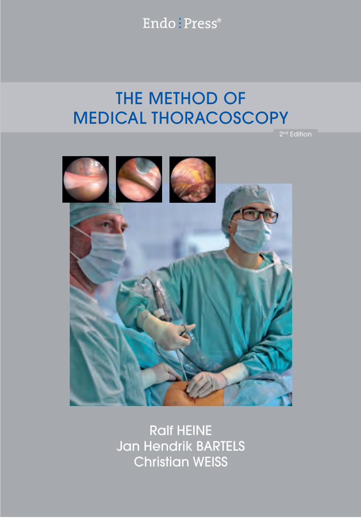
ThE METhod of MEdical Thoracoscopy
®

THE METHOD OF MEDICAL THORACOSCOPY
2nd Edition
Ralf HEINE, MD Jan Hendrik BARTELS, MD
Christian WEISS
Medical Clinic III – Pneumonology, Hematology-Oncology and Palliative Medicine
Hospital of St. Elisabeth and St. Barbara Halle (Saale), Germany
The Method of Medical Thoracoscopy4
Cover image:Andreas Heine
The Method of Medical Thoracoscopy 2nd EditionRalf Heine, MDJan Hendrik Bartels, MDChristian WeissMedical Clinic III – Pneumonology, Hematology-Oncology and Palliative Medicine, Hospital of St. Elisabeth and St. Barbara, Halle (Saale), Germany
Correspondence address of the author: Dr. med. Ralf Heine Facharzt für Innere Medizin, Pneumologie und Notfallmedizin Chefarzt der Medizinischen Klinik III – Pneumologie, Häma tologie-Onkologie und Palliativmedizin Krankenhaus St. Elisabeth und St. Barbara, Halle/Saale Mauerstr. 5 06110 Halle/Saale, Germany
All rights reserved. 2nd edition | 1st edition 2007 © 2015 GmbH P.O. Box, 78503 Tuttlingen, Germany Phone: +49 (0) 74 61/1 45 90 Fax: +49 (0) 74 61/708-529 E-Mail: [email protected]
No part of this publication may be translated, reprinted or reproduced, transmitted in any form or by any means, electronic or mechanical, now known or hereafter invent ed, including photocopying and recording, or utilized in any information storage or retrieval system without the prior written permission of the copyright holder.
Editions in languages other than English and German are in preparation. For up-to-date information, please contact
GmbH at the address shown above.
Design and Composing: GmbH, Germany
Printing and Binding: Straub Druck + Medien AG Max-Planck-Straße 17, 78713 Schramberg, Germany 11.15-1
ISBN 978-3-89756-616-3
Important notes:
Medical knowledge is ever changing. As new research and clinical experience broaden our knowledge, changes in treat ment and therapy may be required. The authors and editors of the material herein have consulted sources believed to be reliable in their efforts to provide information that is complete and in accord with the standards accept ed at the time of publication. However, in view of the possibili ty of human error by the authors, editors, or publisher, or changes in medical knowledge, neither the authors, editors, publisher, nor any other party who has been involved in the prepara-tion of this booklet, warrants that the information contained herein is in every respect accurate or complete, and they are not responsible for any errors or omissions or for the results obtained from use of such information. The information contained within this booklet is intended for use by doctors and other health care professionals. This material is not intended for use as a basis for treatment decisions, and is not a substitute for professional consultation and/or use of peer- reviewed medical literature.
Some of the product names, patents, and re gistered designs referred to in this booklet are in fact registered trademarks or proprietary names even though specific reference to this fact is not always made in the text. Therefore, the appearance of a name without designation as proprietary is not to be construed as a representation by the publisher that it is in the public domain.
The use of this booklet as well as any implementation of the information contained within explicitly takes place at the reader’s own risk. No liability shall be accepted and no guarantee is given for the work neither from the publisher or the editor nor from the author or any other party who has been involved in the preparation of this work. This particularly applies to the content, the timeliness, the correctness, the complete-ness as well as to the quality. Printing errors and omissions cannot be completely excluded. The publisher as well as the author or other copyright holders of this work disclaim any liability, particularly for any damages arising out of or associated with the use of the medical procedures mentioned within this booklet.
Any legal claims or claims for damages are excluded.
In case any references are made in this booklet to any 3rd party publica-tion(s) or links to any 3rd party websites are mentioned, it is made clear that neither the publisher nor the author or other copyright holders of this booklet endorse in any way the content of said publication(s) and/or web sites referred to or linked from this booklet and do not assume any form of liability for any factual inaccuracies or breaches of law which may occur therein. Thus, no liability shall be accepted for content within the 3rd party publication(s) or 3rd party websites and no guarantee is given for any other work or any other websites at all.

5The Method of Medical Thoracoscopy
Table of Contents
1 Historical Background . . . . . . . . . . . . . . . . . . . . . . . . . . . . . . . . . . . . . . 6
2 Indications and Contraindications for Thoracoscopy . . . . . . . . . . . 62.1 Indications. . . . . . . . . . . . . . . . . . . . . . . . . . . . . . . . . . . . . . . . . . . . 62.2 Contraindications. . . . . . . . . . . . . . . . . . . . . . . . . . . . . . . . . . . . . . 7
3 Anesthesia . . . . . . . . . . . . . . . . . . . . . . . . . . . . . . . . . . . . . . . . . . . . . . . . 7
4 Preparations for Thoracoscopy. . . . . . . . . . . . . . . . . . . . . . . . . . . . . . . 84.1 Imaging Studies . . . . . . . . . . . . . . . . . . . . . . . . . . . . . . . . . . . . . . . 84.2 Diagnostic Pneumothorax . . . . . . . . . . . . . . . . . . . . . . . . . . . . . . 84.3 Premedication . . . . . . . . . . . . . . . . . . . . . . . . . . . . . . . . . . . . . . . . 104.3.1 The Evening Before the Procedure . . . . . . . . . . . . . . . . . . . . . . . 104.3.2 The Day of the Procedure . . . . . . . . . . . . . . . . . . . . . . . . . . . . . . . 10
5 Technique of Thoracoscopy. . . . . . . . . . . . . . . . . . . . . . . . . . . . . . . . . . 115.1 Procedure Room . . . . . . . . . . . . . . . . . . . . . . . . . . . . . . . . . . . . . . 115.2 Instruments . . . . . . . . . . . . . . . . . . . . . . . . . . . . . . . . . . . . . . . . . . . 115.3 Positioning the Patient . . . . . . . . . . . . . . . . . . . . . . . . . . . . . . . . . 115.4 Monitoring and Other Measures during the Procedure . . . . . . 125.5 Patient Preparation after Positioning . . . . . . . . . . . . . . . . . . . . . 125.6 Selection of the Entry Site. . . . . . . . . . . . . . . . . . . . . . . . . . . . . . . 125.7 Local Anesthesia . . . . . . . . . . . . . . . . . . . . . . . . . . . . . . . . . . . . . . 125.8 Conscious Sedation. . . . . . . . . . . . . . . . . . . . . . . . . . . . . . . . . . . . 135.9 Trocar Insertion . . . . . . . . . . . . . . . . . . . . . . . . . . . . . . . . . . . . . . . 135.10 Inspection of the Thoracic Cavity . . . . . . . . . . . . . . . . . . . . . . . . 145.11 Thoracoscopic Biopsy and Lysis of Adhesions . . . . . . . . . . . . 165.12 Talc Pleurodesis . . . . . . . . . . . . . . . . . . . . . . . . . . . . . . . . . . . . . . . 175.13 Concluding the Procedure . . . . . . . . . . . . . . . . . . . . . . . . . . . . . . 18
6 Management after Thoracoscopy. . . . . . . . . . . . . . . . . . . . . . . . . . . . . 19
7 Complications . . . . . . . . . . . . . . . . . . . . . . . . . . . . . . . . . . . . . . . . . . . . . 19
8 Summary. . . . . . . . . . . . . . . . . . . . . . . . . . . . . . . . . . . . . . . . . . . . . . . . . . 19
References . . . . . . . . . . . . . . . . . . . . . . . . . . . . . . . . . . . . . . . . . . . . . . . . . . . . 20
Instrument Set for Medical Thoracoscopy . . . . . . . . . . . . . . . . . . . . . . . . . 22
The Method of Medical Thoracoscopy6
Historical Background
The idea of using optical instruments to enter and examine body cavities that cannot be accessed through a natural orifice dates back to the Dresden physician G. Kelling (1866 – 945). As early as 1902, he published a report detailing how he was able to perform ‘coelioscopy’ in a dog after first insufflating air into the abdominal cavity. His optical system consisted of a cystoscope like the one previously developed by M. Nitze (1848 –1906).11
Diagnostic thoracoscopy was first performed in human patients in 1910 by the Swedish internist H. C. Jacobaeus (1879 –1937).10 The creation of a pneumothorax did not pose a new challenge for Jacobaeus, as C. Forlanini (1847–1918) had already developed the procedure in the late 1800s for the collapse therapy to treat tuberculosis.6 Jacobaeus advanced the capabilities of diagnostic thoraco scopy by the introduction of thoracocautery. This technique, which became important in the treatment of tuberculosis, used electrocautery for the lysis of pleural adhesions.9 By the end of the 1950s, thoracoscopy with thoracocautery was widely practiced in the collapse therapy of tuberculosis. Only a few clinicians utilized the diagnostic potential of thoracoscopy
during that time; as a result, the diagnostic capabilities of thoracoscopy were largely forgotten.18
The fascination of being able to look into the chest led R. Korbsch to state in 1927 that ‘in vivo pathology’ could be accomplished if gross visual findings could be supple-mented by the histologic evaluation of biopsy specimens.
The Viennese physician A. Sattler rediscovered the diagnostic value of thoracoscopy in the early 1960s. He performed thoracoscopies in several thousand patients, and we must credit him with making pleural biopsy practical for clinical use.18 He also performed thoracoscopy for therapeutic purposes and described life-saving emergency endoscopies for the treatment of hemothorax.23
With the advent of video-assisted thoracoscopy in the 1980s, it also became possible to use thoracoscopy for surgical indications. Since then, video-assisted thoracic surgery (VATS) has become an established part of the thoracic surgical repertoire. At the same time, video-assisted thoracoscopy continues to be a mainstay in the medical diagnosis of diseases of the pleura, lung, and mediastinum.
Indications and Contraindications for Thoracoscopy
2.1 IndicationsThe range of indications for medical thoracoscopy has changed significantly in recent decades. The standard indications for medical thoracoscopy in the 1980s were unexplained pleural effusion, peripheral lung lesions, lesions near the chest wall, and mediastinal disease.7, 17
Today, tumors of the pleura, mediastinum and peripheral lung are diagnosed by CT- or ultrasound-guided biopsy. Generally these cases are not investigated by thoracoscopy. Interstitial lung diseases and peripheral lung tumors that cannot be diagnosed by bronchoscopic tissue sampling are a domain of thoracoscopic surgery. VATS can provide access for wedge resections that yield adequate material for histologic analysis. It is also used for the complete removal of solid peripheral lung lesions and isolated pleural tumors.
�� Pleural effusions of unknown origin.�� Pleural effusions with negative cytology in lung cancer to exclude M1a disease (pleural carcinomatosis).
�� Staging and histologic confirmation of pleural mesothelioma.
�� Pneumothorax (prior to chest tube placement if indicat ed).
�� Thoracoscopy in surgical cavities for a suspected tumor recurrence or specific infection.
Diagnostic Indications for Thoracoscopy:
In most cases thoracoscopy is performed during the inves-tigation of pleural exudates that are not explained by cyto-logic examination. Interstitial lung diseases may be an indi-cation for medical thoracoscopy in exceptional cases. In this application tissue is sampled from the periphery of the lung with a biopsy forceps. Generally speaking, however, a thoracoscopic wedge excision would be preferred for this indication.
1
2
7The Method of Medical Thoracoscopy
In patients with a pneumothorax, thoracoscopy can supply vital information that is helpful in directing further manage-ment. It can be used to inspect the pleural cavity before the placement of a chest tube.16
�� Talc pleurodesis for rapidly recurring malignant effusions and for transudates unresponsive to medical therapy. In selected cases, the procedure may also be used in the treatment of chylothorax and refractory exudative inflammatory pleural effusions.15,29
�� Talc pleurodesis for recurrent pneumothorax.3, 20
�� Pleural empyema and complicated parapneumonic effusions, where thoracoscopy can be used to evacuate the collection, lyse adhesions, and place a chest tube under vision.5, 12, 22, 25, 30
Therapeutic Indications for Thoracoscopy:
Persistent recurrence of pneumothorax with a chest tube in place is a definite indication for video-assisted thoracic surgery (VATS), which will also establish access for repairing the existing air leak.
Pleural empyema is a potential indication for medical thoracoscopy only if it is in an exudative or fibropurulent stage. Stage III disease is an indication for thoracic surgical intervention.
Thoracoscopy for pneumothorax as well as pleural empyema requires close interdisciplinary cooperation between pneumonology and thoracic surgery in developing a treatment strategy.
2.2 ContraindicationsThoracoscopy is generally well tolerated. We believe that it is contraindicated by conditions in which the creation of a diagnostic pneumothorax would exacerbate existing functional disorders.8, 19 In patients with large pleural effusions that may cause respiratory insufficiency, however, functional status can be improved by aspirating the effusion during the procedure.
Anesthesia
Medical thoracoscopy is usually perform ed under local anesthesia, which should be combined with adequate conscious sedation. It is recommended that con scious sedation and patient monitoring during the procedure be conducted by an anesthesiologist or a physician experienced in conscious sedation who can quickly recognize and manage any threatening situations that may arise.
General anesthesia with muscle relaxation and intubation with a double-lumen endotracheal tube are rarely necessary for medical thoracoscopy and are generally reserved for children and uncooperative patients.
�� Frank cardiac insufficiency.�� Frank pulmonary insufficiency.�� Coagulation disorder (Quick value < 60%, platelets < 80,000 Gpt/L).
�� Dual antiplatelet therapy with aspirin and clopidogrel. A daily aspirin dose of 100 mg is not a contraindication in itself. Clopidogrel should be stopped approximately 1 week before the procedure.
�� Treatment with dabigatran (Pradaxa®), apixaban (Ellquis®), or rivaroxaban (Xarelto®). Cessation of these drugs should follow current recommendations.
�� Anemia (Hb < 6 mmol/L).�� Severe kyphoscoliosis.�� Myocardial infarction during the previous 6 weeks.
Contraindications to Thoracoscopy:
3
The Method of Medical Thoracoscopy8
Preparations for Thoracoscopy
4.1 Imaging Studies
Preprocedure studies for thoracoscopy should always include PA and lateral chest radiographs. Thoracic computed tomography may yield important additional information, depending on the clinical question.
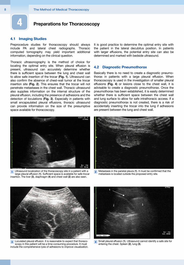
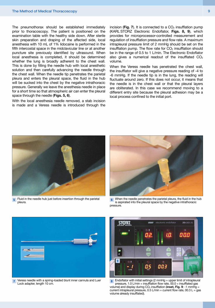
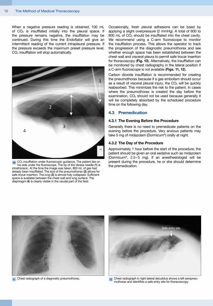
Thoracic ultrasonography is the method of choice for locating the optimal entry site. When pleural effusion is present, ultrasound can accurately determine whether there is sufficient space between the lung and chest wall to allow safe insertion of the trocar (Fig. 1). Ultrasound can also confirm the absence of chest-wall tumor at the trocar insertion site (Fig. 2). This ensures that the trocar will not penetrate metastases in the chest wall. Thoracic ultrasound also supplies information on the internal structure of the pleural effusion, including the presence of adhesions and the detection of loculations (Fig. 3). Especially in patients with small encapsulated pleural effusions, thoracic ultrasound can provide information on the size of the presumptive space available for thoracoscopy.
It is good practice to determine the optimal entry site with the patient in the lateral decubitus position. In patients with larger effusions, the potential entry site can also be determined and marked with bedside ultrasound.
4.2 Diagnostic Pneumothorax
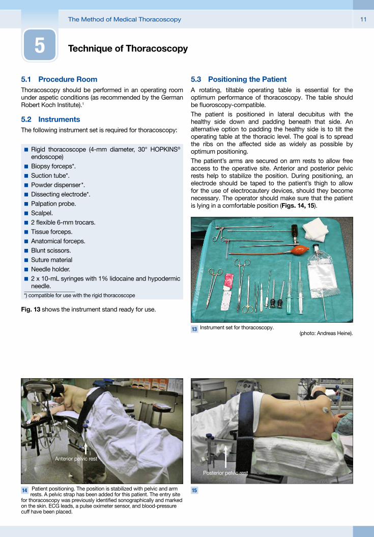
Basically there is no need to create a diagnostic pneumo-thorax in patients with a large pleural effusion. When thoracoscopy is used in the investigation of smaller pleural effusions (Fig. 4) or lesions close to the chest wall, it is advisable to create a diagnostic pneumothorax. Once the pneumothorax has been established, it is easily determined whether there is sufficient space between the chest wall and lung surface to allow for safe intrathoracic access. If a diagnostic pneumothorax is not created, there is a risk of accidentally inserting the trocar into the lung if adhesions are present between the lung and chest wall.
1 Ultrasound localization of the thoracoscopy site in a patient with a large pleural effusion (1). Sufficient space is available for safe trocar
insertion. The liver (3), diaphragm (4) and chest wall (2) are also seen.
2 Metastasis in the parietal pleura (1). It must be confirmed that the metastasis is located outside the proposed entry site.
3 Loculated pleural effusion. It is reasonable to expect that thoraco-scopy in this patient will be a time-consuming procedure. It must
include the comprehensive lysis of adhesions to improve visualization.
4 Small pleural effusion (1). Ultrasound cannot identify a safe site for entering the chest. Spleen (2), lung (3).
4
9The Method of Medical Thoracoscopy
The pneumothorax should be established immediately prior to thoracoscopy. The patient is positioned on the examination table with the healthy side down. After sterile skin preparation and draping of the affected side, local anesthesia with 10 mL of 1% lidocaine is performed in the fifth intercostal space in the midclavicular line or at another puncture site previously identified by ultrasound. When local anesthesia is completed, it should be determined whether the lung is broadly adherent to the chest wall. This is done by filling the needle hub with local anesthetic solution and then carefully advancing the needle through the chest wall. When the needle tip penetrates the parietal pleura and enters the pleural space, the fluid in the hub will be sucked into the chest by the negative intrathoracic pressure. Generally we leave the anesthesia needle in place for a short time so that atmospheric air can enter the pleural space through the needle (Figs. 5, 6).With the local anesthesia needle removed, a stab incision is made and a Veress needle is introduced through the
incision (Fig. 7). It is connected to a CO2 insufflation pump (KARL STORZ Electronic Endoflator, Figs. 8, 9), which provides for microprocessor-controlled measurement and regulation of insufflation pressure and flow rate. A maximum intrapleural pressure limit of 2 mmHg should be set on the insufflation pump. The flow rate for CO2 insufflation should be in the range of 0.5 to 1 L/min. The Electronic Endoflator also gives a numerical readout of the insufflated CO2 volume.When the Veress needle has penetrated the chest wall, the insufflator will give a negative pressure reading of -4 to -6 mmHg. If the needle tip is in the lung, the reading will fluctuate around zero. If this does not occur, it means that the needle is in the chest wall or that the pleural layers are obliterated. In this case we recommend moving to a different entry site because the pleural adhesion may be a local process confined to the initial port.
5 Fluid in the needle hub just before insertion through the parietal pleura.
6 When the needle penetrates the parietal pleura, the fluid in the hub is aspirated into the pleural space by the negative intrathoracic
pressure.
7 Veress needle with a spring-loaded blunt inner cannula and Luer Lock adapter, length 10 cm.
8 Endoflator with initial settings (2 mmHg = upper limit of intrapleural pressure, 1.0 L/min = insufflation flow rate, 00.0 = insufflated gas
volume) and display during CO2 insufflation (inset, Fig. 9: -1 mmHg = current intrapleural pressure, 0.5 L/min = current flow rate, 00.3 L = gas volume already insufflated).
9
The Method of Medical Thoracoscopy10
When a negative pressure reading is obtained, 100 mL of CO2 is insufflated initially into the pleural space. If the pressure remains negative, the insufflation may be continued. During this time the Endoflator will give an intermittent reading of the current intrapleural pressure. If the pressure exceeds the maximum preset pressure level, CO2 insufflation will stop automatically.
10 CO2 insufflation under fluoroscopic guidance. The patient lies on his side under the fluoroscope. The tip of the Veress needle (1) is
intrathoracic. At the time the image was taken, 800 mL of gas had already been insufflated. The size of the pneumothorax (2) allows for safe trocar insertion. The lung (3) is almost fully collapsed. Sufficient space is available between the chest wall and lung surface. The diaphragm (4) is clearly visible in the caudal part of the field.
11 Chest radiograph of a diagnostic pneumothorax. 12 Chest radiograph in right lateral decubitus shows a left seropneu-mothorax and identifies a safe entry site for thoracoscopy.
Occasionally, fresh pleural adhesions can be lysed by applying a slight overpressure (2 mmHg). A total of 600 to 800 mL of CO2 should be insufflated into the chest cavity. We recommend using a C-arm fluoroscope to monitor the insufflation process. This allows the operator to track the progression of the diagnostic pneumothorax and see whether enough space has been established between the chest wall and visceral pleura to permit safe trocar insertion for thoracoscopy (Fig. 10). Alternatively, the insufflation can be monitored by chest radiography in the lateral position if a C-arm fluoroscope is not available (Figs. 11, 12).Carbon dioxide insufflation is recommended for creating the pneumothorax because if a gas embolism should occur as a result of visceral pleural injury, the CO2 will be quickly reabsorbed. This minimizes the risk to the patient. In cases where the pneumothorax is created the day before the examination, CO2 should not be used because generally it will be completely absorbed by the scheduled procedure time on the following day.
4.3 Premedication
4.3.1 The Evening Before the Procedure
Generally there is no need to premedicate patients on the evening before the procedure. Very anxious patients may take 5 mg of midazolam (Dormicum®) orally at night.
4.3.2 The Day of the Procedure
Approximately 1 hour before the start of the procedure, the patient should be given an oral sedative such as midazolam (Dormicum®, 2.5 – 5 mg). If an anesthesiologist will be present during the procedure, he or she should determine the premedication.
Safe entry site
The Method of Medical Thoracoscopy 11
Technique of Thoracoscopy
5.1 Procedure RoomThoracoscopy should be performed in an operating room under aspetic conditions (as recommended by the German Robert Koch Institute).1
5.2 InstrumentsThe following instrument set is required for thoracoscopy:
Fig. 13 shows the instrument stand ready for use.
5.3 Positioning the PatientA rotating, tiltable operating table is essential for the optimum performance of thoracoscopy. The table should be fluoroscopy-compatible.The patient is positioned in lateral decubitus with the healthy side down and padding beneath that side. An alternative option to padding the healthy side is to tilt the operating table at the thoracic level. The goal is to spread the ribs on the affected side as widely as possible by optimum positioning.The patient’s arms are secured on arm rests to allow free access to the operative site. Anterior and posterior pelvic rests help to stabilize the position. During positioning, an electrode should be taped to the patient’s thigh to allow for the use of electrocautery devices, should they become necessary. The operator should make sure that the patient is lying in a comfortable position (Figs. 14, 15).
�� Rigid thoracoscope (4-mm diameter, 30° HOPKINS® endoscope)
�� Biopsy forceps*.�� Suction tube*.�� Powder dispenser *.�� Dissecting electrode*.�� Palpation probe.�� Scalpel.�� 2 flexible 6-mm trocars.�� Tissue forceps.�� Anatomical forceps.�� Blunt scissors.�� Suture material�� Needle holder.�� 2 x 10-mL syringes with 1% lidocaine and hypodermic needle.
*) compatible for use with the rigid thoracoscope
13 Instrument set for thoracoscopy.(photo: Andreas Heine).
5
14 Patient positioning. The position is stabilized with pelvic and arm rests. A pelvic strap has been added for this patient. The entry site
for thoracoscopy was previously identified sonographically and marked on the skin. ECG leads, a pulse oximeter sensor, and blood-pressure cuff have been placed.
15
Anterior pelvic rest
Posterior pelvic rest
The Method of Medical Thoracoscopy12
5.4 Monitoring and Other Measures during the Procedure
Oxygen saturation is continuously monitored by pulse oximetry throughout the procedure. An ECG trace should be recorded to monitor cardiac rhythm, and blood-pressure readings should be taken at 5-minute intervals. Oxygen is administered by oronasal mask at a rate of 3 – 4 L/min. A secure IV line (20-gauge indwelling venous cannula) is placed for administering medications during the procedure.
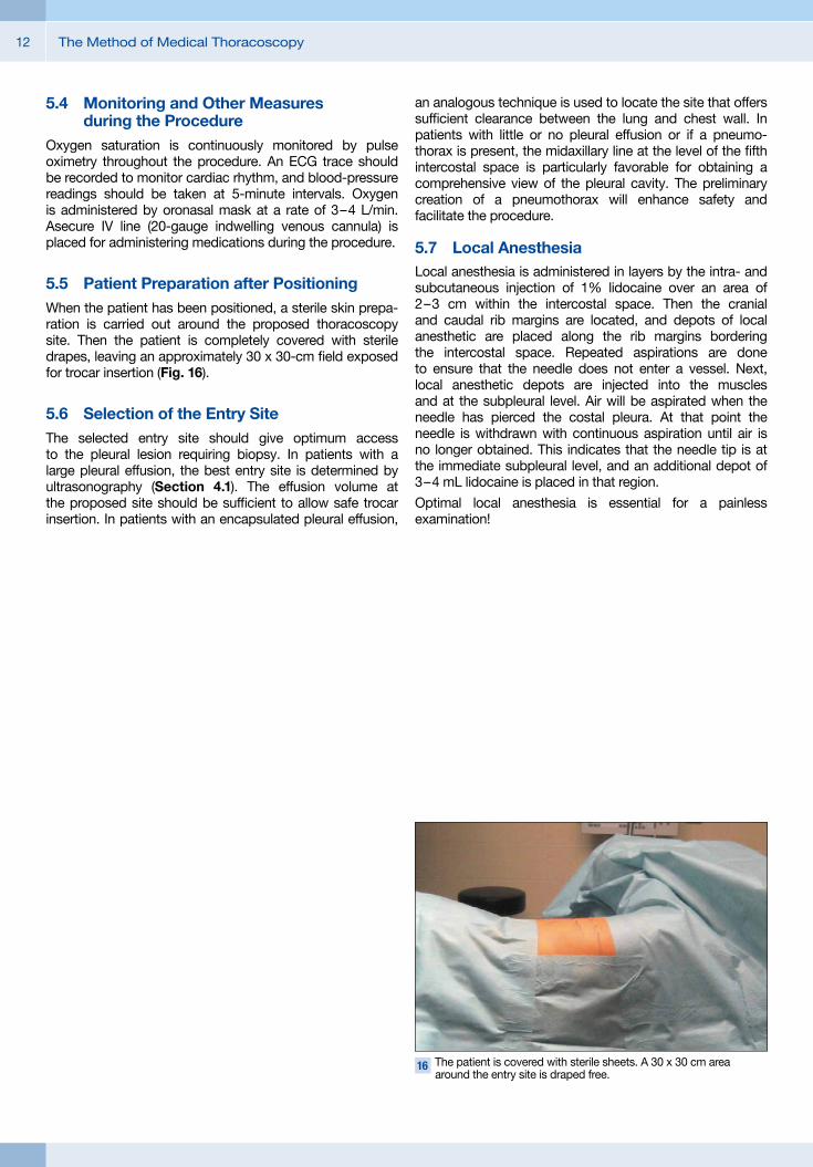
5.5 Patient Preparation after PositioningWhen the patient has been positioned, a sterile skin prepa-ration is carried out around the proposed thoracoscopy site. Then the patient is completely covered with sterile drapes, leaving an approximately 30 x 30-cm field exposed for trocar insertion (Fig. 16).
5.6 Selection of the Entry SiteThe selected entry site should give optimum access to the pleural lesion requiring biopsy. In patients with a large pleural effusion, the best entry site is determined by ultrasonography (Section 4.1). The effusion volume at the proposed site should be sufficient to allow safe trocar insertion. In patients with an encapsulated pleural effusion,
an analogous technique is used to locate the site that offers sufficient clearance between the lung and chest wall. In patients with little or no pleural effusion or if a pneumo-thorax is present, the midaxillary line at the level of the fifth intercostal space is particularly favorable for obtaining a comprehensive view of the pleural cavity. The preliminary creation of a pneumothorax will enhance safety and facilitate the procedure.
5.7 Local AnesthesiaLocal anesthesia is administered in layers by the intra- and subcutaneous injection of 1% lidocaine over an area of 2 – 3 cm within the intercostal space. Then the cranial and caudal rib margins are located, and depots of local anesthetic are placed along the rib margins bordering the intercostal space. Repeated aspirations are done to ensure that the needle does not enter a vessel. Next, local anesthetic depots are injected into the muscles and at the subpleural level. Air will be aspirated when the needle has pierced the costal pleura. At that point the needle is withdrawn with continuous aspiration until air is no longer obtained. This indicates that the needle tip is at the immediate subpleural level, and an additional depot of 3 – 4 mL lidocaine is placed in that region.
Optimal local anesthesia is essential for a painless examination!
16 The patient is covered with sterile sheets. A 30 x 30 cm area around the entry site is draped free.
13The Method of Medical Thoracoscopy
5.8 Conscious SedationConscious sedation should be administered by a physician with comprehensive experience in that area. At our center, this responsibility is assumed by an anesthesiologist. The operator performing the thoracoscopy should not also be responsible for conscious sedation, so that he or she can devote full attention to the procedure. We recommend a titrated dose of midazolam and piritramide for conscious sedation, starting with an initial i.v. dose of 2 – 3 mg midazolam and 5 mg piritramide. An alternative analgesic is ketamine. Also, propofol (depending on response) may be given in combination with an analgesic.28
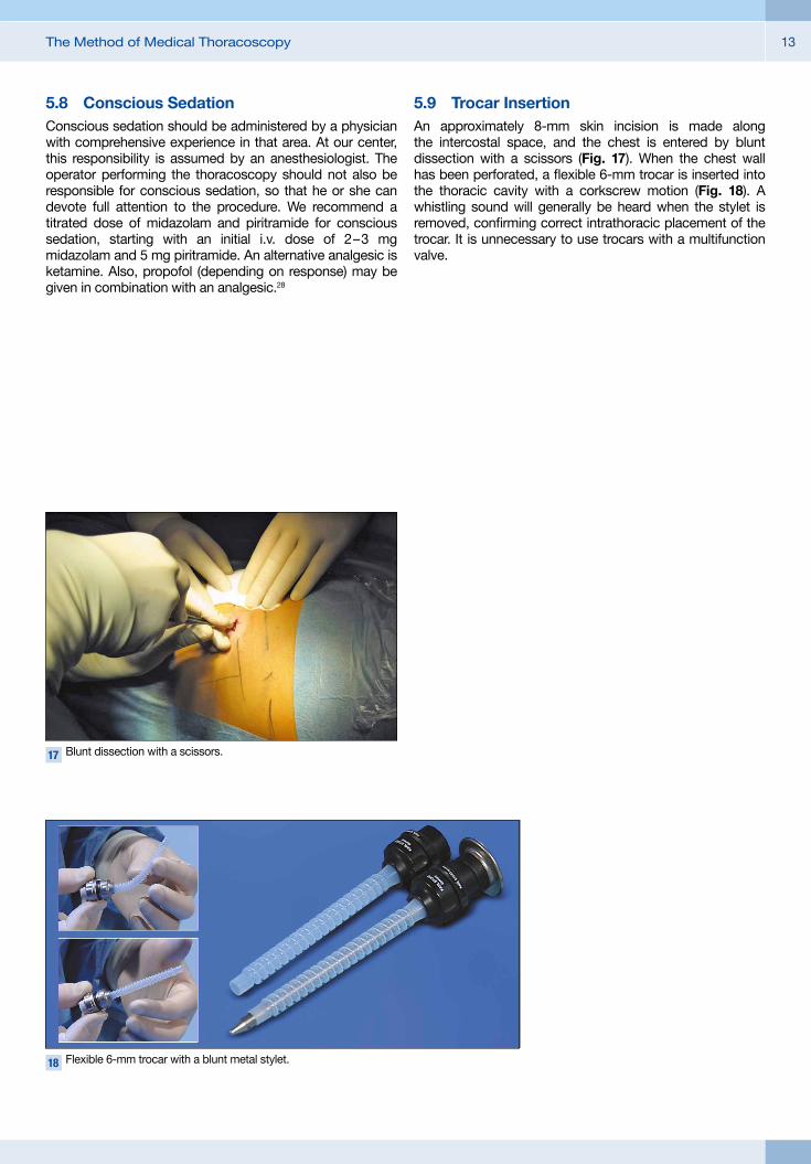
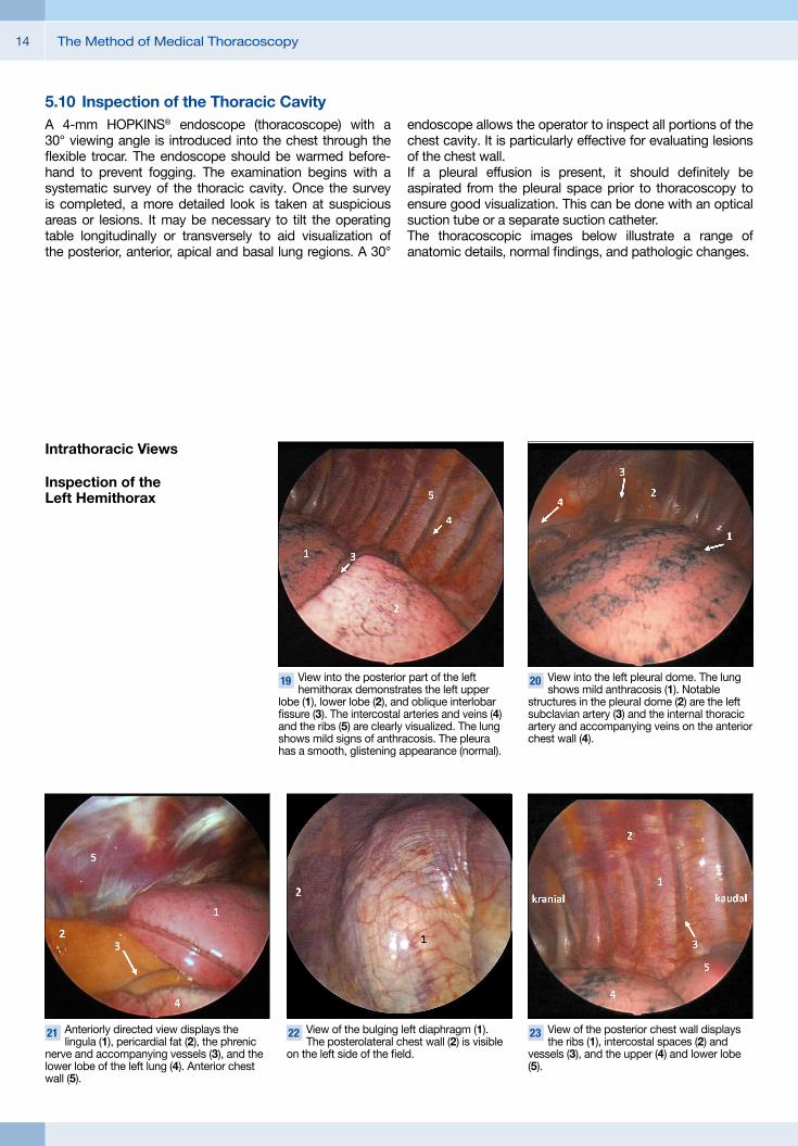
5.9 Trocar InsertionAn approximately 8-mm skin incision is made along the intercostal space, and the chest is entered by blunt dissection with a scissors (Fig. 17). When the chest wall has been perforated, a flexible 6-mm trocar is inserted into the thoracic cavity with a corkscrew motion (Fig. 18). A whistling sound will generally be heard when the stylet is removed, confirming correct intrathoracic placement of the trocar. It is unnecessary to use trocars with a multifunction valve.
17 Blunt dissection with a scissors.
18 Flexible 6-mm trocar with a blunt metal stylet.
The Method of Medical Thoracoscopy14
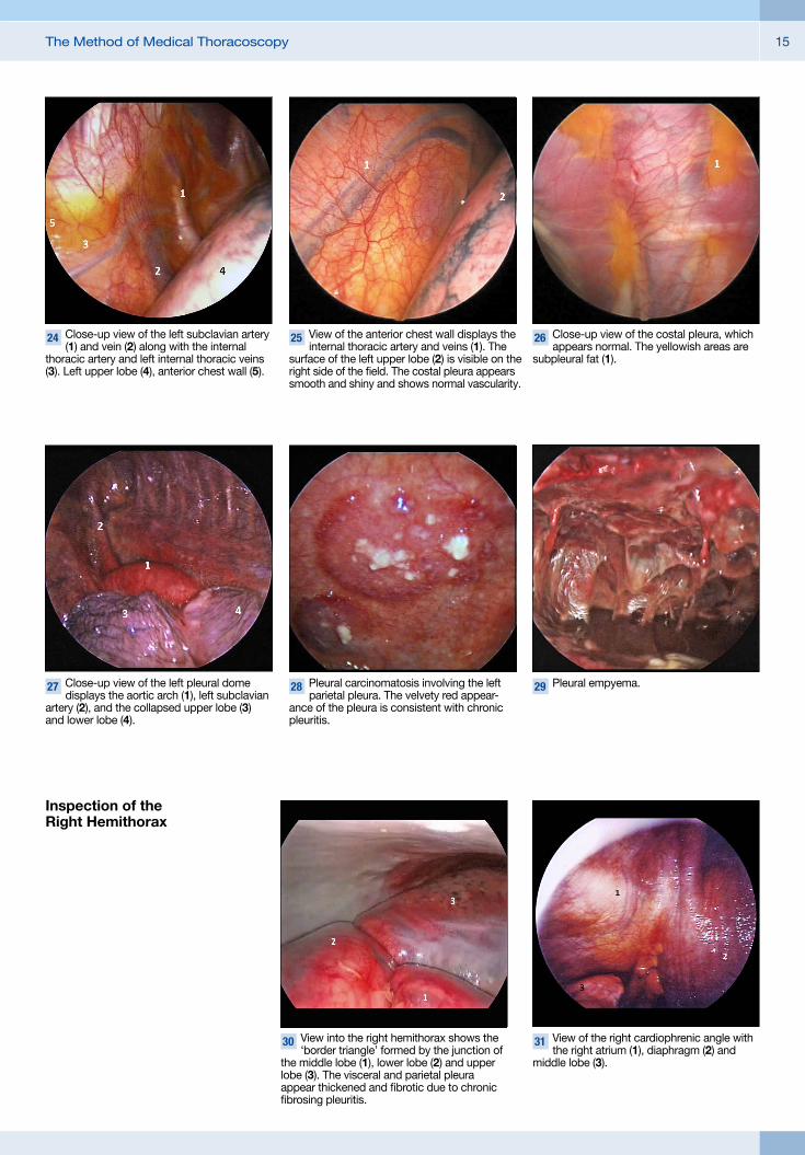
5.10 Inspection of the Thoracic CavityA 4-mm HOPKINS® endoscope (thoracoscope) with a 30° viewing angle is introduced into the chest through the flexible trocar. The endoscope should be warmed before-hand to prevent fogging. The examination begins with a systematic survey of the thoracic cavity. Once the survey is completed, a more detailed look is taken at suspicious areas or lesions. It may be necessary to tilt the operating table longitudinally or transversely to aid visualization of the posterior, anterior, apical and basal lung regions. A 30°
endoscope allows the operator to inspect all portions of the chest cavity. It is particularly effective for evaluating lesions of the chest wall.If a pleural effusion is present, it should definitely be aspirated from the pleural space prior to thoracoscopy to ensure good visualization. This can be done with an optical suction tube or a separate suction catheter.The thoracoscopic images below illustrate a range of anatomic details, normal findings, and pathologic changes.
19 View into the posterior part of the left hemithorax demonstrates the left upper
lobe (1), lower lobe (2), and oblique interlobar fissure (3). The intercostal arteries and veins (4) and the ribs (5) are clearly visualized. The lung shows mild signs of anthracosis. The pleura has a smooth, glistening appearance (normal).
20 View into the left pleural dome. The lung shows mild anthracosis (1). Notable
structures in the pleural dome (2) are the left subclavian artery (3) and the internal thoracic artery and accompanying veins on the anterior chest wall (4).
21 Anteriorly directed view displays the lingula (1), pericardial fat (2), the phrenic
nerve and accompanying vessels (3), and the lower lobe of the left lung (4). Anterior chest wall (5).
22 View of the bulging left diaphragm (1). The posterolateral chest wall (2) is visible
on the left side of the field.
23 View of the posterior chest wall displays the ribs (1), intercostal spaces (2) and
vessels (3), and the upper (4) and lower lobe (5).
Intrathoracic Views
Inspection of the Left Hemi thorax
15The Method of Medical Thoracoscopy
24 Close-up view of the left subclavian artery (1) and vein (2) along with the internal
thoracic artery and left internal thoracic veins (3). Left upper lobe (4), anterior chest wall (5).
25 View of the anterior chest wall displays the internal thoracic artery and veins (1). The
surface of the left upper lobe (2) is visible on the right side of the field. The costal pleura appears smooth and shiny and shows normal vascularity.
26 Close-up view of the costal pleura, which appears normal. The yellowish areas are
subpleural fat (1).
27 Close-up view of the left pleural dome displays the aortic arch (1), left subclavian
artery (2), and the collapsed upper lobe (3) and lower lobe (4).
28 Pleural carcinomatosis involving the left parietal pleura. The velvety red appear-
ance of the pleura is consistent with chronic pleuritis.
29 Pleural empyema.
Inspection of the Right Hemi thorax
30 View into the right hemithorax shows the ‘border triangle’ formed by the junction of
the middle lobe (1), lower lobe (2) and upper lobe (3). The visceral and parietal pleura appear thickened and fibrotic due to chronic fibrosing pleuritis.
31 View of the right cardiophrenic angle with the right atrium (1), diaphragm (2) and
middle lobe (3).
The Method of Medical Thoracoscopy16
5.11 Thoracoscopic Biopsy and Lysis of Adhesions All lesions visible at thoracoscopy should be biopsied. This can be done with an optical biopsy forceps (Fig. 32). With diffuse pleural diseases, multiple pleural biopsies should be taken to obtain ample material from various sites. If the desired biopsy sites are not accessible through the initial portal, a second trocar can be inserted under local anesthesia and thoracoscopic guidance so that the necessary biopsies can be taken from that position. It is rarely necessary to add a second portal, however. Heavy bleeding from biopsy sites can be controlled by electrocautery with an optically guided hook electrode (Figs. 33, 34).Biopsies can be taken from the parietal and visceral pleura as required. When tissue is sampled from the costal pleura, the area about the caudal rib margins should be avoided to prevent injury to intercostal vessels. If the visceral pleura is biopsied and an air leak occurs, it will generally close spontaneously within 7 days.
When adhesions are present, they can be cleared by side-to-side movements of the thoracoscope itself if they are very soft. Firmer adhesions can be divided with the hook electrode. As a note of caution, dense adhesions may transmit vessels of significant size that can bleed profusely if severed. If this complication arises, it may be necessary to create a second portal for hemostasis with a cautery probe or clip. Complicated parapneumonic effusions and pleural empyema may also require a second portal, through which adhesions can be lysed under vision with a probe. As a general rule, however, it is rarely necessary to create a second portal.
32 Biopsy with an optical forceps. 33 Electrocautery with an optically guided hook electrode.
34 Result of hemostasis by electrocautery.
17The Method of Medical Thoracoscopy
5.12 Talc PleurodesisThe indications for talc pleurodesis are described in Sect. 2.1. It must be possible for the lung to expand fully and occupy all of the chest cavity. The expansibility of the lung is tested by introducing the thoracoscope and optical suction tube into the chest. The air is then suctioned from the chest while lung expansion is observed with the thoracoscope. Once the lung has expanded completely, it is ready for talc pleurodesis. Talcum powder is blown into the pleural space with an optical powder dispenser (Figs. 35 – 37). For effective pleurodesis, approximately 4 gr of talc is blown into the pleural cavity under thoracoscopic vision. The operator should make sure that all effusion has been aspirated from the chest prior to talc pleurodesis and that the entire lung and chest wall are coated with a thin film of talcum powder.
The talc should be applied in fractionated doses. Talc insufflation raises the intrathoracic pressure, and a potentially dangerous intrathoracic pressure rise is avoided by intermittently pausing the insufflation and removing the dispenser. Air escaping from the chest cavity makes an audible whistling sound.Talc pleurodesis is a very effective procedure with a reported success rate of 83 – 93%.2, 13, 31 Even long-term studies have documented success rates higher than 80%.In recent years there have been efforts to replace talc pleurodesis with other procedures. Studies in relatively small case numbers have shown that the instillation of silver nitrate solution into the pleura can also induce pleuro desis.21 To date, there are no comprehensive studies showing that this technique is superior to talc pleurodesis.
35 Optical powder dispenser. 36 Talc pleurodesis with a powder dispenser under vision.
37 Intrathoracic view after talc application. The talc dust has been uniformly
distributed over the lung surface.
The Method of Medical Thoracoscopy18
5.13 Concluding the ProcedureAt the end of the procedure, the entire pleural cavity should be carefully reinspected so that any bleeding from a biopsy or adhesiolysis can be detected and controlled.A 24-Charr. drain is then introduced into the pleura under vision. We recommend the preplacement of vertical mattress sutures after trocar removal. Then a 24-Charr. drain is passed through the trocar port into the chest cavity. During this time the drain should be stabilized with a probe passed into its lumen through a side hole in the drain. The probe is removed, and the 4-mm HOPKINS® endoscope is inserted into the drain lumen through a side hole and advanced until the interior of the chest cavity can be seen. Now the drain can be advanced posteriorly along a
para vertebral path under direct visual control (Figs. 38 – 40). Next the thoracoscope is withdrawn, removing it completely from the drain. At this point the thoracoscope and drainage tube are in the original trocar port, so the thoracoscope can again be used to check the drain position and adjust it as needed. After the thoracoscope is removed, the drain can be secured with the preplaced suture. It is advisable to tie one knot, then wind the suture several times around the drain and tie a final knot. Next the chest wall is cleaned and an adhesive dressing is placed around the drain.The drain is connected to a suction pump. We prefer an electronic pump that also indicates airflow (Fig. 41).
38 The thoracoscope is inserted into the drain lumen, and the drain is carefully
advanced under vision.
39 40 Placement of the drain under vision.
41 The drainage tube is connected to the suction pump. In the case shown, the suction was set to 20 cm H2O. The flow rate is still
420 mL/min but should fall to zero within minutes if there is no air leak.
The Method of Medical Thoracoscopy 19
Management after Thoracoscopy
Patients are monitored in the recovery room for 1– 2 hours after thoracoscopy. A chest radiograph is obtained when the patient is returned to the floor. This is necessary to check for lung reexpansion and drain position.Adequate pain management is important after thoraco-scopy. For this purpose, 50 mg pethidine may be given subcutaneously or 0.2 – 0.4 mg buprenorphine sublingually as needed. Following talc pleurodesis, care should be taken that the patient does not receive corticosteroids or NSAIDs as they would suppress the inflammatory
response necessary for pleurodesis. Vital signs (pulse and blood pressure) should be taken hourly for the first 6 hours after the procedure. Nursing staff should make sure that the chest tube remains patent. Drain output and fluid appearance should be recorded and documented. When the output falls below 100 mL/24 hours, the drain may be removed. It is important to ambulate the patient immediately after thoracoscopy. Prophylactic antibiotics are unnecessary.
Complications
The complication rate after thoracoscopy is low. Our own studies indicate a rate of 2.34%. A total of 214 cases were reviewed.7 More recent studies also document the safety of thoracoscopy. Brims et al. (2012)4 report an infection rate of 10.5% in 57 cases (4 cases of pneumonia, 2 empyemas).
This appears relatively high and is not consistent with our experience. The longer the drain remains in place, however, the higher the risk of infection. No thoracoscopy-related deaths have been reported.
Summary
Medical thoracoscopy is an economical, highly effective interventional procedure that can be learned quickly, has few complications, and permits the rapid and safe diagnosis of pleural diseases. This opinion is shared by many other authors.16, 24 If necessary, thoracoscopy can also be used to investigate lesions in the peripheral lung and mediastinum. ‘Minithoracoscopy,’ which employs thoracoscopes 2 – 5 mm in diameter, is also described by other authors as a highly effective, minimally invasive diagnostic procedure.27
In recent years medical thoracoscopy has been increasingly applied for therapeutic purposes. It has a major role in the treatment of complicated parapneumonic pleural effusions and pleural empyema. At present, thoracoscopic talc pleurodesis is the most effective and economical method that we have for inducing pleurodesis.
6
7
8
The Method of Medical Thoracoscopy20
References1. Anforderungen der Hygiene bei Operationen und
anderen invasiven Eingriffen Mitteilung der Kommission für Krankenhaushygiene und Infektionsprävention am Robert-Koch-Institut. Bundesgesundheitsblatt – Gesundheitsforschung – Gesundheitsschutz. 2000;43(8):644-8.
2. ARAPIS K, CALIANDRO R, STERN JB, et al.: Thoraco-scopic palliative treatment of malignant pleural effusions: results in 273 patients. Surg Endosc. 2006;20(6):919–23.
3. BRIDEVAUX PO, TSCHOPP JM, CARDILLO G, et al.: Short-term safety of thoracoscopic talc pleurodesis for recurrent primary spontaneous pneumothorax: a prospective European multicentre study. Eur Respir J. 2011;38(4):770–3.
4. BRIMS FJ, ARIF M, CHAUHAN AJ: Outcomes and complications following medical thoracoscopy. Clin Respir J. 2012;6(3):144–9.
5. BRUTSCHE MH, TASSI GF, GYORIK S, et al.: Treatment of sonographically stratified multiloculated thoracic empyema by medical thoracoscopy. Chest. 2005;128(5):3303–9.
6. FORLANINI C: Zur Behandlung der Lungenschwind-sucht durch künstlich erzeugten Pneumothorax. Deutsche med Wochenschr. 1906;32:1401–5.
7. HEINE R: Methode und Ergebnisse der Pleuroskopie. Diplomarbeit, Martin-Luther-Universität Halle- Wittenberg. 1985.
8. HIEN P: Diagnostische Thorakoskopie. Praktische Pneumologie. Springer Berlin Heidelberg; 2012. p. 71–3.
9. JACOBAEUS HC: Endopleurale Operation unter Leitung des Thorakoskopes. Beitr Klin Tuberk. 1916;35:1–35.
10. JACOBAEUS HC: Kurze Übersicht über meine Erfahrungen mit der Laparo-Thorakoskopie. Münchener med Wochenschr. 1911;58:2017–9.
11. KELLING G: Über Ösophagoskopie, Gastroskopie und Kölioskopie. Münchener med Wochenschr. 1902;49:21–4.
12. KERN L, ROBERT J, BRUTSCHE M: Management of parapneumonic effusion and empyema: medical thoracoscopy and surgical approach. Respiration. 2011;82(2):193–6.
13. KOLSCHMANN S, BALLIN A, GILLISSEN A: Clinical efficacy and safety of thoracoscopic talc pleurodesis in malignant pleural effusions. Chest. 2005;128(3):1431–5.
14. KORBSCH R: Lehrbuch und Atlas der Laparo- und Thorakoskopie. München: J.F. Lehmann; 1927 (Zit. nach Matzel W, 1966).
15. LEE WJ, KIM HJ, PARK JH, et al.: Chemical pleurodesis for the management of refractory hepatic hydrothorax in patients with decompensated liver cirrhosis. Korean J Hepatol. 2011;17(4):292–8.
16. LODDENKEMPER R: Thoracoscopy – state of the art. Eur Respir J. 1998;11(1):213–21.
17. MATZEL W: Diagnostische Thorakoskopie bei intrathorakalen Rundherden. Z Tbk. 1963;120:1–13.
18. MATZEL W: Thorakoskopie. Z Tbk. 1964;122:252–3.
19. MATZEL W: Thorakoskopie. In: Emmrich R, Hrsg. Arbeitsmethoden der inneren Medizin und ihr verwandter Gebiete. 3. Ausg. Jena: Gustav Fischer; 1966. p. 177–92.
20. MORENO-MERINO S, CONGREGADO M, GALLARDO G, et al.: Comparative study of talc poudrage versus pleural abrasion for the treatment of primary spontaneous pneumothorax. Interact Cardiovasc Thorac Surg. 2012;15(1):81–5.
21. PASCHOALINI MDA S, VARGAS FS, MARCHI E, et al.: Prospective randomized trial of silver nitrate vs talc slurry in pleurodesis for symptomatic malignant pleural effusions. Chest. 2005;128(2):684–9.
22. RAVAGLIA C, GURIOLI C, TOMASSETTI S, et al.: Is medical thoracoscopy efficient in the management of multiloculated and organized thoracic empyema? Respiration. 2012;84(3):219–24.
23. SATTLER A: 3. Tagung der wissenschaftlichen Gesellschaft für Tuberkulose und Lungenkrankheiten. Z Tbk. 1964;122:257.
24. SIMPSON G: Medical thoracoscopy in an Australian regional hospital. Intern Med J. 2007;37(4):267–9.
25. SOLER M, WYSER C, BOLLIGER CT, et al.: Treatment of early parapneumonic empyema by "medical" thoracoscopy. Schweiz Med Wochenschr. 1997;127(42): 1748–53.
26. STANZEL F: Instrumentarium für die internistische Thorakoskopie – „Pleuroskopie“. EndoGramm, KARL STORZ GmbH & Co KG. 2005:1–8.
27. TASSI GF, MARCHETTI GP, PINELLI V: Minithoracoscopy: a complementary technique for medical thoracoscopy. Respiration. 2011;82(2):204–6.
28. TSCHOPP JM, PUREK L, FREY JG, et al.: Titrated sedation with propofol for medical thoracoscopy: a feasibility and safety study. Respiration. 2011;82(5): 451–7.
29. TSCHOPP JM, RAMI-PORTA R, NOPPEN M, et al.: Management of spontaneous pneumothorax: state of the art. Eur Respir J. 2006;28(3):637–50.
30. VAZIRI M, ABED O: Management of thoracic empyema: review of 112 cases. Acta Med Iran. 2012;50(3):203–7.
31. WALKER-RENARD PB, VAUGHAN LM, SAHN SA: Chemical pleurodesis for malignant pleural effusions. Ann Intern Med. 1994;120(1):56–64.
21The Method of Medical Thoracoscopy
Instrument Set for Medical Thoracoscopy
Excerpts from the following catalogs:
THORAX and TELEPRESENCE, IMAGING SYSTEMS, DOCUMENTATION AND ILLUMINATION
Die Methode der internistischen Thorakoskopie22
Es wird empfohlen, vor der Verwendung die Eignung der Produkte für den geplanten Eingriff zu überprüfen.Es wird empfohlen, vor der Verwendung die Eignung der Produkte für den geplanten Eingriff zu überprüfen.
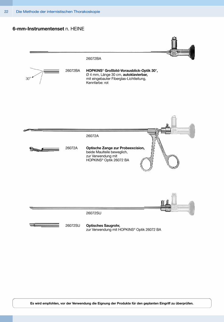
6-mm-Instrumentenset n. HEINE
26072A Optische Zange zur Probeexcision, beide Maulteile beweglich, zur Verwendung mit HOPKINS® Optik 26072 BA
26072A
26072SU Optisches Saugrohr, zur Verwendung mit HOPKINS® Optik 26072 BA
26072SU
26072BA HOPKINS® Großbild-Vorausblick-Optik 30°, Ø 4 mm, Länge 30 cm, autoklavierbar, mit eingebauter Fiberglas-Lichtleitung, Kennfarbe: rot
26072BA
23Die Methode der internistischen Thorakoskopie
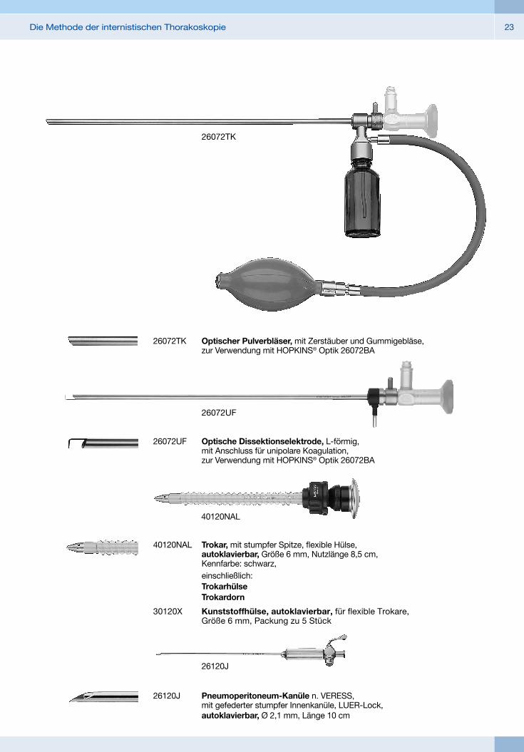
40120NAL Trokar, mit stumpfer Spitze, flexible Hülse, autoklavierbar, Größe 6 mm, Nutzlänge 8,5 cm, Kennfarbe: schwarz,
einschließlich: Trokarhülse Trokardorn
30120X Kunststoffhülse, autoklavierbar, für flexible Trokare, Größe 6 mm, Packung zu 5 Stück
40120NAL
26072UF Optische Dissektionselektrode, L-förmig, mit Anschluss für unipolare Koagulation, zur Verwendung mit HOPKINS® Optik 26072BA
26072UF
26072TK Optischer Pulverbläser, mit Zerstäuber und Gummigebläse, zur Verwendung mit HOPKINS® Optik 26072BA
26072TK
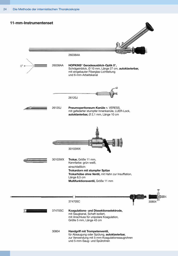
26120J Pneumoperitoneum-Kanüle n. VERESS, mit gefederter stumpfer Innenkanüle, LUER-Lock, autoklavierbar, Ø 2,1 mm, Länge 10 cm
26120J
Die Methode der internistischen Thorakoskopie24
26038AA HOPKINS® Geradeausblick-Optik 0°, Schrägeinblick, Ø 10 mm, Länge 27 cm, autoklavierbar, mit eingebauter Fiberglas-Lichtleitung und 6-mm-Arbeitskanal
26038AA
11-mm-Instrumentenset
26120J Pneumoperitoneum-Kanüle n. VERESS, mit gefederter stumpfer Innenkanüle, LUER-Lock, autoklavierbar, Ø 2,1 mm, Länge 10 cm
26120J
30804 Handgriff mit Trompetenventil, für Absaugung oder Spülung, autoklavierbar, zur Verwendung mit 5-mm-Koagulationssaugrohren und 5-mm-Saug- und Spülrohren
37470SC Koagulations- und Dissektionselektrode, mit Saugkanal, Schaft isoliert, mit Anschluss für unipolare Koagulation, Größe 5 mm, Länge 43 cm
37470SC 30804
30103WX Trokar, Größe 11 mm, Kennfarbe: grün-weiß,
einschließlich: Trokardorn mit stumpfer Spitze Trokarhülse ohne Ventil, mit Hahn zur Insufflation,
Länge 8,5 cm Multifunktionsventil, Größe 11 mm
30103WX

25Die Methode der internistischen Thorakoskopie
40492TK Pulverbläser, mit Gummigebläse, Schaft gerade, Größe 5 mm, Länge 42 cm,
einschließlich: Schaft Gummiball Schlauch Glas
40492TK
26778UF Koagulations- und Dissektionselektrode, L-förmig, mit Anschluss für unipolare Koagulation, Größe 5 mm, Länge 43 cm
26778UF
34421MB
34421MB CLICKLINE Zange zur Probeexzision, drehbar, zerlegbar, isoliert, mit Anschluss für unipolare Koagulation, mit LUER-Lock-Spülanschluss zur Reinigung, ein Maulteil beweglich, Größe 5 mm, Länge 43 cm,
einschließlich: Kunststoff-Handgriff, ohne Raste Metall-Außenschaft Zangeneinsatz
Die Methode der internistischen Thorakoskopie26
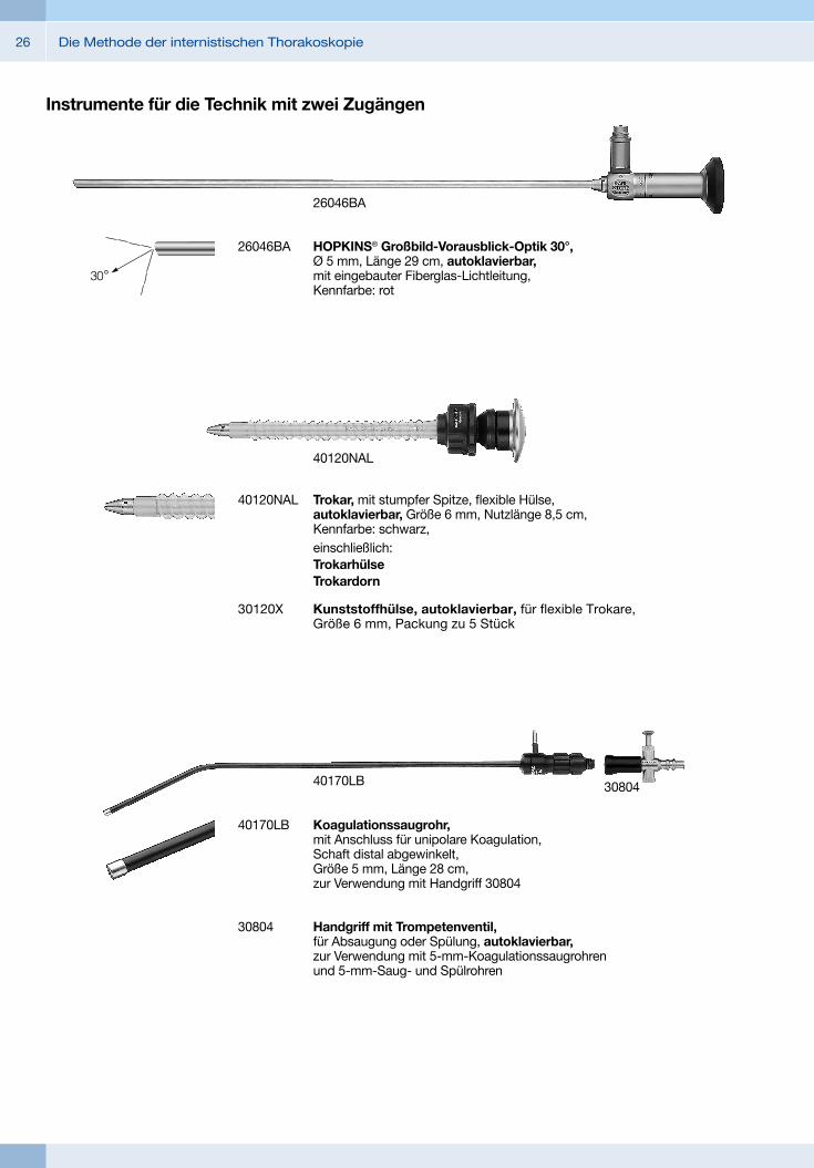
26046BA
26046BA HOPKINS® Großbild-Vorausblick-Optik 30°, Ø 5 mm, Länge 29 cm, autoklavierbar, mit eingebauter Fiberglas-Lichtleitung, Kennfarbe: rot
40120NAL Trokar, mit stumpfer Spitze, flexible Hülse, autoklavierbar, Größe 6 mm, Nutzlänge 8,5 cm, Kennfarbe: schwarz,
einschließlich: Trokarhülse Trokardorn
30120X Kunststoffhülse, autoklavierbar, für flexible Trokare, Größe 6 mm, Packung zu 5 Stück
40120NAL
Instrumente für die Technik mit zwei Zugängen
40170LB Koagulationssaugrohr, mit Anschluss für unipolare Koagulation, Schaft distal abgewinkelt, Größe 5 mm, Länge 28 cm, zur Verwendung mit Handgriff 30804
30804 Handgriff mit Trompetenventil, für Absaugung oder Spülung, autoklavierbar, zur Verwendung mit 5-mm-Koagulationssaugrohren und 5-mm-Saug- und Spülrohren
40170LB 30804
27Die Methode der internistischen Thorakoskopie
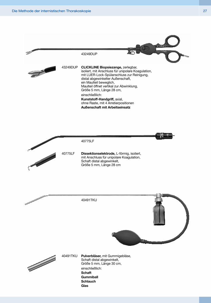
40775LF Dissektionselektrode, L-förmig, isoliert, mit Anschluss für unipolare Koagulation, Schaft distal abgewinkelt, Größe 5 mm, Länge 28 cm
40775LF
40491TKU Pulverbläser, mit Gummigebläse, Schaft distal abgewinkelt, Größe 5 mm, Länge 30 cm,
einschließlich: Schaft Gummiball Schlauch Glas
40491TKU
43249DUP
43249DUP CLICKLINE Biopsiezange, zerlegbar, isoliert, mit Anschluss für unipolare Koagulation, mit LUER-Lock-Spülanschluss zur Reinigung, distal abgewinkelter Außenschaft, ein Maulteil beweglich, Maulteil öffnet vertikal zur Abwinklung, Größe 5 mm, Länge 28 cm,
einschließlich: Kunststoff-Handgriff, axial,
ohne Raste, mit 4 Arretierpositionen Außenschaft mit Arbeitseinsatz
Die Methode der internistischen Thorakoskopie28
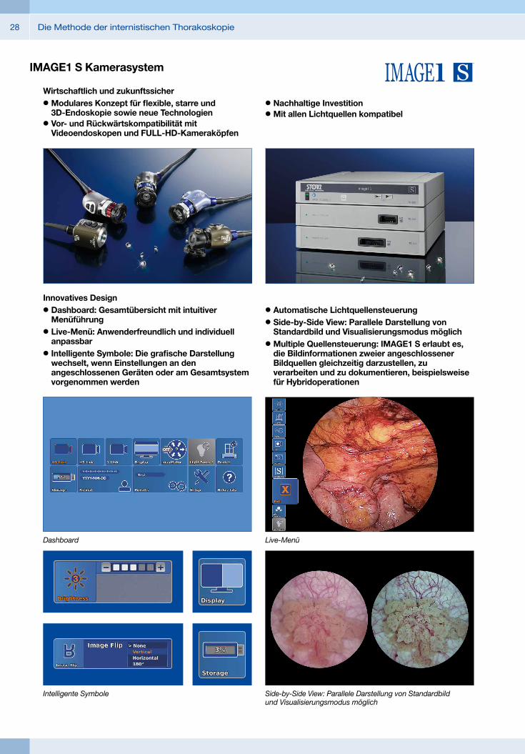
IMAGE1 S Kamerasystem
Wirtschaftlich und zukunftssicher## Modulares Konzept für flexible, starre und 3D-Endoskopie sowie neue Technologien
## Vor- und Rückwärtskompatibilität mit Videoendoskopen und FULL-HD-Kameraköpfen
## Nachhaltige Investition## Mit allen Lichtquellen kompatibel
Innovatives Design## Dashboard: Gesamtübersicht mit intuitiver Menüführung
## Live-Menü: Anwenderfreundlich und individuell anpassbar
## Intelligente Symbole: Die grafische Darstellung wechselt, wenn Einstellungen an den angeschlossenen Geräten oder am Gesamtsystem vorgenommen werden
## Automatische Lichtquellensteuerung## Side-by-Side View: Parallele Darstellung von Standardbild und Visualisierungsmodus möglich
## Multiple Quellensteuerung: IMAGE1 S erlaubt es, die Bildinformationen zweier angeschlossener Bildquellen gleichzeitig darzustellen, zu verarbeiten und zu dokumentieren, beispielsweise für Hybridoperationen
Dashboard Live-Menü
Side-by-Side View: Parallele Darstellung von Standardbild und Visualisierungsmodus möglich
Intelligente Symbole

29Die Methode der internistischen Thorakoskopie
Videoendoskopische Bildgebung## Sehr gute Bildqualität in FULL HD## Natürliche Farbwiedergabe
## Verschiedene IMAGE1 S Technologien für homogene Ausleuchtung, Kontrastanhebung und Farbtonverschiebungen
FULL HD-Bild CHROMA
FULL HD-Bild SPECTRA A*
FULL HD-Bild SPECTRA B **
* SPECTRA A : Nicht für Verkauf in den Vereinigten Staaten** SPECTRA B : Nicht für Verkauf in den Vereinigten Staaten
FULL HD-Bild CLARA
IMAGE1 S Kamerasystem
Die Methode der internistischen Thorakoskopie30
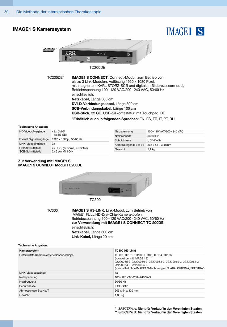
TC200DE* IMAGE1 S CONNECT, Connect-Modul, zum Betrieb von bis zu 3 Link-Modulen, Auflösung 1920 x 1080 Pixel, mit integriertem KARL STORZ-SCB und digitalem Bild prozessor modul, Betriebsspannung 100 – 120 VAC/200 – 240 VAC, 50/60 Hzeinschließlich:Netzkabel, Länge 300 cmDVI-D-Verbindungskabel, Länge 300 cmSCB-Verbindungskabel, Länge 100 cmUSB-Stick, 32 GB, USB-Silikontastatur, mit Touchpad, DE
* Erhältlich auch in folgenden Sprachen: EN, ES, FR, IT, PT, RU
Technische Angaben:
HD-Video-Ausgänge
Format Signalausgänge
LINK-Videoeingänge
USB-Schnittstelle SCB-Schnittstelle
- 2x DVI-D - 1x 3G-SDI
1920 x 1080p, 50/60 Hz
3x
4x USB, (2x vorne, 2x hinten) 2x 6 pin Mini-DIN
Netzspannung
Netzfrequenz
Schutzklasse
Abmessungen B x H x T
Gewicht
100 – 120 VAC/200 – 240 VAC
50/60 Hz
I, CF-Defib
305 x 54 x 320 mm
2,1 kg
TC300 IMAGE1 S H3-LINK, Link-Modul, zum Betrieb von IMAGE1 FULL HD-Drei-Chip-Kamera köpfen, Betriebsspannung 100 – 120 VAC/200 – 240 VAC, 50/60 Hz zur Verwendung mit IMAGE1 S CONNECT TC 200DEeinschließlich:Netzkabel, Länge 300 cmLink-Kabel, Länge 20 cm
TC300
TC300 (H3-Link)
TH100, TH101, TH102, TH103, TH104, TH106 (kompatibel mit IMAGE1 S) 22 2200 55-3, 22 2200 56-3, 22 2200 53-3, 22 2200 60-3, 22 2200 61-3, 22 2200 54-3, 22 2200 85-3 (kompatibel ohne IMAGE1 S-Technologien CLARA, CHROMA, SPECTRA*)
1x
100 – 120 VAC/200 – 240 VAC
50/60 Hz
I, CF-Defib
305 x 54 x 320 mm
1,86 kg
Technische Angaben:
Kamerasystem
Unterstützte Kameraköpfe/Videoendoskope
LINK-Videoausgänge
Netzspannung
Netzfrequenz
Schutzklasse
Abmessungen B x H x T
Gewicht
TC200DE
Zur Verwendung mit IMAGE1 S IMAGE1 S CONNECT Modul TC200DE
IMAGE1 S Kamerasystem
* SPECTRA A : Nicht für Verkauf in den Vereinigten Staaten** SPECTRA B : Nicht für Verkauf in den Vereinigten Staaten
31Die Methode der internistischen Thorakoskopie
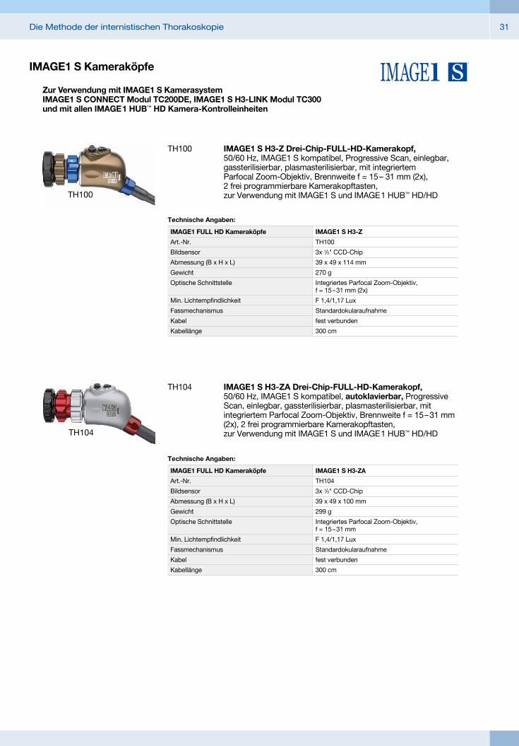
TH100 IMAGE1 S H3-Z Drei-Chip-FULL-HD-Kamerakopf, 50/60 Hz, IMAGE1 S kompatibel, Progressive Scan, einlegbar, gassterilisierbar, plasma sterilisierbar, mit integriertem Parfocal Zoom -Objektiv, Brennweite f = 15 – 31 mm (2x), 2 frei programmierbare Kamera kopftasten, zur Verwendung mit IMAGE1 S und IMAGE 1 HUB™ HD/HD
IMAGE1 S H3-Z
TH100
3x 1/3" CCD-Chip
39 x 49 x 114 mm
270 g
Integriertes Parfocal Zoom-Objektiv, f = 15 – 31 mm (2x)
F 1,4/1,17 Lux
Standardokularaufnahme
fest verbunden
300 cm
Technische Angaben:
IMAGE1 FULL HD Kameraköpfe
Art.-Nr.
Bildsensor
Abmessung (B x H x L)
Gewicht
Optische Schnittstelle
Min. Lichtempfindlichkeit
Fassmechanismus
Kabel
Kabellänge
TH100
TH104
TH104 IMAGE1 S H3-ZA Drei-Chip-FULL-HD-Kamerakopf, 50/60 Hz, IMAGE1 S kompatibel, autoklavierbar, Progressive Scan, einlegbar, gassterilisierbar, plasma sterilisierbar, mit integriertem Parfocal Zoom -Objektiv, Brennweite f = 15 – 31 mm (2x), 2 frei programmierbare Kamera kopftasten, zur Verwendung mit IMAGE1 S und IMAGE 1 HUB™ HD/HD
Technische Angaben:
IMAGE1 FULL HD Kameraköpfe
Art.-Nr.
Bildsensor
Abmessung (B x H x L)
Gewicht
Optische Schnittstelle
Min. Lichtempfindlichkeit
Fassmechanismus
Kabel
Kabellänge
IMAGE1 S H3-ZA
TH104
3x 1/3" CCD-Chip
39 x 49 x 100 mm
299 g
Integriertes Parfocal Zoom-Objektiv, f = 15 – 31 mm
F 1,4/1,17 Lux
Standardokularaufnahme
fest verbunden
300 cm
Zur Verwendung mit IMAGE1 S Kamerasystem IMAGE1 S CONNECT Modul TC200DE, IMAGE1 S H3-LINK Modul TC300 und mit allen IMAGE 1 HUB™ HD Kamera-Kontrolleinheiten
IMAGE1 S Kameraköpfe
Die Methode der internistischen Thorakoskopie32
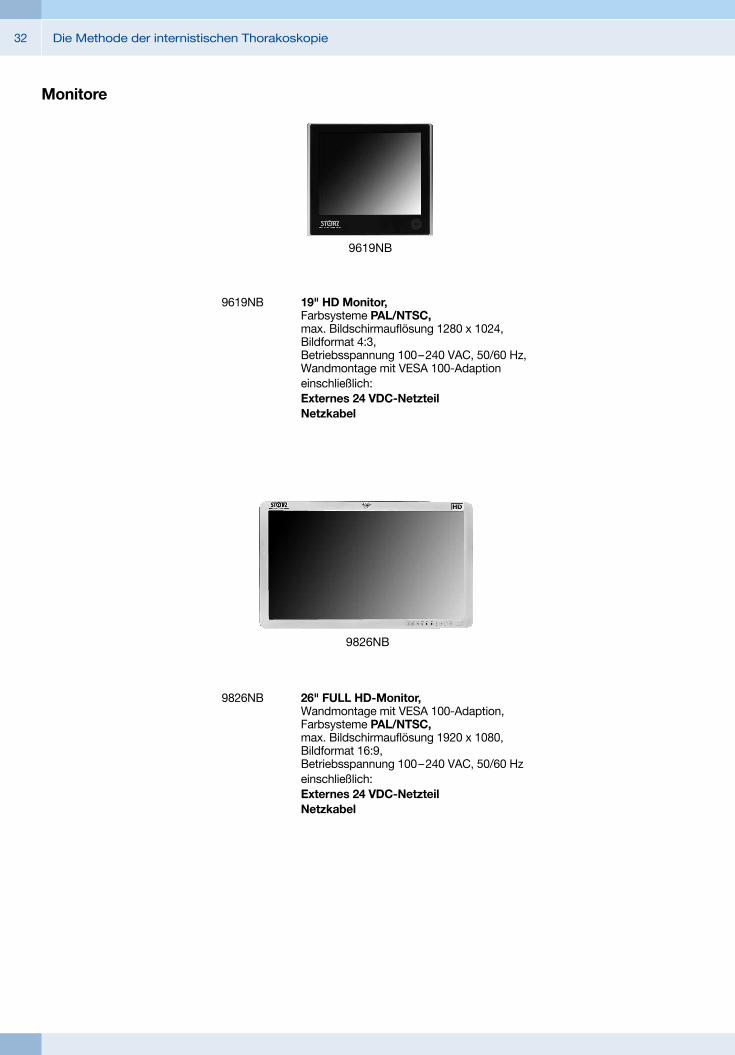
9826NB
9826NB 26" FULL HD-Monitor, Wandmontage mit VESA 100 -Adaption, Farbsysteme PAL/NTSC, max. Bildschirmauflösung 1920 x 1080, Bildformat 16:9, Betriebsspannung 100 – 240 VAC, 50/60 Hzeinschließlich:Externes 24 VDC-NetzteilNetzkabel
9619NB
9619NB 19" HD Monitor, Farbsysteme PAL/NTSC, max. Bildschirmauflösung 1280 x 1024, Bildformat 4:3, Betriebsspannung 100 – 240 VAC, 50/60 Hz, Wandmontage mit VESA 100-Adaptioneinschließlich:
Externes 24 VDC-Netzteil Netzkabel
Monitore
33Die Methode der internistischen Thorakoskopie
Monitore
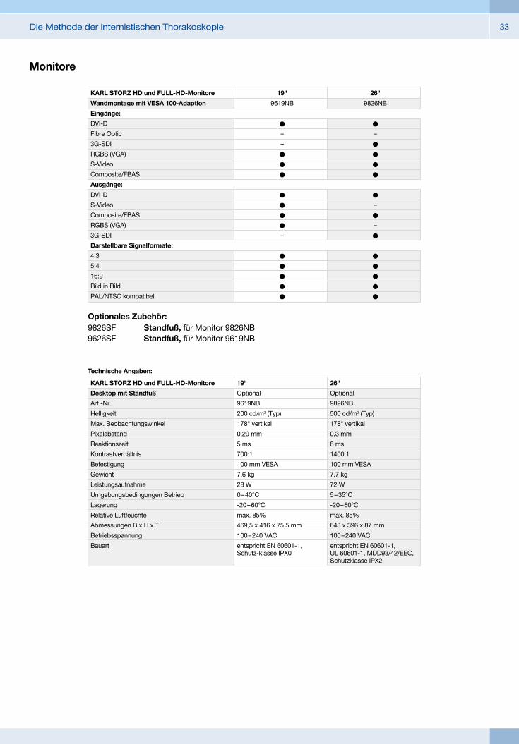
Optionales Zubehör:9826SF Standfuß, für Monitor 9826NB9626SF Standfuß, für Monitor 9619NB
26"
Optional
9826NB
500 cd/m2 (Typ)
178° vertikal
0,3 mm
8 ms
1400:1
100 mm VESA
7,7 kg
72 W
5 – 35°C
-20 – 60°C
max. 85%
643 x 396 x 87 mm
100 – 240 VAC
entspricht EN 60601-1, UL 60601-1, MDD93/42/EEC, Schutzklasse IPX2
19"
Optional
9619NB
200 cd/m2 (Typ)
178° vertikal
0,29 mm
5 ms
700:1
100 mm VESA
7,6 kg
28 W
0 – 40°C
-20 – 60°C
max. 85%
469,5 x 416 x 75,5 mm
100 – 240 VAC
entspricht EN 60601-1, Schutz-klasse IPX0
KARL STORZ HD und FULL-HD-Monitore
Desktop mit Standfuß
Art.-Nr.
Helligkeit
Max. Beobachtungswinkel
Pixelabstand
Reaktionszeit
Kontrastverhältnis
Befestigung
Gewicht
Leistungsaufnahme
Umgebungsbedingungen Betrieb
Lagerung
Relative Luftfeuchte
Abmessungen B x H x T
Betriebsspannung
Bauart
Technische Angaben:
26"
9826NB
l
–
l
l
l
l
l
–
l
–
l
l
l
l
l
l
19"
9619NB
l
–
–
l
l
l
l
l
l
l
–
l
l
l
l
l
KARL STORZ HD und FULL-HD-Monitore
Wandmontage mit VESA 100-Adaption
Eingänge:
DVI-D
Fibre Optic
3G-SDI
RGBS (VGA)
S-Video
Composite/FBAS
Ausgänge:
DVI-D
S-Video
Composite/FBAS
RGBS (VGA)
3G-SDI
Darstellbare Signalformate:
4:3
5:4
16:9
Bild in Bild
PAL/NTSC kompatibel
Die Methode der internistischen Thorakoskopie34
20 1614 20-1 Kaltlicht-Fontäne Power LED 175 SCB, mit integriertem SCB, High-Performance LED und einem KARL STORZ Lichtkabelanschluss, Betriebsspannung 110 – 240 VAC, 50/60 Hz
einschließlich: Netzkabel
Kaltlicht-Fontäne Power LED 175 SCB
ENDOFLATOR® 40 mit KARL STORZ SCBmit Speed-Flow-Insufflation (40 l/min)
UI400S1 ENDOFLATOR® 40 SCB, Set, mit integriertem SCB-Modul, Betriebsspannung 100 – 240 VAC, 50/60 Hz
einschließlich: ENDOFLATOR® 40 Netzkabel, Länge 300 cm SCB-Verbindungskabel, Länge 100 cm Universalschlüssel Insufflationsschlauchset, mit Gasfilter, steril,
zum Einmalgebrauch, Packung zu 5 Stück*
Entsprechend den individuellen Anforderungen des Kunden kann weiteres Zubehör angeboten werden.*
20 3210 08 DUOMAT® Saug- und Spülpumpe,
einschließlich: DUOMAT®,
Betriebsspannung 100 – 120, 230 – 240 VAC, 50/60 Hz
Netzkabel VACUsafe Promotion
Pack Absaugung, 2 l * (nicht abgebildet)
Entsprechend den individuellen Anforderungen des Kunden kann weiteres Zubehör angeboten werden.
DUOMAT®
Saug- und Spülpumpe
*
35Die Methode der internistischen Thorakoskopie
UG540 Monitorschwenkarm, höhen- und seitenverstellbar, links und rechts positionierbar, Schwenkbereich 180°, Ausladung 780 mm, ab Mitte 1170 mm, Tragkraft max. 15 kg, mit Monitorbefestigung VESA 75/100 zur Verwendung mit Gerätewagen UGxxx
UG540
Gerätewagen
UG220
UG220 Gerätewagen, breit, hoch, auf 4 antistatischen und feststellbaren Doppelrollen, Netzhauptschalter an der Abdeckung, Zentralholm mit integrierten elektrischen Unterverteilern mit 12 Steckplätzen, Potentialausgleichsanschlüssen,
Abmessungen in mm ( B x H x T): Gerätewagen: 830 x 1474 x 730, Konsole: 630 x 25 x 510, Rollendurchmesser: 150 mm
einschließlich: Bodenmodul Gerätewagen, breit Abdeckung Gerätewagen, breit Holmpaket Gerätewagen, hoch 3x Konsole, breit Schubladenblock mit Schloss, breit 2x Geräteschiene, lang Kamerahalter
Die Methode der internistischen Thorakoskopie36
Empfohlenes Zubehör für Gerätewagen
UG310 Trenntransformator, 200 – 240 V, 2000 VA, mit 3-fach Spezialsteckdosenleiste, Sicherungsautomat, 3 Potentialausgleichsanschlüssen, Abmessungen in mm (B x H x T): 330 x 90 x 495 mm, zur Verwendung mit Gerätewagen UGxxx
UG310
UG410 Isolationswächter, 200 – 240 V, zur Montage an Gerätewagen, Abmessungen Bedienteil in mm (B x H x T): 44 x 80 x 29 zur Verwendung mit Trenntransformator UG310
UG410
UG510 Monitorhaltearm, höhen- und seitenverstellbar, neigbar, seitlich montierbar links oder rechts, Schwenkbereich bis ca. 320°, Ausladung 530 mm, Tragkraft max. 15 kg, mit Monitorhalterung VESA 75/100, zur Verwendung mit Gerätewagen UGxxx
UG510


with the compliments of
KARL STORZ — ENDOSKOPE