Embed Size (px)
Citation preview
The Microscope and
Discovery of the Cell
The Microscope
An understanding of cells and the ultrastructure of cells had to wait until objects so small could be visualized by magnification.
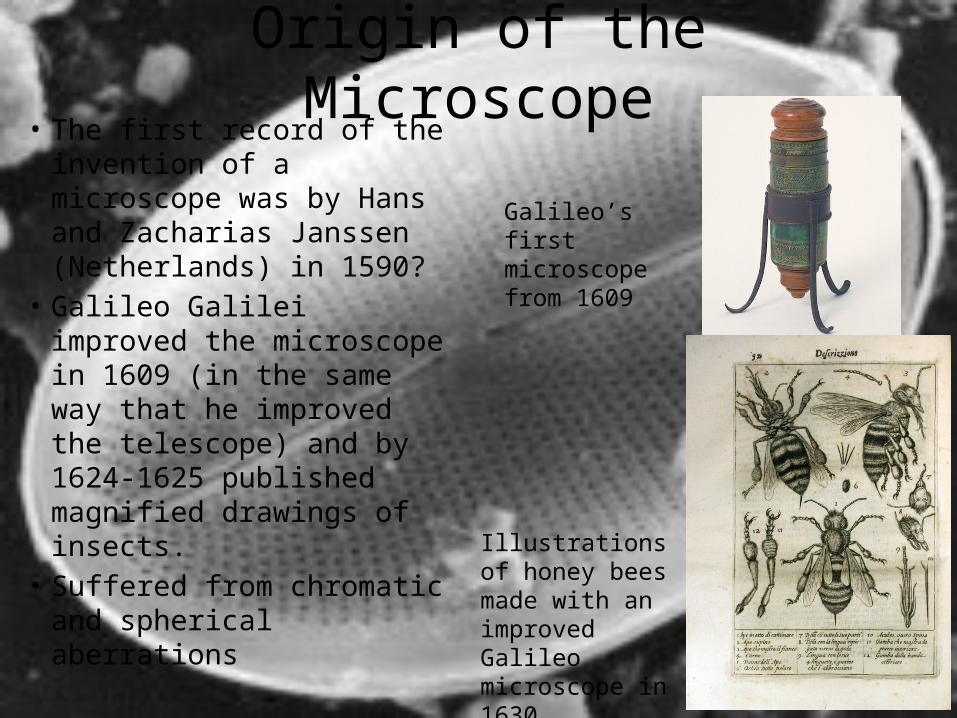
Origin of the Microscope• The first record of the
invention of a microscope was by Hans and Zacharias Janssen (Netherlands) in 1590?
• Galileo Galilei improved the microscope in 1609 (in the same way that he improved the telescope) and by 1624-1625 published magnified drawings of insects.
• Suffered from chromatic and spherical aberrations
Galileo’s first microscope from 1609
Illustrations of honey bees made with an improved Galileo microscope in 1630
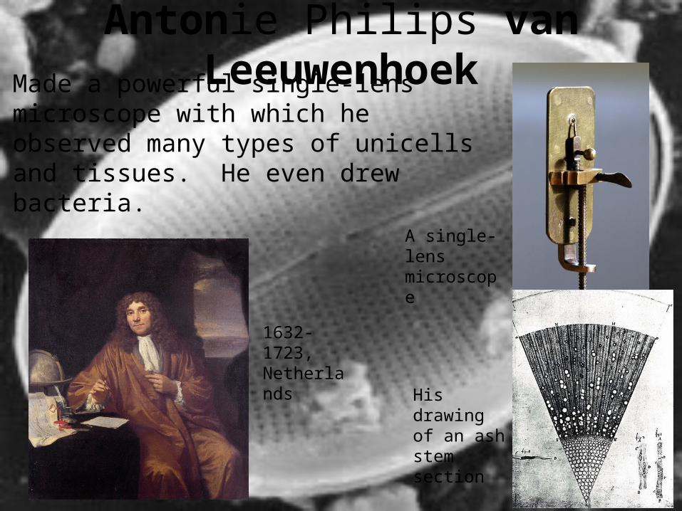
Antonie Philips van LeeuwenhoekMade a powerful single-lens microscope with which he observed many types of unicells and tissues. He even drew bacteria.
A single-lens microscope
His drawing of an ash stem section
1632-1723, Netherlands
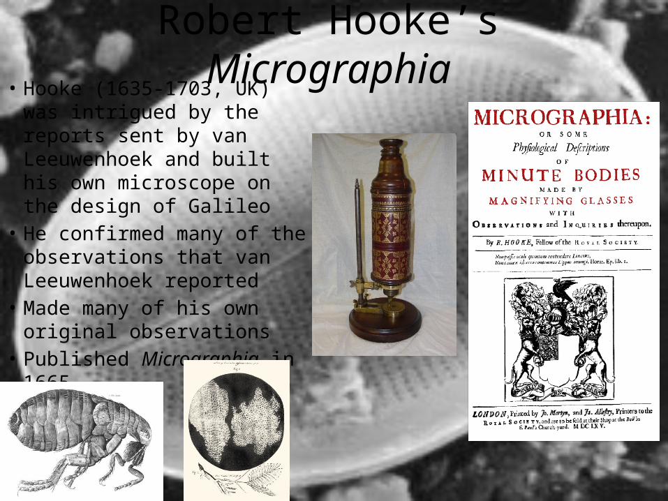
Robert Hooke’s Micrographia• Hooke (1635-1703, UK) was
intrigued by the reports sent by van Leeuwenhoek and built his own microscope on the design of Galileo
• He confirmed many of the observations that van Leeuwenhoek reported
• Made many of his own original observations
• Published Micrographia in 1665
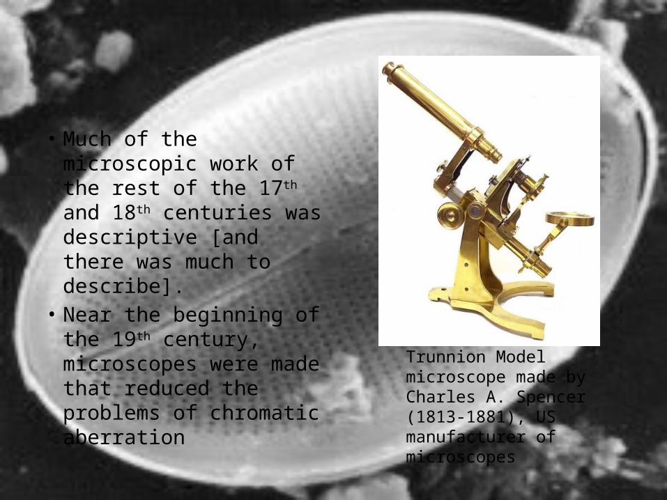
• Much of the microscopic work of the rest of the 17th and 18th centuries was descriptive [and there was much to describe].
• Near the beginning of the 19th century, microscopes were made that reduced the problems of chromatic aberration
Trunnion Model microscope made by Charles A. Spencer (1813-1881), US manufacturer of microscopes
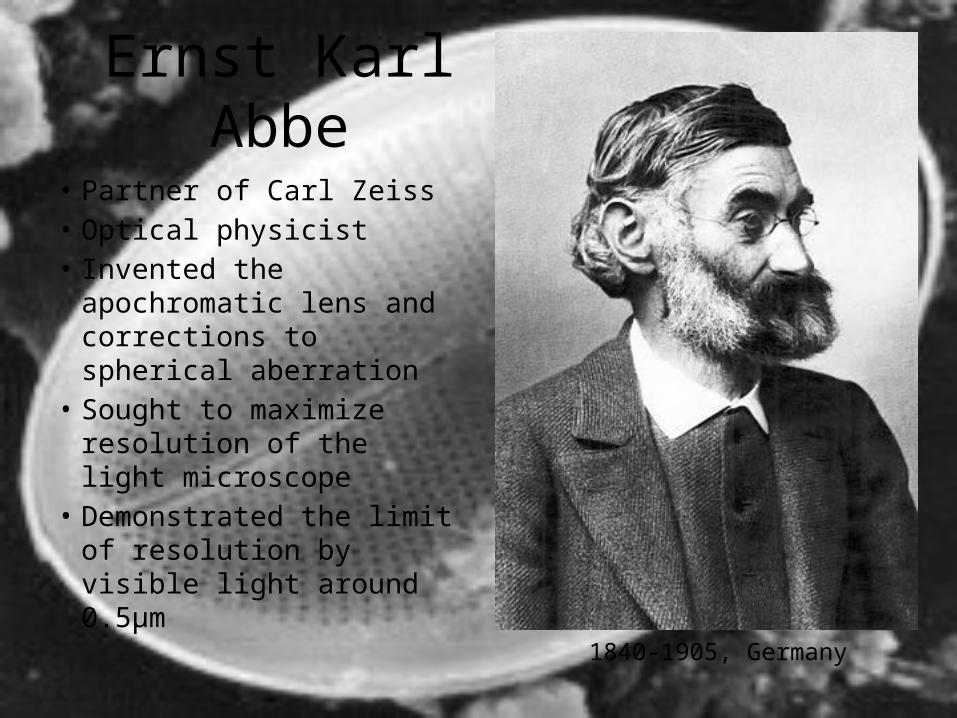
Ernst Karl Abbe
• Partner of Carl Zeiss• Optical physicist• Invented the apochromatic
lens and corrections to spherical aberration
• Sought to maximize resolution of the light microscope
• Demonstrated the limit of resolution by visible light around 0.5μm
1840-1905, Germany
The Cell Theory
…also, by the early part of the 19th century enough biological material had been observed to begin to make generalizations.
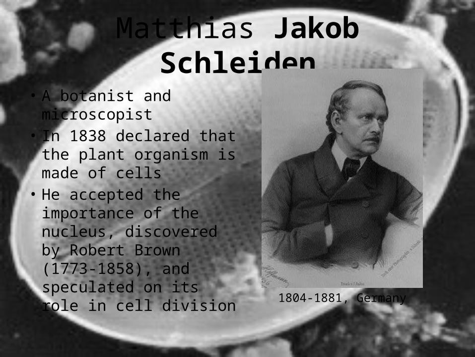
Matthias Jakob Schleiden
• A botanist and microscopist
• In 1838 declared that the plant organism is made of cells
• He accepted the importance of the nucleus, discovered by Robert Brown (1773-1858), and speculated on its role in cell division
1804-1881, Germany
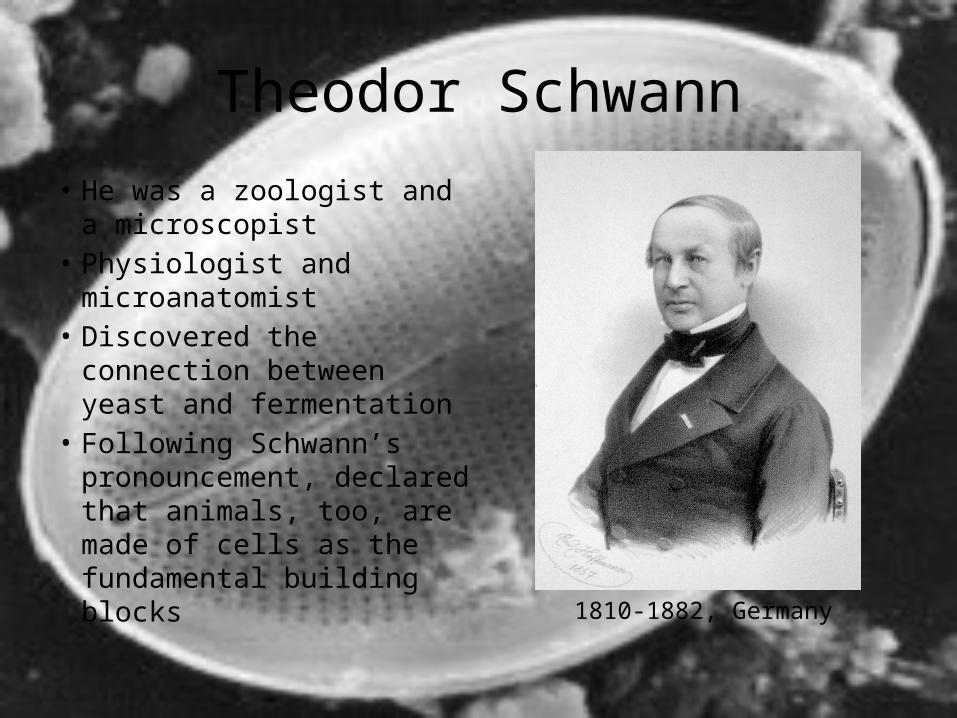
Theodor Schwann
• He was a zoologist and a microscopist
• Physiologist and microanatomist
• Discovered the connection between yeast and fermentation
• Following Schwann’s pronouncement, declared that animals, too, are made of cells as the fundamental building blocks
1810-1882, Germany
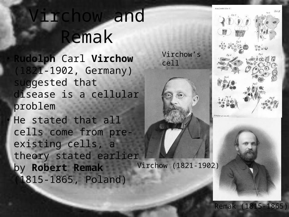
Virchow and Remak
• Rudolph Carl Virchow (1821-1902, Germany) suggested that disease is a cellular problem
• He stated that all cells come from pre-existing cells, a theory stated earlier by Robert Remak (1815-1865, Poland)
Virchow’s cell theory
Virchow (1821-1902)
Remak (1815-1865)
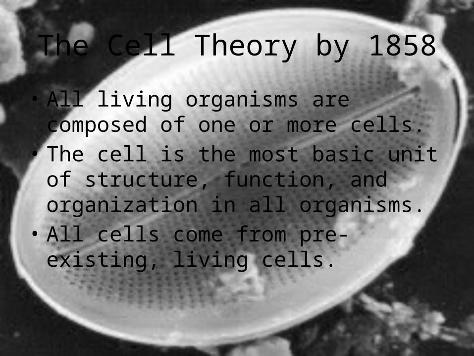
The Cell Theory by 1858
• All living organisms are composed of one or more cells.
• The cell is the most basic unit of structure, function, and organization in all organisms.
• All cells come from pre-existing, living cells.
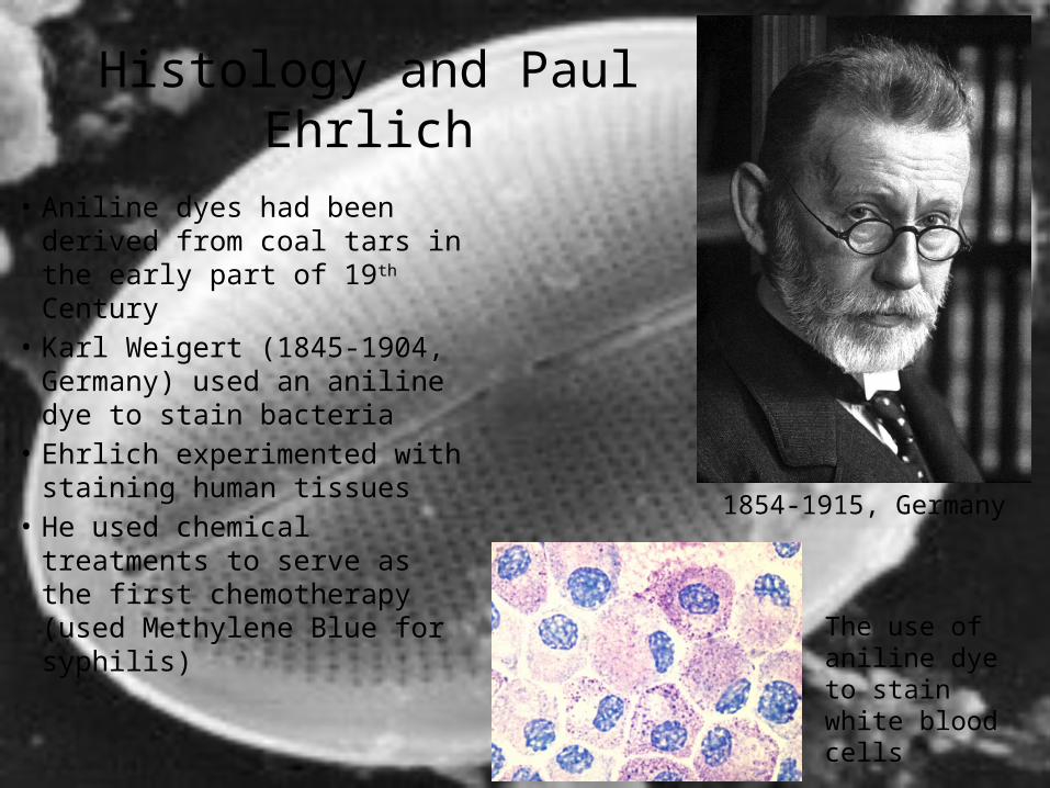
Histology and Paul Ehrlich
• Aniline dyes had been derived from coal tars in the early part of 19th Century
• Karl Weigert (1845-1904, Germany) used an aniline dye to stain bacteria
• Ehrlich experimented with staining human tissues
• He used chemical treatments to serve as the first chemotherapy (used Methylene Blue for syphilis)
1854-1915, Germany
The use of aniline dye to stain white blood cells
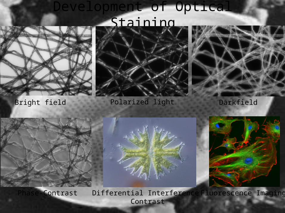
Development of Optical Staining
Bright field Polarized light Darkfield
Phase-Contrast Differential Interference Contrast
Fluorescence Imaging
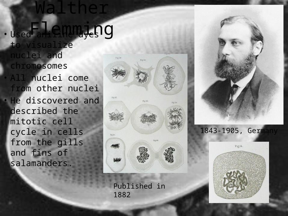
Walther Flemming• Used aniline dyes to
visualize nuclei and chromosomes
• All nuclei come from other nuclei
• He discovered and described the mitotic cell cycle in cells from the gills and fins of salamanders.
1843-1905, Germany
Published in 1882
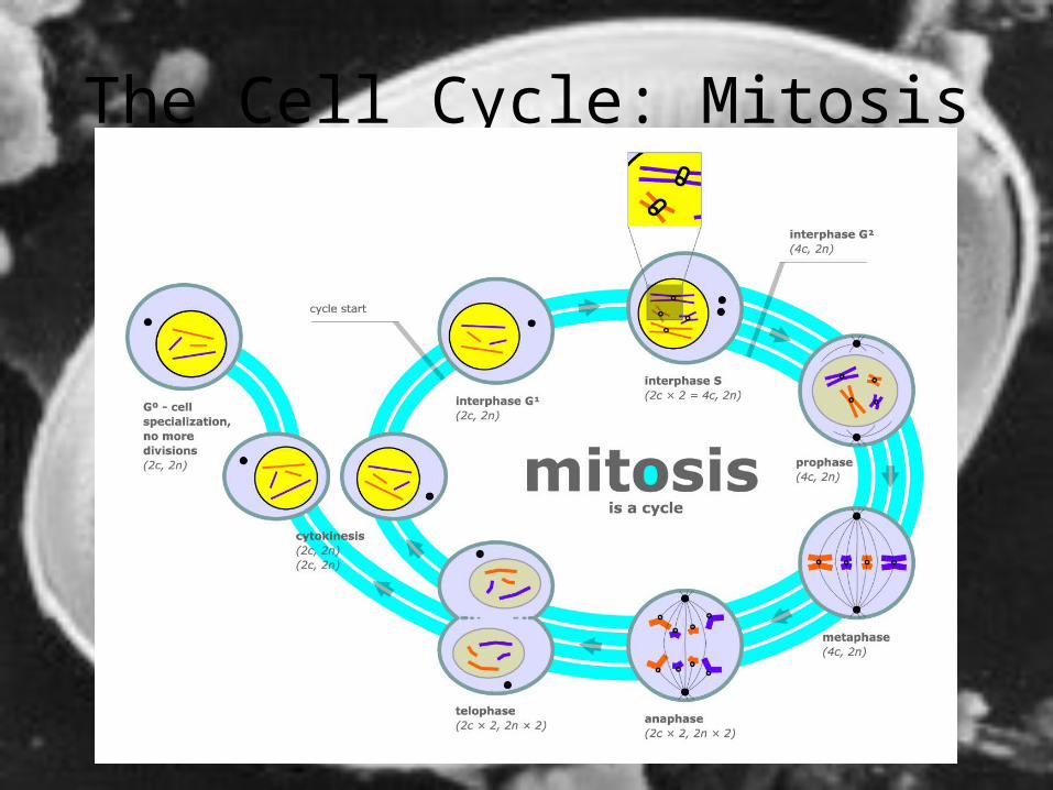
The Cell Cycle: Mitosis
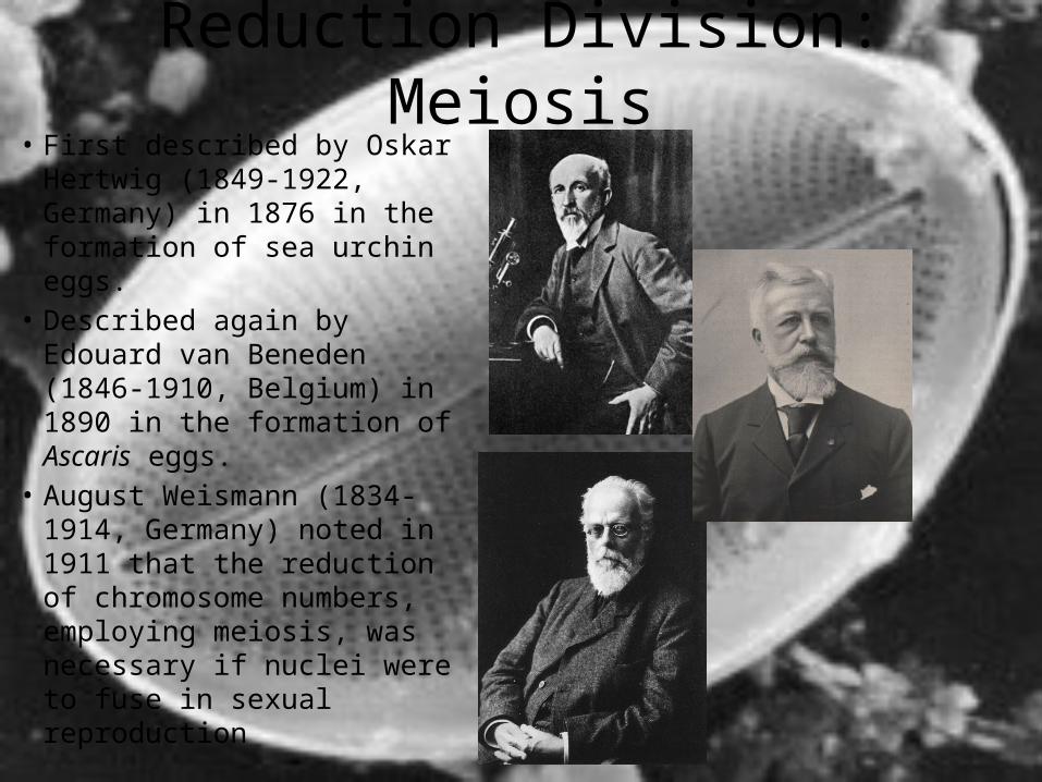
Reduction Division: Meiosis• First described by Oskar
Hertwig (1849-1922, Germany) in 1876 in the formation of sea urchin eggs.
• Described again by Edouard van Beneden (1846-1910, Belgium) in 1890 in the formation of Ascaris eggs.
• August Weismann (1834-1914, Germany) noted in 1911 that the reduction of chromosome numbers, employing meiosis, was necessary if nuclei were to fuse in sexual reproduction
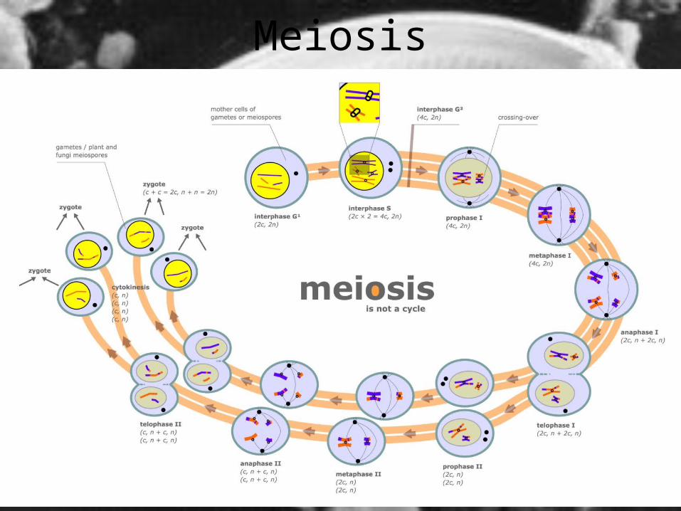
Meiosis
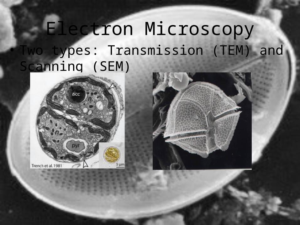
Electron Microscopy• Two types: Transmission (TEM) and Scanning (SEM)
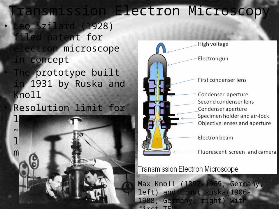
Transmission Electron Microscopy• Leo Szilard (1928) filed patent
for electron microscope in concept
• The prototype built in 1931 by Ruska and Knoll
• Resolution limit for light microscope ~2,000X; but the limit for TEM ~2 millionX
Max Knoll (1897-1969, Germany, left) and Ernst Ruska(1906-1988, Germany, right) with first TEM
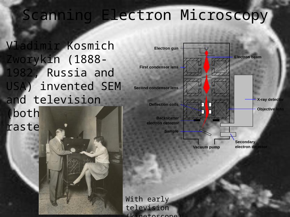
Scanning Electron Microscopy
Vladimir Kosmich Zworykin (1888-1982, Russia and USA) invented SEM and television (both based on raster principle)
With early television (kinetoscope) in 1929
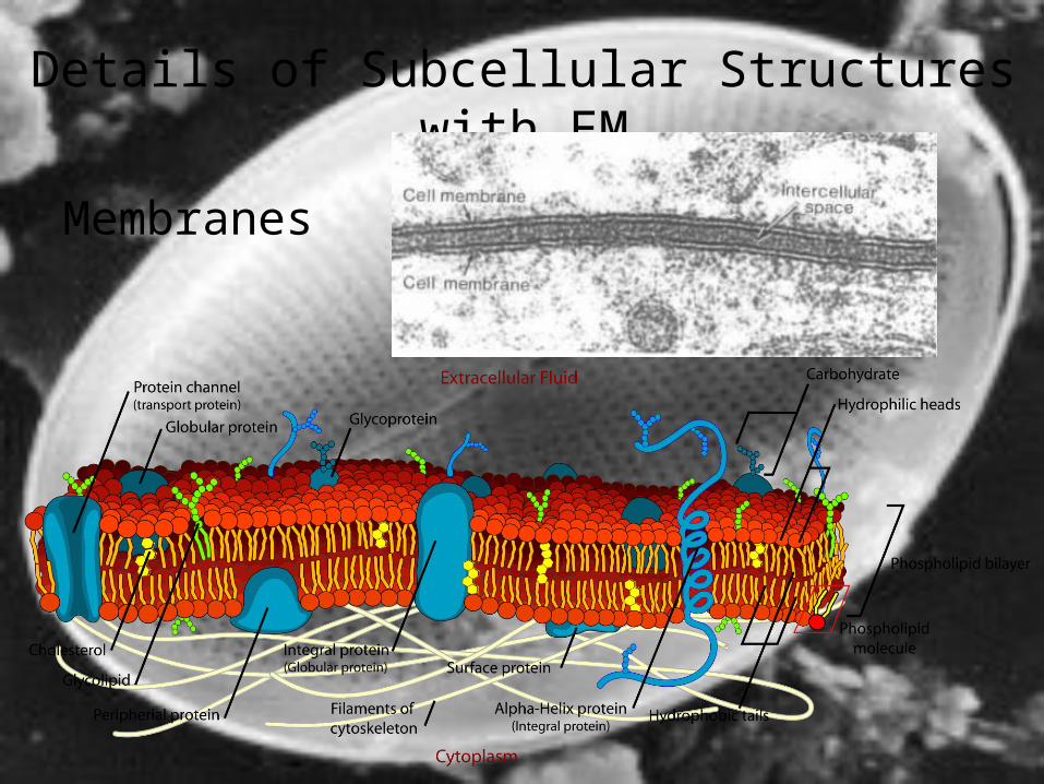
Membranes
Details of Subcellular Structures with EM
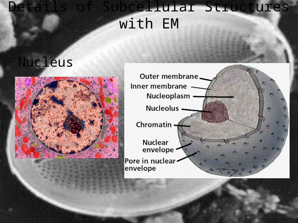
Details of Subcellular Structures with EM
Nucleus
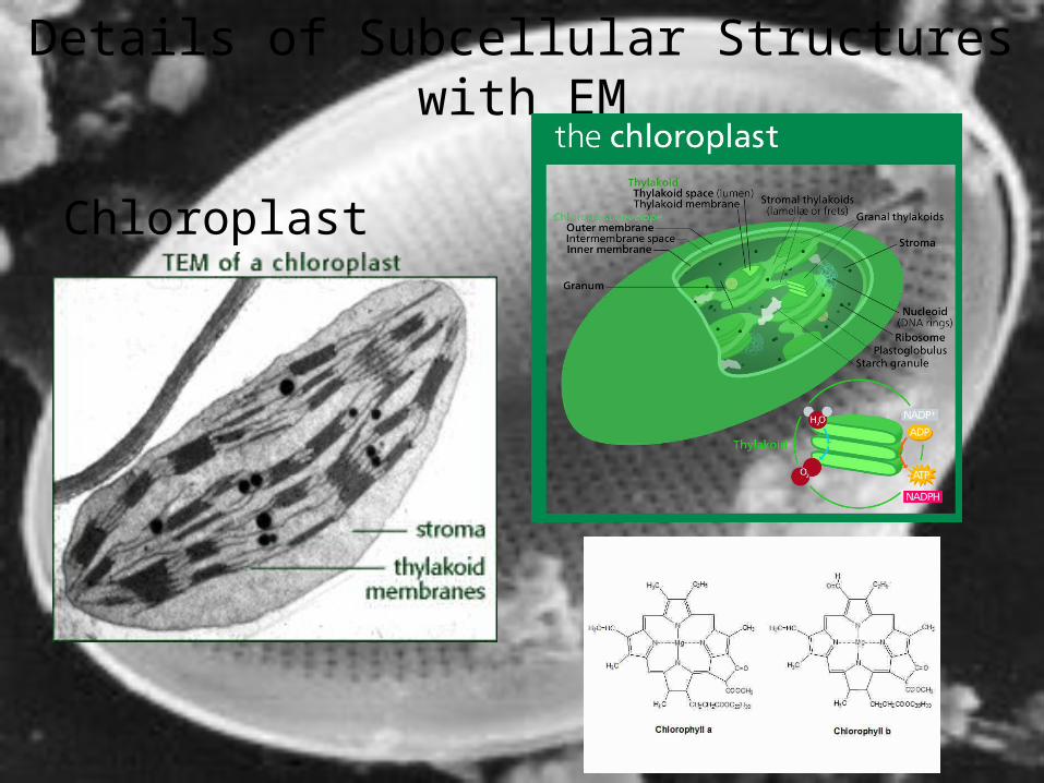
Details of Subcellular Structures with EM
Chloroplast
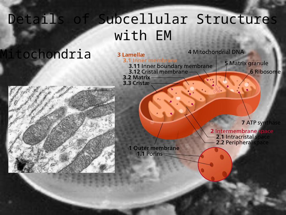
Details of Subcellular Structures with EM
Mitochondria
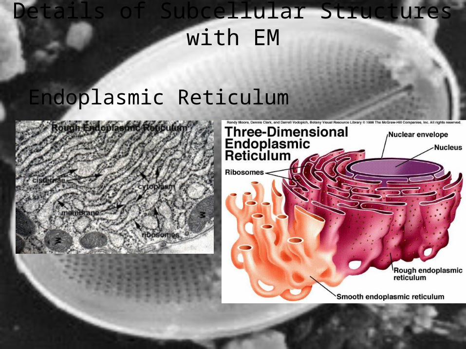
Endoplasmic Reticulum
Details of Subcellular Structures with EM
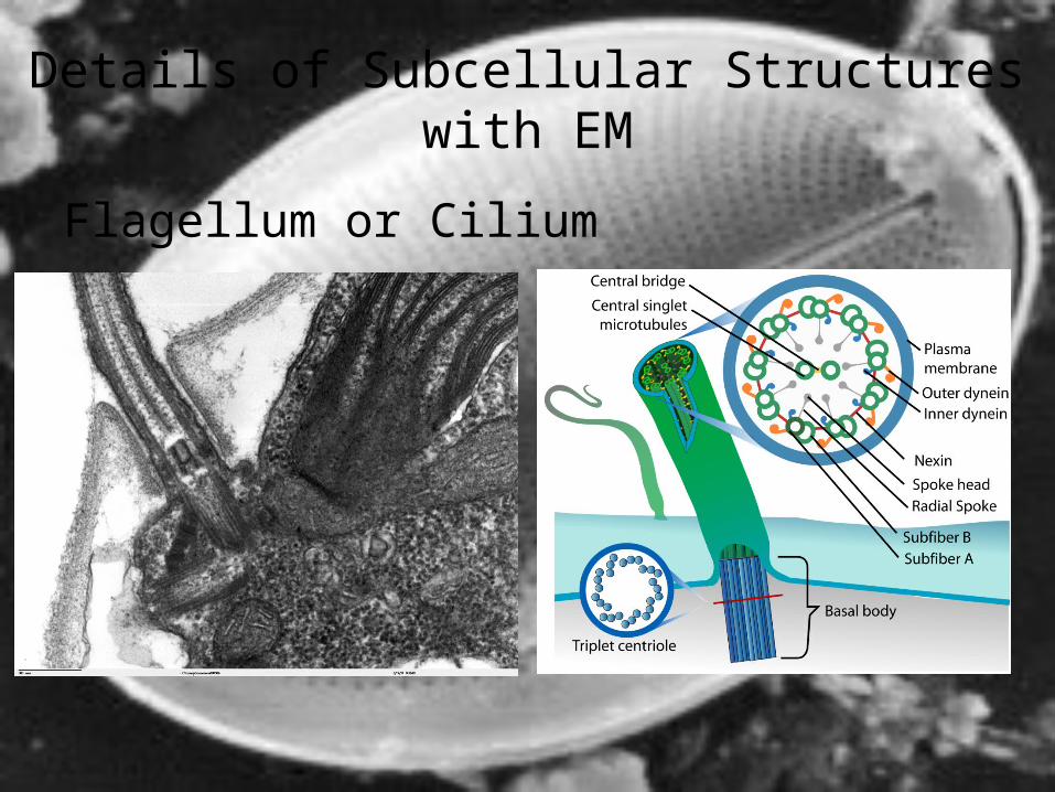
Details of Subcellular Structures with EM
Flagellum or Cilium
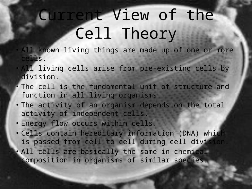
Current View of the Cell Theory
• All known living things are made up of one or more cells.• All living cells arise from pre-existing cells by division.• The cell is the fundamental unit of structure and function in
all living organisms.• The activity of an organism depends on the total activity of
independent cells.• Energy flow occurs within cells.• Cells contain hereditary information (DNA) which is passed
from cell to cell during cell division.• All cells are basically the same in chemical composition in
organisms of similar species.


![[PPT]1.2 Ultrastructure of cells - Fillinghamfillingham.weebly.com/uploads/5/6/7/4/56744911/cell... · Web view1.2 Ultrastructure of cells Last modified by Tanya Fillingham Company](https://img.pdfslide.net/doc/110x75/5ae9ae157f8b9a585f8b56e3/ppt12-ultrastructure-of-cells-view12-ultrastructure-of-cells-last-modified.jpg)