Embed Size (px)
Citation preview
THE NATURAL HISTORY OF PULMONARY TUBERCULOSIS
Dr William Harris, Professor of Clinical Medicine of New York University, School of Medicinedescribes, in this series of slides, the natural history of pulmonary tuberculosis and theimportance of early diagnosis and treatment in achieving cure.
The World Health Organization and other agencies have developed guidelines for thediagnosis and treatment of tuberculosis. Throughout the world where these recommendations areused, the cure rate of TB cases has increased, and control of the disease within communities hasimproved. However, in addition to using these guidelines appropriately, physicians need to befamiliar with the natural history of pulmonary tuberculosis and its varied clinical manifestations, inorder to understand events that may occur in their patients.
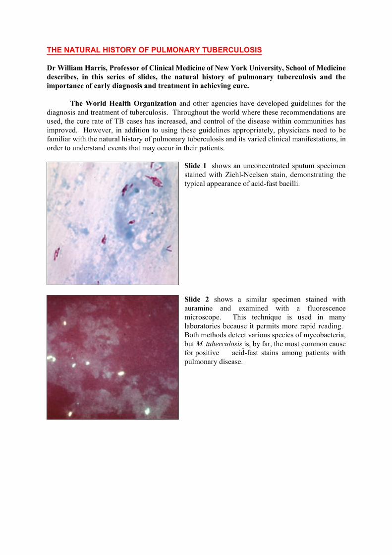
Slide 1 shows an unconcentrated sputum specimenstained with Ziehl-Neelsen stain, demonstrating thetypical appearance of acid-fast bacilli.
Slide 2 shows a similar specimen stained withauramine and examined with a fluorescencemicroscope. This technique is used in manylaboratories because it permits more rapid reading. Both methods detect various species of mycobacteria,but M. tuberculosis is, by far, the most common causefor positive acid-fast stains among patients withpulmonary disease.
THE NATURAL HISTORY OF PULMONARY TUBERCULOSIS
2
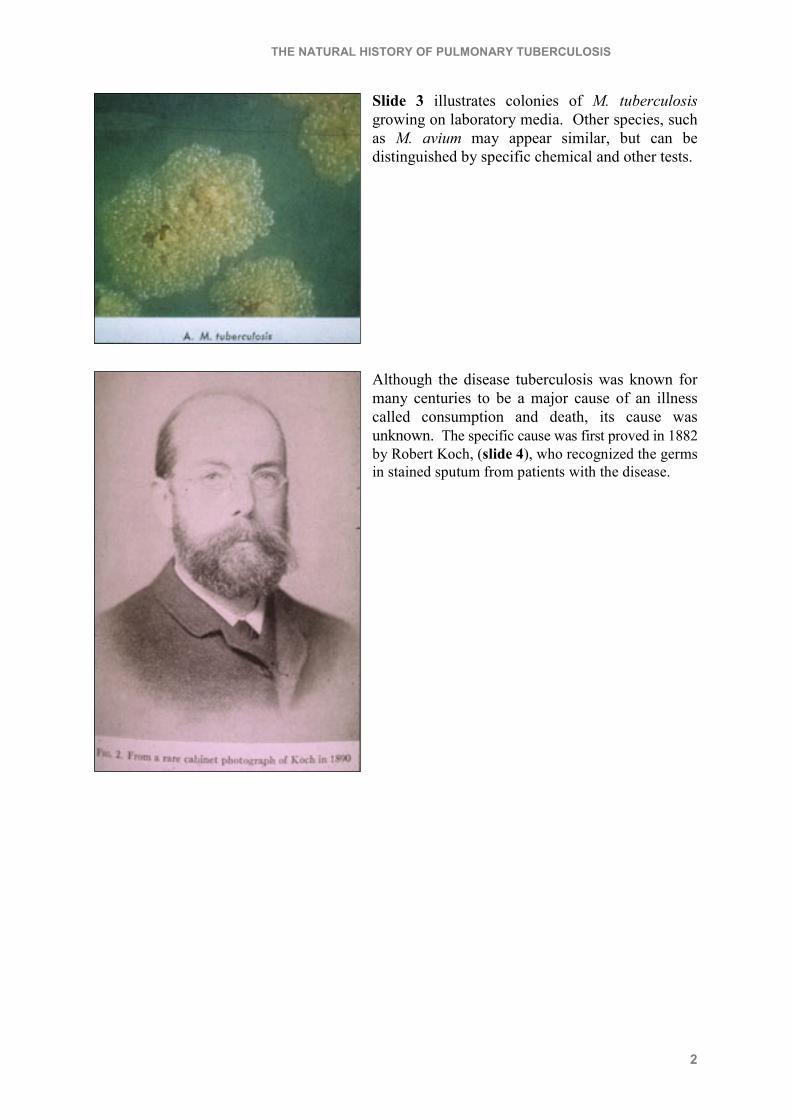
Slide 3 illustrates colonies of M. tuberculosisgrowing on laboratory media. Other species, suchas M. avium may appear similar, but can bedistinguished by specific chemical and other tests.
Although the disease tuberculosis was known formany centuries to be a major cause of an illnesscalled consumption and death, its cause wasunknown. The specific cause was first proved in 1882by Robert Koch, (slide 4), who recognized the germsin stained sputum from patients with the disease.
THE NATURAL HISTORY OF PULMONARY TUBERCULOSIS
3
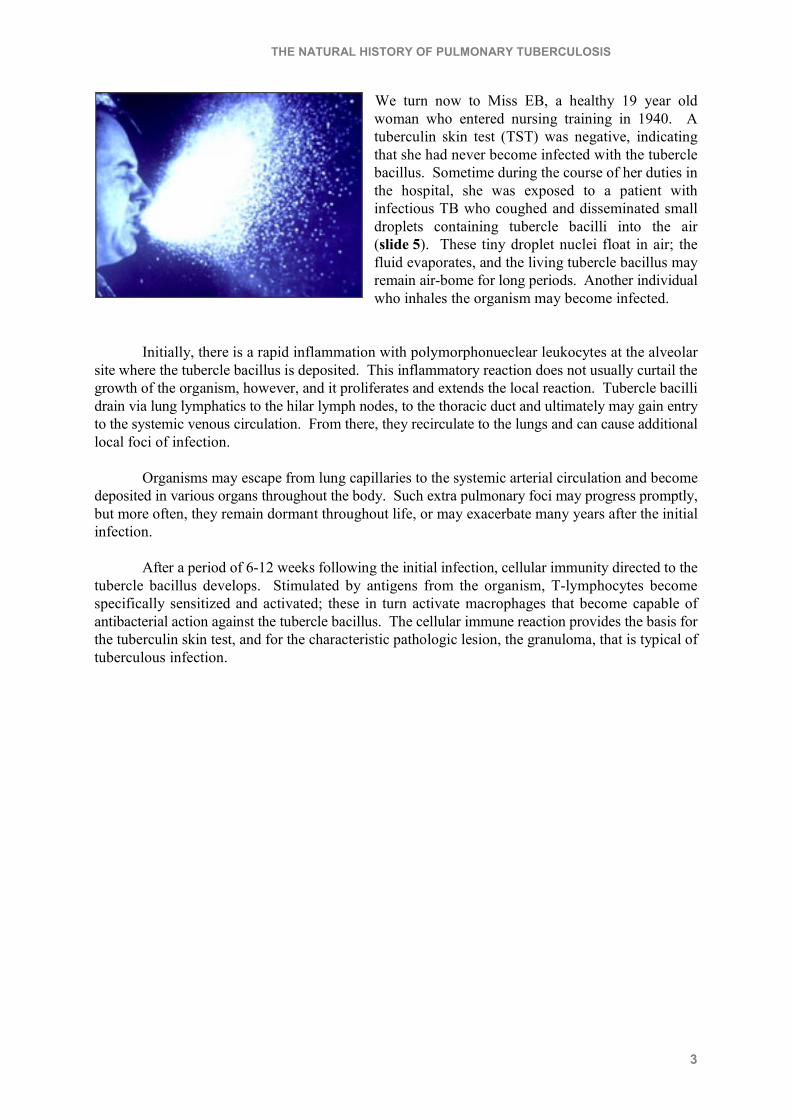
We turn now to Miss EB, a healthy 19 year oldwoman who entered nursing training in 1940. Atuberculin skin test (TST) was negative, indicatingthat she had never become infected with the tuberclebacillus. Sometime during the course of her duties inthe hospital, she was exposed to a patient withinfectious TB who coughed and disseminated smalldroplets containing tubercle bacilli into the air(slide 5). These tiny droplet nuclei float in air; thefluid evaporates, and the living tubercle bacillus mayremain air-bome for long periods. Another individualwho inhales the organism may become infected.
Initially, there is a rapid inflammation with polymorphonueclear leukocytes at the alveolarsite where the tubercle bacillus is deposited. This inflammatory reaction does not usually curtail thegrowth of the organism, however, and it proliferates and extends the local reaction. Tubercle bacillidrain via lung lymphatics to the hilar lymph nodes, to the thoracic duct and ultimately may gain entryto the systemic venous circulation. From there, they recirculate to the lungs and can cause additionallocal foci of infection.
Organisms may escape from lung capillaries to the systemic arterial circulation and becomedeposited in various organs throughout the body. Such extra pulmonary foci may progress promptly,but more often, they remain dormant throughout life, or may exacerbate many years after the initialinfection.
After a period of 6-12 weeks following the initial infection, cellular immunity directed to thetubercle bacillus develops. Stimulated by antigens from the organism, T-lymphocytes becomespecifically sensitized and activated; these in turn activate macrophages that become capable ofantibacterial action against the tubercle bacillus. The cellular immune reaction provides the basis forthe tuberculin skin test, and for the characteristic pathologic lesion, the granuloma, that is typical oftuberculous infection.
THE NATURAL HISTORY OF PULMONARY TUBERCULOSIS
4
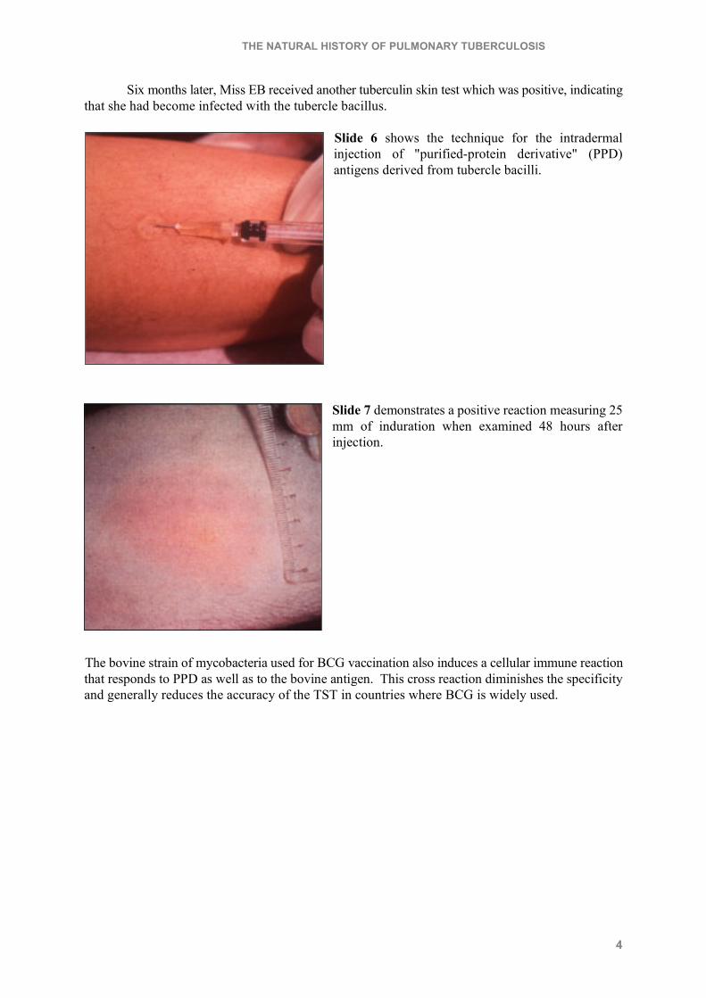
Six months later, Miss EB received another tuberculin skin test which was positive, indicatingthat she had become infected with the tubercle bacillus.
Slide 6 shows the technique for the intradermalinjection of "purified-protein derivative" (PPD)antigens derived from tubercle bacilli.
Slide 7 demonstrates a positive reaction measuring 25mm of induration when examined 48 hours afterinjection.
The bovine strain of mycobacteria used for BCG vaccination also induces a cellular immune reactionthat responds to PPD as well as to the bovine antigen. This cross reaction diminishes the specificityand generally reduces the accuracy of the TST in countries where BCG is widely used.
THE NATURAL HISTORY OF PULMONARY TUBERCULOSIS
5
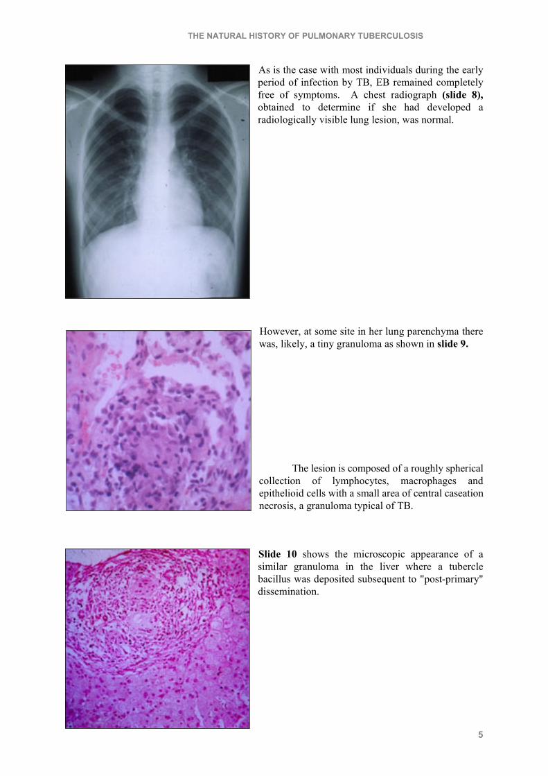
As is the case with most individuals during the earlyperiod of infection by TB, EB remained completelyfree of symptoms. A chest radiograph (slide 8),obtained to determine if she had developed aradiologically visible lung lesion, was normal.
However, at some site in her lung parenchyma therewas, likely, a tiny granuloma as shown in slide 9.
The lesion is composed of a roughly sphericalcollection of lymphocytes, macrophages andepithelioid cells with a small area of central caseationnecrosis, a granuloma typical of TB.
Slide 10 shows the microscopic appearance of asimilar granuloma in the liver where a tuberclebacillus was deposited subsequent to "post-primary"dissemination.
THE NATURAL HISTORY OF PULMONARY TUBERCULOSIS
6
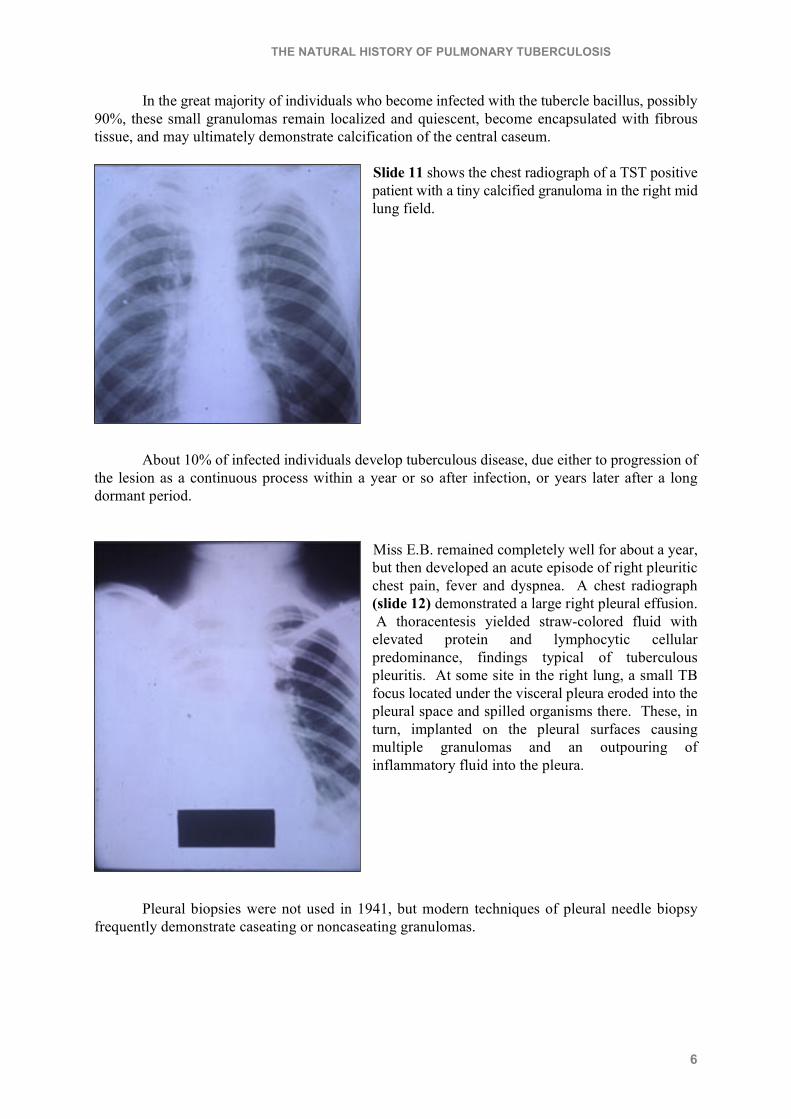
In the great majority of individuals who become infected with the tubercle bacillus, possibly90%, these small granulomas remain localized and quiescent, become encapsulated with fibroustissue, and may ultimately demonstrate calcification of the central caseum.
Slide 11 shows the chest radiograph of a TST positivepatient with a tiny calcified granuloma in the right midlung field.
About 10% of infected individuals develop tuberculous disease, due either to progression ofthe lesion as a continuous process within a year or so after infection, or years later after a longdormant period.
Miss E.B. remained completely well for about a year,but then developed an acute episode of right pleuriticchest pain, fever and dyspnea. A chest radiograph(slide 12) demonstrated a large right pleural effusion. A thoracentesis yielded straw-colored fluid withelevated protein and lymphocytic cellularpredominance, findings typical of tuberculouspleuritis. At some site in the right lung, a small TBfocus located under the visceral pleura eroded into thepleural space and spilled organisms there. These, inturn, implanted on the pleural surfaces causingmultiple granulomas and an outpouring ofinflammatory fluid into the pleura.
Pleural biopsies were not used in 1941, but modern techniques of pleural needle biopsyfrequently demonstrate caseating or noncaseating granulomas.

THE NATURAL HISTORY OF PULMONARY TUBERCULOSIS
7
Slide 13 shows such a lesion in the pleura, amononuclear cellular reaction with an area of caseousnecrosis.
Antituberculous chemotherapy was not available in 1941, but the patient received bed resttherapy for several months while the fluid resorbed, the typical course of most tuberculous pleuraleffusions, even without specific treatment.
Slide 14 shows complete clearing of the pleuraleffusion within a four month period. Her physician,realizing that she was now at greater risk ofdeveloping active TB in the lung, and having no anti-tuberculosis treatment to offer in 1941, obtained chestradiographs every three months.
Slide 15, a chest radiograph taken 6 months later,demonstrates a new rounded shadow in the right upperlobe.
THE NATURAL HISTORY OF PULMONARY TUBERCULOSIS
8
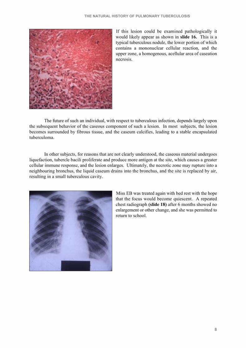
If this lesion could be examined pathologically itwould likely appear as shown in slide 16. This is atypical tuberculous nodule, the lower portion of whichcontains a mononuclear cellular reaction, and theupper zone, a homogenous, acellular area of caseationnecrosis.
The future of such an individual, with respect to tuberculous infection, depends largely uponthe subsequent behavior of the caseous component of such a lesion. In most subjects, the lesionbecomes surrounded by fibrous tissue, and the caseum calcifies, leading to a stable encapsulatedtuberculoma.
In other subjects, for reasons that are not clearly understood, the caseous material undergoesliquefaction, tubercle bacili proliferate and produce more antigen at the site, which causes a greatercellular immune response, and the lesion enlarges. Ultimately, the necrotic zone may rupture into aneighbouring bronchus, the liquid caseum drains into the bronchus, and the site is replaced by air,resulting in a small tuberculous cavity.
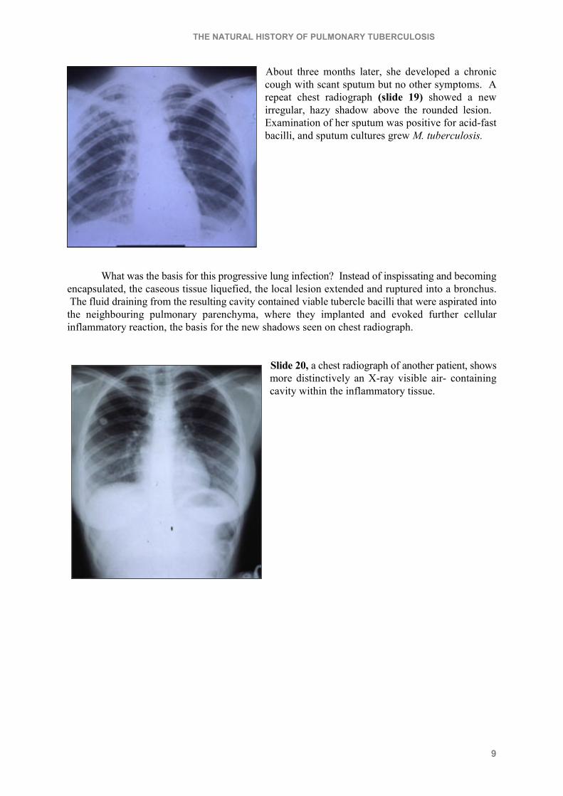
Miss EB was treated again with bed rest with the hopethat the focus would become quiescent. A repeatedchest radiograph (slide 18) after 6 months showed noenlargement or other change, and she was permitted toreturn to school.
THE NATURAL HISTORY OF PULMONARY TUBERCULOSIS
9
About three months later, she developed a chroniccough with scant sputum but no other symptoms. Arepeat chest radiograph (slide 19) showed a newirregular, hazy shadow above the rounded lesion. Examination of her sputum was positive for acid-fastbacilli, and sputum cultures grew M. tuberculosis.
What was the basis for this progressive lung infection? Instead of inspissating and becomingencapsulated, the caseous tissue liquefied, the local lesion extended and ruptured into a bronchus. The fluid draining from the resulting cavity contained viable tubercle bacilli that were aspirated intothe neighbouring pulmonary parenchyma, where they implanted and evoked further cellularinflammatory reaction, the basis for the new shadows seen on chest radiograph.
Slide 20, a chest radiograph of another patient, showsmore distinctively an X-ray visible air- containingcavity within the inflammatory tissue.
THE NATURAL HISTORY OF PULMONARY TUBERCULOSIS
10
The process of bronchial drainage from the cavity,and progressive inflammatory lesions due tobronchial dissemination is further illustrated in slide21 where the lesion is more extensive.
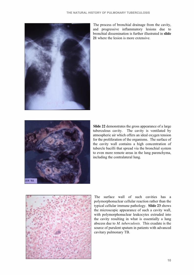
Slide 22 demonstrates the gross appearance of a largetuberculous cavity. The cavity is ventilated byatmospheric air which offers an ideal oxygen tensionfor the proliferation of the organisms. The surface ofthe cavity wall contains a high concentration oftubercle bacilli that spread via the bronchial systemto even more remote areas in the lung parenchyma,including the contralateral lung.
The surface wall of such cavities has apolymorphonuclear cellular reaction rather than thetypical cellular immune pathology. Slide 23 showsthe microscopic appearance of such a cavity wall,with polymorphonuclear leukocytes extruded intothe cavity resulting in what is essentially a lungabscess due to M. tuberculosis. This exudate is thesource of purulent sputum in patients with advancedcavitary pulmonary TB.

THE NATURAL HISTORY OF PULMONARY TUBERCULOSIS
11
Despite all therapy efforts, the disease in Miss EBprogressed to involve both lungs (Slide 24).
Slide 25 demonstrates the pathologic appearance ofsuch extensive inflammation, a pneumonia-like patternof alveolar consolidation due to M. tuberculosis.
Slide 26 illustrates the basis for progressive extensionof the TB lesions because of bronchogenicdissemination. New caseous lesions lead to newcavities which produce airborne infected particles inthe bronchi. These are in turn aspirated into remoteareas of the lung parenchyma.

THE NATURAL HISTORY OF PULMONARY TUBERCULOSIS
12
Miss EB's pulmonary tuberculosis progressed inexorably over the next several years. Shesuffered persistent fever, severe night sweats, chronic cough with sputum production and occasionalbouts of hemoptysis. She was one of the earliest patients in New York City to receive streptomycin(SM) when it became available. Initially, there was a reduction in the number of tubercle bacilli inthe sputum, but this improvement was short-lived as the M. tuberculosis rapidly developed resistanceto the drug.
Slide 27 demonstrates how SM resistant tuberclebacilli emerge when the drug is given alone. Beforetreatment, the M. tuberculosis population, presentpredominantly in cavities, consists of one SMresistant bacillus to every million SM susceptibleorganisms. SM rapidly kills the susceptible but doesnot effect the resistant bacilli. As the number of SMsusceptible bacilli diminishes, they are replaced bySM resistant ones that become the predominant M.tuberculosis in cavities and contained in the sputum.When two effective agents are administeredconcomittantly, each drug kills the cells that areresistant to the companion drug and prevents theemergency of resistance to the other.
Slide 28 shows EB's chest radiograph a few monthsbefore she died of the disease. This shows multiplecavities replacing and destroying most of the lungparenchyima.
This patient had progressive tuberculous disease almost from the beginning of the infection. Other patients develop active TB many years after the initial infection when tubercle bacilli in a longdormant lesion in the lung begin to multiply, and the basic cycle of TB progression, illustrated inslide 26, ensues.
THE NATURAL HISTORY OF PULMONARY TUBERCULOSIS
13
Slide 29 is a chest radiograph of a 55-year-old male,a long time resident in a psychiatric hospital, whichshows a small, densely calcified TB focus in the rightlower lung field. This lesion had been visible onserial chest radiographs's for more than 10 years.
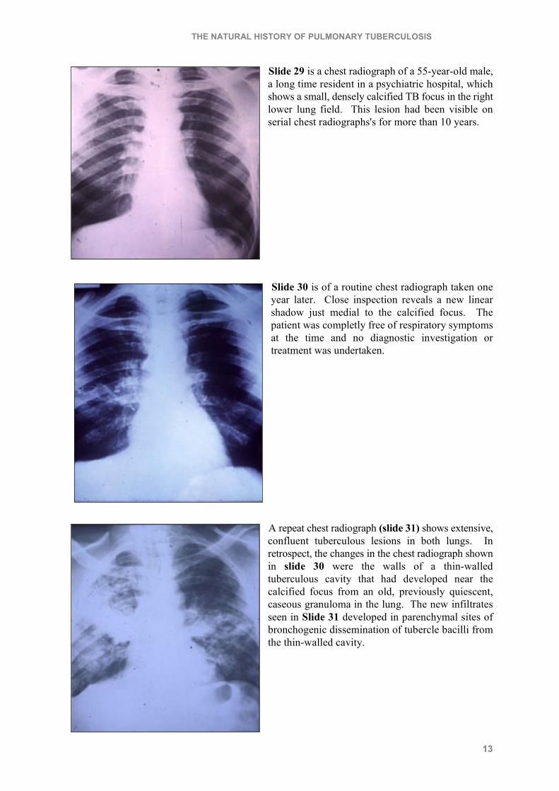
Slide 30 is of a routine chest radiograph taken oneyear later. Close inspection reveals a new linearshadow just medial to the calcified focus. Thepatient was completly free of respiratory symptomsat the time and no diagnostic investigation ortreatment was undertaken.
A repeat chest radiograph (slide 31) shows extensive,confluent tuberculous lesions in both lungs. Inretrospect, the changes in the chest radiograph shownin slide 30 were the walls of a thin-walledtuberculous cavity that had developed near thecalcified focus from an old, previously quiescent,caseous granuloma in the lung. The new infiltratesseen in Slide 31 developed in parenchymal sites ofbronchogenic dissemination of tubercle bacilli fromthe thin-walled cavity.
THE NATURAL HISTORY OF PULMONARY TUBERCULOSIS
14
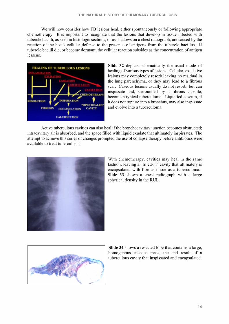
We will now consider how TB lesions heal, either spontaneously or following appropriatechemotherapy. It is important to recognize that the lesions that develop in tissue infected withtubercle bacilli, as seen in histologic sections, or as shadows on a chest radiograph, are caused by thereaction of the host's cellular defense to the presence of antigens from the tubercle bacillus. Iftubercle bacilli die, or become dormant, the cellular reaction subsides as the concentration of antigenlessens.
Slide 32 depicts schematically the usual mode ofhealing of various types of lesions. Cellular, exudativelesions may completely resorb leaving no residual inthe lung parenchyma, or they may lead to a fibrousscar. Caseous lesions usually do not resorb, but caninspissate and, surrounded by a fibrous capsule,become a typical tuberculoma. Liquefied caseum, ifit does not rupture into a bronchus, may also inspissateand evolve into a tuberculoma.
Active tuberculous cavities can also heal if the bronchocavitary junction becomes obstructed;intracavitary air is absorbed, and the space filled with liquid exudate that ultimately inspissates. Theattempt to achieve this series of changes prompted the use of collapse therapy before antibiotics wereavailable to treat tuberculosis.
With chemotherapy, cavities may heal in the samefashion, leaving a "filled-in" cavity that ultimately isencapsulated with fibrous tissue as a tuberculoma. Slide 33 shows a chest radiograph with a largespherical density in the RUL.
Slide 34 shows a resected lobe that contains a large,homogenous caseous mass, the end result of atuberculous cavity that inspissated and encapsulated.
THE NATURAL HISTORY OF PULMONARY TUBERCULOSIS
15
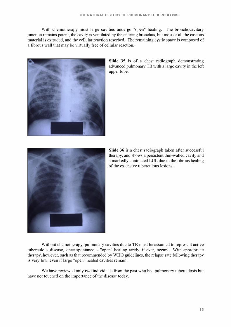
With chemotherapy most large cavities undergo "open" healing. The bronchocavitaryjunction remains patent, the cavity is ventilated by the entering bronchus, but most or all the caseousmaterial is extruded, and the cellular reaction resorbed. The remaining cystic space is composed ofa fibrous wall that may be virtually free of cellular reaction.
Slide 35 is of a chest radiograph demonstratingadvanced pulmonary TB with a large cavity in the leftupper lobe.
Slide 36 is a chest radiograph taken after successfultherapy, and shows a persistent thin-walled cavity anda markedly contracted LUL due to the fibrous healingof the extensive tuberculous lesions.
Without chemotherapy, pulmonary cavities due to TB must be assumed to represent activetuberculous disease, since spontaneous "open" healing rarely, if ever, occurs. With appropriatetherapy, however, such as that recommended by WHO guidelines, the relapse rate following therapyis very low, even if large "open" healed cavities remain.
We have reviewed only two individuals from the past who had pulmonary tuberculosis buthave not touched on the importance of the disease today.
THE NATURAL HISTORY OF PULMONARY TUBERCULOSIS
16
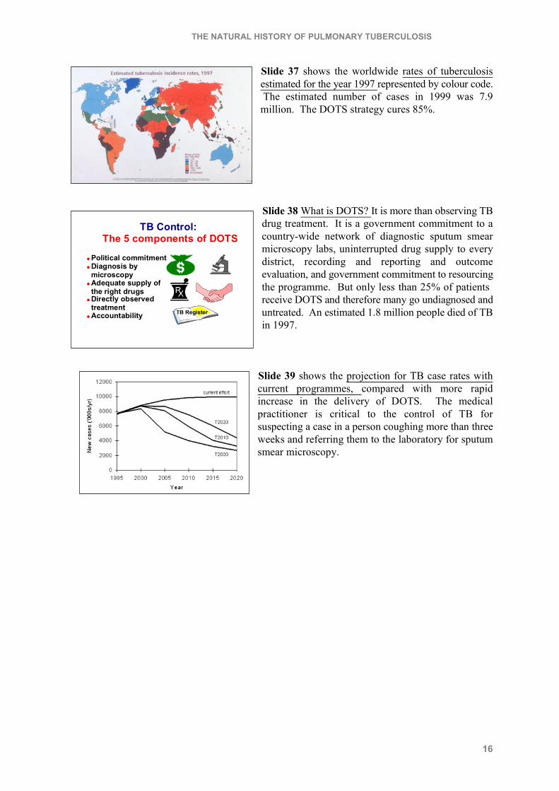
Slide 37 shows the worldwide rates of tuberculosisestimated for the year 1997 represented by colour code. The estimated number of cases in 1999 was 7.9million. The DOTS strategy cures 85%.
Slide 38 What is DOTS? It is more than observing TBdrug treatment. It is a government commitment to acountry-wide network of diagnostic sputum smearmicroscopy labs, uninterrupted drug supply to everydistrict, recording and reporting and outcomeevaluation, and government commitment to resourcingthe programme. But only less than 25% of patients receive DOTS and therefore many go undiagnosed anduntreated. An estimated 1.8 million people died of TBin 1997.
Slide 39 shows the projection for TB case rates withcurrent programmes, compared with more rapidincrease in the delivery of DOTS. The medicalpractitioner is critical to the control of TB forsuspecting a case in a person coughing more than threeweeks and referring them to the laboratory for sputumsmear microscopy.
TB Control:The 5 components of DOTS
TB Register
� Political commitment� Diagnosis by
microscopy� Adequate supply of
the right drugs� Directly observed
treatment� Accountability