Embed Size (px)
Citation preview
The Pathogenesis of Tears of the Retinal Pigment Epithelium
Elaine L. Chuang, M . D . , and Alan C. Bird, M . D .
We compared drusen in the fellow eye of patients with unilateral retinal pigment epithelial tears with those in an age- and sex-matched group of patients with unilateral primary neovascular disciform lesions. In the fellow eye of patients with tears, drusen were more confluent and manifested less fluorescence on angiography than in the comparison group. These observations are in accord with the concept that Bruch's membrane represents a significant barrier to fluid flow, and that fluid beneath detached pigment epithelium is derived in part or wholely from the pigment epithelium.
RIPS OR TEARS of the retinal pigment epithelium have been recognized increasingly as a cause of severe loss of central vision in age-related macular degeneration.116 Patients experience the sudden onset of distortion or a precipitous drop in central acuity coincident with the tear if the fovea is involved. The typical acute fundus appearance is that of an abrupt discontinuity in the pigment epithelium at its border of previous detachment. The consequent defect in the pigmented monolayer and the production of redundant folds of the remaining detached tissue lead to a characteristic acute angiographic appearance of hyperfluo-rescence in the bed of the tear, with adjacent hypofluorescence corresponding to the retracted mound of pigment epithelium. With time, progressive scarring and other alterations render the origin of the lesion more difficult to recognize.15
Recently, tears have been shown to occur
Accepted for publication Jan. 4, 1988. From the Department of Clinical Ophthalmology, In
stitute of Ophthalmology, Moorfields Eye Hospital, London. This study was supported in part by Medical Research Council grant No. G.8320019 N.
Reprint requests to Elaine L. Chuang, M.D., Bascom Palmer Eye Institute, 900 N.W. 17th St., Miami, FL 33134.
bilaterally more frequently than would be predicted by chance alone.16 This could imply that the age-related changes in Bruch's membrane in eyes destined to manifest tears are different from those present in eyes not at risk of sustaining this complication. In this study, we compared the changes in the fellow eye of patients with unilateral tears of the pigment epithelium with those changes seen in the fellow eyes of patients with unilateral neovascular disciform lesions. Our primary goal was to contribute to the understanding of the pathogenesis of pigment epithelial detachments that are destined to tear, and perhaps of pigment epithelial detachments in general.
Patients and Methods
We reviewed our clinical and photographic records between 1976 and 1986 to identify patients in whom the diagnosis of a pigment epithelial tear was made in one eye and in whom the contralateral eye had adequately recorded drusen in the posterior pole. Fellow eyes with evidence of exudative lesions or significant pigmentary atrophy were excluded.
Thirty-one patients with unilateral tears of the retinal pigment epithelium met these criteria. For comparison, a patient group with unilateral neovascular disciform degeneration and contralateral drusen was matched by age and sex as well as by the year of first clinical examination (in order to reduce differences resulting from changes in photographic and angiographic techniques). There were 17 women and 14 men in each group. They ranged in age from 55 to 80 years (mean, 71 and 70 years in women and men, respectively) in the tear group, and 56 to 83 years (mean, 69 and 67 years in women and men, respectively) in the disciform group.
Color photographs and fluorescein angio-grams of each group were analyzed by both of us independently for size, density, and early
©AMERICAN JOURNAL OF OPHTHALMOLOGY 105:285-290, MARCH, 1988 285
286 AMERICAN JOURNAL OF OPHTHALMOLOGY March, 1988
and late angiographic behavior of macular drusen; one of us was masked from the diagnosis in the eye with visual loss. The posterior pole was divided into two areas: outside and within 1,600 \xm of the foveola. The drusen were divided according to size (0 to 50 u.m, 50 to 500 |xm, and greater than 500 (xm), and distribution was classified as scattered, sub-confluent, or confluent. Fluorescence of drusen was assessed as equal to choroidal fluorescence, slightly greater than choroidal fluorescence, or brightly fluorescent. In most cases the drusen were uniform in size, distribution, and fluorescence in each area of the macula; when this was not the case the classification was determined by the largest, most densely packed and most fluorescent lesions. A few eyes were not included in each category of analysis because of the lack of transit or late photographs.
Drusen were present in both eyes of five patients who were examined before the development of a pigment epithelial tear in either eye.
The number of eyes in each group was determined according to the above criteria. The Yates' corrected chi square test was applied to the data to assess the probability of these results occurring by chance alone.
Results
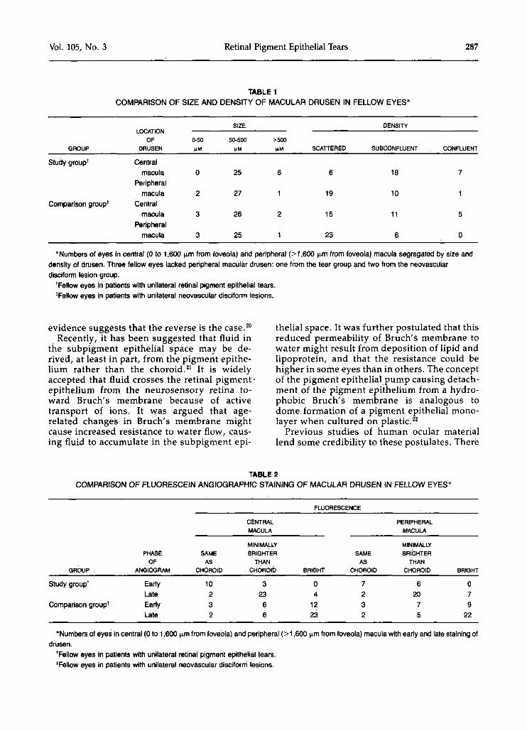
Drusen displayed a greater degree of confluence in fellow eyes of patients with pigment epithelial tears compared to fellow eyes of patients with primary neovascular lesions (Table 1). This was true in the central macula (P = .032), but the difference between the two patient groups did not reach a statistically significant level in the peripheral macula (P = .240). There was a tendency for drusen to be larger in the eye contralateral to eyes with tears in the central macula, but the difference was not statistically significant.
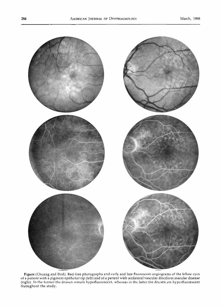
The angiographic behavior of drusen in the setting of tears was found to differ significantly from that in fellow eyes of neovascular disci-form lesions (Table 2). In transit photographs of both the central and peripheral macula, the degree of fluorescence of drusen in fellow eyes of those with rips was less than that in fellow eyes of those with primary neovascular disci-form lesions (P < .001 and P < .006, respectively) (Figure). Similarly, late staining of macular
drusen was less intense in the study patients than in the comparison group centrally and peripherally (P < .0001 and P < .0002, respectively) (Figure).
The findings in the five eyes in which examination of drusen was performed before the development of a pigment epithelial detachment, which then tore, were not different from those of the 31 contralateral study eyes.
Discussion
These results indicate that age-related changes at the level of Bruch's membrane in eyes destined to suffer rips are different from those at risk of developing primary neovascular lesions. The properties of the drusen probably reflect the nature of the changes in Bruch's membrane that place the eye at high risk of suffering a retinal pigment epithelial detachment, which will eventually tear.
It would have been desirable to characterize the nature of these alterations in eyes before development of the pigment epithelial detachment. However, because of the nature of this study, only a limited number of such eyes were observed. It was for this reason that we examined drusen in the fellow eye of patients with tears. The relevance of an analysis of drusen is based on the following rationale. Drusen are the manifestation of age-related change in the region of Bruch's membrane that is most amenable to analysis. It has been shown previously that fellow eyes in patients with unilateral pigment epithelial tears are at high risk of sustaining a similar lesion.16 Furthermore, since there is remarkable symmetry of drusen in the elderly,1718 it is reasonable to assume that drusen in the fellow eye to one with a unilateral tear would be similar to those that existed in the first eye before detachment of the pigment epithelium.
The significance of the hypofluorescence of drusen may be assessed in relationship to concepts of pathogenesis of retinal pigment epithelial detachments. Originally, it was considered likely that detachment was induced by the passage of fluid from the choroid through Bruch's membrane, the physical attachment of the pigment epithelium having been disturbed by the accumulation of debris on the inner surface of Bruch's membrane.19 This presupposes that hydrostatic pressure in the choroid is higher than that in the retina, and yet the
Vol. 105, No. 3 Retinal Pigment Epithelial Tears 287
TABLE 1 COMPARISON OF SIZE AND DENSITY OF MACULAR DRUSEN IN FELLOW EYES*
GROUP
Study group*
Comparison group'
LOCATION OF
DRUSEN
Central macula
Peripheral macula
Central macula
Peripheral macula
0-50 )JLM
0
2
3
3
SIZE
50-500
25
27
26
25
>500 (1M
6
1
2
1
SCATTERED
6
19
15
23
DENSITY
SUBCONFLUENT
18
10
11
6
CONFLUENT
7
1
5
0
'Numbers of eyes in central (0 to 1,600 M.m from foveola) and peripheral (>1,600 p.m from foveola) macula segregated by size and density of drusen. Three fellow eyes lacked peripheral macular drusen: one from the tear group and two from the neovascUlar disciform lesion group.
'Fellow eyes in patients with unilateral retinal pigment epithelial tears. 'Fellow eyes in patients with unilateral neovascUlar disciform lesions.
evidence suggests that the reverse is the case.20
Recently, it has been suggested that fluid in the subpigment epithelial space may be derived, at least in part, from the pigment epithelium rather than the choroid.21 It is widely accepted that fluid crosses the retinal pigment ' epithelium from the neurosensory retina toward Bruch's membrane because of active transport of ions. It was argued that age-related changes in Bruch's membrane might cause increased resistance to water flow, causing fluid to accumulate in the subpigment epi
thelial space. It was further postulated that this reduced permeability of Bruch's membrane to water might result from deposition of lipid and lipoprotein, and that the resistance could be higher in some eyes than in others. The concept of the pigment epithelial pump causing detachment of the pigment epithelium from a hydro-phobic Bruch's membrane is analogous to dome formation of a pigment epithelial mono-layer when cultured on plastic.22
Previous studies of human ocular material lend some credibility to these postulates. There
TABLE 2 COMPARISON OF FLUORESCEIN ANGIOGRAPHIC STAINING OF MACULAR DRUSEN IN FELLOW EYES*
GROUP
Study group*
Comparison group*
PHASE OF
ANGIOGRAM
Early Late Early Late
SAME AS
CHOROID
10 2 3 2
CENTRAL MACULA
MINIMALLY BRIGHTER
tHAN CHOROlb
3 23 6 6
FLUORESCENCE
BRIGHT
0 4
12 23
SAME AS
CHOROID
7 2 3 2
PERIPHERAL MACULA
MINIMALLY BRIGHTER
THAN CHOROID
6 20 7 5
BRIGHT
0 7 9
22
"Numbers of eyes in central (0 to 1,600 |xm from foveola) and peripheral (>1,600 \xm from foveola) macula with early and late staining of drusen.
'Fellow eyes in patients with unilateral retinal pigment epithelial tears. 'Fellow eyes in patients with unilateral neovascular disciform lesions.
288 AMERICAN JOURNAL OF OPHTHALMOLOGY March, 1988
Figure (Chuang and Bird). Red-free photographs and early and late fluorescein angiograms of the fellow eyes of a patient with a pigment epithelial rip (left) and of a patient with unilateral vascular disciform macular disease (right). In the former the drusen remain hypofluorescent, whereas in the latter the drusen are hyperfluorescent throughout the study.
Vol. 105, No. 3 Retinal Pigment Epithelial Tears 289
is evidence of variation in the hydrophobicity of drusen,21 and plaques of lipid have been identified in Bruch's membrane of eyes from the elderly.23 Ultrastructural studies have been interpreted as showing evidence of impedance to fluid flow in the presence of a "basal linear deposit" on the inner surface of Bruch's membrane.24 Finally, investigations into changes in the hydraulic conductivity of Bruch's membrane in human eyes show a marked fall in the ability of water to move across Bruch's membrane with age.25
Retinal pigment epithelial detachments that are destined to tear tend to become progressively larger and more highly detached, thereby generating sufficient tangential stress in the detached tissues to cause a rupture.11 This suggests that it is in these lesions that the resistance to water flow across Bruch's membrane would be highest. The hypofluorescence of drusen observed in this study reflects limited entry of fluorescein into the lesions because of their hydrophobicity.21 The proposed hydrophobicity and confluent tendency of such drusen imply that there would be high resistance to water flow at the level of Bruch's membrane in these eyes.
Much of the evidence concerning the origin of tears and detachments of the retinal pigment epithelium is circumstantial, and many patho-genetic factors are undoubtedly involved. However, the concept of increased resistance to fluid outflow from the subpigment epithelial space is attractive in that it accounts for three observed clinical phenomena that remain unexplained to date. First, it has been shown that in lesions destined to tear, the appearance of fluorescence is slowed during angiography.156
Because it is water soluble, fluorescein would pass slowly through a lipid-laden Bruch's membrane from the choroid into the sub-pigment epithelial space. Secondly, flattening of pigment epithelial detachment occurs in a large percentage of lesions after photocoagula-tion26; by destroying a proportion of retinal pigment epithelial cells, treatment would effectively reduce the quantity of fluid being actively pumped into the subpigment epithelial space. Finally, spontaneous flattening of pigment epithelial detachments in the elderly is accompanied by loss of vision.9 Both would result from metabolic failure of the pigment epithelium as was proposed by Casswell, Kohen, and Bird.9
It has been suggested that subpigment epithelial neovascularization is essential to the pathogenesis of pigment epithelial tears.6 Although new vessels are undoubtedly common
in these lesions, the evidence at present suggests that subpigment epithelial neovascularization is not universal in pigment epithelial detachments.15 Growth of blood vessels from the choroid toward the retina would occur in response to separation of pigment epithelium from Bruch's membrane, if, as has been reported by Glaser and associates,27 the pigment epithelium is responsible for suppressing vascu-larity of neighboring structures. Subpigment epithelial neovascularization may thus occur as a secondary phenomenon. Interestingly, this sequence of events was originally proposed by Gass in his 1967 monograph.19 This is not to deny the potential importance of choroidal neovascularization as a determinant of the clinical behavior of pigment epithelial detachments and tears, but does argue against this process as the primary pathogenetic mechanism in these lesions.
Two further considerations may be relevant. Blood vessels growing on the inner surface of Bruch's membrane would not cause large detachments unless there was significant resistance to water flow out of the cavity at the level of Bruch's membrane. Plasma constituents leaking from these blood vessels into the subpigment epithelial space may compromise further the permeability of Bruch's membrane. Finally, these concepts do not exclude loss of adhesion of pigment epithelium to Bruch's membrane as a possible contributory mechanism in the pathogenesis of pigment epithelial detachments.
Future histopathologic, biochemical, and prospective clinical studies will elicit more information on the nature of the age-related changes at the level of Bruch's membrane. If more evidence for hydrophobicity of Bruch's membrane is found, and further support is derived for the retinal pigment epithelium as a source, or at least, a major contributor to fluid in the subpigment epithelial space, there may be therapeutic implications. Treatment directed at reducing the pigment epithelial pump or increasing the permeability of Bruch's membrane might reduce the risk not only of tears complicating retinal pigment epithelial detachments, but also of pigment epithelial detachments in general.
References
1. Hoskin, A., Bird, A. C , and Sehmi, K.: Tears of detached retinal pigment epithelium. Br. J. Ophthal-mol. 65:417, 1981.
290 AMERICAN JOURNAL OF OPHTHALMOLOGY March, 1988
2. Coscas, G., Quentel, G., Pinon, F., and Soubrane, G.: Dechirure spontanee de I'epithelium pigmentaire dans la region maculaire. Bull. Soc. Ophtalmol. Fr. 82:815, 1982.
3. Cantrill, H. L., Ramsay, R. C , and Knobloch, W. H.: Rips in the pigment epithelium. Arch. Ophthalmol. 101:1074, 1983.
4. Green, S. N., and Yarian, D.: Acute tear of the retinal pigment epithelium. Retina 3:16, 1983.
5. Decker, W. L., Sanborn, G. E., Ridley, M., Annesley, W. H., and Sorr, E. M.: Retinal pigment epithelial tears. Ophthalmology 90:507, 1983.
6. Gass, J. D. M.: Pathogenesis of tears of the retinal pigment epithelium. Br. J. Ophthalmol. 68:513, 1984.
7. Gass, J. D. M.: Retinal pigment epithelial rip during krypton red laser photocoagulation. Am. J. Ophthalmol 98:700, 1984.
8. Swanson, D. E., Kalina, R. E., and Guzak, S. V.: Tears of the retinal pigment epithelium. Retina 4:115, 1984.
9. Casswell, A. G., Kohen, D., and Bird, A. C : Retinal pigment epithelial detachments in the elderly. Classification and outcome. Br. J. Ophthalmol. 69:397, 1985.
10. Sunakawa, M , and Tsukahara, I.: Tear of the retinal pigment epithelium and serous retinal detachment. Am. J. Ophthalmol. 100:488, 1985.
11. Krishan, N. R., Chandra, S. R., and Stevens, T. S.: Diagnosis and pathogenesis of retinal pigment epithelial tears. Am. ]. Ophthalmol. 100:698, 1985.
12. De Laey, J. J., and Riems, D.: Ripping of detached retinal pigment epithelium in senile macular degeneration. Bull. Soc. Beige Ophtalmol. 207:27, 1984.
13. Tutein Nolthenius, P. A., and Deutman, A. F.: Rips of the retinal pigment epithelium. Int. Ophthalmol. 8:19, 1985.
14. Traboulsi, E. I., and Jalkh, A. E.: Retinal pigment epithelium tear as a cause of vitreous hemorrhage. Ann. Ophthalmol. 17:228, 1985.
15. Chuang, E. L., and Bird, A. C : Repair after tears of the retinal pigment epithelium. Eye 2:103, 1988.
16. : Bilaterality of tears of the retinal pigment epithelium. Br. J. Ophthalmol. In press.
17. Leibowitz, H. M., Krueger, D. E., Maunder, L. R., Milton, R. C , Kini, M. M., Kahn, H. A., Nickerson, R. J., Pool, J., Colton, T. L., Ganley, J. P., Loewenstein, J. I., and Dawber, T. R.: Fram-ingham eye study monograph. Surv. Ophthalmol. 24(suppl.):428, 1980.
18. Coffey, A. J. H., and Brownstein, S.: The prevalence of macular drusen in postmortem eyes. Am. J. Ophthalmol. 102:164, 1986.
19. Gass, J. D. M.: Pathogenesis of disciform detachment of the neuroepithelium. 3. Senile disciform macular degeneration. Am. J. Ophthalmol. 63:617, 1967.
20. Foulds, W. S.: Clinical significance of trans-scleral fluid transfer. Doyne memorial lecture. Trans. Ophthalmol. Soc. U.K. 96:290, 1976.
21. Bird, A. C , and Marshall, J.: Retinal pigment epithelial detachments in the elderly. Trans. Ophthalmol. Soc. U.K. 105:674, 1986.
22. Pfeffer, B. A., Usukura, J., and Bok, D.: Ouaba-in and furosemide reversibly suppress domes in cultured human RPE. ARVO Abstracts. Supplement to Invest. Ophthalmol. Vis. Sci. Philadelphia, J. B. Lippincott, 1987, p. 374.
23. Harper, C. A., and Marshall, J.: The deposition of lipid in Bruch's membrane and the implications for age related retinal pathologies. Br. ]. Ophthalmol. In press.
24. Loffler, K. U., and Lee, W. R.: Basal linear deposit in the human macula. Graefes Arch. Clin. Exp. Ophthalmol. 224:493, 1986.
25. Fisher, R. F.: The influence of age on some ocular basement membranes. Eye 1:184, 1987.
26. Moorfields Macular Study Group: Retinal pigment epithelial detachments in the elderly. A controlled trial of argon laser photocoagulation. Br. J. Ophthalmol. 66:1, 1982.
27. Glaser, B. M., Campochiaro, P. A., Davis, J. L., and Sato, M.: Retinal pigment epithelial cells release an inhibitor of neovascularization. Arch. Ophthalmol. 103:1870, 1985.