Embed Size (px)
Citation preview
The respiratory system
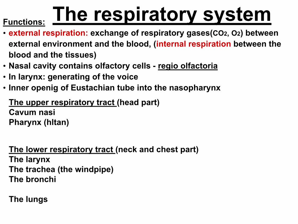
The respiratory system
The upper respiratory tract (head part)
Cavum nasi
Pharynx (hltan)
The lower respiratory tract (neck and chest part)
The larynx
The trachea (the windpipe)
The bronchi
The lungs
Functions:
• external respiration: exchange of respiratory gases(CO2, O2) between
external environment and the blood, (internal respiration between the
blood and the tissues)
• Nasal cavity contains olfactory cells - regio olfactoria
• In larynx: generating of the voice
• Inner openig of Eustachian tube into the nasopharynx
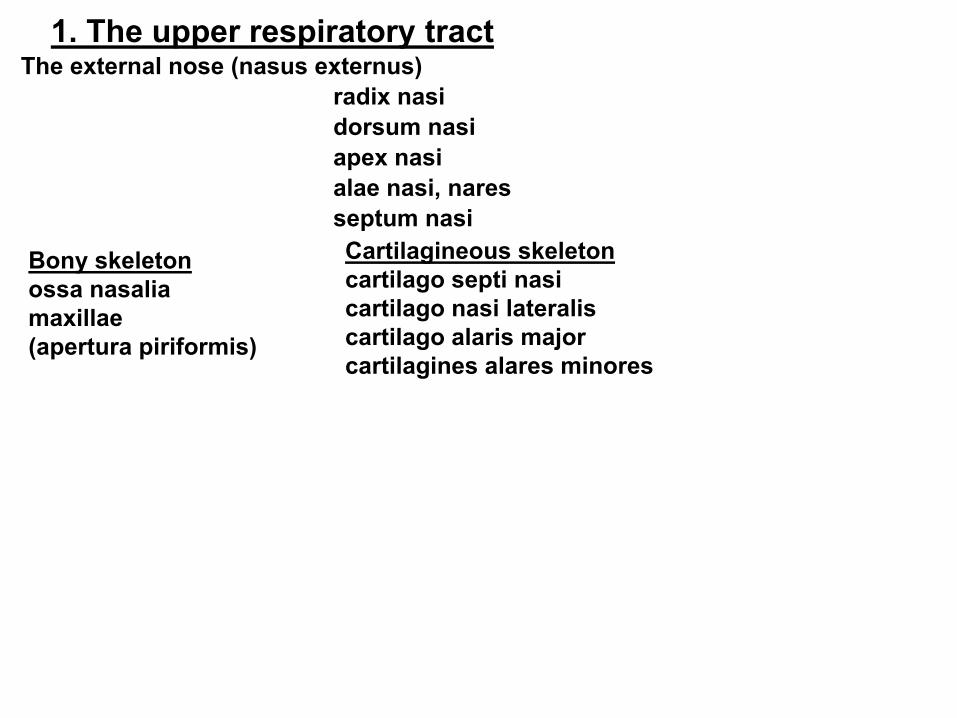
1. The upper respiratory tract The external nose (nasus externus)
radix nasi
dorsum nasi
apex nasi
alae nasi, nares
septum nasi
Cartilagineous skeleton
cartilago septi nasi
cartilago nasi lateralis
cartilago alaris major
cartilagines alares minores
Bony skeleton
ossa nasalia
maxillae
(apertura piriformis)
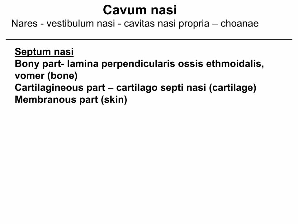
Septum nasi
Bony part- lamina perpendicularis ossis ethmoidalis,
vomer (bone)
Cartilagineous part – cartilago septi nasi (cartilage)
Membranous part (skin)
Cavum nasi Nares - vestibulum nasi - cavitas nasi propria – choanae
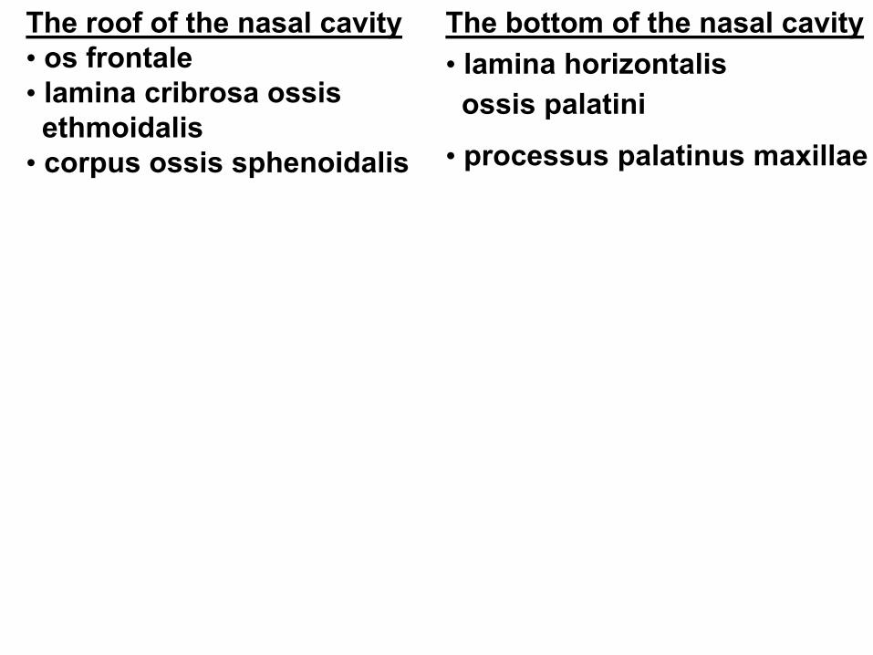
The roof of the nasal cavity
• os frontale
• lamina cribrosa ossis
ethmoidalis
• corpus ossis sphenoidalis
The bottom of the nasal cavity
• lamina horizontalis
ossis palatini
• processus palatinus maxillae
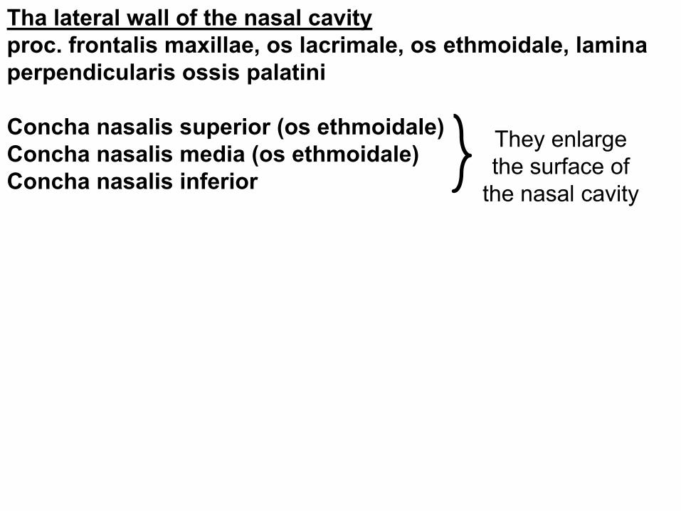
Tha lateral wall of the nasal cavity
proc. frontalis maxillae, os lacrimale, os ethmoidale, lamina
perpendicularis ossis palatini
Concha nasalis superior (os ethmoidale)
Concha nasalis media (os ethmoidale)
Concha nasalis inferior
They enlarge
the surface of
the nasal cavity
The nasal cavity meatus nasi (superior, medius, inferior) – are bordered by
conchae, in elongation of the meatus inf. lies ostium pharyngeum tubae
auditivae
choanae – openings of the nasal cavity to the nasopharynx
The mucosa
Regio olfactoria – on the roof, yellowish colour, olfactory cells (large as a
coin)
Regio respiratoria – grey-pink colour, ciliated epithelium, rich vascular
plexus (plexus cavernosi concharum) - easily bleeds– epistaxis
The paranasal sinuses: open out ino the nasal
cavity, form during the embryonic development Function: resonance chamber
Sinus maxillaris – meatus nasi medius
Sinus frontalis – meatus nasi medius
Sinus ethmoidalis – meatus nasi medius et superior
Sinus sphenoidalis – meatus nasi superior
Ductus nasolacrimalis – meatus nasi inferior
Sinus paranasales have clinical importance– inflammations
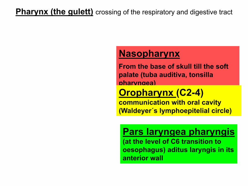
Nasopharynx
From the base of skull till the soft
palate (tuba auditiva, tonsilla
pharyngea)
Oropharynx (C2-4)
communication with oral cavity
(Waldeyer´s lymphoepitelial circle)
Pharynx (the gulett) crossing of the respiratory and digestive tract
Pars laryngea pharyngis
(at the level of C6 transition to
oesophagus) aditus laryngis in its
anterior wall
2. The lower respiratory tract
Larynx
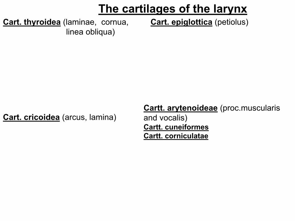
Cart. thyroidea (laminae, cornua,
linea obliqua)
Cart. cricoidea (arcus, lamina)
The cartilages of the larynx Cart. epiglottica (petiolus)
Cartt. arytenoideae (proc.muscularis
and vocalis) Cartt. cuneiformes
Cartt. corniculatae
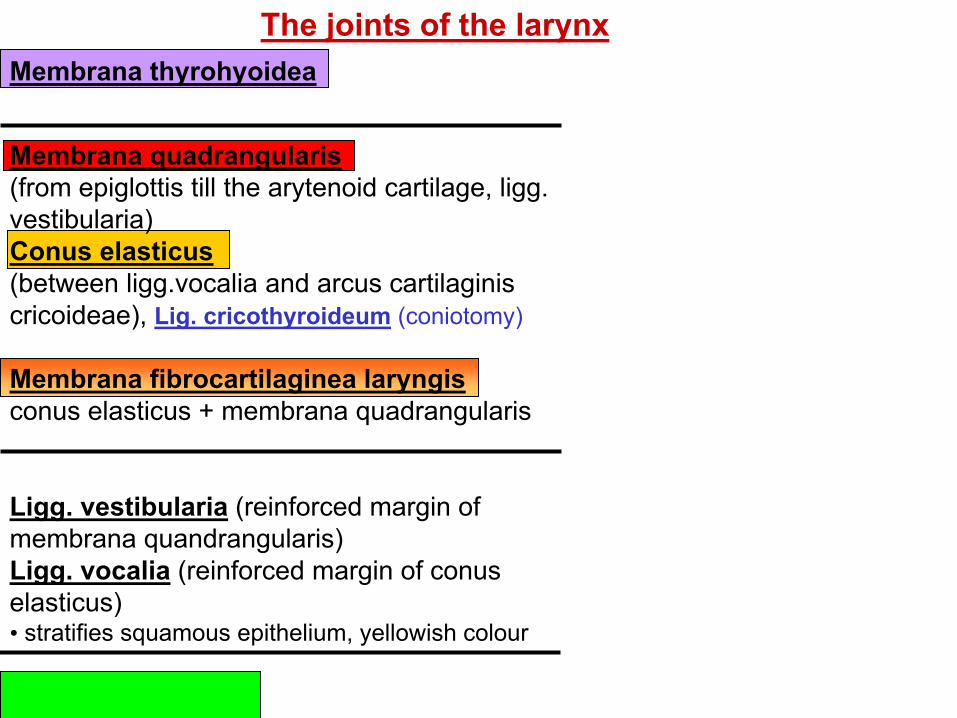
Membrana quadrangularis
(from epiglottis till the arytenoid cartilage, ligg.
vestibularia)
Conus elasticus
(between ligg.vocalia and arcus cartilaginis
cricoideae), Lig. cricothyroideum (coniotomy)
Membrana fibrocartilaginea laryngis
conus elasticus + membrana quadrangularis
Ligg. vestibularia (reinforced margin of
membrana quandrangularis)
Ligg. vocalia (reinforced margin of conus
elasticus) • stratifies squamous epithelium, yellowish colour
Lig. cricotracheale
Membrana thyrohyoidea
The joints of the larynx
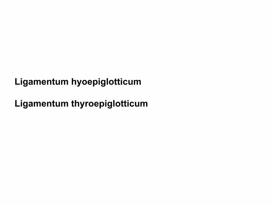
Ligamentum hyoepiglotticum
Ligamentum thyroepiglotticum
Joints:
Art. cricothyroidea (the oscillative motions)
Art. cricoarytenoidea (rotation and sliding movements– base of the
abduction and adduction of the vocal cords)
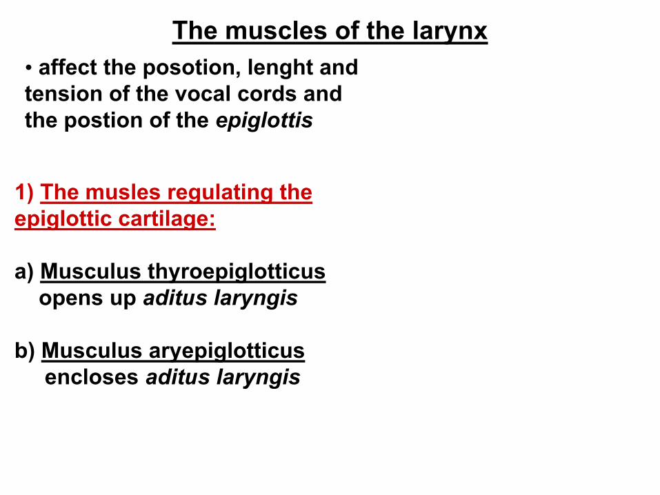
The muscles of the larynx
• affect the posotion, lenght and
tension of the vocal cords and
the postion of the epiglottis
1) The musles regulating the
epiglottic cartilage:
a) Musculus thyroepiglotticus
opens up aditus laryngis
b) Musculus aryepiglotticus
encloses aditus laryngis
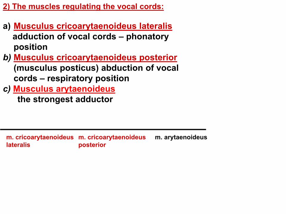
2) The muscles regulating the vocal cords:
a) Musculus cricoarytaenoideus lateralis
adduction of vocal cords – phonatory
position
b) Musculus cricoarytaenoideus posterior
(musculus posticus) abduction of vocal
cords – respiratory position
c) Musculus arytaenoideus
the strongest adductor
m. cricoarytaenoideus
lateralis
m. cricoarytaenoideus
posterior
m. arytaenoideus
L P
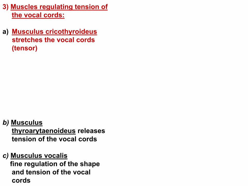
3) Muscles regulating tension of
the vocal cords:
a) Musculus cricothyroideus
stretches the vocal cords
(tensor)
b) Musculus
thyroarytaenoideus releases
tension of the vocal cords
c) Musculus vocalis
fine regulation of the shape
and tension of the vocal
cords
TA
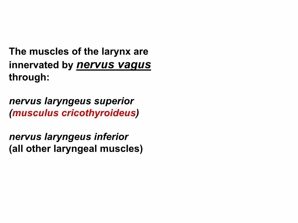
The muscles of the larynx are
innervated by nervus vagus
through:
nervus laryngeus superior
(musculus cricothyroideus)
nervus laryngeus inferior
(all other laryngeal muscles)
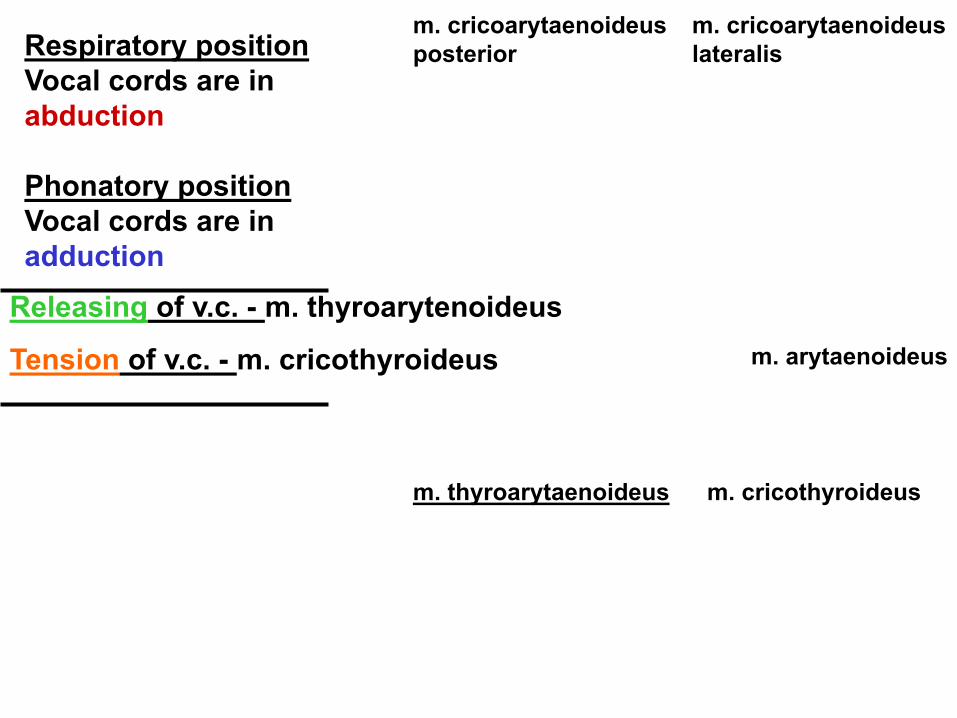
Respiratory position
Vocal cords are in
abduction
Phonatory position
Vocal cords are in
adduction
Releasing of v.c. - m. thyroarytenoideus
Tension of v.c. - m. cricothyroideus
m. cricothyroideus
m. cricoarytaenoideus
posterior
m. cricoarytaenoideus
lateralis
m. arytaenoideus
m. thyroarytaenoideus
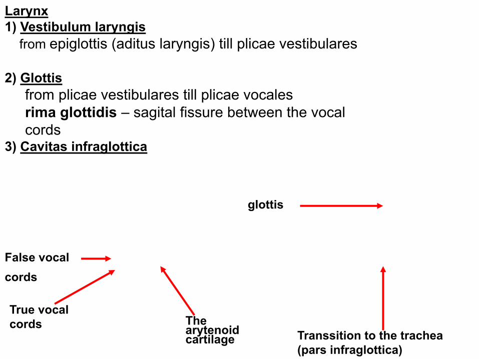
Larynx
1) Vestibulum laryngis
from epiglottis (aditus laryngis) till plicae vestibulares
2) Glottis
from plicae vestibulares till plicae vocales
rima glottidis – sagital fissure between the vocal
cords 3) Cavitas infraglottica
False vocal
cords
True vocal
cords
The arytenoid cartilage
glottis
Transsition to the trachea
(pars infraglottica)
The laryngoskopic view
laryngoscopy
Respiratory
position
Phonatory
position
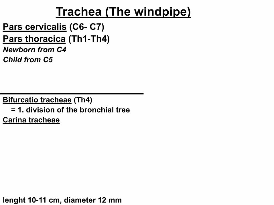
Trachea (The windpipe) Pars cervicalis (C6- C7)
Pars thoracica (Th1-Th4) Newborn from C4
Child from C5
Bifurcatio tracheae (Th4)
= 1. division of the bronchial tree
Carina tracheae
lenght 10-11 cm, diameter 12 mm
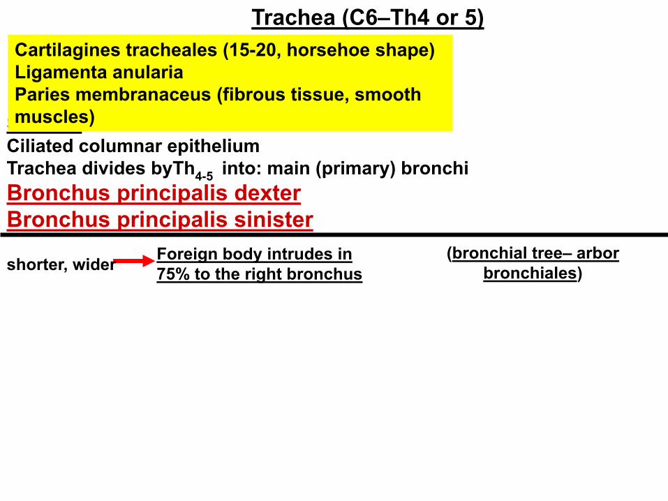
Sliznice:
Ciliated columnar epithelium
Trachea divides byTh4-5
into: main (primary) bronchi
Bronchus principalis dexter
Bronchus principalis sinister
Trachea (C6–Th4 or 5)
shorter, wider Foreign body intrudes in
75% to the right bronchus
(bronchial tree– arbor
bronchiales)
Cartilagines tracheales (15-20, horsehoe shape)
Ligamenta anularia
Paries membranaceus (fibrous tissue, smooth
muscles)
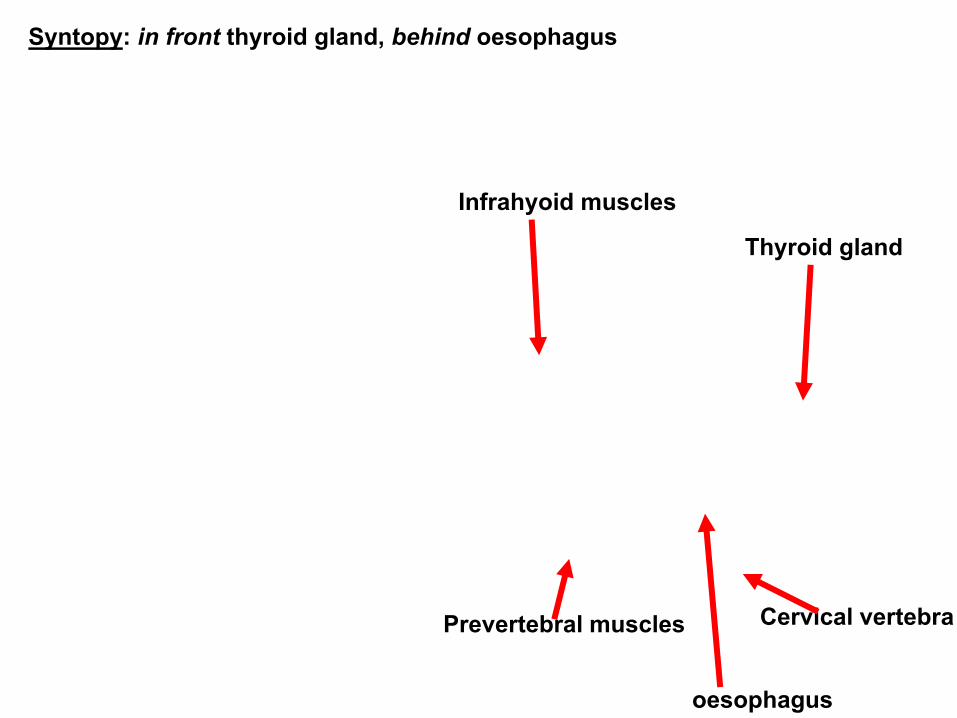
Syntopy: in front thyroid gland, behind oesophagus
Infrahyoid muscles
Thyroid gland
Cervical vertebra Prevertebral muscles
oesophagus
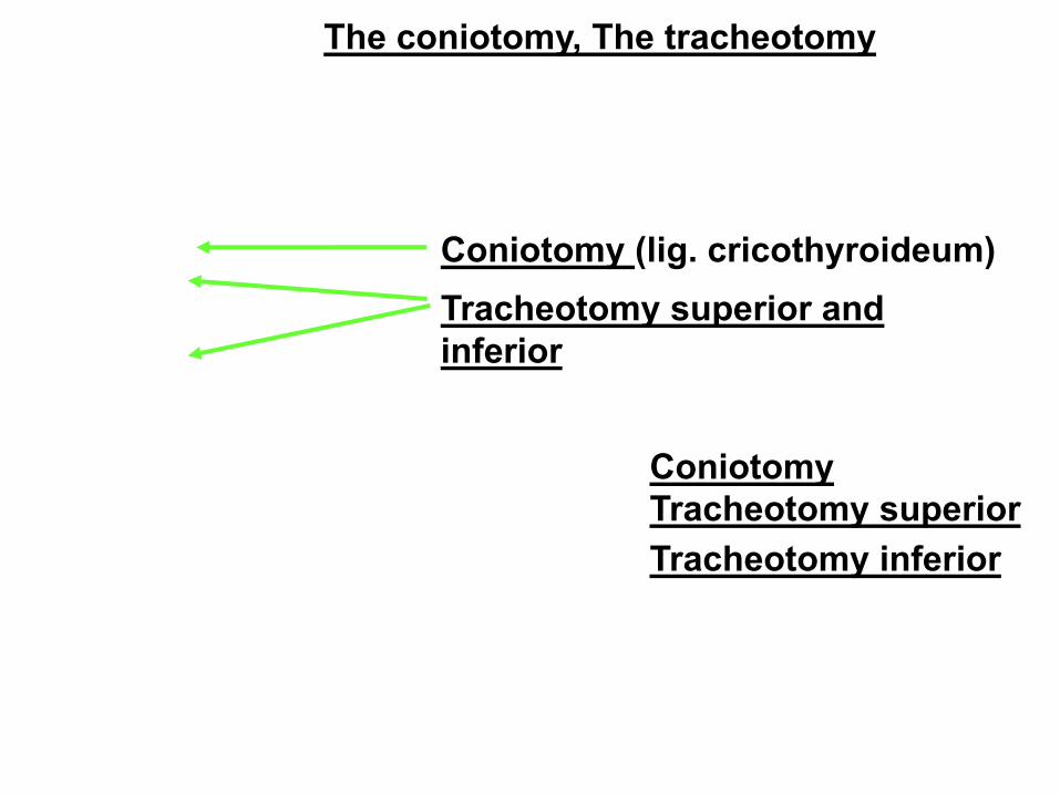
The coniotomy, The tracheotomy
Coniotomy (lig. cricothyroideum)
Tracheotomy superior and
inferior
Coniotomy
Tracheotomy superior
Tracheotomy inferior
Bronchi principales divide into:
bronchi lobares (right 3, left 2) -
secondary bronchi
bronchi segmentales ( 9-10 sin., 10 dx.) –
terciary bronchi
(the pulmonary segment is of pyramidal
shape, the apex is located in the area of the
pulmonary hilus, segmentectomy)
The last section form:
bronchioli terminales (<1mm, have no
reiforcement, the wall is formed by smooth
muscles – they can enclose the bronchial
lumen)
Arbor bronchiales
bronchioli respiratorii
ductuli alveolares – atrium - sacculi alveolares - alveoli pulmonis
Arbor alveolaris – breathing compartment of the lungs–
exchange of breathing gases
alveoli Alveolar sacs Thin-walled tubules
Bronchography Bronchoscopy
The lungs - pulmo
Position of the lungs:
Thorax cavity– pleural cavities
height 20-24 cm, weight 600 – 700g
colour pink– marbling – grey/black
The lungs (Pulmo, Pneumon) Description of the lungs:
facies diaphragmatica - base
apex pulmonis - top (reaches above apertura thoracis superior)
facies costalis - (impressions of the ribs)
facies mediastinalis (hilus pulmonis–dx. BAV, sin. ABV)
radix pulmonis – root of the lung
Impressions
The heart
Aorta
The oesophagus
1. rib
Other vessels
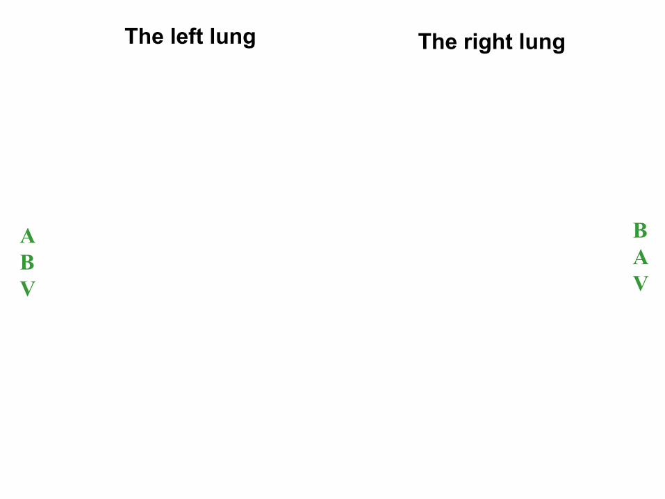
The right lung The left lung
A
B
V
B
A
V
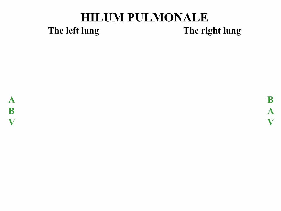
HILUM PULMONALE The left lung The right lung
B
A
V
A
B
V
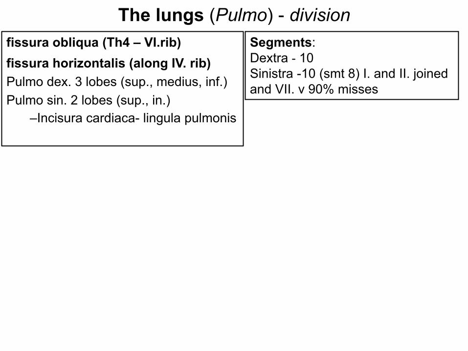
The lungs (Pulmo) - division
fissura obliqua (Th4 – VI.rib)
fissura horizontalis (along IV. rib)
Pulmo dex. 3 lobes (sup., medius, inf.)
Pulmo sin. 2 lobes (sup., in.)
–Incisura cardiaca- lingula pulmonis
Segments:
Dextra - 10
Sinistra -10 (smt 8) I. and II. joined
and VII. v 90% misses
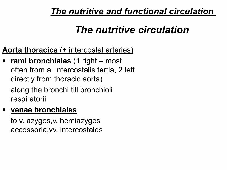
The nutritive circulation
Aorta thoracica (+ intercostal arteries)
rami bronchiales (1 right – most
often from a. intercostalis tertia, 2 left
directly from thoracic aorta)
along the bronchi till bronchioli
respiratorii
venae bronchiales
to v. azygos,v. hemiazygos
accessoria,vv. intercostales
The nutritive and functional circulation
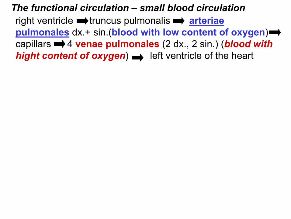
right ventricle truncus pulmonalis arteriae
pulmonales dx.+ sin.(blood with low content of oxygen)
capillars 4 venae pulmonales (2 dx., 2 sin.) (blood with
hight content of oxygen) left ventricle of the heart
The functional circulation – small blood circulation
Pneumothorax
Xray of the thorax
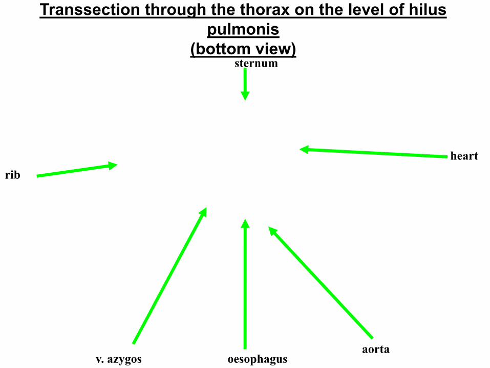
Transsection through the thorax on the level of hilus
pulmonis
(bottom view) sternum
oesophagus aorta
v. azygos
heart
rib
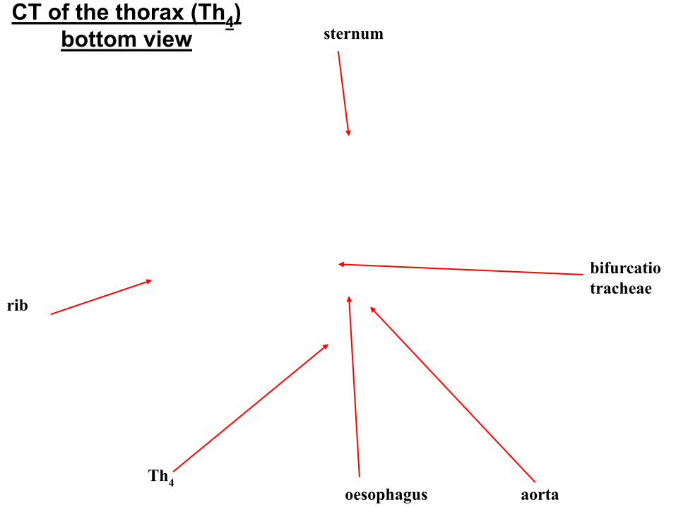
CT of the thorax (Th4)
bottom view
bifurcatio
tracheae
aorta oesophagus Th
4
rib
sternum
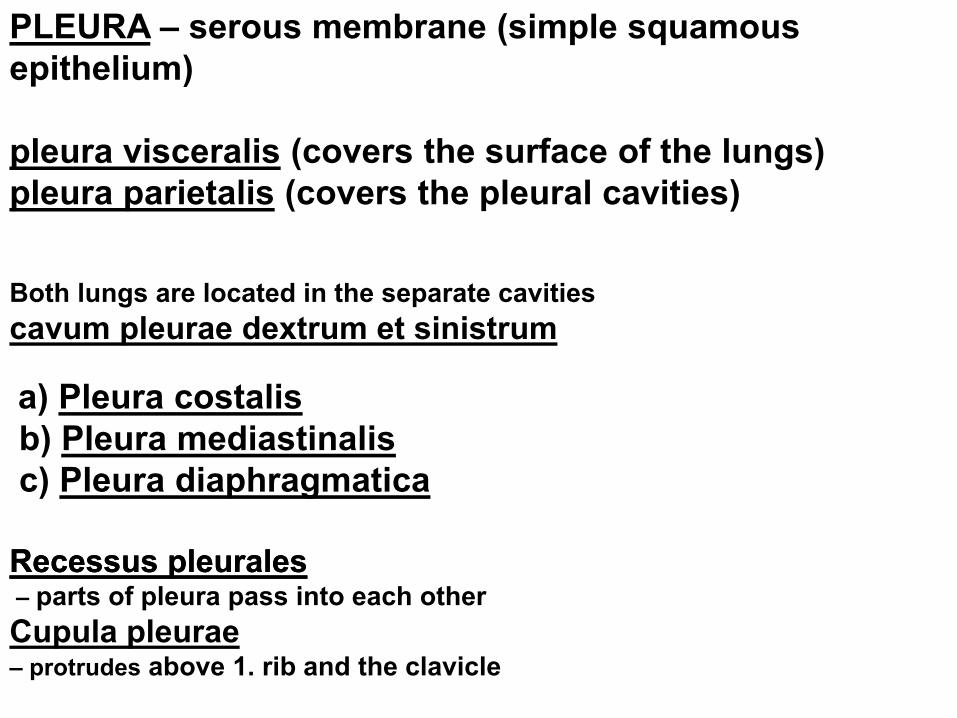
PLEURA – serous membrane (simple squamous
epithelium)
pleura visceralis (covers the surface of the lungs)
pleura parietalis (covers the pleural cavities)
Both lungs are located in the separate cavities
cavum pleurae dextrum et sinistrum
a) Pleura costalis
b) Pleura mediastinalis
c) Pleura diaphragmatica
RecessusRecessus pleuralespleurales – parts of pleura pass into each other
Cupula pleurae
– protrudes above 1. rib and the clavicle
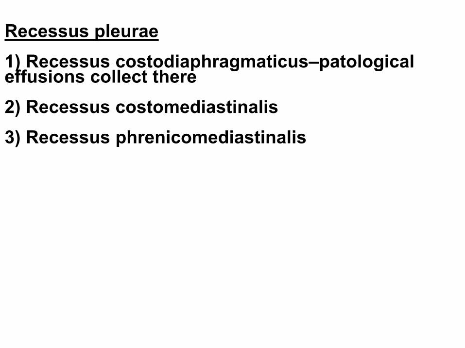
Recessus pleurae 1) Recessus costodiaphragmaticus–patological effusions collect there 2) Recessus costomediastinalis 3) Recessus phrenicomediastinalis
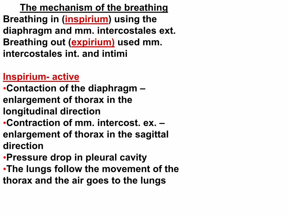
The mechanism of the breathing
Breathing in (inspirium) using the
diaphragm and mm. intercostales ext.
Breathing out (expirium) used mm.
intercostales int. and intimi
Inspirium- active
•Contaction of the diaphragm –
enlargement of thorax in the
longitudinal direction
•Contraction of mm. intercost. ex. –
enlargement of thorax in the sagittal
direction
•Pressure drop in pleural cavity
•The lungs follow the movement of the
thorax and the air goes to the lungs
Expiration - passive
•Relaxation of the
diaphragm(contraction of abdomen
muscles)
•Contraction of mm. intercostales
interni and intimi, decrease of the
ribs – expiration
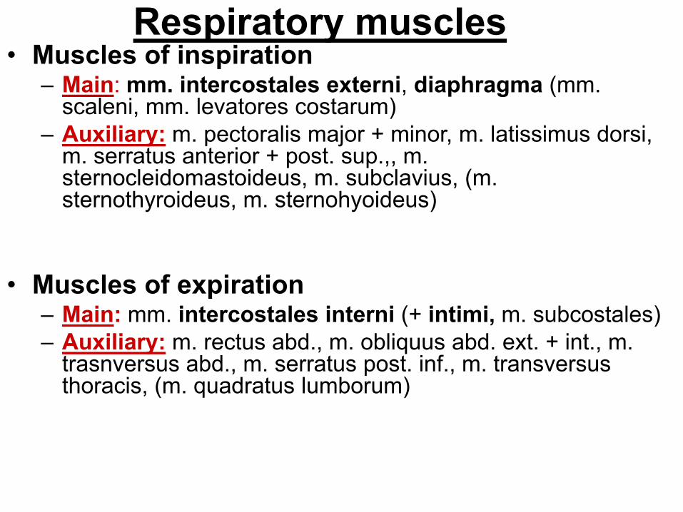
Respiratory muscles • Muscles of inspiration
– Main: mm. intercostales externi, diaphragma (mm. scaleni, mm. levatores costarum)
– Auxiliary: m. pectoralis major + minor, m. latissimus dorsi, m. serratus anterior + post. sup.,, m. sternocleidomastoideus, m. subclavius, (m. sternothyroideus, m. sternohyoideus)
• Muscles of expiration – Main: mm. intercostales interni (+ intimi, m. subcostales)
– Auxiliary: m. rectus abd., m. obliquus abd. ext. + int., m. trasnversus abd., m. serratus post. inf., m. transversus thoracis, (m. quadratus lumborum)
Orientation lines:
linea mediana ant.
linea sternalis
linea parasternalis
linea medioclavicularis
linea axillaris ant., med., post.
linea scapularis
linea paravertebralis
linea mediana post.
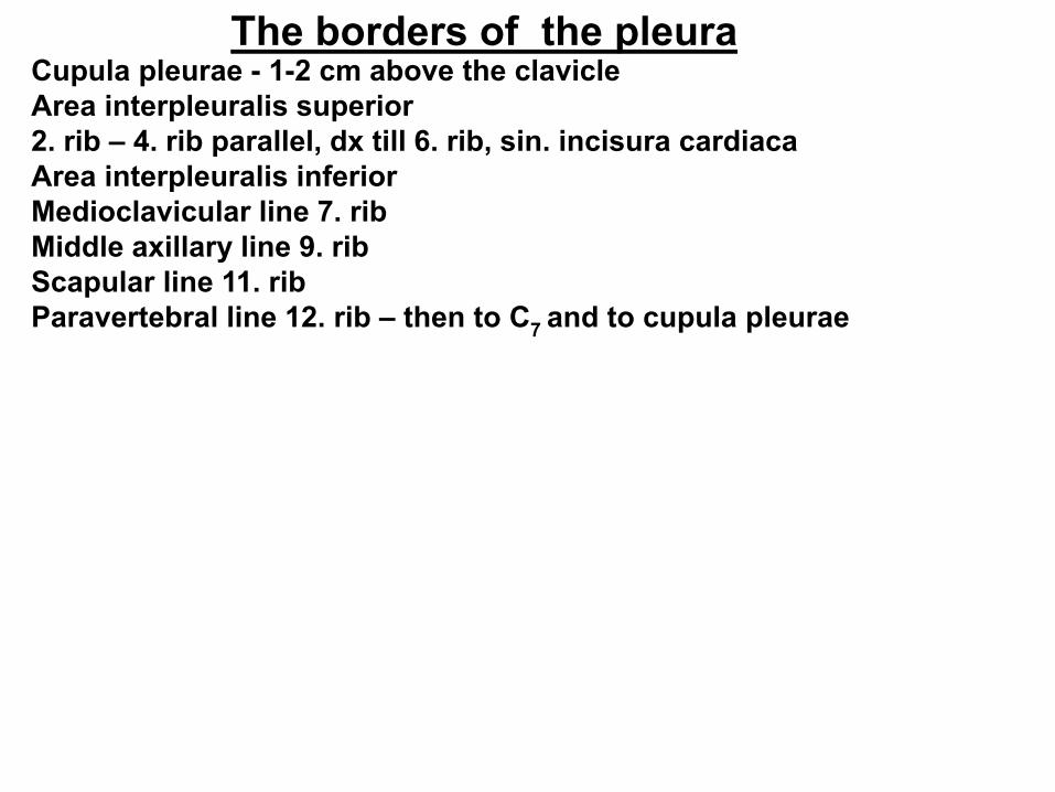
The borders of the pleura Cupula pleurae - 1-2 cm above the clavicle
Area interpleuralis superior
2. rib – 4. rib parallel, dx till 6. rib, sin. incisura cardiaca
Area interpleuralis inferior
Medioclavicular line 7. rib
Middle axillary line 9. rib
Scapular line 11. rib
Paravertebral line 12. rib – then to C7
and to cupula pleurae
The borders of the pleura
• apex pulmonis fills cupula pleurae, reaches 1–2 cm over the
clavicle
• Anterior margin: corresponds during the inspiration with the
anterior margin of the pleura
• Inferior margin: projects about 1 till 2 ribs more cranially than
the inferior border of the pleura
• Posterior margin: projects laterally from the spinous
processes within the range Th11 till Th2.
The borders of the lungs
• a newborn can survive the preterm labor between 24.
and 28. week (formerly lungs are not yet sufficiently
prepared for gas exchange, do not produce
surfactant)
• The lungs of a newborn, who done the inspiration,
keep afloat, the lungs of stillborn sink to the bottom
• The importance in the forensic pathology
The lungs of a newborn
Regional anatomy of the thorax
Pleural cavities
Mediastinum: space between the pleural cavities: it contents organs, vessels and nerves, there is also the loose fibrous connective tissue present
Dividing of the mediastinum:
(border: horizontal plane going through the upper margin of the heart)
1. medistinum superius (thymus, layer of veins, layer of arteries, trachea)
2. mediastinum inferius
mediastinum anterius (lymph nodes)
mediastinum medium (heart)
mediastinum posterius (oesophagus,
aorta, ductus thoracicus, v. azygos et
hemiazygos, truncus sympaticus,
lymph nodes)
Obrázky:
Atlas der Anatomie des Menschen/Sobotta.
Putz,R., und Pabst,R. 20. Auflage. München:
Urban & Schwarzenberg, 1993
Netter: Interactive Atlas of Human Anatomy.
Naňka, Elišková: Přehled anatomie. Galén, Praha 2009.
Čihák: Anatomie I, II, III.
Drake et al: Gray´s Anatomy for Students. 2010