Embed Size (px)
Citation preview
1
THE RESTRICTION FACTORS OF HUMAN IMMUNODEFICIENCY VIRUS
Reuben S. Harris1, Judd F. Hultquist1 and David T. Evans2
1Department of Biochemistry, Molecular Biology and Biophysics, Department of Genetics, Cell Biology and Development, Institute for Molecular Virology
University of Minnesota, Minneapolis, MN, 55455 2Department of Microbiology and Immunobiology, Harvard Medical School, New England
Primate Research Center, One Pine Hill Drive, Southborough, MA 01772-9102
Address correspondence to: Reuben S. Harris, 321 Church Street S.E., 6-155 Jackson Hall, Minneapolis, MN 55455. E-mail: [email protected]
SUMMARY Cellular proteins called ‘restriction factors’ can serve as powerful blockades to HIV replication, but the virus possesses elaborate strategies to circumvent these barriers. First, we discuss general hallmarks of a restriction factor. Then, we review how the viral Vif protein protects the viral genome from lethal levels of cDNA deamination by promoting APOBEC3 protein degradation, how the viral Vpu, Env, and Nef proteins facilitate internalization and degradation of the virus-tethering protein Tetherin/BST-2, and how the viral Vpx protein prevents the premature termination of reverse transcription by degrading the dNTPase SAMHD1. These HIV restriction and counter-restriction mechanisms suggest strategies for new therapeutic interventions. Restriction Factor Hallmarks
Restriction factors have at least four defining characteristics (Fig. 1A). First and foremost, a restriction factor must directly and dominantly cause a significant decrease in HIV infectivity. This is often determined by co-transfecting cell lines such as HEK293 or HeLa with a molecular clone of the virus, with or without a plasmid expressing the restriction factor, and measuring the amount and infectivity of
virus recovered in the cell culture medium after 1 to 2 days of incubation. Such assays are ideally done over a range of restriction factor expression, with the highest levels often imposing log-scale drops in viral infectivity (Fig. 1B). This ‘single cycle’ assay for virus replication is useful for testing the impact of viral mutations and/or restriction factor variations.
Second, if a restriction factor is a true threat to viral replication, then the predecessors of HIV invariably evolved an equally potent counter-restriction mechanism that still exists in the present day virus. For instance, titrating a counter-restriction factor into the aforementioned single cycle infectivity experiment can result in a full recovery of viral infectivity despite the presence of an active restriction factor (Fig. 1B). Viruses lacking these various countermeasures are able to replicate in some, but not all, cell types depending on the expression level of the relevant restriction factor. Cell lines that support replication are termed ‘permissive’ and those that do not are termed ‘non-permissive’. This life/death dichotomy has been elegantly exploited to identify several restriction factors and their corresponding viral antagonists.
Third, because the interaction between restriction and counter-restriction factors occurs through direct protein-protein
http://www.jbc.org/cgi/doi/10.1074/jbc.R112.416925The latest version is at JBC Papers in Press. Published on October 5, 2012 as Manuscript R112.416925
Copyright 2012 by The American Society for Biochemistry and Molecular Biology, Inc.
by guest on May 8, 2018
http://ww
w.jbc.org/
Dow
nloaded from
2
interactions, the restriction factor often shows signatures of rapid evolution. In general, mutations are maintained in a population only if they confer a selective advantage. If a host species experiences iterative rounds of pathogenic pressure, altered variants of host restriction factors that are no longer susceptible to the pathogen’s counteraction mechanism are selected. Over evolutionary time, this results in an overabundance of amino acid substitution mutations in these genes relative to non-amino acid changing, or silent, mutations. These positive selection signatures become apparent by comparing human and primate restriction factor gene sequences. It is important to note that each amino acid substitution could have been selected by an independent pathogen conflict, and that the ancestral pathogen may not have even resembled present day HIV. Thus, a major corollary to the hallmark of positive selection is the strong likelihood that each present day restriction factor has emerged from many ancient host-pathogen conflicts, and has thus been fine-tuned to protect against a formidable number of parasites (i.e., restriction factors elicit broad activity). These hyper-evolved restriction factor protein sequences will undoubtedly continue to change as a result of ongoing interactions with modern pathogens.
Fourth, the expression of each restriction factor is often hard-wired to the innate immune response. For instance, restriction factor expression is often strongly induced by interferon, and is thus tied directly to the host’s innate immune response. Many other genes are also induced by interferons (as well as the interferon genes themselves), with each restriction factor or group of restriction factors comprising a relatively small portion of the much larger immune response. Since the overall composition of innate immune regulon is shaped in part by the pathogens to
which each species and its ancestors were exposed, the various innate immune effector proteins can vary between species (i.e., with one type proving more important for one species in comparison to another). Thus, this membership to the larger innate immune regulon helps rationalize why the numbers of each type of restriction factor often varies between mammalian lineages.
It is also worth noting that restriction factors are relatively rare in contrast to other host proteins that impact virus replication (dependency factors). Following the criteria established above, human cells probably possess only a few HIV restriction factors compared to hundreds of dependency factors and thousands of proteins that are not particularly relevant to HIV replication. Bona fide restriction factors result in log-scale differences in virus infectivity, and interact directly with at least one viral component. Here, we discuss three such HIV restriction factors: the APOBEC3 DNA deaminase sub-family, the BST-2/Tetherin integral membrane protein, and the SAMHD1 dNTP hydrolase. We also encourage readers to see recent reviews on the TRIM5 proteins, which represent an important barrier to HIV replication in other species, but not in humans [e.g., (1)]. Restriction by Hypermutation – APOBEC3 DNA Deaminases Classic studies showed that the HIV Vif protein is dispensable for virus replication in some T cell lines but not others (2,3). Hybrid cells constructed by fusing permissive and non-permissive cell lines inherit the non-permissive phenotype, suggesting the existence of dominant-acting restriction factor (4). This difference led to the molecular identification of APOBEC3G in non-permissive cell types (5). Importantly, expressing APOBEC3G in permissive T cells was sufficient to render
by guest on May 8, 2018
http://ww
w.jbc.org/
Dow
nloaded from
3
them non-permissive for Vif-deficient HIV replication (5). At the same time, APOBEC3G was independently identified and shown to be part of a larger sub-family of seven APOBEC3 proteins that each have the capacity to catalyze DNA cytosine-to-uracil (C-to-U) deamination (6). This biochemical activity not only suggested a mechanism for APOBEC3G-mediated HIV restriction, but also suggested an explanation for the phenomenon of G-to-A hypermutation noted previously in HIV sequences from clinical samples [e.g., (7,8)]. Indeed, three groups demonstrated that APOBEC3G-dependent HIV restriction is characterized by massive levels of viral genomic strand G-to-A hypermutation, which can only be explained by a cDNA C-to-U deamination mechanism (9-11) (Fig. 2). This conclusion is now fortified by biochemical and mutagenesis studies, with the most notable showing that near-physiologic levels of APOBEC3G suppress spreading Vif-deficient virus replication in permissive T cell lines and induce hallmark G-to-A hypermutations, while physiologic levels of a catalytically inactive variant fail to do so (12-14). APOBEC3G has a strong intrinsic preference for deaminating the second cytosine of a 5’CC dinucleotide motif, which results in 5’GG-to-AG hypermutations (9,15,16). HIV sequences from clinical samples frequently bear this pattern, but at near equal levels they also often contain a 5’GA-to-AA pattern [e.g., (7,8)]. Many studies have been dedicated to deducing which of the six other APOBEC3 proteins is responsible for inflicting this additional signature with widely varying results [reviewed in (17,18)]. However, recent studies have brought clarity to this area by strongly implicating APOBEC3D, APOBEC3F, and APOBEC3H in HIV restriction (and simultaneously excluding
APOBEC3A, APOBEC3B, and APOBEC3C) (19,20).
The four HIV-relevant APOBEC3 proteins share the following properties: i) expression in non-permissive CD4+ T cells (the primary HIV target cell), ii) capacity to package into the nucleic acid containing viral core (i.e., encapsidate), iii) potent virus restriction activity, iv) ability to inflict G-to-A mutations, v) susceptibility to HIV Vif, and vi) functional conservation with the homologous proteins of rhesus macaque (19) (Fig. 2). These observations are supported by systematic APOBEC3 knockout and knockdown studies in non-permissive cells that demonstrate the importance of APOBEC3G in causing 5’GG-to-AG hypermutations and reveal overlapping roles for both APOBEC3D and APOBEC3F in Vif-deficient HIV restriction and 5’GA-to-AA hypermutation (20). The contribution of APOBEC3H to Vif-deficient HIV restriction and 5’GA-to-AA hypermutation may vary depending on the stability of the expressed protein, as multiple stable and unstable haplotypes are circulating in the human gene pool (21,22). HIV is a ‘successful’ pathogen, in part because its Vif protein mediates the poly-ubiquitination and subsequent degradation of the four restrictive APOBEC3 proteins by the 26S proteasome (19). This is an ancient and conserved counter-restriction mechanism, since all related lentiviruses, except equine infectious anemia virus (EIAV), express a Vif protein that neutralizes the relevant APOBEC3 proteins of its host species [e.g., (23)]. An excellent recent example is provided by studies on natural SIV infection of four African green monkey (AGM) subspecies (24). As expected, the Vif protein of each SIVagm strain degrades the APOBEC3G protein of its respective host. However, only a few of these Vif proteins are able to degrade the APOBEC3G proteins of other
by guest on May 8, 2018
http://ww
w.jbc.org/
Dow
nloaded from
4
AGM subspecies. Genetic studies deduced that Vif resistance/susceptibility maps to only a few amino acids in APOBEC3G. These studies suggest that SIV may have been transmitted into animals with Vif-resistant APOBEC3G alleles, and then altered its Vif protein to re-gain the capacity to degrade the new host’s APOBEC3G protein. Original biochemical and genetic studies combined to show that Vif recruits an E3 ligase complex consisting of CUL5, ELOB, ELOC, and RBX to mediate APOBEC3G degradation (25,26) (Fig. 2). Despite this knowledge, Vif has resisted purification and biochemical studies suggesting that a co-factor might be missing. Recent proteomic studies identified the transcription factor CBFβ as a Vif-associated protein that also associates with the E3 ligase, but only in the presence of Vif (27,28). Knockdown studies demonstrated that CBFβ is essential for Vif stability and APOBEC3 degradation in human cells (27,28). Importantly, CBFβ permitted the biochemical purification of a tetrameric complex CBFβ-Vif-ELOC-ELOB that can be combined with a CUL5-RBX2 dimer to form a hexameric complex with APOBEC3G poly-ubiquitination activity (27). It is likely that this breakthrough will soon lead to the first atomic structures of Vif, the Vif-APOBEC3 interface, and perhaps an entire Vif-E3-APOBEC3 complex. Such information will undoubtedly expedite the development of drugs to disrupt the Vif-APOBEC3 interaction, and possibly extinguish HIV replication by lethal mutagenesis. This strategy may lead to a new class of therapeutic agents to suppress viral loads, and may have the potential to be curative if combined with ongoing work in the field to purge cells of latently integrated viruses. The fact that the APOBEC3 proteins are pro-mutagenic raises another
provocative possibility. It is conceivable that lentiviruses, including HIV, are using Vif as a mutational rheostat to regulate the overall load and impact of APOBEC3-driven mutations (29,30). In the extreme, it is possible that HIV has become dependent upon the APOBEC3 proteins to achieve the overall high degree of genetic diversity required for the virus to ‘outrun’ antibody and T cell responses. Thus, therapeutic strategies to inhibit APOBEC3 activity and to starve HIV of the fuel driving much of its genetic variability may be worth investigating as a means to constrain immune evasion. Restriction by Particle Tethering – BST-2/Tetherin Integral Membrane Protein Tetherin, also known as BST-2 or CD317, was identified as the cellular protein that accounts for a late-stage defect in the release of vpu-deleted HIV-1 from restrictive cells (31,32). This discovery traces back to original observations that deletion of the HIV-1 vpu gene results in a 5- to 10-fold decrease in virus release from infected T cells without impairing the expression of other viral proteins (33). This defect in virus release was later shown to be cell-type dependent, and to result in a particularly dramatic phenotype observable by electron microscopy, whereby mature virus particles are unable to detach from infected cells in the absence of Vpu, and consequently accumulate on the plasma membrane and within intracellular compartments (34). The explanation for this phenomenon remained enigmatic for several years until heterokaryon fusions of permissive and non-permissive cells revealed a dominant block to the release of vpu-deleted HIV-1 (35). Subsequent work demonstrated that the restriction factor is expressed on the cell surface and is interferon-inducible (36,37), features which ultimately led to the identification of BST-2
by guest on May 8, 2018
http://ww
w.jbc.org/
Dow
nloaded from
5
(dubbed “Tetherin”) as the cellular protein responsible for inhibiting the release of Vpu-deficient HIV-1 (31,32).
The topology of Tetherin, which includes an N-terminal cytoplasmic domain followed by a single-pass transmembrane domain, an extracellular coiled-coil domain, and a C-terminal glycosyl-phosphatidylinositol (GPI) anchor (38), accounts for its broad antiviral activity, not only against HIV-1, but also against many other enveloped viruses (Fig. 3). By virtue of having a membrane-spanning domain and a GPI anchor at opposite ends of the molecule, Tetherin can simultaneously associate with both viral and cellular membranes. As viruses attempt to bud from infected cells, Tetherin becomes incorporated into the viral envelope and physically bridges nascent virions to the cell (39-41). Evidence suggests that Tetherin forms a parallel homodimer, and although either the N- or C-terminal domains can be oriented in the cell (39), at least some of the dimers need to have their N-terminal domains in the cell to interact with the cellular endocytosis machinery. Captured virions are subsequently internalized for degradation in lysosomes (39,42).
There is general consensus that the mechanism of Tetherin counteraction by HIV-1 Vpu begins with a direct physical interaction between the anti-parallel membrane-spanning helices of Vpu and Tetherin (43-45) (Fig. 3). However, the relative importance of alternative cellular pathways leading to the removal of Tetherin from sites of virus release is less clear. A number of studies have shown that casein kinase II phosphorylation of a pair of conserved serine residues (S52 and S56) in the cytoplasmic tail of Vpu recruits β-TrCP-2, a component of the SKP1-CUL-F box E3 ubiquitin ligase complex, which leads to the downregulation and degradation of Tetherin (44,46-48). β-TrCP-2-dependent
degradation involves non-lysine ubiquitination of residues in the cytoplasmic domain of Tetherin (49,50), which serves as a signal for HRS binding and ESCRT-mediated trafficking to lysosomal compartments (51). Vpu-mediated targeting of Tetherin to lysosomes also requires RAB7A (52), a small GTPase essential for the maturation of late endosomes and lysosomal fusion. Other studies have revealed a β-TrCP-2-independent mechanism of Tetherin antagonism by Vpu, which leads to the sequestration of Tetherin in a perinuclear compartment without degradation (48,53,54). This could occur either by trapping newly synthesized Tetherin or by blocking the recycling of Tetherin to the plasma membrane (55,56). The internalization and trafficking of Tetherin by Vpu, whether for sequestration or degradation, depends in part on dynamin 2, a GTPase important for the scission of vesicular membranes (57). Although the relative contribution of pathways leading to the sequestration versus degradation of Tetherin is presently unclear, it is important to recognize that these are not mutually exclusive mechanisms, and both may contribute to the optimal resistance to Tetherin afforded by Vpu. Most primate lentiviruses do not have Vpu, and therefore depend on other viral proteins to counteract Tetherin. In the case of certain HIV-2 and SIV isolates, this activity has been acquired by the viral envelope (Env) glycoprotein (58-61). Indeed, prior to the identification of Tetherin, the envelope glycoproteins of certain HIV-2 isolates were shown to have “Vpu-like” activity that could rescue the release of Vpu-deficient HIV-1 from restrictive cells (62). Tetherin antagonism by Env depends on physical interaction between Env and Tetherin, and on a conserved tyrosine-based endocytosis motif (YXXφ) in the cytoplasmic tail of the Env
by guest on May 8, 2018
http://ww
w.jbc.org/
Dow
nloaded from
6
transmembrane protein gp41 (59,60,63). The residues that contribute to Env-Tetherin interactions are not well defined, but appear to be located in the extracellular domains of both proteins based on analyses of recombinant forms of Env and Tetherin (60,64), and on the identification of defined amino acid changes in the ectodomains of gp41 and Tetherin that disrupt anti-Tetherin activity (64-66). Interaction with Env does not result in the degradation of Tetherin, but instead leads to internalization and sequestration of Tetherin away from sites of virus release at the plasma membrane by a clathrin-dependent pathway (57,60,63).
The majority of primate lentiviruses, including phylogenetically diverse SIVs endemic to chimpanzees (SIVcpz), sooty mangabeys (SIVsmm) and African green monkeys (SIVagm), use Nef to counteract the Tetherin proteins of their non-human primate hosts (61,67,68). In fact, the anti-Tetherin activities of HIV-1 Vpu and HIV-2 Env appear to have been acquired after the respective cross-species transmission of SIVcpz and SIVsmm into humans due to the absence of a five amino acid sequence in human Tetherin required for susceptibility to Nef (61,68). SIV Nef counteracts the Tetherin proteins of apes and Old World monkeys, but not humans, through the recognition of residues in the N-terminal cytoplasmic domain (G/D14DIWK18 in rhesus macaque, sooty mangabey and chimpanzee Tetherin) that are missing from human Tetherin (61,68). SIVcpz, SIVmac and SIVagm Nef down-modulate the Tetherin proteins of their respective hosts from the cell surface by AP-2-dependent endocytosis (69). The nature of the molecular interactions between Nef and Tetherin, and the ultimate fate of Tetherin in SIV-infected cells, remain to be defined. Nevertheless, current evidence suggests that, similar to HIV-1 Vpu and HIV-2 Env, the anti-Tetherin activity of SIV Nef reflects its
ability to remove Tetherin from sites of virus release at the plasma membrane.
In a remarkable instance of convergent evolution, the envelope glycoprotein of a nef-deleted strain of SIV also acquired the ability to counteract Tetherin during serial passage in rhesus macaques. Similar to HIV-2 Env, the anti-Tetherin activity of this Env is dependent on a physical interaction with Tetherin and on the YXXφ motif in gp41 (58). However, in this case, compensatory changes in the cytoplasmic domain of gp41, rather than in the ectodomain, stabilize a physical interaction with rhesus macaque Tetherin (58). These observations imply that Tetherin antagonism is important for efficient virus replication in vivo and ultimately for pathogenesis.
There is now growing interest in the development of novel therapeutic agents to enhance the anti-viral activity of Tetherin as a treatment for HIV-1 infection. NMR structural data and computational modeling have provided a high-resolution picture of the transmembrane interface between Vpu and Tetherin that represents a promising target for drug design (70,71). Pharmaceutical disruption of this interface would, in principle, render HIV-1 susceptible to restriction by Tetherin, significantly attenuating virus replication in vivo. However, features unique to the biology of Tetherin suggest that this may not be so straightforward. Unlike other restriction factors, Tetherin does not impose an absolute block to virus replication; vpu-deleted HIV-1 and nef-deleted SIV still replicate, albeit at significantly reduced rates, in primary lymphocytes and infected animals (37,58). Comparisons of the mechanisms of Tetherin antagonism by HIV-1, HIV-2 and SIV also suggest that the primate lentiviruses have unusual latitude in adapting to the Tetherin proteins of their respective hosts, having evolved to use at
by guest on May 8, 2018
http://ww
w.jbc.org/
Dow
nloaded from
7
least three different proteins to counteract this restriction. Thus, while the development of drugs to interfere with Tetherin antagonism by Vpu certainly represents a worthwhile, and potentially fruitful, avenue of investigation, these considerations suggest that such drugs probably would not fully suppress HIV-1 replication on their own and might rapidly select for viral resistance. Restriction by Starving Reverse Transcripase – SAMHD1 dNTP Hydrolase
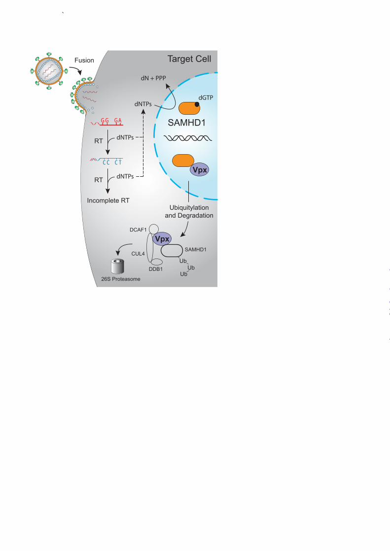
Myeloid cell types such as macrophages and dendritic cells have long been known to be more resistant to HIV-1 infection than CD4+ T lymphocytes (72-74). Major clues to understanding this large phenotypic difference occurred through experiments showing that the Vpx protein of HIV-2 or SIV, naturally absent from HIV-1, improved susceptibility of these cells to HIV-1 infection when delivered by pre-infection with Vpx-containing virus-like particles (VLPs) (74-78). Affinity purification and mass spectrometry studies identified SAMHD1 as a Vpx-interacting protein, and supporting functional studies established that SAMHD1 knockdown renders myeloid cell types more permissive to HIV-1 infection (79,80).
SAMHD1 is comprised of a putative protein-interacting sterile alpha motif (SAM) and a C-terminal deoxynucleotide triphosphate (dNTP) phosphohydrolase domain containing conserved histidine and aspartate residues (HD domain). Biochemical activity was predicted based on homology to the EF1143 protein of Enterococcus faecalis, which was shown previously to elicit dNTPase activity (81,82). This mechanistic possibility was tested and confirmed by biochemical studies with the human and mouse enzymes demonstrating dNTPase activity and a
requirement for dGTP as a co-factor (82,83). A high-resolution crystal structure of the HD domain confirmed strong structural similarities with the Enterococcus enzyme (83).
Current evidence supports a model for HIV-1 restriction in which SAMHD1 causes a diminution of cellular dNTP levels that effectively starves reverse transcriptase of essential building blocks needed for viral cDNA synthesis (82-85) (Fig. 4). A failure to complete cDNA synthesis in a timely manner (4-8 hours) eventually leads to particle disintegration and component degradation by various cellular proteases and nucleases. HIV infection is thought to trigger an innate immune response, which results in interferon production that feeds forward to elevate cellular SAMHD1 levels. It is not clear, however, whether a single incoming viral particle can trigger an interferon response that elevates SAMHD1 to restrictive levels, or whether SAMHD1 up-regulation is a manifestation of a broader interferon-dependent mechanism that serves to protect nearby uninfected cells. Presumably SAMHD1 up-regulation is not harmful to the cell, as macrophages and dendritic cells are terminally differentiated and do not require high levels of dNTPs. The HIV-2 and SIV Vpx proteins counteract restriction by triggering the degradation of SAMHD1 (79,80) (Fig. 4). Vpx serves as a scaffold for the formation of an E3 ubiquitin ligase complex consisting of DCAF1, DDB1, and CUL4, which combine to promote the poly-ubiquitination and degradation of SAMHD1 (79,80,86). Mutagenesis and structural studies are still at early stages, but several reports have already mapped the Vpx-interaction domain to the C-terminus of SAMHD1 and identified residues in Vpx required for functional interaction (87,88). Although Vpx is a relatively new primate lentiviral protein thought to have originated through
by guest on May 8, 2018
http://ww
w.jbc.org/
Dow
nloaded from
8
duplication of the vpr gene, SAMHD1 degradation function predates this duplication. Almost all present day primate Vpx proteins, and many (but not all) Vpr proteins, share SAMHD1 counter-restriction activity (89,90). This evolutionary link is supported by mutagenesis studies that have revealed a conserved hydrophobic motif in Vpx/Vpr proteins required for a functional interaction with SAMHD1 (87). Clusters of positive selection map throughout SAMHD1, including to regions outside of the implicated Vpx/Vpr interacting domain, strongly suggesting past and likely ongoing interactions with a variety of distinct pathogens (88-90). HIV-1 does not have a Vpx protein, nor does it appear to have a Vpr protein capable of interacting with SAMHD1. This deficiency provides a molecular explanation for the resilience of myeloid cell types to HIV-1 infection. It also suggests a provocative hypothesis that could explain why HIV-2 is less pathogenic than HIV-1, and why many SIV strains are non-pathogenic in monkey populations. Greater infection of myeloid cells may shift the balance of antigen presentation to favor reduced immune activation and CD4+ T cell turnover (91). Thus, small molecule inhibitors of SAMHD1 may provide a means to induce better immune responses and natural control of HIV-1 infection. Additional Restriction Factors and The Bigger Picture Undoubtedly, additional HIV restriction factors await discovery. For instance, CD4+ T cells are much more susceptible to infection by Nef-expressing rather than Nef-deficient viruses, suggesting that this accessory protein may serve to counteract other cellular restriction factors (92-94). The cellular target(s) of Vpr and its associated E3 ligase complex is also yet to be identified (17,95). Other cellular proteins
such as human TRIM5α do not inhibit HIV-1, though they are clearly bona fide retrovirus restriction factors (1,96). In fact, a single amino acid substitution in human TRIM5α can endow strong HIV-1 restriction activity suggesting a strategy for gene therapy and/or small molecule mimicry (97,98). Although the examples of restriction and counter-restriction described here are largely binary and involve viral proteins that are dispensable under some growth conditions, it is probable that a subset of viral counter-defense mechanisms could be complicated by overlapping function. For instance, a counter-defense measure might be embedded within an essential retroviral protein such as Gag, reverse transcriptase, or envelope. In these instances, identifying the salient viral protein domain(s) and the relevant host restriction factor may require a combination of biochemical, proteomic, and genetic methods to clearly distinguish between counter-restriction activities and essential viral functions.
Another theme, perhaps best illustrated by Tetherin, is the likelihood that the virus may have multiple ways to block restriction (i.e., functional redundancy). If a restriction factor poses a great enough threat, the virus could very well employ parallel counter-restriction mechanisms. Moreover, there may be major and minor counter-restriction mechanisms, such that if a major pathway is inactivated or nonfunctional the minor pathway may become more prominent. Of potential concern is the possibility that the activation of a minor pathway may only require the virus to make a small number of amino acid changes. Thus, not only should obvious therapeutic possibilities be explored (such as small molecules that promote virus restriction by antagonizing major counter-restriction processes), but less obvious pathways of counter-restriction should be
by guest on May 8, 2018
http://ww
w.jbc.org/
Dow
nloaded from
9
investigated to ensure the virus does not have easy escape routes [e.g., (58,99,100)].
As with the first decade of HIV restriction factors, beginning roughly with the discovery of APOBEC3G in 2002, the second decade is likely to be equally as informative and exciting. One should look forward to the discovery of additional restriction mechanisms, and to the translation of existing knowledge into new therapeutics. HIV is likely to remain at pandemic proportions until a cure is found and applied at the population level. Given the extreme hypervariability of HIV, it would be naïve to assume that one strategy such as vaccination or mono drug therapy
will be curative. Like current combination drug therapies, which simultaneously block multiple viral enzymes, a curative therapy may need to exploit multiple mechanisms. It is conceivable that leveraging a natural restriction factor against the virus, in combination with other methods such as eliminating latent cells and/or boosting antibody responses, may be necessary to extinguish HIV from the human population. Acknowledgements – We thank several lab members and reviewers for thoughtful comments, and we apologize to many whose work could not be cited due to space constraints.
REFERENCES
1. Rahm, N., and Telenti, A. (2012) Curr Opin HIV AIDS 7, 180-186 2. Strebel, K., Daugherty, D., Clouse, K., Cohen, D., Folks, T., and Martin, M. A. (1987)
Nature 328, 728-730 3. Gabuzda, D. H., Lawrence, K., Langhoff, E., Terwilliger, E., Dorfman, T., Haseltine, W. A.,
and Sodroski, J. (1992) J Virol 66, 6489-6495 4. Simon, J. H., Gaddis, N. C., Fouchier, R. A., and Malim, M. H. (1998) Nat Med 4, 1397-
1400 5. Sheehy, A. M., Gaddis, N. C., Choi, J. D., and Malim, M. H. (2002) Nature 418, 646-650 6. Harris, R. S., Petersen-Mahrt, S. K., and Neuberger, M. S. (2002) Mol Cell 10, 1247-1253 7. Vartanian, J. P., Meyerhans, A., Asjo, B., and Wain-Hobson, S. (1991) J Virol 65, 1779-1788 8. Janini, M., Rogers, M., Birx, D. R., and McCutchan, F. E. (2001) J Virol 75, 7973-7986 9. Harris, R. S., Bishop, K. N., Sheehy, A. M., Craig, H. M., Petersen-Mahrt, S. K., Watt, I. N.,
Neuberger, M. S., and Malim, M. H. (2003) Cell 113, 803-809 10. Mangeat, B., Turelli, P., Caron, G., Friedli, M., Perrin, L., and Trono, D. (2003) Nature 424,
99-103 11. Zhang, H., Yang, B., Pomerantz, R. J., Zhang, C., Arunachalam, S. C., and Gao, L. (2003)
Nature 424, 94-98 12. Miyagi, E., Opi, S., Takeuchi, H., Khan, M., Goila-Gaur, R., Kao, S., and Strebel, K. (2007)
J Virol 81, 13346-13353 13. Schumacher, A. J., Haché, G., MacDuff, D. A., Brown, W. L., and Harris, R. S. (2008) J
Virol 82, 2652-2660 14. Browne, E. P., Allers, C., and Landau, N. R. (2009) Virology 387, 313-321 15. Yu, Q., Konig, R., Pillai, S., Chiles, K., Kearney, M., Palmer, S., Richman, D., Coffin, J. M.,
and Landau, N. R. (2004) Nat Struct Mol Biol 11, 435-442 16. Chelico, L., Pham, P., Calabrese, P., and Goodman, M. F. (2006) Nat Struct Mol Biol 13,
392-399 17. Malim, M. H., and Emerman, M. (2008) Cell Host Microbe 3, 388-398 18. Albin, J. S., and Harris, R. S. (2010) Expert Rev Mol Med 12, e4
by guest on May 8, 2018
http://ww
w.jbc.org/
Dow
nloaded from
10
19. Hultquist, J. F., Lengyel, J. A., Refsland, E. W., LaRue, R. S., Lackey, L., Brown, W. L., and Harris, R. S. (2011) J Virol 85, 11220-11234
20. Refsland, E. W., Hultquist, J. F., and Harris, R. S. (2012) PLoS Pathog 8, e1002800 21. OhAinle, M., Kerns, J. A., Li, M. M., Malik, H. S., and Emerman, M. (2008) Cell Host
Microbe 4, 249-259 22. Wang, X., Abudu, A., Son, S., Dang, Y., Venta, P. J., and Zheng, Y. H. (2011) J Virol 85,
3142-3152 23. LaRue, R. S., Lengyel, J., Jónsson, S. R., Andrésdóttir, V., and Harris, R. S. (2010) J Virol
84, 8193-8201 24. Compton, A. A., Hirsch, V. M., and Emerman, M. (2012) Cell Host Microbe 11, 91-98 25. Yu, X., Yu, Y., Liu, B., Luo, K., Kong, W., Mao, P., and Yu, X. F. (2003) Science 302,
1056-1060 26. Mehle, A., Goncalves, J., Santa-Marta, M., McPike, M., and Gabuzda, D. (2004) Genes Dev
18, 2861-2866 27. Jäger, S., Kim, D. Y., Hultquist, J. F., Shindo, K., LaRue, R. S., Kwon, E., Li, M., Anderson,
B. D., Yen, L., Stanley, D., Mahon, C., Kane, J., Franks-Skiba, K., Cimermancic, P., Burlingame, A., Sali, A., Craik, C. S., Harris, R. S., Gross, J. D., and Krogan, N. J. (2012) Nature 481, 371-375
28. Zhang, W., Du, J., Evans, S. L., Yu, Y., and Yu, X. F. (2012) Nature 481, 376-379 29. Harris, R. S. (2008) Nat Biotechnol 26, 1089-1090 30. Pillai, S. K., Wong, J. K., and Barbour, J. D. (2008) Retrovirology 5, 26 31. VanDamme, N., Goff, D., Katsura, C., Jorgenson, R. L., Mitchell, R., Johnson, M. C.,
Stephens, E. B., and Guatelli, J. (2008) Cell Host and Microbe 3, 1-8 32. Neil, S. J. D., Zang, T., and Bieniasz, P. D. (2008) Nature 451, 425-430 33. Strebel, K., Klimkait, T., and Martin, M. A. (1988) Science 241, 1221-1223 34. Geraghty, R. J., Talbot, K. J., Callahan, M., Harper, W., and Panganiban, A. T. (1994)
Journal of Medical Primatology 23, 146-150 35. Varthakavi, V., Smith, R. M., Bour, S. P., Strebel, K., and Spearman, P. (2003) Proc Natl
Acad Sci U S A 100, 15154-15159 36. Neil, S. J., Eastman, S. W., Jouvenet, N., and Bieniasz, P. D. (2006) PLoS Pathog 2, e39 37. Neil, S. J. D., Sandrin, V., Sundquist, W. I., and Bieniasz, P. D. (2007) Cell Host and
Microbe 2, 193-203 38. Kupzig, S., Korolchuk, V., Rollason, R., Sugden, A., Wilde, A., and Banting, G. (2003)
Traffic 4, 694-709 39. Perez-Caballero, D., Zang, T., Ebrahimi, A., McNatt, M. W., Gregory, D. A., Johnson, M.
C., and Bieniasz, P. D. (2009) Cell 139, 499-511 40. Hammonds, J., Wang, J.-J., Yi, H., and Spearman, P. (2010) PLoS Pathogens 6, e1000749 41. Fitzpatrick, K., Skasko, M., Deerinck, T. J., Crum, J., Ellisman, M. H., and Guatelli, J.
(2010) PLoS Pathogens 6, e1000701 42. Miyakawa, K., Ryo, A., Murakami, T., Ohba, K., Yamaoka, S., Fukuda, M., Guatelli, J., and
Yamamoto, N. (2009) PLoS Pathogens 5, e1000700 43. Kobayashi, T., Ode, H., Yoshida, T., Sato, K., Gee, P., Yamamoto, S. P., Ebina, H., Strebel,
K., Sato, H., and Koyanagi, Y. (2011) Journal of Virology 85, 932-945 44. Iwabu, Y., Fujita, H., Kinomoto, M., Kaneko, K., Ishizaka, Y., Tanaka, Y., Sata, T., and
Tokunga, K. (2009) Journal of Biological Chemistry 284, 35060-35072
by guest on May 8, 2018
http://ww
w.jbc.org/
Dow
nloaded from
11
45. Rong, L., Zhang, J., Lu, J., Pan, Q., Lorgeoux, R.-P., Aloysius, C., Guo, F., Liu, S.-L., Wainberg, M. A., and Liang, C. (2009) Journal of Virology 83, 7536-7546
46. Douglas, J. L., Viswanathan, K., McCarroll, M. N., Gustin, J. K., Fruh, K., and Moses, A. V. (2009) Journal of Virology 83, 7931-7947
47. Mangeat, B., Gers-Huber, G., Lehmann, M., Zufferey, M., Luban, J., and Piguet, V. (2009) PLoS Pathogens 5, e1000574
48. Mitchell, R. S., Katsura, C., Skasko, M. A., Fitzpatrick, K., Lau, D., Ruiz, A., Stephens, E. B., Margottin-Goguet, F., Benarous, R., and Guatelli, J. C. (2009) PLoS Pathog 5, e1000450
49. Gustin, J. K., Douglas, J. L., Bai, Y., and Moses, A. V. (2012) Journal of Biological Chemistry 287, 14837-14850
50. Tokarev, A. A., Munguia, J., and Guatelli, J. C. (2011) Journal of Virology 85, 51-63 51. Janvier, K., Pelchen-Matthews, A., Renaud, J.-B., Caillet, M., Marsh, M., and Berlioz-
Torrent, C. (2011) PLoS Pathogens 7, e1001265 52. Caillet, M., Janvier, K., Pelchen-Matthews, A., Delcroix-Genete, D., Camus, G., Marsh, M.,
and Berlioz-Torrent, C. (2011) PLoS Pathogens 7, e1002347 53. Tervo, H.-M., Homann, S., Ambiel, I., Fritz, J. V., Fackler, O. T., and Keppler, O. T. (2011)
Retrovirology 8, 9 54. Dube, M., Roy, B. B., Guiot-Guillain, P., Binette, J., Mercier, J., Chiasson, A., and Cohen, E.
A. (2010) PLoS Pathogens 6, e1000856 55. Schmidt, S., Fritz, J. V., Bitzegeio, J., Fackler, O. T., and Keppler, O. T. (2011) MBio 2,
e00036-00011 56. Dube, M., Paquay, C., Roy, B. B., Bego, M. G., Mercier, J., and Cohen, E. A. (2011) Traffic
12, 1714-1729 57. Lau, D., Kwan, W., and Guatelli, J. (2011) Journal of Virology 85, 9834-9846 58. Serra-Moreno, R., Jia, B., Breed, M., Alvarez, X., and Evans, D. T. (2011) Cell Host and
Microbe 9, 46-57 59. Gupta, R. K., Mlcochova, P., Pelchen-Matthews, A., Petit, S. J., Mattiuzzo, G., Pillay, D.,
Takeuchi, Y., Marsh, M., and Towers, G. (2009) Proceedings of the National Academy of Sciences of the United States of America 106, 20889-20894
60. LeTortorec, A., and Neil, S. J. D. (2009) Journal of Virology 83, 11966-11978 61. Jia, B., Serra-Moreno, R., Neidermyer, W., Rahmberg, A., Mackey, J., Fofana, I. B.,
Johnson, W. E., Westmoreland, S., and Evans, D. T. (2009) PLoS Pathog 5, e1000429 62. Bour, S., Schubert, U., Peden, K., and Strebel, K. (1996) Journal of Virology 70, 820-829 63. Hauser, H., Lopez, L. A., Yang, S. J., Oldenburg, J. E., Exline, C. M., Guatelli, J. C., and
Cannon, P. M. (2010) Retrovirology 7, 51 64. Lopez, L. A., Yang, S. J., Hauser, H., Exline, C. M., Haworth, K. G., Oldenburg, J., and
Cannon, P. M. (2010) Journal of Virology 84, 7243-7255 65. Gupta, R. K., Hue, S., Schaller, T., Verschoor, E., Pillay, D., and Towers, G. J. (2009) PLoS
Pathog 5, e1000443 66. Bour, S., Akari, H., Miyagi, E., and Strebel, K. (2003) Virology 309, 85-98 67. Sauter, D., Schindler, M., Specht, A., Landford, W. N., Munch, J., Kim, K.-A., Votteler, J.,
Schubert, U., Bibollet-Ruche, F., Keele, B. F., Takehisa, J., Ogando, Y., Ochsenbauer, C., Kappes, J. C., Ayouba, A., Peeters, M., Learn, G. H., Shaw, G., Sharp, P. M.,
by guest on May 8, 2018
http://ww
w.jbc.org/
Dow
nloaded from
12
Bieniasz, P., Hahn, B. H., Hatziioannou, T., and Kirchhoff, F. (2009) Cell Host and Microbe 6, 409-421
68. Zhang, F., Wilson, S. J., Landford, W. C., Virgen, B., Gregory, D., Johsnson, M. C., Munch, J., Kirchhoff, F., Bieniasz, P. D., and Hatziioannou, T. (2009) Cell Host and Microbe 6, 1-14
69. Zhang, F., Landford, W. N., Ng, M., McNatt, M. W., Bieniasz, P. D., and Hatziioannou, T. (2011) PLoS Pathogens 7, e1002039
70. Zhou, J., Zhang, Z., Mi, Z., Wang, X., Zhang, Q., Li, X., Liang, C., and Cen, S. (2012) Biochemistry 51, 1288-1296
71. Skasko, M., Wang, Y., Tian, Y., Tokarev, A., Munguia, J., Ruiz, A., Stephens, E. B., Opella, S. J., and Guatelli, J. (2012) Journal of Biological Chemistry 287, 58-67
72. Guyader, M., Emerman, M., Montagnier, L., and Peden, K. (1989) EMBO J 8, 1169-1175 73. Kappes, J. C., Conway, J. A., Lee, S. W., Shaw, G. M., and Hahn, B. H. (1991) Virology 184,
197-209 74. Yu, X. F., Yu, Q. C., Essex, M., and Lee, T. H. (1991) J Virol 65, 5088-5091 75. Negre, D., Mangeot, P. E., Duisit, G., Blanchard, S., Vidalain, P. O., Leissner, P., Winter, A.
J., Rabourdin-Combe, C., Mehtali, M., Moullier, P., Darlix, J. L., and Cosset, F. L. (2000) Gene therapy 7, 1613-1623
76. Goujon, C., Arfi, V., Pertel, T., Luban, J., Lienard, J., Rigal, D., Darlix, J. L., and Cimarelli, A. (2008) J Virol 82, 12335-12345
77. Goujon, C., Riviere, L., Jarrosson-Wuilleme, L., Bernaud, J., Rigal, D., Darlix, J. L., and Cimarelli, A. (2007) Retrovirology 4, 2
78. Kaushik, R., Zhu, X., Stranska, R., Wu, Y., and Stevenson, M. (2009) Cell Host Microbe 6, 68-80
79. Laguette, N., Sobhian, B., Casartelli, N., Ringeard, M., Chable-Bessia, C., Segeral, E., Yatim, A., Emiliani, S., Schwartz, O., and Benkirane, M. (2011) Nature 474, 654-657
80. Hrecka, K., Hao, C., Gierszewska, M., Swanson, S. K., Kesik-Brodacka, M., Srivastava, S., Florens, L., Washburn, M. P., and Skowronski, J. (2011) Nature 474, 658-661
81. Vorontsov, II, Minasov, G., Kiryukhina, O., Brunzelle, J. S., Shuvalova, L., and Anderson, W. F. (2011) J Biol Chem 286, 33158-33166
82. Powell, R. D., Holland, P. J., Hollis, T., and Perrino, F. W. (2011) J Biol Chem 286, 43596-43600
83. Goldstone, D. C., Ennis-Adeniran, V., Hedden, J. J., Groom, H. C., Rice, G. I., Christodoulou, E., Walker, P. A., Kelly, G., Haire, L. F., Yap, M. W., de Carvalho, L. P., Stoye, J. P., Crow, Y. J., Taylor, I. A., and Webb, M. (2011) Nature 480, 379-382
84. Kim, B., Nguyen, L. A., Daddacha, W., and Hollenbaugh, J. A. (2012) J Biol Chem 287, 21570-21574
85. Lahouassa, H., Daddacha, W., Hofmann, H., Ayinde, D., Logue, E. C., Dragin, L., Bloch, N., Maudet, C., Bertrand, M., Gramberg, T., Pancino, G., Priet, S., Canard, B., Laguette, N., Benkirane, M., Transy, C., Landau, N. R., Kim, B., and Margottin-Goguet, F. (2012) Nat Immunol 13, 223-228
86. Ahn, J., Hao, C., Yan, J., DeLucia, M., Mehrens, J., Wang, C., Gronenborn, A. M., and Skowronski, J. (2012) J Biol Chem 287, 12550-12558
87. Wei, W., Guo, H., Han, X., Liu, X., Zhou, X., Zhang, W., and Yu, X. F. (2012) Cell Microbiol
by guest on May 8, 2018
http://ww
w.jbc.org/
Dow
nloaded from
13
88. Laguette, N., Rahm, N., Sobhian, B., Chable-Bessia, C., Munch, J., Snoeck, J., Sauter, D., Switzer, W. M., Heneine, W., Kirchhoff, F., Delsuc, F., Telenti, A., and Benkirane, M. (2012) Cell Host Microbe 11, 205-217
89. Lim, E. S., Fregoso, O. I., McCoy, C. O., Matsen, F. A., Malik, H. S., and Emerman, M. (2012) Cell Host Microbe 11, 194-204
90. Zhang, C., de Silva, S., Wang, J. H., and Wu, L. (2012) PloS one 7, e37477 91. Manel, N., and Littman, D. R. (2011) Cell 147, 271-274 92. Kestler, H. W., Ringler, D. J., Mori, K., Panicali, D. L., Sehgal, P. K., Daniel, M. D., and
Desrosiers, R. C. (1991) Cell 65, 651-662 93. Miller, M. D., Warmerdam, M. T., Gaston, I., Greene, W. C., and Feinberg, M. B. (1994)
Journal of Experimental Medicine 179, 101-113 94. Aiken, C., and Trono, D. (1995) Journal of Virology 69, 5048-5056 95. Casey, L., Wen, X., and de Noronha, C. M. (2010) Cytokine 51, 1-9 96. Stremlau, M., Owens, C. M., Perron, M. J., Kiessling, M., Autissier, P., and Sodroski, J.
(2004) Nature 427, 848-853 97. Yap, M. W., Nisole, S., and Stoye, J. P. (2005) Curr Biol 15, 73-78 98. Li, Y., Li, X., Stremlau, M., Lee, M., and Sodroski, J. (2006) Journal of Virology 80, 6738-
6744 99. Albin, J. S., Hache, G., Hultquist, J. F., Brown, W. L., and Harris, R. S. (2010) J Virol 84,
10209-10219 100. Haché, G., Abbink, T. E., Berkhout, B., and Harris, R. S. (2009) J Virol 83, 5956-5960
FOOTNOTES Supported grants from the National Institutes of Health R01 AI064046 and P01 GM091743 to RSH, and R01 AI098485 and R21 AI087498 to DTE. DTE was also supported in part by P51 RR000168/OD011103. Salary support for JFH was provided by a University of Minnesota Graduate Student Fellowship. The abbreviations used are: APOBEC3A/B/C/D/F/G/H, apolipoprotein mRNA editing catalytic protein-like enzyme 3A/B/C/D/F/G/H; BST2, bone stromal tumor protein 2; dNTP, deoxynucleotide triphosphate; Env, envelope protein; HIV, human immunodeficiency virus; Nef, non-essential factor; SAMHD1, sterile alpha motif and HD domain protein 1; SIV, simian immunodeficiency virus; Vif, viral infectivity factor; Vpr, viral protein R; Vpu, viral protein U; Vpx, viral protein X.
by guest on May 8, 2018
http://ww
w.jbc.org/
Dow
nloaded from
14
FIGURE LEGENDS
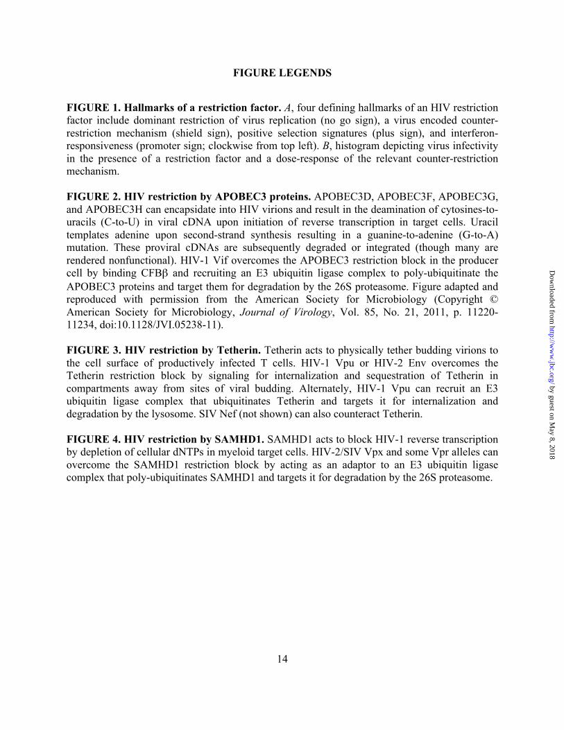
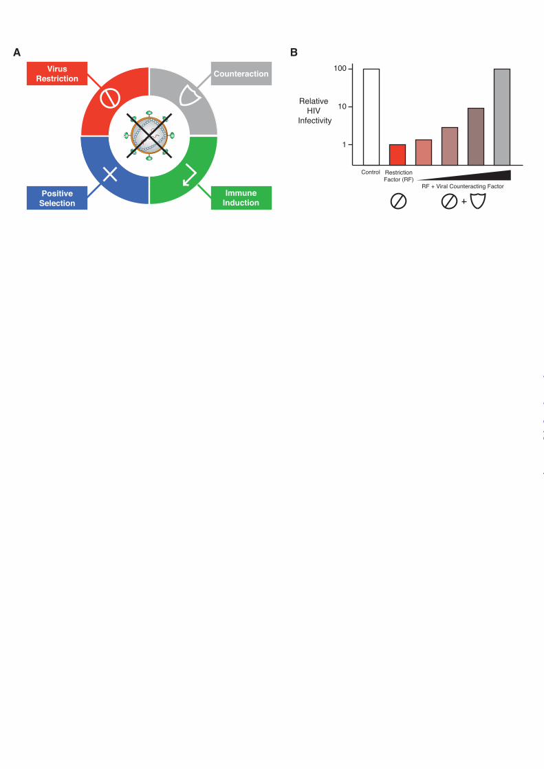
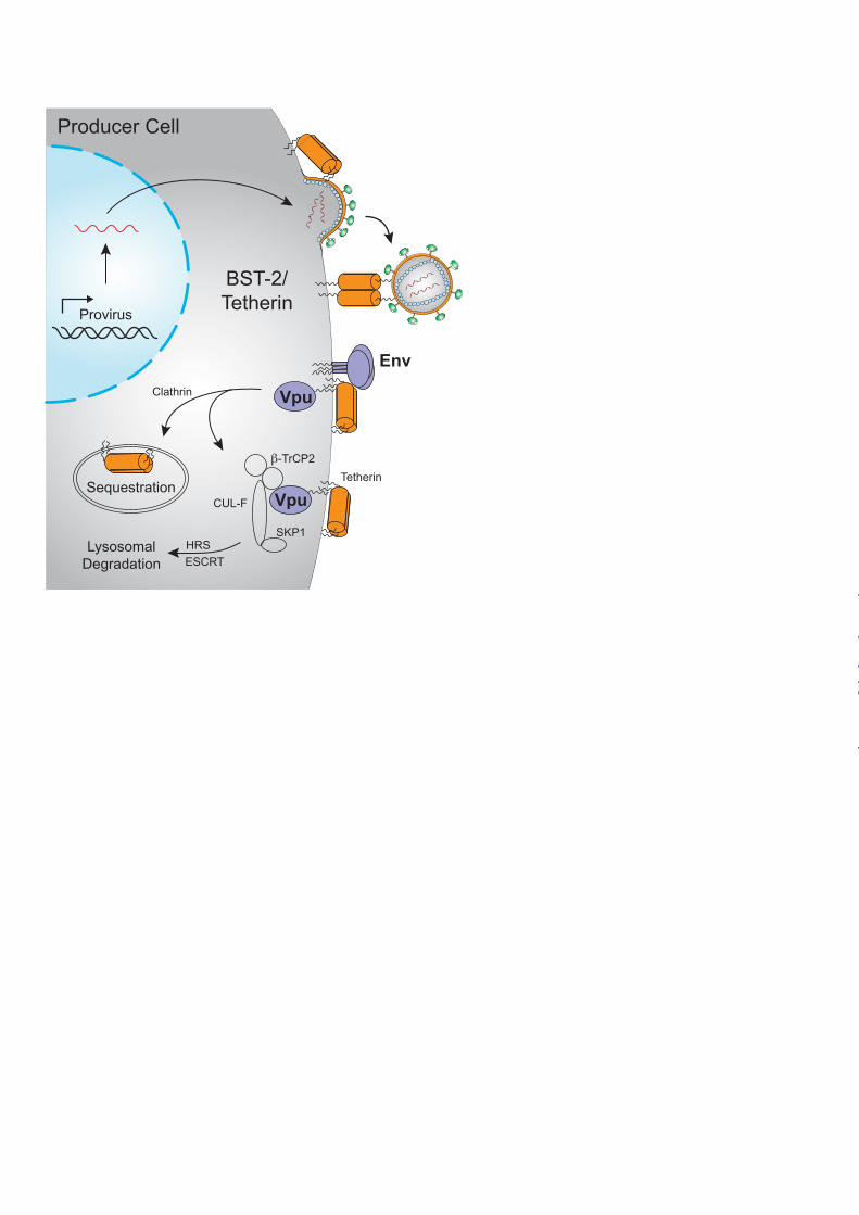
FIGURE 1. Hallmarks of a restriction factor. A, four defining hallmarks of an HIV restriction factor include dominant restriction of virus replication (no go sign), a virus encoded counter-restriction mechanism (shield sign), positive selection signatures (plus sign), and interferon-responsiveness (promoter sign; clockwise from top left). B, histogram depicting virus infectivity in the presence of a restriction factor and a dose-response of the relevant counter-restriction mechanism. FIGURE 2. HIV restriction by APOBEC3 proteins. APOBEC3D, APOBEC3F, APOBEC3G, and APOBEC3H can encapsidate into HIV virions and result in the deamination of cytosines-to-uracils (C-to-U) in viral cDNA upon initiation of reverse transcription in target cells. Uracil templates adenine upon second-strand synthesis resulting in a guanine-to-adenine (G-to-A) mutation. These proviral cDNAs are subsequently degraded or integrated (though many are rendered nonfunctional). HIV-1 Vif overcomes the APOBEC3 restriction block in the producer cell by binding CFBβ and recruiting an E3 ubiquitin ligase complex to poly-ubiquitinate the APOBEC3 proteins and target them for degradation by the 26S proteasome. Figure adapted and reproduced with permission from the American Society for Microbiology (Copyright © American Society for Microbiology, Journal of Virology, Vol. 85, No. 21, 2011, p. 11220-11234, doi:10.1128/JVI.05238-11). FIGURE 3. HIV restriction by Tetherin. Tetherin acts to physically tether budding virions to the cell surface of productively infected T cells. HIV-1 Vpu or HIV-2 Env overcomes the Tetherin restriction block by signaling for internalization and sequestration of Tetherin in compartments away from sites of viral budding. Alternately, HIV-1 Vpu can recruit an E3 ubiquitin ligase complex that ubiquitinates Tetherin and targets it for internalization and degradation by the lysosome. SIV Nef (not shown) can also counteract Tetherin. FIGURE 4. HIV restriction by SAMHD1. SAMHD1 acts to block HIV-1 reverse transcription by depletion of cellular dNTPs in myeloid target cells. HIV-2/SIV Vpx and some Vpr alleles can overcome the SAMHD1 restriction block by acting as an adaptor to an E3 ubiquitin ligase complex that poly-ubiquitinates SAMHD1 and targets it for degradation by the 26S proteasome.
by guest on May 8, 2018
http://ww
w.jbc.org/
Dow
nloaded from
A B100
10
1
RelativeHIV
Infectivity
Virus Restriction Counteraction
Positive Selection
Immune Induction
RF + Viral Counteracting Factor
RestrictionFactor (RF)
Control
by guest on May 8, 2018
http://ww
w.jbc.org/
Dow
nloaded from

C
U U
GG AG
Provirus
Incorporation
G
FD
H
APOBEC3
Ubiquitylation and Degradation
26S Proteasome
C C T
C T
U UC TAAG A
Producer Cell
RT
APOBEC3
RT
Target CellFusion
Budding
Integration
Degradation
G/C A/THypermutation
Provirus
Vif
UbUb
Ub
APOBEC3
CUL5
ELOB/C
RBX2
CBFβ
by guest on May 8, 2018
http://ww
w.jbc.org/
Dow
nloaded from
Provirus
BST-2/Tetherin
Lysosomal Degradation
Producer Cell
UbUb
Ub
CUL-F
β-TrCP2
SKP1
Vpu
Env
SequestrationVpu
HRSESCRT
Clathrin
Tetherin
by guest on May 8, 2018
http://ww
w.jbc.org/
Dow
nloaded from
GG AG
CC C T
RT
Target CellFusion
Incomplete RT
RT
dNTPs
dNTPs
dNTPs
dN + PPP
SAMHD1
Vpx
Vpx
UbUb
Ub
CUL4
DCAF1
DDB1
SAMHD1
Ubiquitylation and Degradation
GGG ACCC T
26S Proteasome
dGTP
by guest on May 8, 2018
http://ww
w.jbc.org/
Dow
nloaded from
Reuben S. Harris, Judd F. Hultquist and David T. EvansTHE RESTRICTION FACTORS OF HUMAN IMMUNODEFICIENCY VIRUS
published online October 5, 2012J. Biol. Chem.
10.1074/jbc.R112.416925Access the most updated version of this article at doi:
Alerts:
When a correction for this article is posted•
When this article is cited•
to choose from all of JBC's e-mail alertsClick here
http://www.jbc.org/content/suppl/2012/11/29/R112.416925.DCAuthor_profileRead an Author Profile for this article at
by guest on May 8, 2018
http://ww
w.jbc.org/
Dow
nloaded from