Embed Size (px)
Citation preview
FEBS Letters 585 (2011) 952–957
journal homepage: www.FEBSLetters .org
Review
The role of LKB1 and AMPK in cellular responses to stress and damage
Angela Alexander, Cheryl L. Walker ⇑Department of Molecular Carcinogenesis, The University of Texas, M.D. Anderson Cancer Center, Smithville, TX 78957, United StatesGraduate School of Biomedical Sciences, University of Texas Health Science Center at Houston, Houston, TX 77030, United States
a r t i c l e i n f o
Article history:Received 14 February 2011Revised 3 March 2011Accepted 4 March 2011Available online 12 March 2011
Edited by Wilhelm Just
Keywords:LKB1AMPKApoptosisSurvivalStressDNA damage
0014-5793/$36.00 � 2011 Federation of European Biodoi:10.1016/j.febslet.2011.03.010
⇑ Corresponding author at: Department of MoUniversity of Texas, M.D. Anderson Cancer Center, SStates. Fax: +1 512 237 2475.
E-mail address: [email protected] (C.L. W
a b s t r a c t
The LKB1 and AMPK proteins participate in an energy sensing cascade that responds to depletion ofATP, serving as a master regulator of metabolism that inhibits anabolic processes and stimulatescatabolic processes. However in recent years, LKB1 and AMPK have been implicated in a variety ofother cellular processes, both cytoplasmic and nuclear, such as control of cell polarity and regula-tion of gene transcription. In this review, we summarize the most recent discoveries regarding par-ticipation of LKB1 and AMPK in signaling pathways that respond to cellular stress and damage, andthe relevance of this signaling for disease and therapy.� 2011 Federation of European Biochemical Societies. Published by Elsevier B.V. All rights reserved.
1. Introduction
The LKB1–AMPK pathway has until fairly recently been thoughtof as primarily an energy sensing pathway engaged by cells in re-sponse to low energy levels. More recently, LKB1 and AMPK havebeen linked to many other fundamental cellular processes includ-ing regulation of cell proliferation, cell polarity, migration, tran-scription and cellular stress and damage responses, the focus ofthis mini-review.
2. Discovery and characterization of LKB1 and AMPK
Liver kinase B1 (LKB1) was discovered only 12 years ago, as a ser-ine–threonine kinase that is mutated in Peutz-Jeghers Syndrome [1].Peutz-Jeghers Syndrome is a rare autosomal dominant hamartomasyndrome, predisposing patients to multiple benign and malignanttumors including gastrointestinal, pancreatic and lung tumors [2].Initially, studies to identify the function of LKB1 were difficult, asits sequence gave few clues as to its activity. Overall it had littlesimilarity to other protein kinases. However, studies in cell linesincluding HeLa S3 and G361 melanoma cells, which lack endogenousLKB1, provided insights into LKB1’s role as a tumor suppressor,including early evidence of its role in stress and damage responses.
chemical Societies. Published by E
lecular Carcinogenesis, Themithville, TX 78957, United
alker).
2.1. LKB1 regulation of p53 activity
LKB1 physically associates with p53 upon DNA damage (UVCradiation), and localizes to the p21 promoter region in tandemwith p53 [3]. Additional interactions with this damage-responsetumor suppressor occur at the level of post-translational modifica-tion of p53, with both LKB1 and AMPK acting as kinases for p53 atthe serine (Ser) 15 phosphorylation site; LKB1 also phosphorylatesp53 at Ser 392, which increases p53 protein stability [3,4].
2.2. LKB1 signaling to AMPK
More recently, LKB1 has been appreciated to be a signaling pro-tein, integrating cellular energy sensing with growth and prolifer-ation, functioning both in the nucleus and the cytoplasm. It isubiquitously expressed throughout the body, and functions as aheterotrimer with STRAD (sterile-20-related adaptor) and MO25(mouse protein-25) in cells [5]. LKB1 is post-translationally modi-fied by kinases in several signaling pathways including cAMP–PKA,the ERK–RSK pathway and ATM, a DNA damage sensor, as re-viewed in [6]. Despite the fact that it is constitutively active in cells[7], it has also been noted that LKB1 catalytic activity is enhancedwhen STRAD and MO25 are present in the complex [5].
AMP-activated protein kinase (AMPK) is one of the best charac-terized substrates of LKB1. AMPK is a heterotrimer that consists ofa catalytic subunit, AMPKa, and 2 regulatory subunits, AMPKb andAMPKc. There are 2 distinct isoforms of the AMPKa subunitdesignated AMPKa1 and AMPKa2, which differ in their tissue
lsevier B.V. All rights reserved.
A. Alexander, C.L. Walker / FEBS Letters 585 (2011) 952–957 953
specificity, subcellular localization and mechanisms of activation.While AMPKa1 is widely expressed, AMPKa2 is restricted mainlyto skeletal muscle, cardiac muscle and liver cells, where it is highlyexpressed [8]. On a cellular level under basal conditions, AMPKa1appears mainly cytoplasmic whereas AMPKa2 is primarily local-ized to the nucleus. The yeast AMPK homolog, Snf1, is also predom-inantly located in the nucleus [9]. It is currently unclear how theAMPK complex containing a2 localizes to the nucleus, since noclassical nuclear localization sequence has been found, and the�63 kDa protein is probably too large to passively diffuse throughthe nuclear pore.
2.3. How is AMPK activated?
At present, a number of stimuli are known to activate AMPK,some of which are related to energy sensing (such as glucose depri-vation), as well as others that are not (such as hyperosmoticstress). When cellular energy levels are depleted, and the AMP:ATPratio rises, AMPK is activated via allosteric binding of AMP to theAMPKc subunit, which has been proposed to induce a conforma-tional change in the complex, improving the ability of AMPKa-sub-unit to serve as a substrate for upstream kinases [6]. The primaryactivation site on the catalytic component AMPKa is threonine(Thr) 172, and while both a1 and a2 can be phosphorylated at thissite by the same upstream kinases, a2 subunit phosphorylation ismore AMP-dependent [9].
LKB1 has now been shown to be one of the primary kinases thatphosphorylates AMPK. AMPK activation by agonists such as 5-aminoimidazole-4-carboxamide ribonucleoside (AICAR), metfor-min or phenformin or in response to stress is absent in LKB1-defi-cient cells [5], identifying LKB1 as an obligatory AMPK kinase inthis setting. Activation of AMPK by AICAR occurs via mono-phos-phorylation of AICAR to its active form, ZMP which can mimicthe effect of AMP on AMPK, i.e., allosteric activation and enhance-ment of phosphorylation by upstream kinase(s) [10].
In addition to LKB1, the calmodulin-dependent protein kinasesCaMKKa and CaMKKb also function as kinases for Thr 172 of AMPK[11–13]. This activation pathway is calcium-responsive, does notrequire an increase in AMP, and is thought to be particularlyimportant for endocrine hormone regulation, for example adipo-nectin activation of AMPK in vascular endothelial cells [14]. CaM-kinases are most highly expressed in neural tissue, where they playa role in activating AMPK in response to neuronal depolarizationcaused by K+-increases and Ca2+ influx [11].
2.4. AMPK signaling to mTORC1 and growth suppression
How does activation of LKB1 and AMPK result in growth sup-pression? One of the key targets of these kinases is the TSC2–mTORC1 signaling node. The tuberous sclerosis complex 2 (TSC2)tumor suppressor is directly phosphorylated by AMPK at Thr1227 and Ser 1345, which enhances its GTP-ase activity towardsRheb, inactivating this small GTPase to decrease mTORC1 signaling[15]. In addition to this TSC2-dependent pathway, AMPK has beenshown to directly phosphorylate raptor (a component of themTORC1 complex) to inhibit mTORC1 signaling [16]. The reciprocalrelationship between AMPK activity and mTORC1 activity allowsthe cell to coordinate energy requiring anabolic processes with en-ergy availability – under conditions of energy deprivation, activa-tion of AMPK can limit energy-consuming processes such asprotein synthesis via repression of mTORC1.
In addition to protein synthesis and cell growth, mTORC1 is alsoa key negative regulator of autophagy, the catabolic process ofbreaking down cell components via the fusion of autophagosomeswith lysosomes, degrading the contents autophagosomes for recy-cling by the cell [17]. Activation of AMPK induces autophagy by
negatively regulating mTORC1 via TSC2 or raptor, and thereby as-sists the cell in generating building blocks for energy productionand other cellular processes. Recently, ULK1 has been identifiedas a bona fide AMPK substrate when cells undergo nutrient depri-vation, providing a molecular link between this energy-sensingpathway and regulation of autophagy [18,19].
In contrast to energy stress, DNA damage, which activates p53via ATM/ATR phosphorylation, can both promote and inhibitautophagy. In the nucleus, p53 functions as a transcription factor,where it regulates the expression of many genes, including severalthat participate in autophagy [20]. In the cytoplasm, p53 can inhi-bit AMPK and autophagy, indicating that the relationship betweenp53 and AMPK in regulation of autophagy is very complex andlikely context dependent [21].
A second pathway of AMPK-mediated regulation of autophagymay be via the eEF-2 kinase (also known as Ca2+/calmodulin-dependent kinase III) pathway. AMPK directly phosphorylatesand activates eEF-2 kinase, which inhibits protein synthesis atthe elongation stage [22]. The precise role that eEF-2 plays inautophagy is still not known, however it was shown that knock-down of eEF-2 inhibited autophagy in glioblastoma cells, whereasoverexpression increased autophagy. Finally, another less studiedAMPK-kinase, TAK1 (transforming growth factor b-activatedkinase 1) was shown to be induced in response to the apoptosis-inducing agent TRAIL, resulting in a cytoprotective autophagyresponse, which is dependent upon AMPKa1 [23]. It is clear fromthese studies that AMPK plays important roles in signaling to reg-ulate autophagy in response to diverse stress stimuli.
2.5. Other AMPK-related proteins
Analysis of the human kinome reveals a family of AMPK-relatedproteins that contain activation loops that are homologous toAMPK, suggesting they could also be targets of LKB1. This familyof proteins is now known to contain at least 12 validated LKB1 sub-strates (not including AMPK) and 8 non-LKB1 substrates [6,24].One of these AMPK-family members, SNARK, was discovered as agene induced in response to UV damage in rodent keratinocytes;SNARK also expressed in a number of human tissues [25,26]. Uponadditional characterization, a broad range of stressors (includingglucose and glutamine deprivation, hydrogen peroxide, hyperos-motic stress, ATP synthesis inhibitors such as oligomycin andarsenite) were capable of activating SNARK kinase activity in acell-type specific manner. These results suggest that other AMPK-family members also may function as stress responsive proteins.
It is unclear whether AMP directly activates all AMPK-relatedproteins, although SNARK is activated by AICAR or glucose depriva-tion in cells [25]. It is likely there are some cell-type differences inactivation mechanisms, since in contrast to the first report onSNARK (NUAK2) activation in neonatal rat keratinocytes and babyhamster kidney cells [25], later studies showed that in mouse em-bryo fibroblasts (MEFs), AICAR and phenformin were unable toactivate SNARK [24]. While AMPK-related proteins are less wellstudied than AMPK, it is already clear that they vary in their sub-strate specificity, although not all these substrates have been char-acterized. Future research on AMPK-related proteins will likelyexpand our knowledge about how they are regulated, includingidentification of additional upstream kinases and regulatory phos-phorylation sites, their physiological and pathological functions,and identify additional substrates.
3. Involvement in DNA damage response pathways
The kinase ATM lies upstream of LKB1 in damage response sig-naling. ATM, the gene mutated in the disease ataxia-telangiectasia,
954 A. Alexander, C.L. Walker / FEBS Letters 585 (2011) 952–957
is a critical early damage response protein that plays multipleimportant roles in sensing and responding to different types ofdamage. In the nucleus, it is well appreciated that ATM is activatedby DNA double-strand breaks, leading to induction of cell cyclecheckpoints, DNA repair and if necessary, apoptosis if the damageis too severe to be repaired. However, ATM has also been localizedto the cytoplasm, where it can be activated in the absence of DNAdamage [27,28]. Dario Alessi’s group in 2002 identified a series ofphosphorylation sites on LKB1, none of which affected LKB1’s ki-nase activity towards p53, one of which, threonine 366 (Thr366), lay within an ATM consensus phosphorylation motif. Theywent on to show that ionizing radiation (which activates ATM)led to phosphorylation of LKB1 at this site [29]. The function of thisphosphorylation site remained somewhat elusive since mutationof this site to alanine had little affect on cell growth when transfec-ted into LKB1-deficient cells. However until recently, the impact ofphosphorylation at Thr 366 on downstream signaling to AMPK ormTORC1 had not been studied further.
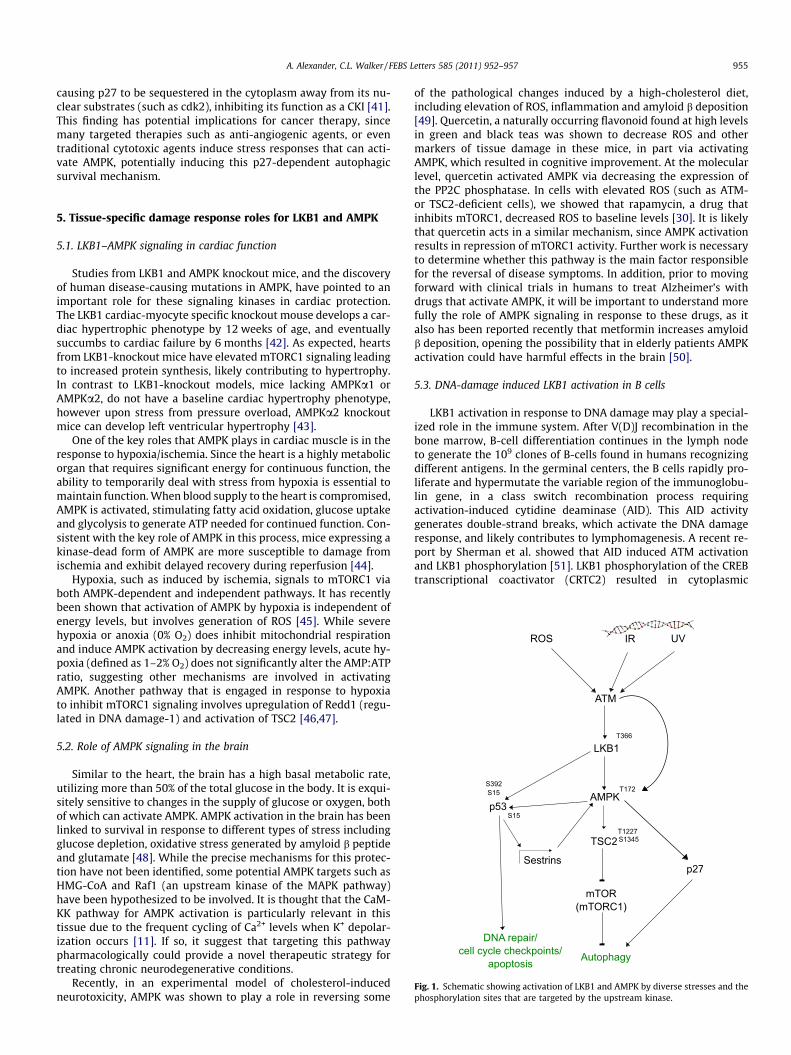
We have shown that a damage response signaling pathway ex-ists from ATM to LKB1 that regulates AMPK in the cytoplasm [30].In response to oxidative damage, cytoplasmic ATM is rapidly acti-vated, phosphorylating LKB1 in the cytoplasm, activating AMPK,TSC2 and suppressing mTORC1. As a consequence of this mTORC1suppression, autophagy is induced, suggesting this pathway mayfunction as a mechanism to maintain redox homeostasis.
ATM has also been shown to directly phosphorylate AMPKaindependently of LKB1 on T172 in response to DNA damage (eto-poside), which has been linked to mitochondrial biogenesis in avariety of mammalian cell types [31]. While not definitively shownin this paper, the authors proposed that in response to DNA dam-age, AMPK was activated both by a decrease in ATP (increase inAMP:ATP ratio) as well as by ATM phosphorylation. Induction ofmitochondrial biogenesis in response to DNA damage by a varietyof agents including chemotherapeutics and radiation (which candamage mitochondria and release ROS, inducing oxidative dam-age), coupled with autophagy of damaged mitochondria, may bea mechanism for limiting cellular damage while ensuring sufficientenergy to carry out the important repair pathways necessary forsurvival. Apart from the DNA damage response, AMPK has alsobeen linked to mitochondrial biogenesis in skeletal muscle in re-sponse to chronic energy deprivation or endurance training exer-cises [32]. The mechanisms responsible involve master regulatorsof mitochondrial biogenesis including the peroxisome proliferatoractivator receptor c coactivator 1a, and calcium/calmodulin-dependent protein kinase IV [32].
A second DNA damage-induced pathway for AMPK activationinvolves p53 [33]. Two recently-identified p53 transcriptional tar-gets, sestrin 1 and sestrin 2 were shown to mediate AMPK activa-tion and mTORC1 suppression by DNA damage (specificallycamptothecin in vitro and the hepatocarcinogen diethylnitros-amine in vivo). While the precise mechanism of AMPK activationby sestrins was not analyzed, this pathway was p53-dependent,and resulted in the formation of large protein complexes contain-ing sestrins, AMPK, TSC2, and TSC1, suggesting that the sestrinsmay function as adaptor proteins to bring together these proteins,promoting AMPK activation of TSC2/1 and mTORC1 repression.
4. Regulation of cell death pathways
4.1. Interactions with p53 family members
Apart from autophagy, AMPK activation in response to DNAdamage may regulate apoptosis or other cell death pathways. Forexample, the chemotherapeutic drug cisplatin, which damagesDNA by forming intrastrand crosslinks, has been shown to activateAMPK in several tumor types including colon cancer, gastric cancer
and glioma [34,35]. In one study, AMPK activation by cisplatin cor-related with ROS generation, and resulted in enhanced survival. Inthis setting, as would be predicted, inhibition of AMPK sensitizedthe cells to cisplatin-induced apoptosis, which was preceded by adramatic decrease in ATP levels [34]. This pathway was dependenton p53, and involved hyper-phosphorylation of p53 at Ser 15 byERK, which occurred in the presence of AMPK inhibition. Interest-ingly, when a panel of different tumor lines were treated with arelatively high dose of cisplatin, highly chemosensitive cells suchas HeLa, which are LKB1-null, were refractory to AMPK activation,whereas more resistant cells exhibited AMPK activation, suggest-ing a role for LKB1 signaling to AMPK in a survival pathway. Thusin the context of cytotoxic therapy, it is possible that inhibition ofLKB1/AMPK in p53-proficient cells may reduce survival and im-prove cell killing.
In addition to p53, other members of this transcription factorfamily are phosphorylated by AMPK in response to DNA damage.For example, AMPKa interacts with both p73a and p63a, withmultiple functional consequences, including altering subcellularlocalization of AMPKa subunits [36]. Under normal conditions,endogenous AMPKa1 is mainly cytoplasmic, whereas AMPKa2 isnuclear [9,37,38]. However, when exogenous p73a is overexpres-sed, both AMPKa1 and AMPKa2 localize to the nucleus, whereAMPK:p73a complexes act as transcriptional co-repressors. Thiswas directly shown by overexpressing AMPKa1 or AMPKa2, andmeasuring luciferase activity at p53-family responsive promoterssuch as the Bax promoter. Using these luciferase reporters as read-outs of transcriptional activity, Lee et al. showed that increasingexpression of AMPKa1 or AMPKa2 resulted in inhibition ofp73a-mediated repression of gene expression. Interestingly, the ki-nase activity of AMPK was not required for this activity. Furtherevidence of in vivo relevance of this observation was obtainedusing ChIP assays of the endogenous p21 promoter, which couldimmunoprecipitated with antibodies directed against AMPKa2.Importantly, this AMPK-mediated p73a transcriptional repressionwas shown to enhance survival in response to DNA damage, withcells overexpressing AMPK2a resistant to cisplatin-induced apop-tosis [36].
4.2. Other nuclear functions of AMPK
Histone H2B has also been identified recently as an AMPKa2stress-response substrate [39]. When AMPK is activated by a vari-ety of DNA damaging agents and stressors such as 2-deoxyglucose,histone H2B is phosphorylated at Ser 36, inducing transcription ofa large number of stress-inducible genes. This pathway for directmodification of chromatin by AMPK allows this kinase to partici-pate in a global transcriptional response in addition to directlyphosphorylating specific transcription factors, which may allowcells to fine-tune gene expression to survive damage and stress.
4.3. Regulation of p27 stability and localization
Both cell death (apoptosis) and survival (autophagy) are regu-lated by AMPK in response to the stress of glucose deprivation.One of the key regulators of this ‘‘life or death’’ decision down-stream of AMPK is p27, a cyclin-dependent kinase inhibitor (CKI)and a direct substrate for AMPK in response to serum starvationor 2-deoxyglucose treatment [40]. AMPK phosphorylation of p27at the carboxy-terminal threonine (Thr 198 in humans) results inincreased protein stability and combined with cytoplasmic locali-zation of this CKI, promotes autophagy. However, in cells depletedof p27, glucose deprivation induces apoptosis, demonstrating thatp27-dependent autophagy functions downstream of AMPK as asurvival mechanism under conditions of energetic stress. AMPKalso phosphorylates p27 at its nuclear localization signal (NLS),
A. Alexander, C.L. Walker / FEBS Letters 585 (2011) 952–957 955
causing p27 to be sequestered in the cytoplasm away from its nu-clear substrates (such as cdk2), inhibiting its function as a CKI [41].This finding has potential implications for cancer therapy, sincemany targeted therapies such as anti-angiogenic agents, or eventraditional cytotoxic agents induce stress responses that can acti-vate AMPK, potentially inducing this p27-dependent autophagicsurvival mechanism.
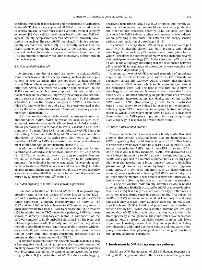
Fig. 1. Schematic showing activation of LKB1 and AMPK by diverse stresses and thephosphorylation sites that are targeted by the upstream kinase.
5. Tissue-specific damage response roles for LKB1 and AMPK
5.1. LKB1–AMPK signaling in cardiac function
Studies from LKB1 and AMPK knockout mice, and the discoveryof human disease-causing mutations in AMPK, have pointed to animportant role for these signaling kinases in cardiac protection.The LKB1 cardiac-myocyte specific knockout mouse develops a car-diac hypertrophic phenotype by 12 weeks of age, and eventuallysuccumbs to cardiac failure by 6 months [42]. As expected, heartsfrom LKB1-knockout mice have elevated mTORC1 signaling leadingto increased protein synthesis, likely contributing to hypertrophy.In contrast to LKB1-knockout models, mice lacking AMPKa1 orAMPKa2, do not have a baseline cardiac hypertrophy phenotype,however upon stress from pressure overload, AMPKa2 knockoutmice can develop left ventricular hypertrophy [43].
One of the key roles that AMPK plays in cardiac muscle is in theresponse to hypoxia/ischemia. Since the heart is a highly metabolicorgan that requires significant energy for continuous function, theability to temporarily deal with stress from hypoxia is essential tomaintain function. When blood supply to the heart is compromised,AMPK is activated, stimulating fatty acid oxidation, glucose uptakeand glycolysis to generate ATP needed for continued function. Con-sistent with the key role of AMPK in this process, mice expressing akinase-dead form of AMPK are more susceptible to damage fromischemia and exhibit delayed recovery during reperfusion [44].
Hypoxia, such as induced by ischemia, signals to mTORC1 viaboth AMPK-dependent and independent pathways. It has recentlybeen shown that activation of AMPK by hypoxia is independent ofenergy levels, but involves generation of ROS [45]. While severehypoxia or anoxia (0% O2) does inhibit mitochondrial respirationand induce AMPK activation by decreasing energy levels, acute hy-poxia (defined as 1–2% O2) does not significantly alter the AMP:ATPratio, suggesting other mechanisms are involved in activatingAMPK. Another pathway that is engaged in response to hypoxiato inhibit mTORC1 signaling involves upregulation of Redd1 (regu-lated in DNA damage-1) and activation of TSC2 [46,47].
5.2. Role of AMPK signaling in the brain
Similar to the heart, the brain has a high basal metabolic rate,utilizing more than 50% of the total glucose in the body. It is exqui-sitely sensitive to changes in the supply of glucose or oxygen, bothof which can activate AMPK. AMPK activation in the brain has beenlinked to survival in response to different types of stress includingglucose depletion, oxidative stress generated by amyloid b peptideand glutamate [48]. While the precise mechanisms for this protec-tion have not been identified, some potential AMPK targets such asHMG-CoA and Raf1 (an upstream kinase of the MAPK pathway)have been hypothesized to be involved. It is thought that the CaM-KK pathway for AMPK activation is particularly relevant in thistissue due to the frequent cycling of Ca2+ levels when K+ depolar-ization occurs [11]. If so, it suggest that targeting this pathwaypharmacologically could provide a novel therapeutic strategy fortreating chronic neurodegenerative conditions.
Recently, in an experimental model of cholesterol-inducedneurotoxicity, AMPK was shown to play a role in reversing some
of the pathological changes induced by a high-cholesterol diet,including elevation of ROS, inflammation and amyloid b deposition[49]. Quercetin, a naturally occurring flavonoid found at high levelsin green and black teas was shown to decrease ROS and othermarkers of tissue damage in these mice, in part via activatingAMPK, which resulted in cognitive improvement. At the molecularlevel, quercetin activated AMPK via decreasing the expression ofthe PP2C phosphatase. In cells with elevated ROS (such as ATM-or TSC2-deficient cells), we showed that rapamycin, a drug thatinhibits mTORC1, decreased ROS to baseline levels [30]. It is likelythat quercetin acts in a similar mechanism, since AMPK activationresults in repression of mTORC1 activity. Further work is necessaryto determine whether this pathway is the main factor responsiblefor the reversal of disease symptoms. In addition, prior to movingforward with clinical trials in humans to treat Alzheimer’s withdrugs that activate AMPK, it will be important to understand morefully the role of AMPK signaling in response to these drugs, as italso has been reported recently that metformin increases amyloidb deposition, opening the possibility that in elderly patients AMPKactivation could have harmful effects in the brain [50].
5.3. DNA-damage induced LKB1 activation in B cells
LKB1 activation in response to DNA damage may play a special-ized role in the immune system. After V(D)J recombination in thebone marrow, B-cell differentiation continues in the lymph nodeto generate the 109 clones of B-cells found in humans recognizingdifferent antigens. In the germinal centers, the B cells rapidly pro-liferate and hypermutate the variable region of the immunoglobu-lin gene, in a class switch recombination process requiringactivation-induced cytidine deaminase (AID). This AID activitygenerates double-strand breaks, which activate the DNA damageresponse, and likely contributes to lymphomagenesis. A recent re-port by Sherman et al. showed that AID induced ATM activationand LKB1 phosphorylation [51]. LKB1 phosphorylation of the CREBtranscriptional coactivator (CRTC2) resulted in cytoplasmic
956 A. Alexander, C.L. Walker / FEBS Letters 585 (2011) 952–957
sequestration and inhibition of its ability to block B cell prolifera-tion and promote B cell differentiation. Interestingly, CRTC2 hasalso been shown to be phosphorylated by the AMPK-family mem-bers MARK2 and SIK2 in pancreatic beta cells, highlighting thispathway as an important mediator of LKB1/AMPK activity [52].
6. Summary
Taken together, it is apparent that LKB1 and AMPK, initiallyidentified as participating in energy sensing in the cell, also play di-verse roles in cellular responses to many types of stress. As illus-trated in Fig. 1, the LKB1–AMPK pathway plays critical roles inregulating important life-and-death processes including apoptosisand autophagy, which may function in a tissue-specific manner,such as cardioprotection from damage in the heart. As we developa more detailed understanding of how these kinases function innormal physiology, and are their role in pathophysiology and dis-ease, it is likely that their utility as therapeutic targets will in-crease. Thus, both LKB1 and its target AMPK, hold promise astherapeutic targets to treat, and possibly prevent, many diseasesincluding neurodegeneration, cardiovascular disease and cancer.
Acknowledgements
This work was supported by grants from NIH (to C.L.W.), theOvarian Cancer Research Foundation (to C.L.W.) and the Sowell-Huggins Fellowship (to A.A.).
References
[1] Jenne, D.E. et al. (1998) Peutz-Jeghers syndrome is caused by mutations in anovel serine threonine kinase. Nat. Genet. 18, 38–43.
[2] Hemminki, A. et al. (1998) A serine/threonine kinase gene defective in Peutz-Jeghers syndrome. Nature 391, 184–187.
[3] Zeng, P.Y. and Berger, S.L. (2006) LKB1 is recruited to the p21/WAF1 promoterby p53 to mediate transcriptional activation. Cancer Res. 66, 10701–10708.
[4] Jones, R.G., Plas, D.R., Kubek, S., Buzzai, M., Mu, J., Xu, Y., Birnbaum, M.J. andThompson, C.B. (2005) AMP-activated protein kinase induces a p53-dependentmetabolic checkpoint. Mol. Cell 18, 283–293.
[5] Hawley, S.A., Boudeau, J., Reid, J.L., Mustard, K.J., Udd, L., Makela, T.P., Alessi,D.R. and Hardie, D.G. (2003) Complexes between the LKB1 tumor suppressor,STRAD alpha/beta and MO25 alpha/beta are upstream kinases in the AMP-activated protein kinase cascade. J. Biol. 2, 28.
[6] Alessi, D.R., Sakamoto, K. and Bayascas, J.R. (2006) LKB1-dependent signalingpathways. Annu. Rev. Biochem. 75, 137–163.
[7] Woods, A. et al. (2003) LKB1 is the upstream kinase in the AMP-activatedprotein kinase cascade. Curr. Biol. 13, 2004–2008.
[8] Stapleton, D. et al. (1996) Mammalian AMP-activated protein kinasesubfamily. J. Biol. Chem. 271, 611–614.
[9] Salt, I., Celler, J.W., Hawley, S.A., Prescott, A., Woods, A., Carling, D. and Hardie,D.G. (1998) AMP-activated protein kinase: greater AMP dependence, andpreferential nuclear localization, of complexes containing the alpha2 isoform.Biochem. J. 334 (Pt. 1), 177–187.
[10] Corton, J.M., Gillespie, J.G., Hawley, S.A. and Hardie, D.G. (1995) 5-Aminoimidazole-4-carboxamide ribonucleoside. A specific method foractivating AMP-activated protein kinase in intact cells? Eur. J. Biochem. 229,558–565.
[11] Hawley, S.A., Pan, D.A., Mustard, K.J., Ross, L., Bain, J., Edelman, A.M., Frenguelli,B.G. and Hardie, D.G. (2005) Calmodulin-dependent protein kinase kinase-beta is an alternative upstream kinase for AMP-activated protein kinase. CellMetab. 2, 9–19.
[12] Hurley, R.L., Anderson, K.A., Franzone, J.M., Kemp, B.E., Means, A.R. andWitters, L.A. (2005) The Ca2+/calmodulin-dependent protein kinase kinasesare AMP-activated protein kinase kinases. J. Biol. Chem. 280, 29060–29066.
[13] Woods, A., Dickerson, K., Heath, R., Hong, S.P., Momcilovic, M., Johnstone, S.R.,Carlson, M. and Carling, D. (2005) Ca2+/calmodulin-dependent protein kinasekinase-beta acts upstream of AMP-activated protein kinase in mammaliancells. Cell Metab. 2, 21–33.
[14] Hattori, Y., Nakano, Y., Hattori, S., Tomizawa, A., Inukai, K. and Kasai, K. (2008)High molecular weight adiponectin activates AMPK and suppresses cytokine-induced NF-kappaB activation in vascular endothelial cells. FEBS Lett. 582,1719–1724.
[15] Inoki, K., Zhu, T. and Guan, K.L. (2003) TSC2 mediates cellular energy responseto control cell growth and survival. Cell 115, 577–590.
[16] Gwinn, D.M., Shackelford, D.B., Egan, D.F., Mihaylova, M.M., Mery, A., Vasquez,D.S., Turk, B.E. and Shaw, R.J. (2008) AMPK phosphorylation of raptor mediatesa metabolic checkpoint. Mol. Cell 30, 214–226.
[17] Jung, C.H., Ro, S.H., Cao, J., Otto, N.M. and Kim, D.H. (2010) MTOR regulation ofautophagy. FEBS Lett. 584, 1287–1295.
[18] Kim, J., Kundu, M., Viollet, B. and Guan, K.L. (2011) AMPK and mTOR regulateautophagy through direct phosphorylation of Ulk1. Nat. Cell Biol. 13,132–141.
[19] Zhao, M. and Klionsky, D.J. (2011) AMPK-dependent phosphorylation of ULK1induces autophagy. Cell Metab. 13, 119–120.
[20] Tasdemir, E. et al. (2008) A dual role of p53 in the control of autophagy.Autophagy 4, 810–814.
[21] Tasdemir, E. et al. (2008) Regulation of autophagy by cytoplasmic p53. Nat.Cell Biol. 10, 676–687.
[22] Browne, G.J., Finn, S.G. and Proud, C.G. (2004) Stimulation of the AMP-activated protein kinase leads to activation of eukaryotic elongation factor 2kinase and to its phosphorylation at a novel site, serine 398. J. Biol. Chem. 279,12220–12231.
[23] Herrero-Martin, G., Hoyer-Hansen, M., Garcia-Garcia, C., Fumarola, C., Farkas,T., Lopez-Rivas, A. and Jaattela, M. (2009) TAK1 activates AMPK-dependentcytoprotective autophagy in TRAIL-treated epithelial cells. EMBO J. 28, 677–685.
[24] Lizcano, J.M. et al. (2004) LKB1 is a master kinase that activates 13 kinases ofthe AMPK subfamily, including MARK/PAR-1. EMBO J. 23, 833–843.
[25] Lefebvre, D.L., Bai, Y., Shahmolky, N., Sharma, M., Poon, R., Drucker, D.J. andRosen, C.F. (2001) Identification and characterization of a novel sucrose-non-fermenting protein kinase/AMP-activated protein kinase-related proteinkinase, SNARK. Biochem. J. 355, 297–305.
[26] Lefebvre, D.L. and Rosen, C.F. (2005) Regulation of SNARK activity in responseto cellular stresses. Biochim. Biophys. Acta 1724, 71–85.
[27] Li, J., Han, Y.R., Plummer, M.R. and Herrup, K. (2009) Cytoplasmic ATM inneurons modulates synaptic function. Curr. Biol. 19, 2091–2096.
[28] Shen, K., Wang, Y., Brooks, S.C., Raz, A. and Wang, Y.A. (2006) ATM is activatedby mitotic stress and suppresses centrosome amplification in primary but notin tumor cells. J. Cell. Biochem. 99, 1267–1274.
[29] Sapkota, G.P. et al. (2002) Ionizing radiation induces ataxia telangiectasiamutated kinase (ATM)-mediated phosphorylation of LKB1/STK11 at Thr-366.Biochem. J. 368, 507–516.
[30] Alexander, A. et al. (2010) ATM signals to TSC2 in the cytoplasm to regulatemTORC1 in response to ROS. Proc. Natl. Acad. Sci. USA 107, 4153–4158.
[31] Fu, X., Wan, S., Lyu, Y.L., Liu, L.F. and Qi, H. (2008) Etoposide induces ATM-dependent mitochondrial biogenesis through AMPK activation. PLoS ONE 3,e2009.
[32] Zong, H., Ren, J.M., Young, L.H., Pypaert, M., Mu, J., Birnbaum, M.J. andShulman, G.I. (2002) AMP kinase is required for mitochondrial biogenesis inskeletal muscle in response to chronic energy deprivation. Proc. Natl. Acad.Sci. USA 99, 15983–15987.
[33] Budanov, A.V. and Karin, M. (2008) p53 target genes sestrin1 and sestrin2connect genotoxic stress and mTOR signaling. Cell 134, 451–460.
[34] Kim, H.S. et al. (2008) Inhibition of AMP-activated protein kinase sensitizescancer cells to cisplatin-induced apoptosis via hyper-induction of p53. J. Biol.Chem. 283, 3731–3742.
[35] Harhaji-Trajkovic, L., Vilimanovich, U., Kravic-Stevovic, T., Bumbasirevic, V.and Trajkovic, V. (2009) AMPK-mediated autophagy inhibits apoptosis incisplatin-treated tumour cells. J. Cell. Mol. Med. 13, 3644–3654.
[36] Lee, Y.G., Lee, S.W., Sin, H.S., Kim, E.J. and Um, S.J. (2009) Kinase activity-independent suppression of p73alpha by AMP-activated kinase alpha(AMPKalpha). Oncogene 28, 1040–1052.
[37] Turnley, A.M., Stapleton, D., Mann, R.J., Witters, L.A., Kemp, B.E. and Bartlett,P.F. (1999) Cellular distribution and developmental expression of AMP-activated protein kinase isoforms in mouse central nervous system. J.Neurochem. 72, 1707–1716.
[38] da Silva Xavier, G., Leclerc, I., Salt, I.P., Doiron, B., Hardie, D.G., Kahn, A. andRutter, G.A. (2000) Role of AMP-activated protein kinase in the regulation byglucose of islet beta cell gene expression. Proc. Natl. Acad. Sci. USA 97, 4023–4028.
[39] Bungard, D. et al. (2010) Signaling kinase AMPK activates stress-promotedtranscription via histone H2B phosphorylation. Science 329, 1201–1205.
[40] Liang, J. et al. (2007) The energy sensing LKB1–AMPK pathway regulatesp27(kip1) phosphorylation mediating the decision to enter autophagy orapoptosis. Nat. Cell Biol. 9, 218–224.
[41] Short, J.D. et al. (2008) AMP-activated protein kinase signaling results incytoplasmic sequestration of p27. Cancer Res. 68, 6496–6506.
[42] Ikeda, Y., Sato, K., Pimentel, D.R., Sam, F., Shaw, R.J., Dyck, J.R. and Walsh, K.(2009) Cardiac-specific deletion of LKB1 leads to hypertrophy anddysfunction. J. Biol. Chem. 284, 35839–35849.
[43] Zhang, P. et al. (2008) AMP activated protein kinase-alpha2 deficiencyexacerbates pressure-overload-induced left ventricular hypertrophy anddysfunction in mice. Hypertension 52, 918–924.
[44] Russell III, R.R. et al. (2004) AMP-activated protein kinase mediates ischemicglucose uptake and prevents postischemic cardiac dysfunction, apoptosis, andinjury. J. Clin. Invest. 114, 495–503.
[45] Emerling, B.M., Weinberg, F., Snyder, C., Burgess, Z., Mutlu, G.M., Viollet, B.,Budinger, G.R. and Chandel, N.S. (2009) Hypoxic activation of AMPK isdependent on mitochondrial ROS but independent of an increase in AMP/ATPratio. Free Radic. Biol. Med. 46, 1386–1391.
[46] Sofer, A., Lei, K., Johannessen, C.M. and Ellisen, L.W. (2005) Regulation of mTORand cell growth in response to energy stress by REDD1. Mol. Cell. Biol. 25,5834–5845.

A. Alexander, C.L. Walker / FEBS Letters 585 (2011) 952–957 957
[47] Schneider, A., Younis, R.H. and Gutkind, J.S. (2008) Hypoxia-induced energystress inhibits the mTOR pathway by activating an AMPK/REDD1 signalingaxis in head and neck squamous cell carcinoma. Neoplasia 10, 1295–1302.
[48] Culmsee, C., Monnig, J., Kemp, B.E. and Mattson, M.P. (2001) AMP-activatedprotein kinase is highly expressed in neurons in the developing rat brain andpromotes neuronal survival following glucose deprivation. J. Mol. Neurosci.17, 45–58.
[49] Lu, J. et al. (2010) Quercetin activates AMP-activated protein kinase byreducing PP2C expression protecting old mouse brain against highcholesterol-induced neurotoxicity. J. Pathol. 222, 199–212.
[50] Chen, Y. et al. (2009) Antidiabetic drug metformin (GlucophageR) increasesbiogenesis of Alzheimer’s amyloid peptides via up-regulating BACE1transcription. Proc. Natl. Acad. Sci. USA 106, 3907–3912.
[51] Sherman, M.H. et al. (2010) AID-induced genotoxic stress promotes B celldifferentiation in the germinal center via ATM and LKB1 signaling. Mol. Cell39, 873–885.
[52] Jansson, D., Ng, A.C., Fu, A., Depatie, C., Al Azzabi, M. and Screaton, R.A. (2008)Glucose controls CREB activity in islet cells via regulated phosphorylation ofTORC2. Proc. Natl. Acad. Sci. USA 105, 10161–10166.


![Equilibrative nucleoside transporter 1 inhibition rescues ......the cellular level of AMP, alters the AMP/ATP ratio, and subsequently activates AMPK [32]. Together, these 31, observations](https://img.pdfslide.net/doc/110x75/6138214f0ad5d20676491218/equilibrative-nucleoside-transporter-1-inhibition-rescues-the-cellular-level.jpg)