Embed Size (px)
Citation preview
Physiologia Plantarum 129: 253–266. 2007 Copyright ª Physiologia Plantarum 2007, ISSN 0031-9317
REVIEW
The role of the mitochondrion in plant responses tobiotic stressSasan Amirsadeghi, Christine A. Robson and Greg C. Vanlerberghe*
Department of Life Sciences and Department of Cell and Systems Biology, University of Toronto, Scarborough, 1265 Military Trail, Toronto,
ON M1C 1A4, Canada
Correspondence
*Corresponding author,
e-mail: [email protected]
Received 24 April 2006; revised 23 May 2006
doi: 10.1111/j.1399-3054.2006.00775.x
Recent studies suggest that the plant mitochondrion may play a role during
biotic stress responses, such as those occurring during incompatible plant–
pathogen interactions. There are indications that signal molecules or pathwaysinitiated by such interactions may directly or indirectly target mitochondrial
components and that an important consequence of this targeting is an early
disruption of mitochondrial homeostasis, resulting in an increased generation
of mitochondrial reactive oxygen species (mROS). These mROS may then
initiate further mitochondrial dysfunction and further mROS generation in
a self-amplifying manner. The mROS, as well as the graded dysfunction of
the mitochondrion may act as cellular signals that initiate graded cellular
responses ranging from defense gene induction to initiation of programmedcell death. However, these events may be attenuated by the unique
components of the plant electron transport chain that act to substitute for
dysfunctional components, dampen mROS generation or facilitate in defining
the cellular level of ROS and antioxidant defense systems.
Introduction
Upon recognition of a pathogen, plants mount a resis-
tance response meant to cease pathogen growth and
disease development (Dangl and Jones 2001, Greenbergand Yao 2004, Lam et al. 2001). The resistance response
can include activation of local and systemic defenses
(e.g. expression of pathogenesis-related proteins) and
induction of a localized plant cell death at the site of
infection called the hypersensitive response (HR). The
HR is a form of programmed cell death (PCD) and shares
some molecular and biochemical similarities with
animal apoptosis.Salicylic acid (SA), nitric oxide (NO) and reactive
oxygen species (ROS) (particularly H2O2) increase in
abundance following pathogen recognition and each are
important signaling molecules that promote and co-
ordinate defense and HR responses (Alvarez 2000,
Delledonne 2005, Laloi et al. 2004, Neill et al. 2002,
Torres and Dangl 2005, Wendehenne et al. 2004). The
increase in ROS (the so-called oxidative burst) involvesactivation of a plasma membrane-localized nicotinamide
adenine dinucleotide phosphate (NADPH) oxidase.
During the HR, this is accompanied by an active down-
regulation of ROS-scavenging systems to further promote
ROS accumulation (Mittler et al. 1998, Vacca et al. 2004).
There are also complex synergistic (and possibly antag-
onistic) interactions between SA, NO and ROS that define
the responses to biotic stress (Delledonne 2005).
Abbreviations – AA, antimycin A; ANT, adenine nucleotide translocator; AOX, alternative oxidase; BA, bongkrekic acid; CsA,
cyclosporin A; cyt, cytochrome;DCm,mitochondrial transmembrane potential; DPI, diphenylene iodonium; ETC, electron transport
chain; GDC, glycine decarboxylase; HR, hypersensitive response; IMM, innermitochondrial membrane; IMS, intermembrane space;
mROS, mitochondrial reactive oxygen species; NO, nitric oxide; OMM, outer mitochondrial membrane; PCD, programmed cell
death; PPIX, protoporphyrin IX; PTP, permeability transition pore; ROS, reactive oxygen species; SA, salicylic acid; VDAC, voltage-
dependent anion channel.
Physiol. Plant. 129, 2007 253
It is hypothesized that plant mitochondria act in the
perception of biotic stress and take part in initiating
responses such as the HR (Jones 2000, Lam et al. 2001). In
part, this hypothesis derives from studies of animal
apoptosis, where mitochondria play an active role (see
reviews by Bratton and Cohen 2001, Crompton 1999,Kuwana and Newmeyer 2003, Ly et al. 2003, Newmeyer
and Ferguson-Miller 2003, van Loo et al. 2002). Animal
apoptosis involves activation of an aspartate-specific
cysteine protease (caspase) cascade. Activation is
achieved by the release of mitochondrial intermembrane
space (IMS) proteins, in particular the electron transport
chain (ETC) component cytochrome (cyt) c, to the cytosol.
Cyt c then combines with other cytosolic components toform a caspase-activating complex. The caspase cascade
acts to amplify the original death-inducing signal and
participates in the ordered disassembly of the cell. Cyt c
release is tightly regulated: antiapoptotic Bcl-2 family
members present on the outer mitochondrial membrane
(OMM) act to prevent cyt c release, whereas proapoptotic
Bcl-2 members can translocate from cytosol to the OMM
and promote cyt c release.The mechanism by which IMS proteins are released to
the cytosol during animal apoptosis remains a topic of
debate (Ly et al. 2003). Potential mechanisms are
broadly divided into three types: (1) the inner mitochon-
drial membrane (IMM) experiences a large increase in
permeability because of opening of the permeability
transition pore (PTP). The PTP resides at contact sites
between the inner and outer membranes and itscore components include the IMM-localized adenine
nucleotide translocator (ANT), the OMM-localized
voltage-dependent anion channel (VDAC) and the
matrix-localized cyclophilin-D. Pore opening results in
a loss of mitochondrial transmembrane potential (DCm),
which is followed by an influx of water and solutes to the
matrix. This causes matrix swelling and selective rupture
of the OMM (because of its smaller surface area incomparison to the IMM), allowing the release of IMS
proteins. Cyclosporin A (CsA) and bongkrekic acid (BA)
are pharmacological inhibitors of PTP opening, acting
by interaction with cyclophilin-D or ANT, respectively.
A key requirement for pore opening is the accumulation
of Ca21 in the mitochondrial matrix and susceptibility to
Ca21-induced opening is influenced by numerous other
aspects of mitochondrial status (Crompton 1999). Also,the pro- and antiapoptotic proteins may act by pro-
moting or inhibiting PTPopening; (2) proteins residing in
and/or recruited to the OMM can produce a pore that
allows release of IMS proteins to the cytosol. VDAC, as
well as proapoptotic proteins (e.g. Bax) may be
components of this pore, whereas antiapoptotic proteins
(e.g. Bcl-2) may inhibit pore formation; (3) the VDAC
closes in response to death stimuli and because VDAC
and ANT coordinately shuttle adenosine diphosphate
(ADP) into the matrix in exchange for adenosine
triphosphate (ATP), this closure depletes matrix ADP.
This leads to an initial increase in DCm that promotes
enhanced generation of ROS by the ETC (see below).These factors damage the IMM, leading to an influx of
solutes and water, followed by swelling and rupture of
the OMM.
A distinct feature of plant mitochondria is the presence
of several unique ETC components beside those com-
ponents associated with the usual cyt pathway (that
consists of Complexes I–IV and cyt c). Besides Complex I
(the rotenone-sensitive NADH dehydrogenase oxidizingmatrix NADH), the IMM contains alternative rotenone-
resistant NAD(P)H dehydrogenases (Finnegan et al. 2004,
Rasmusson et al. 2004). These include both ‘internal’
enzymes oxidizing matrix NAD(P)H and ‘external’
enzymes that oxidize NAD(P)H on the external side of
the IMM. The alternative dehydrogenases reduce the
energy yield of respiration because they are non-proton
pumping and bypass the proton-pumping Complex I.Several alternative NAD(P)H dehydrogenases possess
EF-hand motifs for Ca21 binding, consistent with the
observation that their activity is modulated by Ca21.
The IMM also contains an additional terminal oxidase
(beside Complex IV or cyt oxidase) called alternative
oxidase (AOX) that catalyzes the oxidation of ubiquinone
and reduction of O2 to H2O (Finnegan et al. 2004). AOX
also reduces the energy yield of respiration because it isnon-proton pumping and bypasses proton-pumping
Complexes III and IV.
Mitochondrial electron transport is associated with the
generation of ROS such as superoxide and H2O2, which
are referred to in this review specifically as mitochondrial
ROS (mROS). Because ROS can damage macromole-
cules, their cellular levels are managed through avoid-
ance and scavenging mechanisms (Mittler et al. 2004). Asin animals, Complexes I and III likely represent the
primary sites of mROS generation (Møller 2001). The
relative importance of these two sites of mROS generation
and the factors influencing their rates of mROS pro-
duction are largely unknown but an important gener-
alization is that mROS formation increases as the ETC
becomes more highly reduced. mROS generation by
isolated mitochondria is therefore increased under ADP-limiting conditions that increase DCm and decreased by
uncouplers that dissipate DCm. mROS formation is also
increased by inhibition of specific sites in the ETC such
as inhibition of Complex III by antimycin A (AA) or inhibi-
tion of Complex I by rotenone. These inhibitors presum-
ably promote mROS formation by promoting overreduction
of specific ETC components (Møller 2001).
254 Physiol. Plant. 129, 2007
The alternative dehydrogenases and AOX may impact
the rate of mROS production. By accepting electrons
from ubiquinone, AOX may prevent overreduction at
Complex I and/or III. This route of electron transport could
be important in dampening mROS formation under
conditions in which cyt pathway components havesuffered stress-induced damage or, because AOX respi-
ration is less tightly coupled to ATP production, under
conditions in which ADP availability is limiting. Such
a role for AOX is supported by the finding that transgenic
cells lacking AOX have more ROS emanating from the
mitochondrion (Maxwell et al. 1999). How the alterna-
tive NAD(P)H dehydrogenases impact ROS generation
is unknown. On the one hand, they may themselvesrepresent sites of ROS generation. Alternatively, they may
act to dampen ROS generation because (1) their activity
will bypass Complex I, a known ROS producer and (2)
unlike Complex I, their activity will not contribute
to DCm.
Below, we review recent literature investigating the
potential role of plant mitochondria in biotic stress
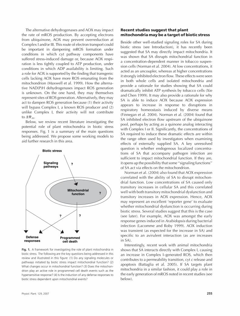
responses. Fig. 1 is a summary of the main questionsbeing addressed. We propose some working models to
aid further research in this area.
Recent studies suggest that plantmitochondriamaybe a target of biotic stress
Beside other well-studied signaling roles for SA during
biotic stress (see Introduction), it has recently beensuggested that SA may directly impact mitochondria. It
was shown that SA disrupts mitochondrial function in
a concentration-dependent manner in tobacco suspen-
sion cells (Norman et al. 2004). At low concentrations, it
acted as an uncoupler, whereas at higher concentrations
it strongly inhibited electron flow. These effects were seen
in both whole cells and isolated mitochondria and
provide a rationale for studies showing that SA coulddramatically inhibit ATP synthesis by tobacco cells (Xie
and Chen 1999). It may also provide a rationale for why
SA is able to induce AOX because AOX expression
appears to increase in response to disruptions in
respiratory homeostasis induced by diverse means
(Finnegan et al. 2004). Norman et al. (2004) found that
SA inhibited electron flow upstream of the ubiquinone
pool, perhaps by acting as a quinone analog interactingwith Complex I or II. Significantly, the concentrations of
SA required to induce these dramatic effects are within
the range often used by investigators when examining
effects of externally supplied SA. A key unresolved
question is whether endogenous localized concentra-
tions of SA that accompany pathogen infection are
sufficient to impact mitochondrial function. If they are,
it opens up the possibility that some ‘‘signaling functions’’of SA act via effects on the mitochondrion.
Norman et al. (2004) also found that AOX expression
correlated with the ability of SA to disrupt mitochon-
drial function. Low concentrations of SA caused only
transitory increases in cellular SA and this correlated
well with both transitory mitochondrial dysfunction and
transitory increases in AOX expression. Hence, AOX
may represent an excellent ‘reporter gene’ to evaluatewhether mitochondrial dysfunction is occurring during
biotic stress. Several studies suggest that this is the case
(see later). For example, AOX was amongst the early
response genes induced in Arabidopsis during bacterial
infection (Lacomme and Roby 1999). AOX induction
was transient (as expected for the increase in SA) and
specific to an avirulent interaction (as are increases
in SA).Interestingly, recent work with animal mitochondria
shows that SA interacts directly with Complex I, causing
an increase in Complex I–generated ROS, which then
contributes to a permeability transition, cyt c release and
apoptosis (Battaglia et al. 2005). If SA targets plant
mitochondria in a similar fashion, it could play a role in
the early generation of mROS noted in recent studies (see
below).
Biotic stress
Programmedcell death
Mitochondrialfunction
Defenseresponses
?
?
??
Signalingpathways
Fig. 1. A framework for investigating the role of plant mitochondria in
biotic stress. The following are the key questions being addressed in this
review and illustrated in this figure: (1) Do any signaling molecules or
pathways initiated by biotic stress impact mitochondrial function? (2)
What changes occur in mitochondrial function? (3) Does the mitochon-
drion play an active role in programmed cell death events such as the
hypersensitive response? (4) Is the induction of any defense responses to
biotic stress dependent upon mitochondrial events?
Physiol. Plant. 129, 2007 255
Another signal molecule during biotic stress is NO,
which along with SA and ROS, has been shown to
promote the HR (see Introduction). In animals, NO is
a modulator of mitochondrial-mediated apoptosis, in part
because it causes a strong reversible inhibition of cyt
oxidase (Vieira and Kroemer 2003). Plant cyt oxidase issimilarly sensitive to NO but whether the physiological
NO concentrations generated during plant–pathogen
interactions are sufficient to inhibit cyt oxidase and
whether such inhibition contributes to defense responses
or the HR remains unknown. An important factor in this
regard may be the cellular source of NO. Animals have
a mitochondrial-localized NO synthase. The situation in
plants has been less clear but a recent publication hasidentified a NO synthase localizing to mitochondria (Guo
and Crawford 2005). Under some conditions, the plant
ETC may also generate NO from nitrite (Planchet et al.
2005). These studies provide potential means by which
NO could be generated in close proximity to cyt oxidase,
hence perturbing mitochondrial function.
An important set of virulence factors in pathogenic
fungi is the so-called host-selective toxins that interactwith host molecules to cause plant cell death and
contribute to disease development. One such toxin,
victorin, was shown to bind to and inhibit mitochondrial
glycine decarboxylase (GDC), suggesting that GDC
inhibition acted to promote cell death (Curtis and
Wolpert 2002). Victorin treatment of oat leaves resulted
in a loss of DCm, followed by an ability of victorin to gain
access to the mitochondrial matrix. This was interpretedto indicate that a permeability transition had occurred
and that victorin used the PTP to gain access to matrix
GDC. However, more recent results suggest that cell
death precedes access of victorin to the cell interior and
that victorin likely interacts with a cell surface protein to
initiate defense responses and cell death (Curtis and
Wolpert 2004, Tada et al. 2005). In this respect, the
virulence of the toxin may reside in its ability to elicita plant PCD pathway. These results shed doubt on the
importance of victorin-induced GDC inhibition in pro-
moting cell death, but they do not preclude a role for the
mitochondrion in this cell death. In particular, Yao et al.
(2002) have shown that victorin induces a burst of mROS
preceding death (see below).
Ceramides are lipids that act as important second
messengers in animals, where the balance betweenceramides and their phosphorylated derivatives may
regulate apoptosis. Animal studies indicate that ceramide
can cause a direct inhibition of Complex III, which, by
promoting mROS generation, initiates apoptosis (Gudz
et al. 1997, Quillet-Mary et al. 1997). Interestingly, an
Arabidopsis mutant defective in ceramide kinase (and
hence accumulating ceramide) shows excessive PCD in
response to bacterial infection (Liang et al. 2006). It will
be interesting to examine whether this enhanced cell
death is because of ceramide targeting of the ETC.
In summary, a number of molecules commonly
associated with biotic stress may have a direct impact
on ETC components such as Complexes I, III and IV. Asdiscussed next, a common consequence of this targeting
may be an increase in mROS.
Recent studies suggest that an increase inmROS formation is an early consequence ofbiotic stress
Earlier studies showed that intracellular sources of ROSmight contribute to the pathogen-induced oxidative burst
(e.g. Allan and Fluhr 1997, Naton et al. 1996) and a review
by Bolwell and Wojtaszek (1997) suggested a need to
investigate whether the mitochondrion represented such
a source. A few recent studies have now directly addressed
this question by using ROS-sensitive fluorescent dyes and
other imaging techniques to localize ROS generation in
vivo and in response to pathogens or their elicitors.Harpins are virulence factors produced by bacterial
pathogens such as Pseudomonas syringae. Application of
purified harpin to plant tissue can elicit a rapid HR-like
cell death and some studies have examined the impact
of such harpin treatments on mitochondria. By double
staining Arabidopsis cell cultures with both a mitochon-
drial-specific dye and a ROS-indicating dye, it was shown
that a large and early ROS burst associated with harpintreatment emanated specifically from the mitochondrion,
suggesting the ETC as the likely source of ROS (Krause and
Durner 2004). This burst of mROS was associated with
a decline in DCm and cellular ATP levels and the
appearance of cytosol-localized cyt c. All these events
preceded PCD by several hours. The results are consistent
with those of another study in which harpin was shown to
dramatically inhibit ATP synthesis in tobacco cell cultures(Xie and Chen 2000). That study found that the early
harpin-induced burst of ROS could be completely
inhibited by diphenylene iodonium (DPI), a finding
usually interpreted to indicate that ROS production is
occurring via the DPI-sensitive NADPH oxidase. How-
ever, DPI is also a potent inhibitor of Complex I (Møller
2001). Hence, another interpretation of the DPI result
could be that ROS is being generated by the mitochon-drion in response to harpin and that this ROS generation
can be dampened by DPI inhibition of Complex I. The
study of Xie and Chen (2000) also found that harpin
treatment dramatically reduced the in vivo capacity for
cyt pathway electron transport downstream of ubiqui-
none. This would be consistent with a loss of cyt c from
the mitochondrion, although this was not examined.
256 Physiol. Plant. 129, 2007
The above studies show that harpin has a rapid and
dramatic impact on mitochondria, an interesting obser-
vation in light of the recent proposal that most P. syringae
virulence factors likely function by targeting the plasma
membrane, chloroplast or mitochondrion of host cells
(Greenberg and Vinatzer 2003).Greenberg and colleagues have studied mitochondrial
events associated with HR induction by P. syringae as
well as PCD induced by protoporphyrin IX (PPIX) or by
light treatment of theArabidopsis accelerated cell death 2
(ACD2) mutant. ACD2 encodes a protein that attenuates
PCD, probably by sequestering or metabolizing porphy-
rin-related molecules (such as PPIX) that can be photo-
activated, leading to the production of ROS. Interestingly,the localization of ACD2 shifts from being largely
chloroplastic to including the mitochondrion during
PCD-inducing treatments. Yao and Greenberg (2006)
reported that a very early event (1.5 h) associated with
death-inducing treatment of wild-type or ACD2 plants
was a burst of mROS, localized using ROS-sensitive
fluorescent dyes. This was followed slightly later by a loss
of DCm (quantified using flow cytometry) that, if blockedby CsA or ROS scavengers, was able to attenuate the PCD
(Yao and Greenberg 2006, Yao et al. 2004). These elegant
studies provide the most convincing data to date that
mitochondrial events precede and contribute toward
plant PCD.
In another interesting study,DCm and mROS generation
of camptothecin-treated and digitonin-permeabilized pro-
toplasts were monitored by flow cytometry (Weir et al.2003). This study also found an early (1.5 h) burst in
mROS and this corresponded closely with an increase of
DCm. This was then followed slightly later by a decrease
in both these parameters. The initial increase in DCm
(similar to that reported in an early study by Naton et al.
1996) is of particular interest. It is in keeping with animal
models in which impaired ATP/ADP exchange between
the cytosol and matrix (perhaps because of VDACclosure) promotes an initial increase in DCm that, by
promoting overreduction of the ETC, promotes mROS
generation and mitochondrial dysfunction. The decreased
expression of ANT during heat shock or senescence
associated PCD of Arabidopsis cells provides another hint
that impaired ATP/ADPexchange may be an early event in
PCD (Swidzinski et al. 2002). In another study, victorin was
shown to elicit a very rapid (30 min) increase in mROS(Yao et al. 2002). In this case, localization of the ROS was
based on a cytochemical assay that showed H2O2
eruptions at specific sites on the OMM.
The above studies indicate that increased mROS is an
early event that clearly precedes PCD and likely also
precedes other documented mitochondrial events such
as loss of DCm and cyt c release (see later). As well, the
results suggest that the mROS released is obligatory to
PCD in that, in some cases, it was shown that scavenging
of the ROS attenuated PCD. We suggest that the early
burst of mROS being noted in these studies is because of
a disruption of metabolic homeostasis in the mitochon-
drion, possibly because of molecules (such as thosedescribed in the previous section) that target the ETC.
Also, we suggest that an important consequence of this
mROS burst will be a self-amplifying cycle in which the
increased mROS leads to mitochondrial damage, result-
ing in further increases in mROS and further damage. The
culmination of these events will be the catastrophic
mitochondrial dysfunction associated with changes in the
permeability or integrity of the mitochondrial membranes(see later). This hypothesis is outlined in Fig. 2.
Several studies have documented the sensitivity of
mitochondria (particularly components of energy metab-
olism) to oxidative stress, suggesting that ROS accumu-
lation can promote damage and dysfunction (Bartoli et al.
2004, Kristensen et al. 2004, Sweetlove et al. 2002, Taylor
et al. 2002). Some of the identified components that
appear particularly susceptible to oxidative stress includeaconitase, GDC, ATP synthase, cyt c and VDAC. As
outlined more later, the self-amplifying cycle of mROS
generation and mitochondrial dysfunction may be an
important feature promoting PCD.
There is also evidence that the ROS-scavenging
capacity of the mitochondrion is modulated in response
to pathogen infection. In particular, increases in mito-
chondrial superoxide-scavenging capacity combinedwith decreases in the H2O2-scavenging components of
the organelle were seen during Botrytis cinerea infection
of tomato leaves and it was hypothesized that this could
promote accumulation of mitochondrial H2O2 (Kuzniak
and Skłodowska 2004). Such results imply an active
mechanism to ensure accumulation of specific ROS
species at the mitochondrion.
Recent studies suggest thatmitochondria doplay an active role in plant PCD
A possible role of plant mitochondria in PCD was
indicated by studies showing that when pro- or antiapop-
totic animal proteins such as Bax or Bcl-2 were expressed
in plants, they were able to, respectively, promote or
inhibit PCD (Lam et al. 2001). Plants lack clear homologsof these proteins and so the functional relevance of these
observations remains speculative. However, the studies
did emphasize that manipulation of components at the
OMM impacted PCD, implying that plant mitochondria
could play an active role in the process.
Table 1 summarizes some recent literature in which
mitochondrial events were examined during PCD and the
Physiol. Plant. 129, 2007 257
reader is referred to this literature for a more in-depth
analysis of this topic. An understanding of how mito-
chondria contribute to PCD will depend upon elucidating
the timing of mitochondrial events, of which we still have
only a rudimentary knowledge. As summarized in
Table 1, numerous studies have documented decreases
in DCm that precede PCD. In some cases (but not all), this
decrease (and in some cases PCD itself) can be attenuatedby CsA, consistent with the drop in DCm representing
a permeability transition. Often closely associated with
the loss of DCm is a loss of cyt c to the cytosol. This might
also be consistent with a permeability transition because
many animal models of cyt c release are dependent upon
the permeability transition (see Introduction). However,
interpretations of such data remain difficult because the
mechanism of cyt c release in plants has not beeninvestigated. As noted in the previous section, a break-
through in our understanding may reside with studies that
have shown a very early and localized increase in mROS.
If, as we suggest, this mROS promotes a self-amplifying
cycle of mitochondrial dysfunction, then this could lead
to the often-documented (and often slightly later) events
of declining DCm and cyt c release. Interestingly, a recent
article shows that cyt c release can be blocked by
antioxidants, perhaps evidence that cyt c release is
dependent upon mROS generation (Vacca et al. 2006).
A central feature of many models of mitochondrial
dysfunction and release of IMS proteins during animal
apoptosis is opening of the PTP (see Introduction). Plant
mitochondria are known to contain the key components
(VDAC, ANT, cyclophilin-D) that constitute the animalPTP. Hence, a key question is whether a similar perme-
ability transition occurs in plants. The elegant study of
Arpagaus et al. (2002) strongly suggests that such
a permeability transition can indeed occur in plants and
that conditions promoting PTP opening are similar to
those described in animals. Under PTP-inducing con-
ditions, swelling of purified potato mitochondria pro-
ceeded with kinetics similar to that in animals and thisresulted in selective rupture of the OMM and release of
IMS proteins, including cyt c. Similar to animals, these
events were absolutely dependent upon the presence of
Ca21 (other cations such as Mg21 were not effective) and
were potently inhibited by CsA. Similar to animals, the
ability of Ca21 to induce pore opening was modulated by
other key factors. For example, the presence of Pi was
Defense geneexpression
PCD
Bio
tic
stre
ss
SA, NO,ROS, Ca2+,ceramide,virulence factors,toxinsunknown factors
Increased ROSfrom ETC
Self-amplifying
loop
Membrane disruptionMembrane poresRelease of IMS proteinRespiratory collapse
Alternative mitochondrialETC components+
Catastrophicmitochondrialdysfunction
Early disruptionof mitochondrial
homeostasis
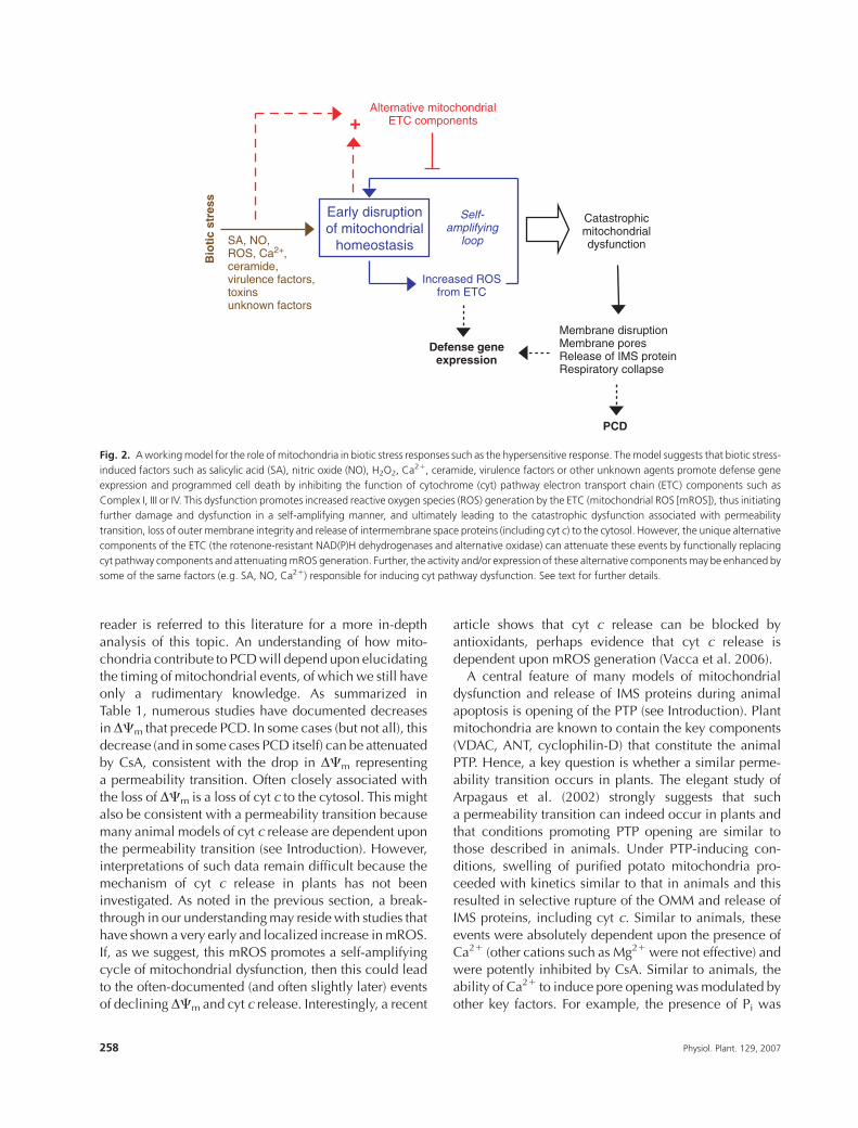
Fig. 2. Aworkingmodel for the role ofmitochondria in biotic stress responses such as the hypersensitive response. Themodel suggests that biotic stress-
induced factors such as salicylic acid (SA), nitric oxide (NO), H2O2, Ca21, ceramide, virulence factors or other unknown agents promote defense gene
expression and programmed cell death by inhibiting the function of cytochrome (cyt) pathway electron transport chain (ETC) components such as
Complex I, III or IV. This dysfunction promotes increased reactive oxygen species (ROS) generation by the ETC (mitochondrial ROS [mROS]), thus initiating
further damage and dysfunction in a self-amplifying manner, and ultimately leading to the catastrophic dysfunction associated with permeability
transition, loss of outer membrane integrity and release of intermembrane space proteins (including cyt c) to the cytosol. However, the unique alternative
components of the ETC (the rotenone-resistant NAD(P)H dehydrogenases and alternative oxidase) can attenuate these events by functionally replacing
cyt pathway components and attenuatingmROS generation. Further, the activity and/or expression of these alternative componentsmay be enhanced by
some of the same factors (e.g. SA, NO, Ca21) responsible for inducing cyt pathway dysfunction. See text for further details.
258 Physiol. Plant. 129, 2007
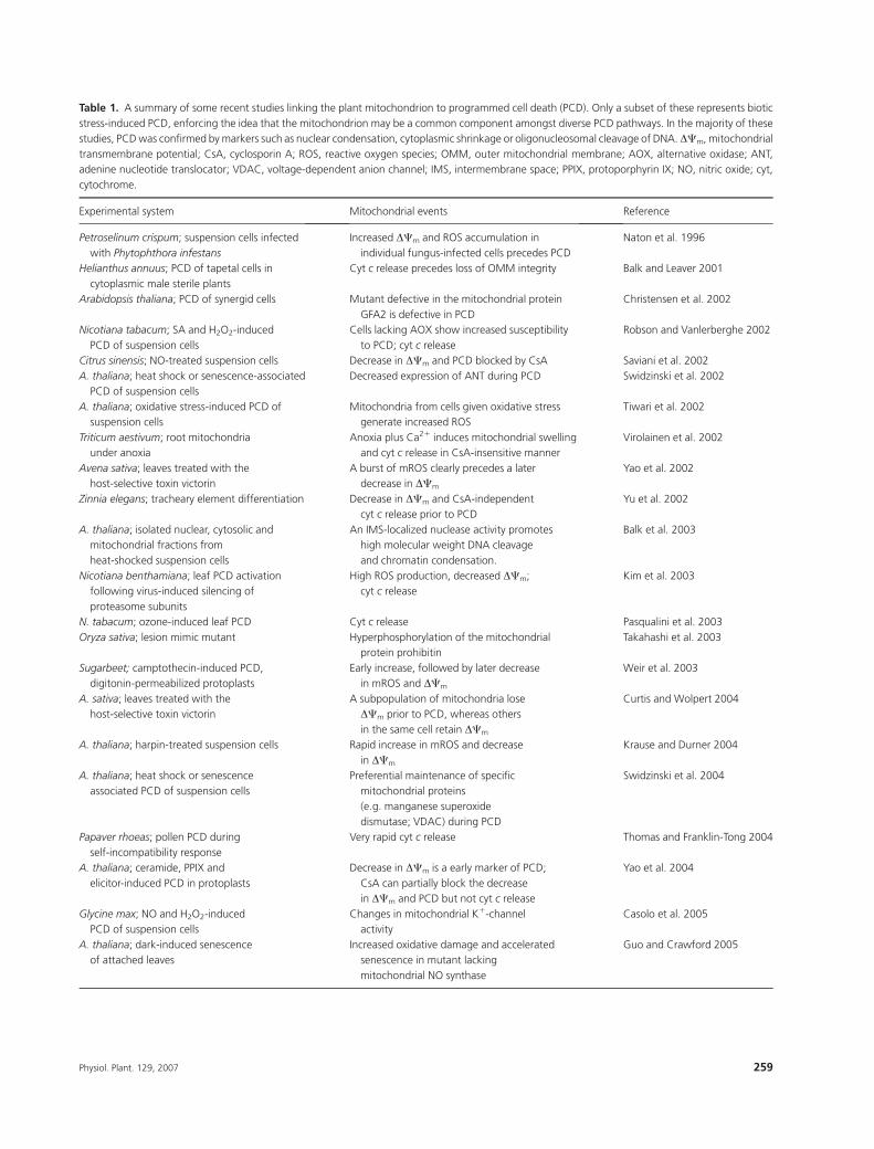
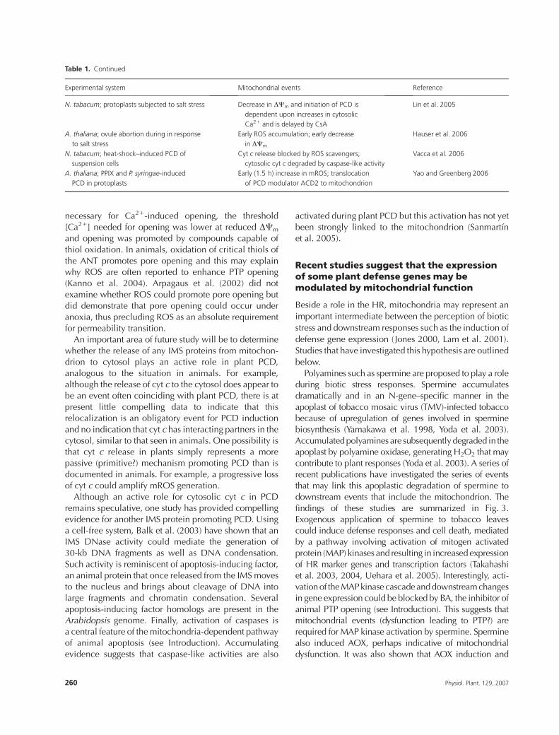
Table 1. A summary of some recent studies linking the plant mitochondrion to programmed cell death (PCD). Only a subset of these represents biotic
stress-induced PCD, enforcing the idea that the mitochondrion may be a common component amongst diverse PCD pathways. In the majority of these
studies, PCDwas confirmed bymarkers such as nuclear condensation, cytoplasmic shrinkage or oligonucleosomal cleavage of DNA.DCm,mitochondrial
transmembrane potential; CsA, cyclosporin A; ROS, reactive oxygen species; OMM, outer mitochondrial membrane; AOX, alternative oxidase; ANT,
adenine nucleotide translocator; VDAC, voltage-dependent anion channel; IMS, intermembrane space; PPIX, protoporphyrin IX; NO, nitric oxide; cyt,
cytochrome.
Experimental system Mitochondrial events Reference
Petroselinum crispum; suspension cells infected
with Phytophthora infestans
Increased DCm and ROS accumulation in
individual fungus-infected cells precedes PCD
Naton et al. 1996
Helianthus annuus; PCD of tapetal cells in
cytoplasmic male sterile plants
Cyt c release precedes loss of OMM integrity Balk and Leaver 2001
Arabidopsis thaliana; PCD of synergid cells Mutant defective in the mitochondrial protein
GFA2 is defective in PCD
Christensen et al. 2002
Nicotiana tabacum; SA and H2O2-induced
PCD of suspension cells
Cells lacking AOX show increased susceptibility
to PCD; cyt c release
Robson and Vanlerberghe 2002
Citrus sinensis; NO-treated suspension cells Decrease in DCm and PCD blocked by CsA Saviani et al. 2002
A. thaliana; heat shock or senescence-associated
PCD of suspension cells
Decreased expression of ANT during PCD Swidzinski et al. 2002
A. thaliana; oxidative stress-induced PCD of
suspension cells
Mitochondria from cells given oxidative stress
generate increased ROS
Tiwari et al. 2002
Triticum aestivum; root mitochondria
under anoxia
Anoxia plus Ca21 induces mitochondrial swelling
and cyt c release in CsA-insensitive manner
Virolainen et al. 2002
Avena sativa; leaves treated with the
host-selective toxin victorin
A burst of mROS clearly precedes a later
decrease in DCm
Yao et al. 2002
Zinnia elegans; tracheary element differentiation Decrease in DCm and CsA-independent
cyt c release prior to PCD
Yu et al. 2002
A. thaliana; isolated nuclear, cytosolic and
mitochondrial fractions from
heat-shocked suspension cells
An IMS-localized nuclease activity promotes
high molecular weight DNA cleavage
and chromatin condensation.
Balk et al. 2003
Nicotiana benthamiana; leaf PCD activation
following virus-induced silencing of
proteasome subunits
High ROS production, decreased DCm;
cyt c release
Kim et al. 2003
N. tabacum; ozone-induced leaf PCD Cyt c release Pasqualini et al. 2003
Oryza sativa; lesion mimic mutant Hyperphosphorylation of the mitochondrial
protein prohibitin
Takahashi et al. 2003
Sugarbeet; camptothecin-induced PCD,
digitonin-permeabilized protoplasts
Early increase, followed by later decrease
in mROS and DCm
Weir et al. 2003
A. sativa; leaves treated with the
host-selective toxin victorin
A subpopulation of mitochondria lose
DCm prior to PCD, whereas others
in the same cell retain DCm
Curtis and Wolpert 2004
A. thaliana; harpin-treated suspension cells Rapid increase in mROS and decrease
in DCm
Krause and Durner 2004
A. thaliana; heat shock or senescence
associated PCD of suspension cells
Preferential maintenance of specific
mitochondrial proteins
(e.g. manganese superoxide
dismutase; VDAC) during PCD
Swidzinski et al. 2004
Papaver rhoeas; pollen PCD during
self-incompatibility response
Very rapid cyt c release Thomas and Franklin-Tong 2004
A. thaliana; ceramide, PPIX and
elicitor-induced PCD in protoplasts
Decrease in DCm is a early marker of PCD;
CsA can partially block the decrease
in DCm and PCD but not cyt c release
Yao et al. 2004
Glycine max; NO and H2O2-induced
PCD of suspension cells
Changes in mitochondrial K1-channel
activity
Casolo et al. 2005
A. thaliana; dark-induced senescence
of attached leaves
Increased oxidative damage and accelerated
senescence in mutant lacking
mitochondrial NO synthase
Guo and Crawford 2005
Physiol. Plant. 129, 2007 259
necessary for Ca21-induced opening, the threshold
[Ca21] needed for opening was lower at reduced DCm
and opening was promoted by compounds capable of
thiol oxidation. In animals, oxidation of critical thiols of
the ANT promotes pore opening and this may explainwhy ROS are often reported to enhance PTP opening
(Kanno et al. 2004). Arpagaus et al. (2002) did not
examine whether ROS could promote pore opening but
did demonstrate that pore opening could occur under
anoxia, thus precluding ROS as an absolute requirement
for permeability transition.
An important area of future study will be to determine
whether the release of any IMS proteins from mitochon-drion to cytosol plays an active role in plant PCD,
analogous to the situation in animals. For example,
although the release of cyt c to the cytosol does appear to
be an event often coinciding with plant PCD, there is at
present little compelling data to indicate that this
relocalization is an obligatory event for PCD induction
and no indication that cyt c has interacting partners in the
cytosol, similar to that seen in animals. One possibility isthat cyt c release in plants simply represents a more
passive (primitive?) mechanism promoting PCD than is
documented in animals. For example, a progressive loss
of cyt c could amplify mROS generation.
Although an active role for cytosolic cyt c in PCD
remains speculative, one study has provided compelling
evidence for another IMS protein promoting PCD. Using
a cell-free system, Balk et al. (2003) have shown that anIMS DNase activity could mediate the generation of
30-kb DNA fragments as well as DNA condensation.
Such activity is reminiscent of apoptosis-inducing factor,
an animal protein that once released from the IMS moves
to the nucleus and brings about cleavage of DNA into
large fragments and chromatin condensation. Several
apoptosis-inducing factor homologs are present in the
Arabidopsis genome. Finally, activation of caspases isa central feature of the mitochondria-dependent pathway
of animal apoptosis (see Introduction). Accumulating
evidence suggests that caspase-like activities are also
activated during plant PCD but this activation has not yet
been strongly linked to the mitochondrion (Sanmartın
et al. 2005).
Recent studies suggest that the expressionof some plant defense genes may bemodulated by mitochondrial function
Beside a role in the HR, mitochondria may represent an
important intermediate between the perception of biotic
stress and downstream responses such as the induction of
defense gene expression ( Jones 2000, Lam et al. 2001).
Studies that have investigated this hypothesis are outlined
below.
Polyamines such as spermine are proposed to play a role
during biotic stress responses. Spermine accumulates
dramatically and in an N-gene–specific manner in the
apoplast of tobacco mosaic virus (TMV)-infected tobacco
because of upregulation of genes involved in sperminebiosynthesis (Yamakawa et al. 1998, Yoda et al. 2003).
Accumulated polyamines are subsequently degraded in the
apoplast by polyamine oxidase, generating H2O2 that may
contribute to plant responses (Yoda et al. 2003). A series of
recent publications have investigated the series of events
that may link this apoplastic degradation of spermine to
downstream events that include the mitochondrion. The
findings of these studies are summarized in Fig. 3.Exogenous application of spermine to tobacco leaves
could induce defense responses and cell death, mediated
by a pathway involving activation of mitogen activated
protein (MAP) kinases and resulting in increased expression
of HR marker genes and transcription factors (Takahashi
et al. 2003, 2004, Uehara et al. 2005). Interestingly, acti-
vation of the MAP kinase cascade and downstream changes
in gene expression could be blocked by BA, the inhibitor ofanimal PTP opening (see Introduction). This suggests that
mitochondrial events (dysfunction leading to PTP?) are
required for MAP kinase activation by spermine. Spermine
also induced AOX, perhaps indicative of mitochondrial
dysfunction. It was also shown that AOX induction and
Table 1. Continued
Experimental system Mitochondrial events Reference
N. tabacum; protoplasts subjected to salt stress Decrease in DCm and initiation of PCD is
dependent upon increases in cytosolic
Ca21 and is delayed by CsA
Lin et al. 2005
A. thaliana; ovule abortion during in response
to salt stress
Early ROS accumulation; early decrease
in DCm
Hauser et al. 2006
N. tabacum; heat-shock–induced PCD of
suspension cells
Cyt c release blocked by ROS scavengers;
cytosolic cyt c degraded by caspase-like activity
Vacca et al. 2006
A. thaliana; PPIX and P. syringae-induced
PCD in protoplasts
Early (1.5 h) increase in mROS; translocation
of PCD modulator ACD2 to mitochondrion
Yao and Greenberg 2006
260 Physiol. Plant. 129, 2007

activation of the MAP kinases could be blocked by the
antioxidant flavone and by the Ca21 channel blocker La21
suggesting that increases in ROS and influx of Ca21 are
cellular events upstream of the mitochondrial events.
Critically, increased ROS and especially increased mito-chondrial Ca21 are thought to be critical factors promoting
PTP opening (Arpagaus et al. 2002, Crompton 1999). The
requirement of spermine and mitochondrial events for
changes in defense gene induction should now be
evaluated using recently isolated spermine-deficient mu-
tants of Arabidopsis (Imai et al. 2004).
As noted above, one source of the ROS generated
during biotic stress could derive from the metabolism ofamines by cell wall–localized amine oxidases. Interest-
ingly, animals have an amine oxidase that localizes to the
OMM and which is able to induce the mitochondrial
pathway of PCD via H2O2 generation (Maccarrone et al.
2001). To our knowledge, there has been no report of
a similar activity in plant mitochondria but this might
represent a fruitful area for study.
A similar BA-sensitive pathway to that outlined above
was described by Maxwell et al. (2002) (Fig. 3). They
found that treatment of tobacco cells with AA resulted in
the rapid expression of eight different genes, as identifiedusing differential display. Only one of these genes
encoded an ETC component (AOX), whereas the others
encoded proteins more generally implicated in either
senescence or (biotic) stress responses. All eight genes
were also induced by treatment of cells with SA or H2O2.
Each of the gene-inducing treatments was associated with
increased cellular levels of ROS and gene induction
could be partially blocked by antioxidants that loweredROS levels, suggesting ROS as an important intermediary
in gene induction. The authors also showed that pre-
treatment of cells with BA blocked induction of all eight
genes regardless of whether AA, SA or H2O2 was used as
the inducing agent (Maxwell et al. 2002).
Interpretation of the above studies is still somewhat
speculative because it is not well established that the
plant PTP is BA sensitive. Nonetheless, the mostparsimonious explanation of results to date is that a plant
BA-sensitive PTPexists, that this pore opens in response to
signaling molecules commonly associated with biotic
stress (spermine, SA, H2O2) or mitochondrial dysfunction
(AA) and that the state of this pore affects defense gene
expression (Fig. 3). If this is the case, then plant
mitochondria may indeed act as a focal point for the
perception of and response to biotic stress.Further evidence that mitochondria may act in the
perception of and response to stress (albeit abiotic stress
in this case) comes from studies of the fro1 mutant of
Arabidopsis (Lee et al. 2002). Mutant plants are unable
to induce a set of cold-responsive genes, thus reducing
their capacity to cold acclimate. The defect is specific
to cold stress in that expression of the genes in response
to other treatments (abseisic acid, NaCl) is normal.Interestingly, FRO1 encodes a protein similar to the 18-
kDa Fe-S subunit of complex I from diverse organisms
and was shown to localize to Arabidopsis mitochondria.
Fro1 plants also displayed constitutively higher levels of
ROS, even in the absence of stress. Because Complex I is
considered a major site of mROS production, one
potential explanation is that mutation of the 18-kDa
subunit has increased the ROS-generating activity ofComplex I. The authors hypothesize that the constitutive
generation of ROS makes mutant plants less responsive
to what would normally be a cold-induced increase in
ROS generation. Presumably, this cold-induced ROS
generation would also involve Complex I, thus repre-
senting part of a cold-stress–activated signal path from
PTP opening
SIPK / WIPKactivation
Changes in gene expression
Defense / death responses
Bongkrekic acid
Ca2+ ROS
ETCdysfunction
Flavone
TMV Spermine
AA, SA, ROS, cold
Ca2+ influx
ROS
Apoplast
Symplast
La2+
Polyamine oxidase
Mitochondrion
Fig. 3. A working model for how mitochondria may act as a focal point
for the perception of and response to biotic stress. Several studies have
shown that stress-induced changes in gene expression can be blocked by
bongkrekic acid, suggesting that opening of amitochondrial permeability
transition pore (PTP) is a necessary step for gene induction. Opening of the
PTPmay be promoted by stress-induced changes in Ca21, reactive oxygen
species or mitochondrial function. The results outlined here are primarily
based upon thework of Lee et al. (2002),Maxwell et al. (2002), Takahashi
et al. (2003b, 2004) and Uehara et al. (2005). See text for further details.
Physiol. Plant. 129, 2007 261
mitochondrion to nucleus, with mROS as a key inter-
mediate (Fig. 3).
Other mutations of Complex I or Complex IV (but in
these case mutations that dramatically compromise their
activity) have been shown to increase stress gene
expression and/or stress tolerance (Dutilleul et al. 2003,Kuzmin et al. 2004). These mutations illustrate that the
consequence of a major mitochondrial deficiency is not
limited to PCD (perhaps because of compensatory
activities; see below) but can also be linked to protective
(defense) responses.
Several studies investigated a possible role for the
mitochondrion in the SA-mediated development of local
and systemic resistance to viruses. Interest in this areastemmed from studies suggesting that AOX played some
active role in the induction of resistance. Results from the
use of transgenic plants with increased and decreased
levels of AOX have largely negated any direct role for
AOX in the development of resistance (Gilliland et al.
2003, Ordog et al. 2002).
Recent studies suggest that the uniquecomponents of the plant mitochondrial ETCplay a complex role in biotic stress responses
Although there is now a large body of circumstantial
evidence that plant mitochondria play a regulatory role in
PCD (Table 1), analogous to the relatively well-defined
active role of mitochondria in animal apoptosis, it is also
evident that the regulation in plants must differ from thatin animals. This is best exemplified by the lack of plant
homologs of many of the key pro- and antiapoptotic
proteins described in animals (Lam et al. 2001). Hence,
an important next step will be to define the pro- and
antiapoptotic players in a plant mitochondria-dependent
PCD pathway.
As discussed earlier, the generation of mROS and
mitochondrial dysfunction are early events associatedwith biotic stress and preceding PCD. Dysfunction may
be the result of key mitochondrial components (e.g.
Complexes I, III, IV) being targeted by biotic stress signals,
leading to the self-amplifying cycle of increased mROS
and increased dysfunction described earlier (Fig. 2).
However, a striking feature of plant mitochondria in
comparison to their animal counterparts is the existence
of additional ETC components that increase the points ofentry and exit of electrons in the respiratory chain as well
as providing a high degree of flexibility in terms of the
coupling of electron transport to oxidative phosphoryla-
tion (see Introduction). This is significant because these
components (the rotenone-resistant alternative dehydro-
genases and AOX) represent a potential means to
modulate a mitochondria-dependent PCD because they
could compensate for dysfunctional ETC components, as
well as providing a means to dampen the mROS
generation associated with escalating dysfunction
(Fig. 2). In other words, they may represent antiapoptotic
components of plant mitochondria, the levels of which
could define cell fate (e.g. defense vs death). It is certainlyclear, for example, that AOX can prevent the PCD
initiated by a loss of cyt pathway function downstream
of ubiquinone (Vanlerberghe et al. 2002).
Interestingly, numerous studies have shown increases in
AOX expression in response to pathogen infection, sig-
naling molecules (SA, NO, H2O2) or elicitors (Bruggman
et al. 2005, Huang et al. 2002, Krause and Durner
2004, Maxwell et al. 2002, Vanlerberghe and McIntosh1996, Mizuno et al. 2005, Murphy et al. 2001, Lacomme
and Roby 1999, Ordog et al. 2002, Takahashi et al. 2003,
Simons et al. 1999, Zottini et al. 2002). As discussed
earlier, one interpretation is that increased AOX expression
simply represents an all-purpose response to disruptions of
respiratory homeostasis. However, another interpretation
is that it represents a defensive response against the
initiation of PCD, much like increases in expression ofROS-scavenging systems. Such a response could be
important in limiting cell death progression and in this
regard it is interesting that overexpression of AOX has been
shown to reduce the size of HR lesions induced by TMV
(Ordog et al. 2002). In the case of ROS-scavenging
systems, however, it has also been shown that an active
decline in their capacity may be an important means to
commit a cell to PCD (see Introduction). It would beinteresting to examine whether capacity of the alternative
ETC components might also be declining in such
instances. It is also possible that once a threshold level of
ROS is reached in the mitochondrion, AOX might be
inactivated by oxidation of critical sulfhydryl residues
involved in a-keto acid activation. This has been
demonstrated to occur when cells are treated exogenously
with H2O2 (Vanlerberghe et al. 1999). Such inactivationcould again act to amplify mROS levels.
Chloroplasts are a large source of ROS and so it is often
assumed that the steady-state level of cellular ROS as well
as the capacity of cellular ROS-scavenging systems is
largely determined by this organelle. However, as
reviewed by Foyer and Noctor (2003), recent studies
indicate an unexpectedly influential role for mitochon-
dria in determining the cellular level of ROS and capacityof both intra- and extramitochondrial antioxidant de-
fenses. Hence, another role for the alternative compo-
nents of the mitochondrial ETC during biotic stress
(beside a role in compensating for dysfunctional ETC
components and/or controlling mROS production after
imposition of the stress) is that they may play a key role in
constitutively defining the cellular level of ROS and
262 Physiol. Plant. 129, 2007
capacity of antioxidant defenses in the plant. This could
dramatically impact the plant response to biotic stress
(e.g. defense vs death) if in fact this response is modulated
by the cellular level of ROS and capacity of antioxidant
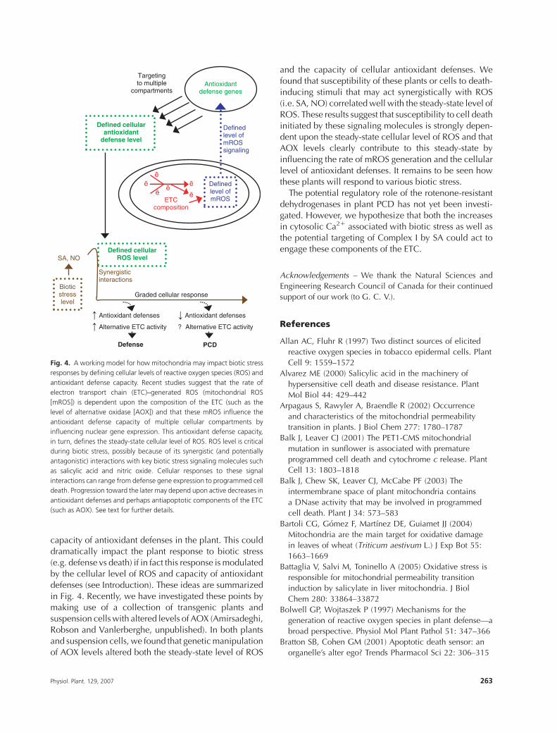
defenses (see Introduction). These ideas are summarized
in Fig. 4. Recently, we have investigated these points bymaking use of a collection of transgenic plants and
suspension cells with altered levels of AOX (Amirsadeghi,
Robson and Vanlerberghe, unpublished). In both plants
and suspension cells, we found that genetic manipulation
of AOX levels altered both the steady-state level of ROS
and the capacity of cellular antioxidant defenses. We
found that susceptibility of these plants or cells to death-
inducing stimuli that may act synergistically with ROS
(i.e. SA, NO) correlated well with the steady-state level of
ROS. These results suggest that susceptibility to cell death
initiated by these signaling molecules is strongly depen-dent upon the steady-state cellular level of ROS and that
AOX levels clearly contribute to this steady-state by
influencing the rate of mROS generation and the cellular
level of antioxidant defenses. It remains to be seen how
these plants will respond to various biotic stress.
The potential regulatory role of the rotenone-resistant
dehydrogenases in plant PCD has not yet been investi-
gated. However, we hypothesize that both the increasesin cytosolic Ca21 associated with biotic stress as well as
the potential targeting of Complex I by SA could act to
engage these components of the ETC.
Acknowledgements – We thank the Natural Sciences and
Engineering Research Council of Canada for their continued
support of our work (to G. C. V.).
References
Allan AC, Fluhr R (1997) Two distinct sources of elicited
reactive oxygen species in tobacco epidermal cells. Plant
Cell 9: 1559–1572
Alvarez ME (2000) Salicylic acid in the machinery of
hypersensitive cell death and disease resistance. Plant
Mol Biol 44: 429–442
Arpagaus S, Rawyler A, Braendle R (2002) Occurrence
and characteristics of the mitochondrial permeability
transition in plants. J Biol Chem 277: 1780–1787
Balk J, Leaver CJ (2001) The PET1-CMS mitochondrial
mutation in sunflower is associated with premature
programmed cell death and cytochrome c release. Plant
Cell 13: 1803–1818
Balk J, Chew SK, Leaver CJ, McCabe PF (2003) The
intermembrane space of plant mitochondria contains
a DNase activity that may be involved in programmed
cell death. Plant J 34: 573–583
Bartoli CG, Gomez F, Martınez DE, Guiamet JJ (2004)
Mitochondria are the main target for oxidative damage
in leaves of wheat (Triticum aestivum L.) J Exp Bot 55:
1663–1669
Battaglia V, Salvi M, Toninello A (2005) Oxidative stress is
responsible for mitochondrial permeability transition
induction by salicylate in liver mitochondria. J Biol
Chem 280: 33864–33872
Bolwell GP, Wojtaszek P (1997) Mechanisms for the
generation of reactive oxygen species in plant defense—a
broad perspective. Physiol Mol Plant Pathol 51: 347–366
Bratton SB, Cohen GM (2001) Apoptotic death sensor: an
organelle’s alter ego? Trends Pharmacol Sci 22: 306–315
ETCcomposition
Definedlevel ofmROS
Definedlevel ofmROSsignaling
Antioxidantdefense genes
Defined cellularantioxidant
defense level
Targetingto multiple
compartments
Defined cellularROS levelSA, NO
Synergisticinteractions
Graded cellular response
Antioxidant defenses
Alternative ETC activity
Antioxidant defenses
? Alternative ETC activity
Defense PCD
Bioticstresslevel
Fig. 4. A working model for how mitochondria may impact biotic stress
responses by defining cellular levels of reactive oxygen species (ROS) and
antioxidant defense capacity. Recent studies suggest that the rate of
electron transport chain (ETC)–generated ROS (mitochondrial ROS
[mROS]) is dependent upon the composition of the ETC (such as the
level of alternative oxidase [AOX]) and that these mROS influence the
antioxidant defense capacity of multiple cellular compartments by
influencing nuclear gene expression. This antioxidant defense capacity,
in turn, defines the steady-state cellular level of ROS. ROS level is critical
during biotic stress, possibly because of its synergistic (and potentially
antagonistic) interactions with key biotic stress signaling molecules such
as salicylic acid and nitric oxide. Cellular responses to these signal
interactions can range from defense gene expression to programmed cell
death. Progression toward the later may depend upon active decreases in
antioxidant defenses and perhaps antiapoptotic components of the ETC
(such as AOX). See text for further details.
Physiol. Plant. 129, 2007 263
Bruggman R, Abderhalden O, Reymond P, Dudler R (2005)
Analysis of epidermis- and mesophyll-specific transcript
accumulation in powdery mildew-inoculated wheat
leaves. Plant Mol Biol 58: 247–267
Casolo V, Petrussa E, Krajnakova J, Macri F, Vianello A
(2005) Involvement of the mitochondrial K1ATP channel
in H2O2- or NO-induced programmed cell death of
soybean suspension cell cultures. J Exp Bot 56: 997–1006
Christensen CA, Gorsich SW, Brown RH, Jones LG, Brown J,
Shaw JM, Drews GN (2002) Mitochondrial GFA2 is
required for synergid cell death in Arabidopsis. Plant Cell
14: 2215–2232
Crompton M (1999) The mitochondrial permeability transition
pore and its role in cell death. Biochem J 341: 233–249
Curtis MJ, Wolpert TJ (2002) The oat mitochondrial
permeability transition and its implication in victorin
binding and induced cell death. Plant J 29: 295–312
Curtis MJ, Wolpert TJ (2004) The victorin-induced
mitochondrial permeability transition precedes cell
shrinkage and biochemical markers of cell death, and
shrinkage occurs without loss of membrane integrity. Plant
J 38: 244–259
Dangl JL, Jones JDG (2001) Plant pathogens and integrated
defense responses to infection. Nature 411: 826–833
Delledonne M (2005) NO news is good news for plants.
Curr Opin Plant Biol 8: 390–396
Dutilleul C, Garmier M, Noctor G, Mathieu C, Chetrit P,
Foyer CH, de Paepe R (2003) Leaf mitochondria modulate
whole cell redox homeostasis, set antioxidant capacity,
and determine stress resistance through altered signaling
and diurnal regulation. Plant Cell 15: 1212–1226
Finnegan PM, Soole KL, Umbach AL (2004) Alternative
mitochondrial electron transport proteins in higher plants.
In: Day DA, Millar AH, Whelan J (eds) Plant Mitochondria:
From Genome to Function. Kluwer Academic Publishers,
Great Britain, pp 163–230
Foyer CH, Noctor G (2003) Redox sensing and signaling
associated with reactive oxygen in chloroplasts,
peroxisomes and mitochondria. Physiol Plant 119: 355–364
Gilliland A, Singh DP, Hayward JM, Moore CA, Murphy AM,
York CJ, Slator J, Carr JP (2003) Genetic modification of
alternative respiration has differential effects on antimycin
A–induced versus salicylic acid–induced resistance to
Tobacco mosaic virus. Plant Physiol 132: 1518–1528
Greenberg JT, Vinatzer BA (2003) Identifying type III effectors
of plant pathogens and analyzing their interaction with
plant cells. Curr Opin Microbiol 6: 20–28
Greenberg JT, Yao N (2004) The role and regulation of
programmed cell death in plant–pathogen interactions.
Cell Microbiol 6: 201–211
Gudz TI, Tserng K-Y, Hoppel CL (1997) Direct inhibition
of mitochondrial respiratory chain Complex III by
cell-permeable ceramide. J Biol Chem 272: 24154–24158
Guo F-Q, Crawford NM (2005) Arabidopsis nitric oxide
synthase 1 is targeted to mitochondria and protects against
oxidative damage and dark-induced senescence. Plant
Cell 17: 3436–3450
Hauser BA, Sun K, Oppenheimer DG, Sage TL (2006)
Changes in mitochondrial membrane potential and
accumulation of reactive oxygen species precede
ultrastructural changes during ovule abortion. Planta
223: 492–499
Huang X, von Rad U, Durner J (2002) Nitric oxide induces
transcriptional activation of the nitric oxide-tolerant
alternative oxidase in Arabidopsis suspension cells. Plant
215: 914–923
Imai A, Akiyama T, Kato T, Sato S, Tabata S, Yamamoto KT,
Takahashi T (2004) Spermine is not essential for survival
of Arabidopsis. FEBS Lett 556: 148–152
Jones A (2000) Does the plant mitochondrion integrate
cellular stress and regulate programmed cell death? Trends
Plant Sci 5: 225–230
Kanno T, Sato EF, Muranaka S, Fujita H, Fujiwara T, Utsumi T,
Inoue M, Utsumi K (2004) Oxidative stress underlies the
mechanism for Ca21-induced permeability transition of
mitochondria. Free Radic Res 38: 27–35
Kim M, Ahn J-W, Jin U-H, Choi D, Paek K-H, Pai H-S (2003)
Activation of the programmed cell death pathway by
inhibition of proteasome function in plants. J Biol Chem
278: 19406–19415
Krause M, Durner J (2004) Harpin inactivates mitochondria
in Arabidopsis suspension cells. Mol Plant Microbe
Interact 17: 131–139
Kristensen BK, Askerlund P, Bykova NV, Egsgaard H, Møller
IM (2004) Identification of oxidized proteins in the matrix
of rice leaf mitochondria by immunoprecipitation and
two-dimensional liquid chromatography–tandem mass
spectrometry. Phytochemistry 65: 1839–1851
Kuwana T and Newmeyer DD (2003) Bcl-2 family proteins
and the role of mitochondria in apoptosis. Curr Opin Plant
Biol 15: 691–699
Kuzmin EV, Karpova OV, Elthon TE, Newton KJ (2004)
Mitochondrial respiratory deficiencies signal up-regulation
of genes for heat shock proteins. J Biol Chem 279:
20672–20677
Kuzniak E, Skłodowska M (2004) The effect of Botrytis
cinerea infection on the antioxidant profile of
mitochondria from tomato leaves. J Exp Bot 55:
605–612
Lacomme C, Roby D (1999) Identification of new early
markers of the hypersensitive response in Arabidopsis
thaliana. FEBS Lett 459: 149–153
Laloi C, Apel K, Danon A (2004) Reactive oxygen signaling:
the latest news. Curr Opin Plant Biol 7: 323–328
Lam E, Kato N, Lawton M (2001) Programmed cell death,
mitochondria and the plant hypersensitive response.
Nature 411: 848–853
Lee B, Lee H, Xiong L, Zhu J-K (2002) A mitochondrial
Complex I defect impairs cold-regulated nuclear gene
expression. Plant Cell 14: 1235–1251
264 Physiol. Plant. 129, 2007
Liang H, Yao N, Song JT, Luo S, Lu H, Greenberg JT (2006)
Ceramides modulate programmed cell death in plants.
Genes Dev 17: 2636–2641
Lin J, Wang Y, Wang G (2005) Salt stress-induced
programmed cell death via Ca21-mediated mitochondrial
permeability transition in tobacco protoplasts. Plant
Growth Regul 45: 243–250
Ly JD, Grubb DR, Lawen A (2003) The mitochondrial
membrane potential (DCm) in apoptosis; an update.
Apoptosis 8: 115–128
Maccarrone M, Bari M, Battista N, Di Rienzo M, Falciglia K,
Agro AF (2001) Oxidation products of polyamines
induce mitochondrial uncoupling and cytochrome
c release. FEBS Lett 507: 30–34
Maxwell DP, Wang Y, McIntosh L (1999) The alternative
oxidase lowers mitochondrial reactive oxygen production
in plant cells. Proc Natl Acad Sci USA 96: 8271–8276
Maxwell DP, Nickels R, McIntosh L (2002) Evidence of
mitochondrial involvement in the transduction of signals
required for the induction of genes associated with
pathogen attack and senescence. Plant J 29: 269–279
Mittler R, Feng X, Cohen M (1998) Post-transcriptional
suppression of cytosolic ascorbate peroxidase expression
during pathogen-induced programmed cell death in
tobacco. Plant Cell 10: 461–473
Mittler R, Vanderauwera S, Gollery M, Van Breusegem F
(2004) Reactive oxygen gene network of plants. Trends
Plant Sci 9: 490–498
Mizuno M, Tada Y, Uchii K, Kawakami S, Mayama S (2005)
Catalase and alternative oxidase cooperatively regulate
programmed cell death induced by b-glucan elicitor in
potato suspension cultures. Planta 220: 849–853
Møller IM (2001) Plant mitochondria and oxidative stress:
electron transport, NADPH turnover, and metabolism of
reactive oxygen species. Annu Rev Plant Physiol Plant
Mol Biol 52: 561–591
Murphy AM, Gilliland A, Wong CE, West J, Singh DP, Carr JP
(2001) Signal transduction in resistance to plant viruses.
Eur J Plant Pathol 107: 121–128
Naton B, Hahlbrock K, Schmelzer E (1996) Correlation of
rapid cell death with metabolic changes in fungus-
infected, cultured parsley cells. Plant Physiol 112:
433–444
Neill S, Desikan R, Hancock J (2002) Hydrogen peroxide
signaling. Curr Opin Plant Biol 5: 388–395
Newmeyer DD, Ferguson-Miller S (2003) Mitochondria:
releasing power for life and unleashing the machineries
of death. Cell 112: 481–490
Norman C, Howell KA, Millar AH, Whelan JM, Day DA
(2004) Salicylic acid is an uncoupler and inhibitor of
mitochondrial electron transport. Plant Physiol 134:
492–501
Ordog SH, Higgins VJ, Vanlerberghe GC (2002)
Mitochondrial alternative oxidase is not a critical
component of plant viral resistance but may play a role in
the hypersensitive response. Plant Physiol 129: 1858–1865
Pasqualini S, Piccioni C, Reale L, Ederli L, Della Torre G,
Ferranti F (2003) Ozone-induced cell death in tobacco
cultivar Bel W3 plants. The role of programmed cell death
in lesion formation. Plant Physiol 133: 1122–1134
Planchet E, Gupta KJ, Sonoda M, Kaiser WM (2005) Nitric
oxide emission from tobacco leaves and cell suspensions:
rate limiting factors and evidence for the involvement of
mitochondrial electron transport. Plant J 41: 732–743
Quillet-Mary A, Jaffrezou J-P, Mansat V, Bordier C, Naval J,
Laurent G (1997) Implication of mitochondrial hydrogen
peroxide generation in ceramide-induced apoptosis.
J Biol Chem 272: 21388–21395
Rasmusson AG, Soole KL, Elthon TE (2004) Alternative
NAD(P)H dehydrogenases of plant mitochondria. Annu
Rev Plant Biol 55: 23–39
Robson CA, Vanlerberghe GC (2002) Transgenic plant
cells lacking mitochondrial alternative oxidase have
increased susceptibility to mitochondria-dependent
and -independent pathways of programmed cell
death. Plant Physiol 129: 1908–1920
Sanmartın M, Jaroszewski L, Raikhel N, Rojo E (2005)
Caspases. Regulating death since the origin of life. Plant
Physiol 137: 841–847
Saviani EE, Orsi CH, Oliveira JFP, Pinto-Maglio CAF,
Salgado I (2002) Participation of the mitochondrial
permeability transition pore in nitric oxide-induced plant
cell death. FEBS Lett 510: 136–140
Simons BH, Millenaar FF, Mulder L, Van Loon LC, Lambers H
(1999) Enhanced expression and activation of the
alternative oxidase during infection of Arabidopsis with
Pseudomonas syringae pv tomato. Plant Physiol 120:
529–538
Sweetlove L, Heazlewood J, Herald V, Holtzapffel R, Day
DA, Leaver C, Millar AH (2002) The impact of oxidative
stress on Arabidopsis mitochondria. Plant J 32: 891–904
Swidzinski JA, Sweetlove LJ, Leaver CJ (2002) A custom
microarray analysis of gene expression during
programmed cell death in Arabidopsis thaliana. Plant J 30:
431–446
Swidzinski JA, Leaver CJ, Sweetlove LJ (2004) A proteomic
analysis of plant programmed cell death. Phytochem 65:
1829–1838
Tada Y, Kusaka K, Betsuyaku S, Shinoyi T, Sakamoto M,
Ohura Y, Hata S, Mori T, Tosa Y, Mayama S (2005) Victorin
triggers programmed cell death and the defense response
via interaction with a cell surface mediator. Plant Cell
Physiol 46: 1787–1798
Takahashi A, Kawasaki T, Wong HL, Suharsono U, Hirano H,
Shimamoto K (2003a) Hyperphosphorylation of
a mitochondrial protein, prohibitin, is induced by
calyculin A in a rice lesion-mimic mutant cdr1. Plant
Physiol 132: 1861–1869
Physiol. Plant. 129, 2007 265
Takahashi Y, Berberich T, Miyazaki A, Seo S, Ohashi Y,
Kusano T (2003b) Spermine signaling in tobacco:
activation of mitogen-activated protein kinases by
spermine is mediated through mitochondrial dysfunction.
Plant J 36: 820–829
Takahashi Y, Uehara Y, Berberich T, Ito A, Saitoh H, Miyazaki
A, Terauchi R, Kusano T (2004) A subset of hypersensitive
response marker genes, including HSR203J, is the
downstream target of a spermine signal transduction
pathway in tobacco. Plant J 40: 586–595
Taylor NL, Day DA, Millar AH (2002) Environmental stress
causes oxidative damage to plant mitochondria leading to
inhibition of glycine decarboxylase. J Biol Chem 277:
42663–42668
Thomas SG, Franklin-Tong VE (2004) Self-incompatibility
triggers programmed cell death in Papaver pollen. Nature
429: 305–309
Tiwari BS, Belenghi B, Levine A (2002) Oxidative stress
increased respiration and generation of reactive oxygen
species, resulting in ATP depletion, opening of
mitochondrial permeability transition, and programmed
cell death. Plant Physiol 128: 1271–1281
Torres MA, Dangl JL (2005) Functions of the respiratory burst
oxidase in biotic interactions, abiotic stress and
development. Curr Opin Plant Biol 8: 397–403
Uehara Y, Takahashi Y, Berberich T, Miyazaki A, Takahashi
H, Matsui K, Ohme-Takagi M, Saitoh H, Terauchi R,
Kusano T (2005) Tobacco ZFT1, a transcriptional repressor
with a Cys2/His2 type zinc finger motif that functions
in spermine-signaling pathway. Plant Mol Biol 59:
435–448
Vacca RA, de Pinto MC, Valenti D, Passarella S, Marra E,
De Gara L (2004) Production of reactive oxygen species,
alteration of cytosolic ascorbate peroxidase, and
impairment of mitochondrial metabolism are early events
in heat-shock–induced programmed cell death in tobacco
Bright-Yellow 2 cells. Plant Physiol 134: 1100–1112
Vacca RA, Valenti D, Bobba A, Merafina RS, Passarella S,
Marra E (2006) Cytochrome c is released in a reactive
oxygen species-dependent manner and is degraded via
caspase-like proteases in tobacco BY-2 cells en route to
heat-shock induced cell death. Plant Physiol 141: 208–219
Vanlerberghe GC, McIntosh L (1996) Signals regulating the
expression of the nuclear gene encoding alternative
oxidase of plant mitochondria. Plant Physiol 111: 589–595
Vanlerberghe GC, Yip JYH, Parsons HL (1999) In organello
and in vivo evidence of the importance of the regulatory
sulfhydryl/disulfide system and pyruvate for alternative
oxidase activity in tobacco. Plant Physiol 121: 793–803
Vanlerberghe GC, Robson CA, Yip JYH (2002) Induction of
mitochondrial alternative oxidase in response to a cell
signal pathway down-regulating the cytochrome pathway
prevents programmed cell death. Plant Physiol 129:
1829–1842
van Loo G, Saelens X, van Gurp M, MacFarlane M, Martin SJ,
Vandenabeele P (2002) The role of mitochondrial factors
in apoptosis: a Russian roulette with more than one bullet.
Cell Death Differ 9: 1031–1042
Vieira H, Kroemer G (2003) Mitochondria as targets of
apoptosis regulation by nitric oxide. IUBMB Life 55:
613–616
Virolainen E, Blokhina O, Fagerstedt K (2002) Ca21-induced
high amplitude swelling and cytochrome c release from
wheat (Triticum aestivum L.) mitochondria under anoxic
stress. Ann Bot 90: 509–516
Weir IE, Pham N-A, Hedley DW (2003) Oxidative stress is
generated via the mitochondrial respiratory chain during
plant cell apoptosis. Cytometry Part A 54A: 109–117
Wendehenne D, Durner J, Klessig DF (2004) Nitric oxide:
a new player in plant signaling and defense responses.
Curr Opin Plant Biol 7: 449–455
Xie Z, Chen Z (1999) Salicylic acid induces rapid inhibition
of mitochondrial electron transport and oxidative
phosphorylation in tobacco cells. Plant Physiol 120:
217–226
Xie Z, Chen Z (2000) Harpin-induced hypersensitive cell
death is associated with altered mitochondrial functions in
tobacco cells. Mol Plant Microbe Interact 13: 183–190
Yamakawa H, Kamada H, Satoh M, Ohashi Y (1998)
Spermine is a salicylate-independent endogenous inducer
for both tobacco acidic pathogenesis-related proteins and
resistance against tobacco mosaic virus infection. Plant
Physiol 118: 1213–1222
Yao N, Greenberg JT (2006) Arabidopsis ACCELERATED
CELL DEATH2 modulates programmed cell death. Plant
Cell 18: 397–411
Yao N, Tada Y, Sakamoto M, Nakayashiki H, Park P, Tosa Y,
Mayama S (2002) Mitochondrial oxidative burst involved
in apoptotic response in oats. Plant J 30: 567–579
Yao N, Eisfelder BJ, Marvin J, Greenberg JT (2004) The
mitochondrion—an organelle commonly involved in
programmed cell death in Arabidopsis thaliana. Plant J
40: 596–610
Yoda H, Yamaguchi Y, Sano H (2003) Induction of
hypersensitive cell death by hydrogen peroxide produced
through polyamine degradation in tobacco plants. Plant
Physiol 132: 1973–1981
Yu X-H, Perdue TD, Heimer YM, Jones AM (2002)
Mitochondrial involvement in tracheary element
programmed cell death. Cell Death Differ 9:
189–198
Zottini M, Formentin E, Scattolin M, Carimi F, Schiavo FL,
Terzi M (2002) Nitric oxide affects plant mitochondrial
functionality in vivo. FEBS Lett 515: 75–78
Edited by P. Gardestrom
266 Physiol. Plant. 129, 2007