Embed Size (px)
Citation preview
Copyright 2002 by the Genetics Society of America
The Scw1 RNA-Binding Domain Protein Regulates Septation andCell-Wall Structure in Fission Yeast
Jim Karagiannis, Rena Oulton1 and Paul G. Young2
Department of Biology, Queen’s University, Kingston, Ontario K7L-3N6, Canada
Manuscript received March 4, 2002Accepted for publication June 6, 2002
ABSTRACTLoss of the nonessential RNA-binding domain protein, Scw1, increases resistance to cell-wall-degrading
enzymes in fission yeast. Surprisingly, scw1 null mutations also suppress the lethality of mutations (cdc11-136,cdc7-24, cdc14-118, sid1-239, sid2-250, sid3-106, sid4-A1, and mob1-1) at all levels of the sid pathway. Thispathway forms part of the septation initiation network (SIN), which regulates the onset of septum formationand ensures the proper coupling of mitosis to cytokinesis. In contrast, scw1� mutations do not suppressts alleles of the rng genes, cdc12 or cdc15. These mutations also prevent the formation of a septum andin addition block assembly and/or function of the contractile acto-myosin ring. sid mutants exhibit ahyper-sensitivity to cell-wall-degrading enzymes that is suppressed by loss of Scw1. Furthermore, scw1�-mediated rescue of sid mutants is abolished in the presence of calcofluor white, a compound that interfereswith cell-wall synthesis. These data suggest that Scw1 acts in opposition to the SIN as a negative regulatorof cell-wall/septum deposition. Unlike components of the SIN, Scw1 is predominantly a cytoplasmicprotein and is not localized to the spindle pole body.
CYTOKINESIS requires a large network of genes Marks et al. 1992; Schmidt et al. 1997; Balasubraman-that function in a highly coordinated manner (Le ian et al. 1998; Sparks et al. 1999; Chang and Gould
Goff et al. 1999; Balasubramanian et al. 2000). One 2000; Guertin et al. 2000; Hou et al. 2000; Salimova etof our best models of cytokinesis is the fission yeast al. 2000); and the sep mutants defective in cell separationSchizosaccharomyces pombe, in which the process of cyto- (Sipiczki et al. 1993; Grallert et al. 1999; Ribar et al.plasmic division can be separated into four phases: (1) 1999; Zilahi et al. 2000a,b).the selection of the site of division; (2) the assembly of The sid class of mutants belongs to a regulatory mod-an acto-myosin-based medial ring; (3) contraction of ule (referred to as the septation initiation network, orthe ring concurrent with the deposition of a primary SIN) that localizes to the spindle pole body and func-septum and two flanking secondary septa; and (4) degra- tions in signaling the onset of septum formation oncedation of the primary septum leading to the physical per cell cycle. This network consists of the products ofseparation of two daughter cells. the cdc16, byr4, cdc11, cdc7, cdc14, spg1/sid3, sid1, sid2,
Mutants specifically defective in each of these pro- sid4, and mob1 genes (reviewed in Sawin 2000; McCol-cesses have been identified and constitute the pos, mid, lum and Gould 2001).and pom mutants defective in positioning of the division Sid4 and Cdc11 are the most upstream members ofsite (Edamatsu and Toyoshima 1996; Sohrmann et al. this network and are localized constitutively to the spin-1996; Bahler and Pringle 1998; Bahler et al. 1998a; dle pole body (SPB) throughout the cell cycle. Sid4 isPaoletti and Chang 2000); the rng mutants defective required for the recruitment of all other tested SINin assembly of the medial acto-myosin ring (Balasubra- components (Cdc11, Byr4, Spg1, Cdc7, Sid1, Sid2,manian et al. 1992, 1994, 1998; McCollum et al. 1995; Mob1, and Cdc14) to this subcellular location, whereasChang et al. 1996; Bezanilla et al. 1997; Chang et al. Cdc11 is required for the recruitment of Spg1, Sid2,1997; Gould and Simanis 1997; Kitayama et al. 1997; Cdc7, Mob1, and Byr4, but not Sid4. Sid4 and Cdc11May et al. 1997; Motegi et al. 1997; Eng et al. 1998; physically interact, suggesting that the Sid4-Cdc11 com-Naqvi et al. 1999; Wong et al. 2000); the sid (septation plex may act as a scaffold (Chang and Gould 2000;initiation defective) mutants defective in the initiation Sawin 2000; Krapp et al. 2001).of septum deposition (Nurse and Thuriaux 1976; The Spg1 GTPase, which acts as an on/off switch for
septum formation, is also constitutively localized to theSPB but is kept in an inactive guanosine diphosphate-
1Present address: Ontario Cancer Institute, Department of Medical bound state by the GTPase activating protein (GAP)Biophysics, University of Toronto, Toronto, ON M5G-2C1, Canada. activity of a complex formed between Byr4 and Cdc16
2Corresponding author: Department of Biology, Rm. 2443, Biosciences (Furge et al. 1998, 1999). Upon mitotic entry Spg1 isComplex, Queen’s University, Kingston, ON K7L-3N6, Canada.E-mail: [email protected] activated at both SPBs and in this form is able to recruit
Genetics 162: 45–58 (September 2002)
46 J. Karagiannis, R. Oulton and P. G. Young
was cloned into the unique SalI and SphI sites of the pGEMTthe protein kinase Cdc7. Intriguingly, Spg1 becomesplasmid (Promega, Madison, WI). The HindIII-NcoI fragmentdeactivated at one SPB during anaphase, leaving onlyencompassing the translational start and 85% of the scw1 open
the other SPB colocalized with Cdc7 (Schmidt et al. reading frame (including the highly conserved RNP1 and1997; Sohrmann et al. 1998). The Cdc7 kinase is then RNP2 subdomains required for RNA binding) was then re-
placed with a 1.8-kb ura4� fragment PCR amplified to incorpo-required, after cyclin proteolysis at the end of anaphase,rate HindIII and NcoI sites at its termini (forward: 5�-ggg ggafor the recruitment of the Sid1-Cdc14 protein kinaseagc tta gct aca aat ccc act gg-3�; reverse: 5�-ggg ggc cat ggg cttcomplex to the SPB (Guertin et al. 2000). This in turn isgtg ata ttg acg aaa ctt ttt gac-3�). The plasmid was then digested
required for the translocation of the Sid2-Mob1 protein with SalI and SphI releasing the disruption construct, whichkinase complex from the SPB to the acto-myosin ring was purified and used to transform a ura4-D18 h�/ura4-D18
h� diploid strain using the LiAc-DMSO method (Bahler etwhere it is thought to phosphorylate targets that triggeral. 1998b). Stable ura� transformants were sporulated and thering constriction and septum deposition (Sparks et al.progeny examined by tetrad analysis. One clone displaying 2:21999).segregation of wild type (associated with uracil auxotrophy) to
Here we describe the initial characterization of an a mutant phenotype indistinguishable from the original scw1-1RNA-binding domain protein, Scw1, whose loss results mutant (associated with uracil prototrophy) was isolated and
analyzed by PCR (forward: 5�-gta ttc tag gtt gtt gcc ctt ctain increased resistance to cell-wall-degrading enzymes.att-3�; reverse: 5�-gta tcc taa aaa ttc ttg tat gta agc-3�) to confirmscw1 null mutations are also able to suppress the lethalitythe disruption (data not shown). The disruption of the scw1and septation defects associated with sid mutants. Ingene was also confirmed genetically by constructing a scw1-1/
this report we demonstrate that sid mutants themselves scw1::ura4� diploid (which was observed to display a scw1�
exhibit a decrease in resistance to cell-wall-degrading phenotype; data not shown).Overexpression of the scw1 gene: The scw1 gene was PCRenzymes and, in addition, that loss of scw1 counteracts
amplified (forward: 5�-gga att cca tat gtt tgt ggg atc acc gagthis effect. Furthermore, we show that scw1�-mediatedc-3�; reverse: 5�-acg cgt cga cct att tgc cat aca tta gat tat tacsuppression of sid mutants is abolished in the presenceccc-3�) using the Expand high-fidelity PCR system (Roche,
of the cell-wall synthesis inhibitor, calcofluor white. Indianapolis) and cloned into the unique NdeI and SalI sitesThese data suggest that scw1 acts as a negative regulator of the pREP1, pREP41, or pREP81 vectors (Basi et al. 1993;of cell-wall/septum deposition acting in opposition to Maundrell 1993) using standard molecular techniques
(Ausubel et al. 1995). Plasmids were transformed into boththe SIN.scw1-1 leu1-32 and leu1-32 strains and leu� transformants exam-ined on EMM media in the presence or absence of 10 �mthiamine added after autoclaving.
MATERIALS AND METHODS Fluorescence microscopy: For 4�,6-diamidino-2-phenylin-dole [DAPI; Sigma (St. Louis) D1388] and calcofluor stainingStrains, media, and growth conditions: All S. pombe strains(Polysciences, No. 4359) cells were grown to mid-log phase,used in this study (Table 1) were derived from strains 972 h�
collected by centrifugation (3000 rpm, 5 min), washed, andor 975 h�. Cells were grown in yeast extract medium supple-then fixed by adding one-tenth volume 37% formaldehyde tomented with adenine (YEA; Alfa et al. 1993) or in appropri-cells resuspended in 1� PEM (100 mm PIPES, 1 mm EGTA,ately supplemented Edinburgh minimal medium (EMM) ad-and 1 mm MgSO4). Cells were incubated with rotation forjusted to pH 5.5 or 3.5 (Saleki et al. 1997). Strains were20 min, collected by centrifugation, and then washed andstreaked to YEA supplemented with 0.5 mg/ml calcofluorresuspended in 1� PEM. One microliter of cell suspensionwhite M2R (Polysciences, No. 4359) where indicated in thewas then mixed on a microscope slide, with 1 �l of DAPItext. All genetic crosses and general yeast techniques wereat a concentration of 1 �g/ml and 1 �l of calcofluor at aperformed using standard methods (Moreno et al. 1991).concentration of 2 �g/ml. Images were acquired using a LeitzIsolation and cloning of the scw1 gene: The scw1-1 (strongDMRB fluorescence microscope (Leica Microsystems) and acell wall) mutation was isolated in a screen analogous to Salekihigh-performance cooled CCD camera (Cooke Sensicam) op-et al. (1997) aimed at identifying mutants blocked for cellerated by Slidebook image analysis software (Intelligent Imagecycle progression under conditions of lowered extracellularInnovations). Methyl blue (Sigma M6900) staining was per-pH. The scw1 gene was cloned by functional complementationformed according to the protocol of Kippert and Lloydof the cell size and colony morphology phenotype displayed(1995). Cells were collected (3000 rpm, 5 min) and washedby a scw1-1 leu1-32 h� strain when plated on EMM pH 3.5 atwith 10 mm Tris pH 7.6 buffer. Methyl blue was then added36�. Cells were transformed with a HindIII partial genomicto the cell suspension at a concentration of 0.5 mg/ml. Imageslibrary in pWH5 (P. G. Young and D. Beach, unpublishedwere acquired as described above. Alternatively, cells weredata) and a single leu� transformant displaying a wild-typefixed (by adding 2 volumes of ice-cold methanol to 1 volumecolony morphology was isolated after visually screening �18,000of culture) before being washed with 10 mm Tris pH 7.6 buffer.transformants. Plasmid co-loss experiments showed that the
Scw1-GFP fusion: The scw1 gene was PCR amplified (for-complementing activity was due to the presence of the plasmidward: 5�-gga att cca tat gtt tgt ggg atc acc gag c-3�; reverse:(p5ARES) and integrative mapping showed that the plasmid5�-acg cgt cga cga ttt gcc ata cat tag att att acc cca ac-3�) usinghad integrated at or near the scw1 locus (indicating that itthe Expand high-fidelity PCR system (Roche) and cloned intocontained the scw1� gene and not a multicopy suppressor).the unique NdeI and SalI sites of the pREP41-GFP(S65T) plas-Recovery of the plasmid, followed by sequencing from eithermid (Taricani et al. 2002) to create a C-terminal Scw1-greenend of the genomic insert, comparison to Sanger Centre se-fluorescent protein (GFP) fusion. The construct was trans-quence, subcloning, and plasmid complementation, identi-formed into a scw1-1 leu1-32 strain and was able to complementfied the scw1� open reading frame (ORF) as SPCC16C4.07.the strong cell-wall, colony morphology, cell length at septa-Disruption of the scw1 gene: The scw1 gene was disruptedtion, and increased septation index phenotypes of the mutantusing the one-step gene disruption method (Rothstein
1983). A 2.1-kb SalI-SphI fragment containing the scw1� gene in the absence of thiamine (data not shown). The plasmid
47Scw1 Regulates Septation in S. pombe
TABLE 1
Strain list
Strain Genotype Source
Q250 972 h� Lab stockQ2106 scw1-1 leu1-32 h� This studyQ2107 scw1::ura4� ura4-D18 ade6-216 h� This studyQ2108 scw1-1 leu1-32 h� (p5ARES int.) This studyQ2109 scw1::ura4� ura4-D18 leu1-32 ade6-210 h� (pREP81-scw1�) This studyQ2110 leu1-32 (pREP1) h� This studyQ2111 leu1-32 (pREP41-scw1�) h� This studyQ2112 leu1-32 (pREP1-scw1�) h� This studyQ2113 cdc11-136 ura4-D18 h� Lab stockQ2114 cdc7-24 ura4-D18 leu1-32 h� Lab stockYDM670 mob1-1 ura4-D18 leu1-32 ade6-216 his3-D1 (pBEmob1-ts) h� D. McCollumQ2115 cdc12-112 ura4-D18 ade6-210 h� Lab stockQ2116 cdc15-140 ura4-D18 leu1-32 h� Lab stockQ2117 cdc14-118 ura4-D18 ade6-216 h� Lab stockMBY 152 sid1-239 ade6-210 ura4-D18 leu1-32 h� M. BalasubramanianMBY 322 sid4-A1 ade6-216 ura4-D18 leu1-32 h� M. BalasubramanianMBY 338 sid3-106 ade6-210 ura4-D18 leu1-32 h� M. BalasubramanianMBY 503 sid2-250 ade6-216 leu1-32 ura4-D18 h� M. BalasubramanianQ2118 cdc16-116 ura4-D18 ade6-210 h� Lab stockQ2119 cdc11-136 scw1::ura4� ura4-D18 ade6-210 h� This studyQ2120 cdc7-24 scw1::ura4� ura4-D18 leu1-32 ade6-210 h� This studyQ2121 mob1-1 scw1::ura4� ura4-D18 his3-D1 h� This studyQ2122 cdc12-112 scw1::ura4� ura4-D18 ade6-210 h� This studyQ2123 cdc15-140 scw1::ura4� ura4-D18 ade6-216 h� This studyQ2124 cdc14-118 scw1::ura4� ura4-D18 ade6-216 h� This studyQ2125 sid1-239 scw1::ura4� ura4-D18 ade6-216 h� This studyQ2126 sid4-A1 scw1::ura4� ura4-D18 ade6-216 leu1-32 h� This studyQ2127 sid3-106 scw1::ura4� ura4-D18 ade6-216 h� This studyQ2128 sid2-250 scw1::ura4� ura4-D18 ade6-210 h� This studyQ2129 cdc16-116 scw1::ura4� ura4-D18 ade6-216 h� This studyQ2130 sid2-250 cdc16-116 scw1::ura4� ura4-D18 ade6-210 h� This studyQ2131 cdc7-24 scw1::ura4� ura4-D18 leu1-32 ade6-210 h� (pREP81-scw1�) This studyQ2132 scw1-1 leu1-32 (pREP41-scw1GFP int.) h� This studyQ2133 cdc10-129 leu1-32 (pREP3-cdc7) h� This studyQ2134 cdc2-33 leu1-32 (pREP3-cdc7) h� This studyQ2135 leu1-32 (pREP3-cdc7) h� This studyQ2136 scw1-1 leu1-32 h�/scw1::ura4� ura4-D18 ade6-210 h� This study
was integrated by homologous recombination and a stable was then incubated with constant shaking at 36� and cell lysiswas monitored by measuring the optical density at 600 nm atwild-type leu� clone whose GFP expression was independent
of thiamine was chosen for analysis (this selected for clones the time points indicated in the text. In experiments usingtemperature-sensitive (ts) SIN mutants, cultures were grownin which the GFP-tagged version was under control of its native
promoter and the mutant version was under control of the in YEA at 25� and then transferred to 36� for 4 hr prior tosuspension in the digestion buffer.nmt41 promoter; see Decottignies et al. 2001). Integrative
mapping confirmed that the plasmid had integrated at ornear the scw1 locus. Localization was monitored in EMM inthe presence of 10 �m thiamine. Images were acquired as RESULTSdescribed above.
Cell-wall digestion: Cell-wall digestion experiments were Phenotypic characterization of the scw1-1 mutant: Theperformed as described in Levin and Bishop (1990). Cells recessive scw1-1 mutation was isolated in a screen forwere grown to mid-log phase in EMM at 30�, collected by
mutants with pH-sensitive cell cycle defects (see materi-centrifugation (3000 rpm, 5 min), washed, lightly sonicatedals and methods). The mutation conferred increased(to prevent cell clumping), and then resuspended to an optical
density of �1.0 in 50 mm sodium phosphate/50 mm sodium cell size at division as well as an unusual, disorganizedcitrate buffer pH 5.6 containing 0.2–0.5 mg/ml Novozym 234 colony morphology. The phenotype was exacerbated by(�-glucanase; Interspex Products) or in TE buffer pH 7.5 increased temperatures and by lowered external pHs(10 mm Tris-HCl, 1 mm EDTA) containing 0.5 mg/ml Zymoly-
(Figure 1A, Table 2). Logarithmically growing popula-ase 20T (�-glucanase; ICN Biomedicals). Digestion bufferscontained �-mercaptoethanol at 0.01%. The cell suspension tions of scw1-1 mutants also displayed an abnormally
48 J. Karagiannis, R. Oulton and P. G. Young
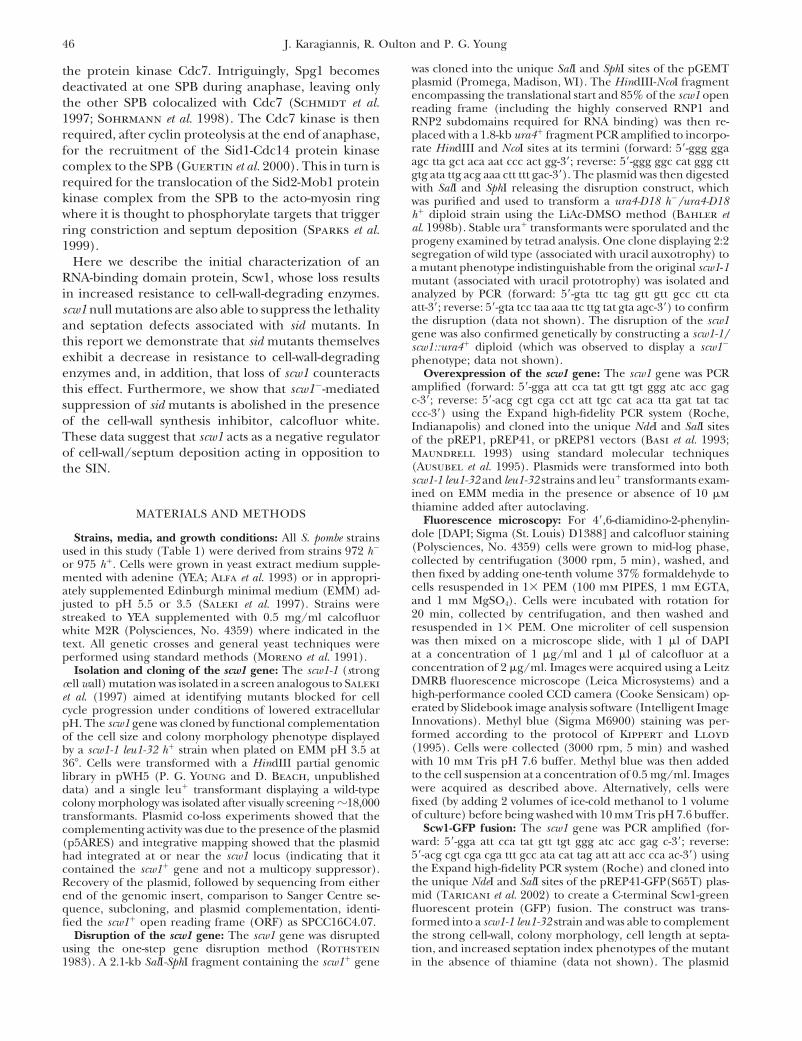
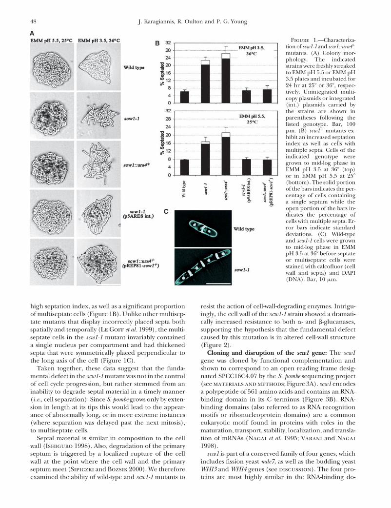
Figure 1.—Characteriza-tion of scw1-1 and scw1::ura4�
mutants. (A) Colony mor-phology. The indicatedstrains were freshly streakedto EMM pH 5.5 or EMM pH3.5 plates and incubated for24 hr at 25� or 36�, respec-tively. Unintegrated multi-copy plasmids or integrated(int.) plasmids carried bythe strains are shown inparentheses following thelisted genotype. Bar, 100�m. (B) scw1� mutants ex-hibit an increased septationindex as well as cells withmultiple septa. Cells of theindicated genotype weregrown to mid-log phase inEMM pH 3.5 at 36� (top)or in EMM pH 5.5 at 25�(bottom). The solid portionof the bars indicates the per-centage of cells containinga single septum while theopen portion of the bars in-dicates the percentage ofcells with multiple septa. Er-ror bars indicate standarddeviations. (C) Wild-typeand scw1-1 cells were grownto mid-log phase in EMMpH 3.5 at 36� before septateor multiseptate cells werestained with calcofluor (cellwall and septa) and DAPI(DNA). Bar, 10 �m.
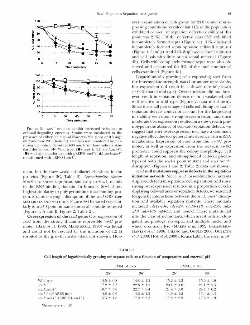
high septation index, as well as a significant proportion resist the action of cell-wall-degrading enzymes. Intrigu-ingly, the cell wall of the scw1-1 strain showed a dramati-of multiseptate cells (Figure 1B). Unlike other multisep-
tate mutants that display incorrectly placed septa both cally increased resistance to both �- and �-glucanases,supporting the hypothesis that the fundamental defectspatially and temporally (Le Goff et al. 1999), the multi-
septate cells in the scw1-1 mutant invariably contained caused by this mutation is in altered cell-wall structure(Figure 2).a single nucleus per compartment and had thickened
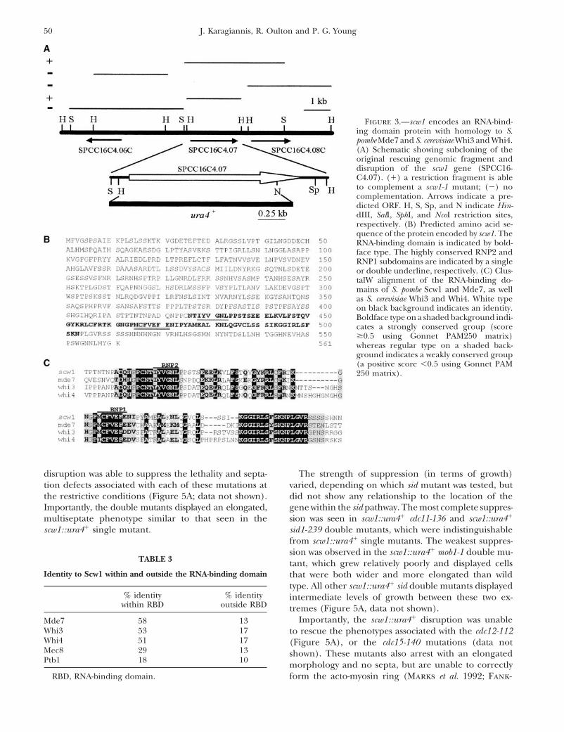
septa that were symmetrically placed perpendicular to Cloning and disruption of the scw1 gene: The scw1gene was cloned by functional complementation andthe long axis of the cell (Figure 1C).
Taken together, these data suggest that the funda- shown to correspond to an open reading frame desig-nated SPCC16C4.07 by the S. pombe sequencing projectmental defect in the scw1-1 mutant was not in the control
of cell cycle progression, but rather stemmed from an (see materials and methods; Figure 3A). scw1 encodesa polypeptide of 561 amino acids and contains an RNA-inability to degrade septal material in a timely manner
(i.e., cell separation). Since S. pombe grows only by exten- binding domain in its C terminus (Figure 3B). RNA-binding domains (also referred to as RNA recognitionsion in length at its tips this would lead to the appear-
ance of abnormally long, or in more extreme instances motifs or ribonucleoprotein domains) are a commoneukaryotic motif found in proteins with roles in the(where separation was delayed past the next mitosis),
to multiseptate cells. maturation, transport, stability, localization, and transla-tion of mRNAs (Nagai et al. 1995; Varani and NagaiSeptal material is similar in composition to the cell
wall (Ishiguro 1998). Also, degradation of the primary 1998).scw1 is part of a conserved family of four genes, whichseptum is triggered by a localized rupture of the cell
wall at the point where the cell wall and the primary includes fission yeast mde7, as well as the budding yeastWHI3 and WHI4 genes (see discussion). The four pro-septum meet (Sipiczki and Bozsik 2000). We therefore
examined the ability of wild-type and scw1-1 mutants to teins are most highly similar in the RNA-binding do-
49Scw1 Regulates Septation in S. pombe
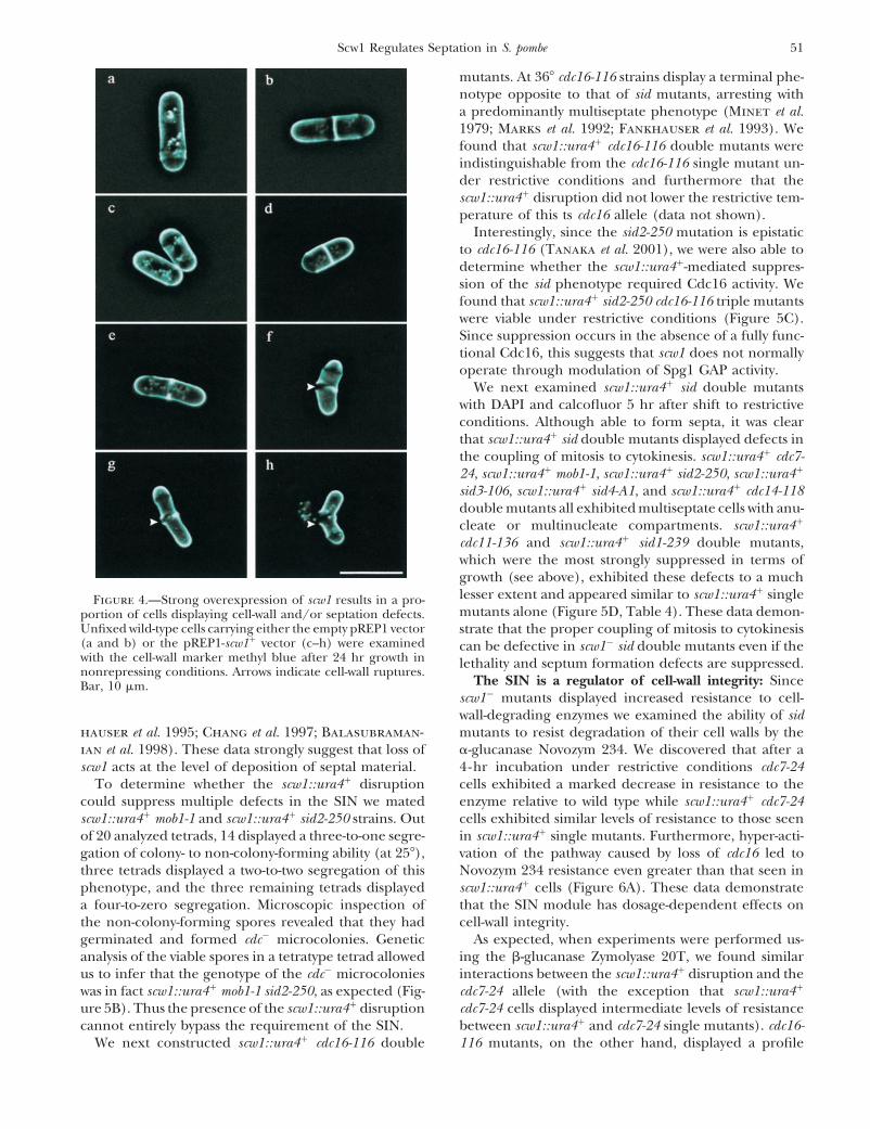
ever, examination of cells grown for 24 hr under nonre-pressing conditions revealed that 11% of the populationexhibited cell-wall or septation defects (viability at thispoint was 21%). Of the defective class 28% exhibitedincompletely formed septa (Figure 4e), 41% displayedincompletely formed septa opposite cell-wall ruptures(Figure 4, f and g), and 31% displayed cell-wall rupturesand cell lysis with little or no septal material (Figure4h). Cells with completely formed septa were also ob-served and accounted for 5% of the total number ofcells examined (Figure 4d).
Logarithmically growing cells expressing scw1 fromthe intermediate strength nmt41 promoter were viable,but expression did result in a slower rate of growth(�60% that of wild type). Overexpression did not, how-ever, result in septation defects or in a weakened cellwall relative to wild type (Figure 2; data not shown).Since the small percentage of cells exhibiting cell-wall/septation defects could not account for the large dropin viability seen upon strong overexpression, and sincemoderate overexpression resulted in a slow-growth phe-notype in the absence of cell-wall/septation defects, we
Figure 2.—scw1� mutants exhibit increased resistance tosuggest that scw1 overexpression may have a dominantcell-wall-degrading enzymes. Strains were incubated in thenegative effect due to a general interference with mRNApresence of either 0.5 mg/ml Novozym 234 (top) or 0.5 mg/
ml Zymolyase 20T (bottom). Cell lysis was monitored by mea- metabolism. Expression of scw1 from the nmt41 pro-suring the optical density at 600 nm. Error bars indicate stan- moter, as well as expression from the weakest nmt81dard deviations. (�) Wild type; (�) scw1-1; (�) scw1::ura4�; promoter, could suppress the colony morphology, cell(�) wild type transformed with pREP41-scw1�; (�) scw1::ura4�
length at septation, and strengthened cell-wall pheno-transformed with pREP81-scw1�.types of both the scw1-1 point mutant and scw1::ura4�
disruption (Figures 1 and 2; Table 2; data not shown).scw1 null mutations suppress defects in the septationmain, but do show weaker similarity elsewhere in the
initiation network: Since scw1 loss-of-function mutantsproteins (Figure 3C, Table 3). Caenorhabditis elegansdisplayed defects in septation/cell separation, and sinceMec8 also shows significant similarity to Scw1, mainlystrong overexpression resulted in a proportion of cellsin the RNA-binding domain. In humans, Scw1 showsdisplaying cell-wall and/or septation defects, we searchedhighest similarity to poly-pyrimidine tract binding pro-for genetic interactions between the scw1::ura4� disrup-tein. Strains carrying a disruption of the scw1 ORF (seetion and available septation mutants. These mutantsmaterials and methods; Figure 3A) behaved very simi-included cdc11-136, cdc7-24, cdc14-118, sid1-239, sid2-larly to scw1-1 point mutants under all conditions tested250, sid3-106, sid4-A1, and mob1-1. These mutants fall(Figure 1, A and B; Figure 2; Table 2).into the class of sid mutants, which arrest with an elon-Overexpression of the scw1 gene: Overexpression ofgated morphology, no septa, and multiple nuclei andscw1 from the strong thiamine repressible nmt1 pro-which eventually lyse (Marks et al. 1992; Balasubra-moter (Basi et al. 1993; Maundrell 1993) was lethalmanian et al. 1998; Chang and Gould 2000; Guertinand could not be rescued by the inclusion of 1.2 m
sorbitol to the growth media (data not shown). How- et al. 2000; Hou et al. 2000). Remarkably, the scw1::ura4�
TABLE 2
Cell length of logarithmically growing uni-septate cells as a function of temperature and external pH
EMM pH 5.5 EMM pH 3.5
25� 36� 25� 36�
Wild type 14.5 0.8 14.8 1.2 15.2 1.3 15.0 1.0scw1-1 17.2 3.3 22.8 4.5 20.1 4.6 24.1 5.5scw1::ura4 � 18.7 3.0 22.7 5.4 21.4 5.0 24.7 6.2scw1-1 (p5ARES int.) 14.8 0.9 14.6 1.2 14.9 1.3 15.4 1.4scw1::ura4 � (pREP81-scw1 �) 15.5 1.8 17.9 2.3 17.8 2.9 17.6 1.9
Micrometers SD.
50 J. Karagiannis, R. Oulton and P. G. Young
Figure 3.—scw1 encodes an RNA-bind-ing domain protein with homology to S.pombe Mde7 and S. cerevisiae Whi3 and Whi4.(A) Schematic showing subcloning of theoriginal rescuing genomic fragment anddisruption of the scw1 gene (SPCC16-C4.07). (�) a restriction fragment is ableto complement a scw1-1 mutant; (�) nocomplementation. Arrows indicate a pre-dicted ORF. H, S, Sp, and N indicate Hin-dIII, SalI, SphI, and NcoI restriction sites,respectively. (B) Predicted amino acid se-quence of the protein encoded by scw1. TheRNA-binding domain is indicated by bold-face type. The highly conserved RNP2 andRNP1 subdomains are indicated by a singleor double underline, respectively. (C) Clus-talW alignment of the RNA-binding do-mains of S. pombe Scw1 and Mde7, as wellas S. cerevisiae Whi3 and Whi4. White typeon black background indicates an identity.Boldface type on a shaded background indi-cates a strongly conserved group (score0.5 using Gonnet PAM250 matrix)whereas regular type on a shaded back-ground indicates a weakly conserved group(a positive score �0.5 using Gonnet PAM250 matrix).
disruption was able to suppress the lethality and septa- The strength of suppression (in terms of growth)varied, depending on which sid mutant was tested, buttion defects associated with each of these mutations at
the restrictive conditions (Figure 5A; data not shown). did not show any relationship to the location of thegene within the sid pathway. The most complete suppres-Importantly, the double mutants displayed an elongated,
multiseptate phenotype similar to that seen in the sion was seen in scw1::ura4� cdc11-136 and scw1::ura4�
sid1-239 double mutants, which were indistinguishablescw1::ura4� single mutant.from scw1::ura4� single mutants. The weakest suppres-sion was observed in the scw1::ura4� mob1-1 double mu-
TABLE 3 tant, which grew relatively poorly and displayed cellsIdentity to Scw1 within and outside the RNA-binding domain that were both wider and more elongated than wild
type. All other scw1::ura4� sid double mutants displayed% identity % identity intermediate levels of growth between these two ex-
within RBD outside RBD tremes (Figure 5A, data not shown).Importantly, the scw1::ura4� disruption was unableMde7 58 13
Whi3 53 17 to rescue the phenotypes associated with the cdc12-112Whi4 51 17 (Figure 5A), or the cdc15-140 mutations (data notMec8 29 13 shown). These mutants also arrest with an elongatedPtb1 18 10 morphology and no septa, but are unable to correctly
RBD, RNA-binding domain. form the acto-myosin ring (Marks et al. 1992; Fank-
51Scw1 Regulates Septation in S. pombe
mutants. At 36� cdc16-116 strains display a terminal phe-notype opposite to that of sid mutants, arresting witha predominantly multiseptate phenotype (Minet et al.1979; Marks et al. 1992; Fankhauser et al. 1993). Wefound that scw1::ura4� cdc16-116 double mutants wereindistinguishable from the cdc16-116 single mutant un-der restrictive conditions and furthermore that thescw1::ura4� disruption did not lower the restrictive tem-perature of this ts cdc16 allele (data not shown).
Interestingly, since the sid2-250 mutation is epistaticto cdc16-116 (Tanaka et al. 2001), we were also able todetermine whether the scw1::ura4�-mediated suppres-sion of the sid phenotype required Cdc16 activity. Wefound that scw1::ura4� sid2-250 cdc16-116 triple mutantswere viable under restrictive conditions (Figure 5C).Since suppression occurs in the absence of a fully func-tional Cdc16, this suggests that scw1 does not normallyoperate through modulation of Spg1 GAP activity.
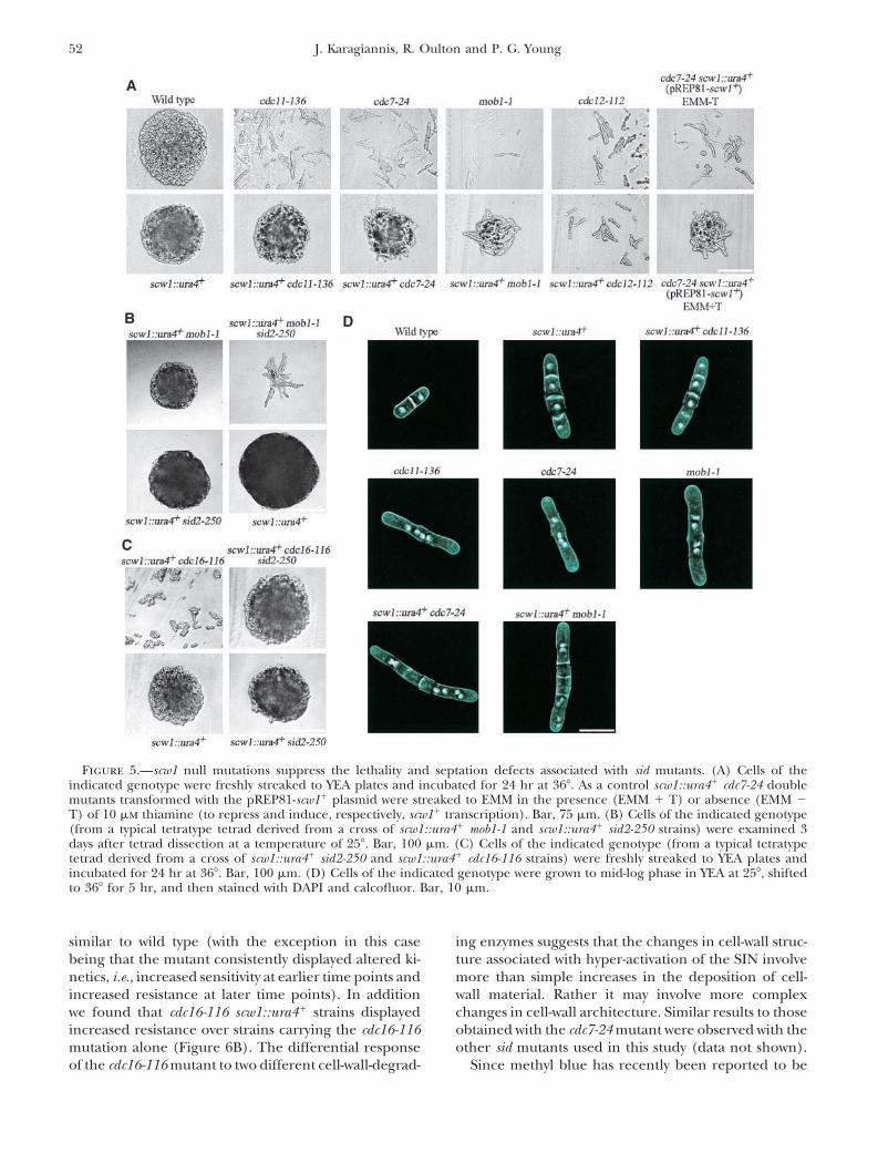
We next examined scw1::ura4� sid double mutantswith DAPI and calcofluor 5 hr after shift to restrictiveconditions. Although able to form septa, it was clearthat scw1::ura4� sid double mutants displayed defects inthe coupling of mitosis to cytokinesis. scw1::ura4� cdc7-24, scw1::ura4� mob1-1, scw1::ura4� sid2-250, scw1::ura4�
sid3-106, scw1::ura4� sid4-A1, and scw1::ura4� cdc14-118double mutants all exhibited multiseptate cells with anu-cleate or multinucleate compartments. scw1::ura4�
cdc11-136 and scw1::ura4� sid1-239 double mutants,which were the most strongly suppressed in terms ofgrowth (see above), exhibited these defects to a muchlesser extent and appeared similar to scw1::ura4� singleFigure 4.—Strong overexpression of scw1 results in a pro-mutants alone (Figure 5D, Table 4). These data demon-portion of cells displaying cell-wall and/or septation defects.
Unfixed wild-type cells carrying either the empty pREP1 vector strate that the proper coupling of mitosis to cytokinesis(a and b) or the pREP1-scw1� vector (c–h) were examined can be defective in scw1� sid double mutants even if thewith the cell-wall marker methyl blue after 24 hr growth in lethality and septum formation defects are suppressed.nonrepressing conditions. Arrows indicate cell-wall ruptures.
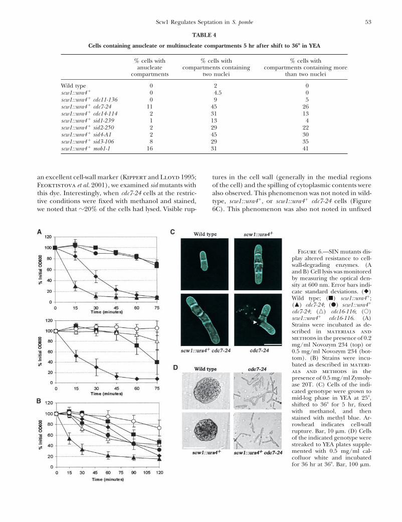
The SIN is a regulator of cell-wall integrity: SinceBar, 10 �m.scw1� mutants displayed increased resistance to cell-wall-degrading enzymes we examined the ability of sidmutants to resist degradation of their cell walls by thehauser et al. 1995; Chang et al. 1997; Balasubraman-
ian et al. 1998). These data strongly suggest that loss of �-glucanase Novozym 234. We discovered that after a4-hr incubation under restrictive conditions cdc7-24scw1 acts at the level of deposition of septal material.
To determine whether the scw1::ura4� disruption cells exhibited a marked decrease in resistance to theenzyme relative to wild type while scw1::ura4� cdc7-24could suppress multiple defects in the SIN we mated
scw1::ura4� mob1-1 and scw1::ura4� sid2-250 strains. Out cells exhibited similar levels of resistance to those seenin scw1::ura4� single mutants. Furthermore, hyper-acti-of 20 analyzed tetrads, 14 displayed a three-to-one segre-
gation of colony- to non-colony-forming ability (at 25�), vation of the pathway caused by loss of cdc16 led toNovozym 234 resistance even greater than that seen inthree tetrads displayed a two-to-two segregation of this
phenotype, and the three remaining tetrads displayed scw1::ura4� cells (Figure 6A). These data demonstratethat the SIN module has dosage-dependent effects ona four-to-zero segregation. Microscopic inspection of
the non-colony-forming spores revealed that they had cell-wall integrity.As expected, when experiments were performed us-germinated and formed cdc� microcolonies. Genetic
analysis of the viable spores in a tetratype tetrad allowed ing the �-glucanase Zymolyase 20T, we found similarinteractions between the scw1::ura4� disruption and theus to infer that the genotype of the cdc� microcolonies
was in fact scw1::ura4� mob1-1 sid2-250, as expected (Fig- cdc7-24 allele (with the exception that scw1::ura4�
cdc7-24 cells displayed intermediate levels of resistanceure 5B). Thus the presence of the scw1::ura4� disruptioncannot entirely bypass the requirement of the SIN. between scw1::ura4� and cdc7-24 single mutants). cdc16-
116 mutants, on the other hand, displayed a profileWe next constructed scw1::ura4� cdc16-116 double
52 J. Karagiannis, R. Oulton and P. G. Young
Figure 5.—scw1 null mutations suppress the lethality and septation defects associated with sid mutants. (A) Cells of theindicated genotype were freshly streaked to YEA plates and incubated for 24 hr at 36�. As a control scw1::ura4� cdc7-24 doublemutants transformed with the pREP81-scw1� plasmid were streaked to EMM in the presence (EMM � T) or absence (EMM �T) of 10 �m thiamine (to repress and induce, respectively, scw1� transcription). Bar, 75 �m. (B) Cells of the indicated genotype(from a typical tetratype tetrad derived from a cross of scw1::ura4� mob1-1 and scw1::ura4� sid2-250 strains) were examined 3days after tetrad dissection at a temperature of 25�. Bar, 100 �m. (C) Cells of the indicated genotype (from a typical tetratypetetrad derived from a cross of scw1::ura4� sid2-250 and scw1::ura4� cdc16-116 strains) were freshly streaked to YEA plates andincubated for 24 hr at 36�. Bar, 100 �m. (D) Cells of the indicated genotype were grown to mid-log phase in YEA at 25�, shiftedto 36� for 5 hr, and then stained with DAPI and calcofluor. Bar, 10 �m.
similar to wild type (with the exception in this case ing enzymes suggests that the changes in cell-wall struc-ture associated with hyper-activation of the SIN involvebeing that the mutant consistently displayed altered ki-
netics, i.e., increased sensitivity at earlier time points and more than simple increases in the deposition of cell-wall material. Rather it may involve more complexincreased resistance at later time points). In addition
we found that cdc16-116 scw1::ura4� strains displayed changes in cell-wall architecture. Similar results to thoseobtained with the cdc7-24 mutant were observed with theincreased resistance over strains carrying the cdc16-116
mutation alone (Figure 6B). The differential response other sid mutants used in this study (data not shown).Since methyl blue has recently been reported to beof the cdc16-116 mutant to two different cell-wall-degrad-
53Scw1 Regulates Septation in S. pombe
TABLE 4
Cells containing anucleate or multinucleate compartments 5 hr after shift to 36� in YEA
% cells with % cells with % cells withanucleate compartments containing compartments containing more
compartments two nuclei than two nuclei
Wild type 0 2 0scw1::ura4 � 0 4.5 0scw1::ura4 � cdc11-136 0 9 5scw1::ura4 � cdc7-24 11 45 26scw1::ura4 � cdc14-114 2 31 13scw1::ura4 � sid1-239 1 13 4scw1::ura4 � sid2-250 2 29 22scw1::ura4 � sid4-A1 2 45 30scw1::ura4 � sid3-106 8 29 35scw1::ura4 � mob1-1 16 31 41
an excellent cell-wall marker (Kippert and Lloyd 1995; tures in the cell wall (generally in the medial regionsof the cell) and the spilling of cytoplasmic contents wereFeoktistova et al. 2001), we examined sid mutants with
this dye. Interestingly, when cdc7-24 cells at the restric- also observed. This phenomenon was not noted in wild-type, scw1::ura4�, or scw1::ura4� cdc7-24 cells (Figuretive conditions were fixed with methanol and stained,
we noted that �20% of the cells had lysed. Visible rup- 6C). This phenomenon was also not noted in unfixed
Figure 6.—SIN mutants dis-play altered resistance to cell-wall-degrading enzymes. (Aand B) Cell lysis was monitoredby measuring the optical den-sity at 600 nm. Error bars indi-cate standard deviations. (�)Wild type; (�) scw1::ura4�;(�) cdc7-24; (�) scw1::ura4�
cdc7-24; (�) cdc16-116; (�)scw1::ura4� cdc16-116. (A)Strains were incubated as de-scribed in materials andmethods in the presence of 0.2mg/ml Novozym 234 (top) or0.5 mg/ml Novozym 234 (bot-tom). (B) Strains were incu-bated as described in materi-als and methods in thepresence of 0.5 mg/ml Zymoly-ase 20T. (C) Cells of the indi-cated genotype were grown tomid-log phase in YEA at 25�,shifted to 36� for 5 hr, fixedwith methanol, and thenstained with methyl blue. Ar-rowhead indicates cell-wallrupture. Bar, 10 �m. (D) Cellsof the indicated genotype werestreaked to YEA plates supple-mented with 0.5 mg/ml cal-cofluor white and incubatedfor 36 hr at 36�. Bar, 100 �m.
54 J. Karagiannis, R. Oulton and P. G. Young
cdc7-24 cells or in cdc7-24 cells fixed with formaldehyde Zymolyase 20T we noted that exponentially growing andG1-arrested cells displayed decreased resistance, but that(data not shown). Therefore, although the phenome-
non is artifactual, most likely due to the harsher nature G2 arrest partially abrogated this effect (Figure 7). Thesedata show that the effects of SIN hyper-activation onof methanol fixation, it does reveal an inherent weak-
ness in the cell wall of cdc7-24 cells. Similar results were cell-wall integrity are modulated by cell cycle position.Zymolyase 20T resistance assays were somewhat sur-obtained with the other sid mutants used in this study
(data not shown). Otherwise, results obtained using prising since cdc7 loss-of-function mutants displayed asimilar resistance to that demonstrated by cells overex-methyl blue were similar to those obtained using the
more widely used cell-wall marker, calcofluor white pressing cdc7. We interpret these results to indicate thatgross disturbances of Cdc7 function (either by loss or(data not shown).
Taking all data together, our results suggested that by strong overexpression) cause drastic changes in cell-wall structure, which manifest as a decrease in Zymolyasethe stronger cell wall conferred by the scw1::ura4� dis-
ruption played a key role in the observed suppression 20T resistance. This is to say that although the loss andoverexpression of Cdc7 have opposing effects, the netof sid mutations. If this were true, then one would expect
conditions that act against cell-wall integrity to nega- result is a poorly structured cell wall vulnerable to attackby Zymolyase 20T.tively affect this rescue. We thus examined the growth of
wild-type, scw1::ura4�, cdc7-24, and scw1::ura4� cdc7-24 Subcellular localization of Scw1: Since members ofthe SIN localize to the spindle pole body we created acells at 36� in the presence of 0.5 mg/ml calcofluor
white. This compound (in addition to acting as a cell- C-terminal Scw1-GFP fusion to determine its subcellularlocalization (see materials and methods). Unlikewall marker) inhibits the formation of glucan and chitin
microfibrils and thus interferes with cell-wall synthesis members of the SIN, Scw1 was predominantly a cyto-plasmic protein (Figure 8). Localization was not altered(Haigler et al. 1980; Roncero et al. 1988; Ram et al.
1994; Lussier et al. 1997). Although growth in media as a function of cell cycle position by changes in temper-ature or by external pH conditions (data not shown).containing calcofluor did not affect the multiseptate
phenotype of the scw1::ura4� mutant (data not shown),scw1::ura4� cdc7-24 double mutants did behave as cdc7-24
DISCUSSIONcells and displayed a lethal cdc� phenotype (Figure 6D).Similar interactions were observed among the scw1::ura4� The cell wall of S. pombe is composed mainly of poly-
mers of 1,3-�-glucan, 1,3-�-glucan, and �-galactomannan.disruption and other sid mutants in media containingcalcofluor (data not shown). These data strongly suggest It serves a wide variety of functions including protection
from environmental stresses, cell adhesion during conju-that the scw1::ura4�-mediated rescue of sid mutants isachieved through altered cell-wall synthesis and/or gation and mating, and the maintenance of cellular mor-
phology (Ishiguro 1998). Many different proteins, actingstructure.SIN-mediated effects on cell-wall integrity are influ- as regulators of a wide variety of pathways, have been
shown to affect cell-wall structure. These include the rho1enced by cell cycle position: The effects of cdc7 overex-pression on septation are reduced in cells arrested at and rho2 GTP-binding proteins, the pmk1/spm1 mitogen-
activated protein kinase, the pab1 protein phosphatasethe G2/M transition (Fankhauser and Simanis 1994).Furthermore, following mitotic entry, translocation of 2A regulatory B subunit, and the pck2 protein kinase C
homolog, among many others (Ishiguro 1998).the Sid1/Cdc14 complex to the SPB is promoted by theinactivation of Cdc2 through cyclin proteolysis (Guer- In this report we show that loss of the scw1 RNA-
binding domain protein also affects the cell wall as evi-tin et al. 2000). If activation of the SIN affects cell-wallstructure, then changes in cell-wall integrity mediated denced by increased resistance to both �- and �-gluca-
nases. The mechanism by which loss of scw1 causes theseby cdc7 overexpression should be influenced by cell cycleposition. We thus examined the effects of increased differences is unknown, but on the basis of scw1 encod-
ing an RNA-binding domain protein, the differencesCdc7 levels on cell-wall integrity in populations arrestedat different points of the cell cycle. Wild type, as well most likely result from an indirect means related to the
regulation of mRNAs. RNA-binding domain proteinsas cdc10-129 and cdc2-33 mutants (which arrest predomi-nantly at the G1/S and G2/M transitions, respectively, have been shown to affect many distinct cellular path-
ways through their effects on mRNA maturation, trans-at 36�), were transformed with the pREP3-cdc7 plasmid(which places cdc7 under control of the nmt1 promoter). port, stability, localization, and translation (see Nagai
et al. 1995 and Varani and Nagai 1998 for reviews).Consistent with our previous results, hyper-activationof the SIN in exponentially growing cells resulted in The scw1 RNA-binding domain protein isolated in
this study is part of a family of four genes that includeincreased resistance to Novozym 234. A similar effect wasseen in cells arrested at the G1/S transition. Intriguingly, S. pombe mde7, as well as S. cerevisiae WHI3 and WHI4.
mde7 has not been characterized in detail, but is knownhowever, the increased resistance was partially abro-gated in cells arrested predominantly in G2 (Figure 7). to be one of nine meiosis-dependent transcripts under
the control of the Mei4 forkhead transcription factorWhen analogous experiments were performed using
55Scw1 Regulates Septation in S. pombe
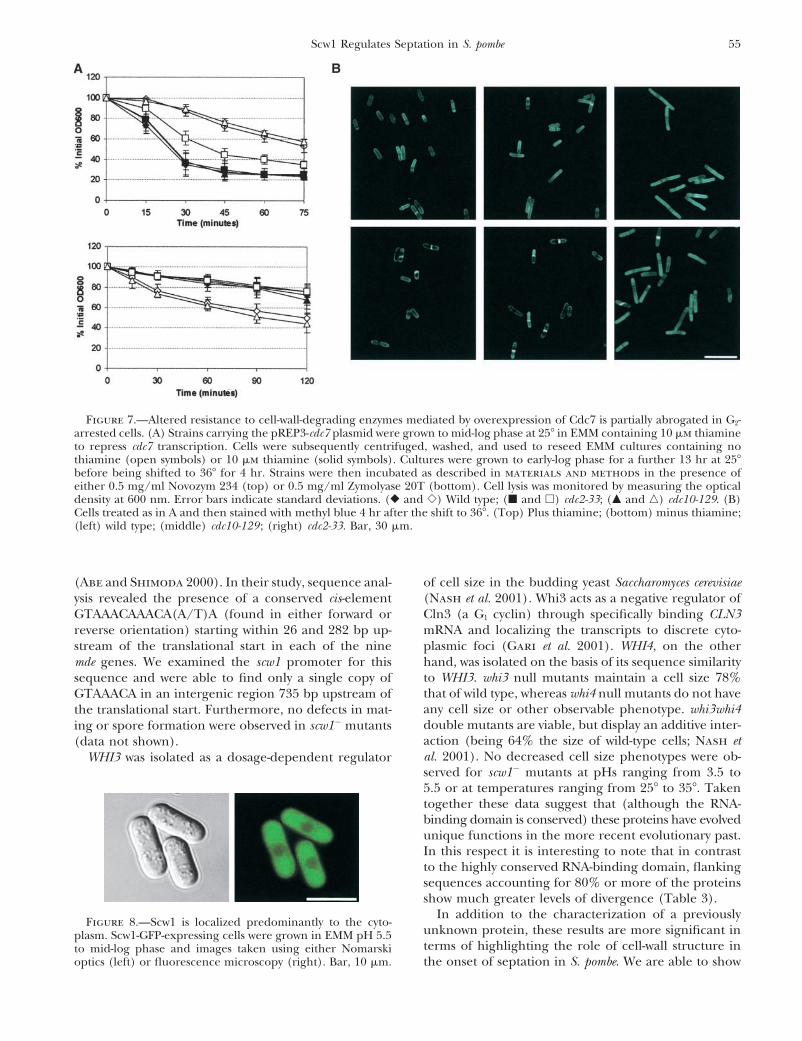
Figure 7.—Altered resistance to cell-wall-degrading enzymes mediated by overexpression of Cdc7 is partially abrogated in G2-arrested cells. (A) Strains carrying the pREP3-cdc7 plasmid were grown to mid-log phase at 25� in EMM containing 10 �m thiamineto repress cdc7 transcription. Cells were subsequently centrifuged, washed, and used to reseed EMM cultures containing nothiamine (open symbols) or 10 �m thiamine (solid symbols). Cultures were grown to early-log phase for a further 13 hr at 25�before being shifted to 36� for 4 hr. Strains were then incubated as described in materials and methods in the presence ofeither 0.5 mg/ml Novozym 234 (top) or 0.5 mg/ml Zymolyase 20T (bottom). Cell lysis was monitored by measuring the opticaldensity at 600 nm. Error bars indicate standard deviations. (� and �) Wild type; (� and �) cdc2-33; (� and �) cdc10-129. (B)Cells treated as in A and then stained with methyl blue 4 hr after the shift to 36�. (Top) Plus thiamine; (bottom) minus thiamine;(left) wild type; (middle) cdc10-129 ; (right) cdc2-33. Bar, 30 �m.
(Abe and Shimoda 2000). In their study, sequence anal- of cell size in the budding yeast Saccharomyces cerevisiae(Nash et al. 2001). Whi3 acts as a negative regulator ofysis revealed the presence of a conserved cis-element
GTAAACAAACA(A/T)A (found in either forward or Cln3 (a G1 cyclin) through specifically binding CLN3mRNA and localizing the transcripts to discrete cyto-reverse orientation) starting within 26 and 282 bp up-
stream of the translational start in each of the nine plasmic foci (Gari et al. 2001). WHI4, on the otherhand, was isolated on the basis of its sequence similaritymde genes. We examined the scw1 promoter for thisto WHI3. whi3 null mutants maintain a cell size 78%sequence and were able to find only a single copy ofthat of wild type, whereas whi4 null mutants do not haveGTAAACA in an intergenic region 735 bp upstream ofany cell size or other observable phenotype. whi3whi4the translational start. Furthermore, no defects in mat-double mutants are viable, but display an additive inter-ing or spore formation were observed in scw1� mutantsaction (being 64% the size of wild-type cells; Nash et(data not shown).al. 2001). No decreased cell size phenotypes were ob-WHI3 was isolated as a dosage-dependent regulatorserved for scw1� mutants at pHs ranging from 3.5 to5.5 or at temperatures ranging from 25� to 35�. Takentogether these data suggest that (although the RNA-binding domain is conserved) these proteins have evolvedunique functions in the more recent evolutionary past.In this respect it is interesting to note that in contrastto the highly conserved RNA-binding domain, flankingsequences accounting for 80% or more of the proteinsshow much greater levels of divergence (Table 3).
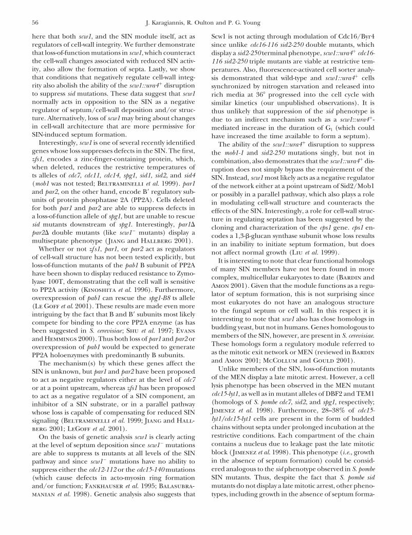
In addition to the characterization of a previouslyFigure 8.—Scw1 is localized predominantly to the cyto-
unknown protein, these results are more significant inplasm. Scw1-GFP-expressing cells were grown in EMM pH 5.5terms of highlighting the role of cell-wall structure into mid-log phase and images taken using either Nomarski
optics (left) or fluorescence microscopy (right). Bar, 10 �m. the onset of septation in S. pombe. We are able to show
56 J. Karagiannis, R. Oulton and P. G. Young
here that both scw1, and the SIN module itself, act as Scw1 is not acting through modulation of Cdc16/Byr4regulators of cell-wall integrity. We further demonstrate since unlike cdc16-116 sid2-250 double mutants, whichthat loss-of-function mutations in scw1, which counteract display a sid2-250 terminal phenotype, scw1::ura4� cdc16-the cell-wall changes associated with reduced SIN activ- 116 sid2-250 triple mutants are viable at restrictive tem-ity, also allow the formation of septa. Lastly, we show peratures. Also, fluorescence-activated cell sorter analy-that conditions that negatively regulate cell-wall integ- sis demonstrated that wild-type and scw1::ura4� cellsrity also abolish the ability of the scw1::ura4� disruption synchronized by nitrogen starvation and released intoto suppress sid mutations. These data suggest that scw1 rich media at 36� progressed into the cell cycle withnormally acts in opposition to the SIN as a negative similar kinetics (our unpublished observations). It isregulator of septum/cell-wall deposition and/or struc- thus unlikely that suppression of the sid phenotype isture. Alternatively, loss of scw1 may bring about changes due to an indirect mechanism such as a scw1::ura4�-in cell-wall architecture that are more permissive for mediated increase in the duration of G1 (which couldSIN-induced septum formation. have increased the time available to form a septum).
Interestingly, scw1 is one of several recently identified The ability of the scw1::ura4� disruption to suppressgenes whose loss suppresses defects in the SIN. The first, the mob1-1 and sid2-250 mutations singly, but not inzfs1, encodes a zinc-finger-containing protein, which, combination, also demonstrates that the scw1::ura4� dis-when deleted, reduces the restrictive temperatures of ruption does not simply bypass the requirement of thets alleles of cdc7, cdc11, cdc14, spg1, sid1, sid2, and sid4 SIN. Instead, scw1 most likely acts as a negative regulator(mob1 was not tested; Beltraminelli et al. 1999). par1 of the network either at a point upstream of Sid2/Mob1and par2, on the other hand, encode B� regulatory sub- or possibly in a parallel pathway, which also plays a roleunits of protein phosphatase 2A (PP2A). Cells deleted in modulating cell-wall structure and counteracts thefor both par1 and par2 are able to suppress defects in effects of the SIN. Interestingly, a role for cell-wall struc-a loss-of-function allele of spg1, but are unable to rescue ture in regulating septation has been suggested by thesid mutants downstream of spg1. Interestingly, par1� cloning and characterization of the cps1 gene. cps1 en-par2� double mutants (like scw1� mutants) display a codes a 1,3-�-glucan synthase subunit whose loss resultsmultiseptate phenotype (Jiang and Hallberg 2001). in an inability to initiate septum formation, but does
Whether or not zf s1, par1, or par2 act as regulators not affect normal growth (Liu et al. 1999).of cell-wall structure has not been tested explicitly, but It is interesting to note that clear functional homologsloss-of-function mutants of the pab1 B subunit of PP2A of many SIN members have not been found in morehave been shown to display reduced resistance to Zymo- complex, multicellular eukaryotes to date (Bardin andlyase 100T, demonstrating that the cell wall is sensitive
Amon 2001). Given that the module functions as a regu-to PP2A activity (Kinoshita et al. 1996). Furthermore,
lator of septum formation, this is not surprising sinceoverexpression of pab1 can rescue the spg1-B8 ts allelemost eukaryotes do not have an analogous structure(Le Goff et al. 2001). These results are made even moreto the fungal septum or cell wall. In this respect it isintriguing by the fact that B and B� subunits most likelyinteresting to note that scw1 also has close homologs incompete for binding to the core PP2A enzyme (as hasbudding yeast, but not in humans. Genes homologous tobeen suggested in S. cerevisiae ; Shu et al. 1997; Evansmembers of the SIN, however, are present in S. cerevisiae.and Hemmings 2000). Thus both loss of par1 and par2 orThese homologs form a regulatory module referred tooverexpression of pab1 would be expected to generateas the mitotic exit network or MEN (reviewed in BardinPP2A holoenzymes with predominantly B subunits.and Amon 2001; McCollum and Gould 2001).The mechanism(s) by which these genes affect the
Unlike members of the SIN, loss-of-function mutantsSIN is unknown, but par1 and par2 have been proposedof the MEN display a late mitotic arrest. However, a cellto act as negative regulators either at the level of cdc7lysis phenotype has been observed in the MEN mutantor at a point upstream, whereas zfs1 has been proposedcdc15-lyt1, as well as in mutant alleles of DBF2 and TEM1to act as a negative regulator of a SIN component, an(homologs of S. pombe cdc7, sid2, and spg1, respectively;inhibitor of a SIN substrate, or in a parallel pathwayJimenez et al. 1998). Furthermore, 28–38% of cdc15-whose loss is capable of compensating for reduced SINlyt1/cdc15-lyt1 cells are present in the form of buddedsignaling (Beltraminelli et al. 1999; Jiang and Hall-chains without septa under prolonged incubation at theberg 2001; LeGoff et al. 2001).restrictive conditions. Each compartment of the chainOn the basis of genetic analysis scw1 is clearly actingcontains a nucleus due to leakage past the late mitoticat the level of septum deposition since scw1� mutationsblock (Jimenez et al. 1998). This phenotype (i.e., growthare able to suppress ts mutants at all levels of the SINin the absence of septum formation) could be consid-pathway and since scw1� mutations have no ability toered analogous to the sid phenotype observed in S. pombesuppress either the cdc12-112 or the cdc15-140 mutationsSIN mutants. Thus, despite the fact that S. pombe sid(which cause defects in acto-myosin ring formationmutants do not display a late mitotic arrest, other pheno-and/or function; Fankhauser et al. 1995; Balasubra-
manian et al. 1998). Genetic analysis also suggests that types, including growth in the absence of septum forma-
57Scw1 Regulates Septation in S. pombe
Rng2p, a protein required for cytokinesis in fission yeast, is ation and cell-wall integrity defects, are applicable to atcomponent of the actomyosin ring and the spindle pole body.
least some MEN mutants. Curr. Biol. 8: 611–621.Evans, D. R., and B. A. Hemmings, 2000 Mutation of the C-terminalWe thank D. McCollum, M. K. Balasubramanian, and V. Simanis
leucine residue of PP2Ac inhibits PR55/B subunit binding andfor strains and/or plasmids, as well as L. Taricani for constructingconfers supersensitivity to microtubule destabilization in Saccharo-
the pREP41-GFP (S65T) plasmid. This research was supported by the myces cerevisiae. Mol. Gen. Genet. 264: 425–432.Natural Science and Engineering Council of Canada through grants Fankhauser, C., and V. Simanis, 1994 The cdc7 protein kinase isto P.G.Y. J.K. was supported by the Bauman Foundation and by an a dosage dependent regulator of septum formation in fissionOntario Graduate Scholarship. yeast. EMBO J. 13: 3011–3019.
Fankhauser, C., J. Marks, A. Reymond and V. Simanis, 1993 TheS. pombe cdc16 gene is required both for maintenance of p34cdc2
kinase activity and regulation of septum formation: A link be-tween mitosis and cytokinesis? EMBO J. 12: 2697–2704.LITERATURE CITED
Fankhauser, C., A. Reymond, L. Cerutti, S. Utzig, K. HofmannAbe, H., and C. Shimoda, 2000 Autoregulated expression of Schizo- et al., 1995 The S. pombe cdc15 gene is a key element in the
saccharomyces pombe meiosis-specific transcription factor Mei4 and reorganization of F-actin at mitosis. Cell 82: 435–444.a genome-wide search for its target genes. Genetics 154: 1497– Feoktistova, A., P. Magnelli, C. Abeijon, P. Perez, R. L. Lester1508. et al., 2001 Coordination between fission yeast glucan formation
Alfa, C., P. Fantes, J. Hyams, M. McLeod and E. Warbrick, 1993 and growth requires a sphingolipase activity. Genetics 158: 1397–Experiments with Fission Yeast: A Laboratory Course Manual. Cold 1411.Spring Harbor Laboratory Press, Cold Spring Harbor, NY. Furge, K. A., K. Wong, J. Armstrong, M. K. Balasubramanian and
Ausubel, F., R. Brent, R. E. Kingston, D. D. Moore, J. G. Seidman C. F. Albright, 1998 Byr4 and Cdc16 form a two-componentet al., 1995 Short Protocols in Molecular Biology, Ed. 3. John Wiley & GTPase-activating protein for the Spg1 GTPase that controls sep-Sons, New York. tation in fission yeast. Curr. Biol. 8: 947–954.
Bahler, J., and J. R. Pringle, 1998 Pom1p, a fission yeast protein Furge, K. A., Q. Cheng, M. Jwa, S. Shin, K. C. Song et al., 1999kinase that provides positional information for both polarised Regions of Byr4, a regulator of septation in fission yeast, thatgrowth and cytokinesis. Genes Dev. 12: 1356–1370. bind spg1 or cdc16 and form a two-component GTPase-activating
Bahler, J., A. B. Steever, S. Wheatley, Y. L. Wang, J. R. Pringle protein with cdc16. J. Biol. Chem. 274: 11339–11343.et al., 1998a Role of polo kinase and Mid1p in determining the Gari, E., T. Volpe, H. Wang, C. Gallego, B. Futcher et al., 2001site of cell division in fission yeast. J. Cell Biol. 143: 1603–1616. Whi3 binds the mRNA of the G1 cyclin CLN3 to modulate cell
Bahler, J., J. Wu, M. S. Longtine, N. G. Shah, A. McKenzie, III et fate in budding yeast. Genes Dev. 15: 2803–2808.al., 1998b Heterologous modules for efficient and versatile PCR- Gould, K. L., and V. Simanis, 1997 The control of septum formationbased gene targeting in Schizosaccharomyces pombe. Yeast 14: 943– in fission yeast. Genes Dev. 11: 2939–2951.951. Grallert, A., B. Grallert, E. Zilahi, Z. Szilagyi and M. Sipiczki,
Balasubramanian, M. K., D. M. Helfman and S. M. Hemmingsen, 1999 Eleven novel sep genes of Schizosaccharomyces pombe re-1992 A new tropomyosin essential for cytokinesis in the fission quired for efficient cell separation and sexual differentiation.yeast S. pombe. Nature 360: 84–87. Yeast 15: 669–686.
Balasubramanian, M. K., B. R. Hirani, J. D. Burke and K. L. Gould, Guertin, D. A., L. Chang, F. Irshad, K. L. Gould and D. McCollum,1994 The Schizosaccharomyces pombe cdc3 gene encodes a profilin 2000 The role of the sid1p kinase and cdc14p in regulating theessential for cytokinesis. J. Cell Biol. 125: 1289–1301. onset of cytokinesis in fission yeast. EMBO J. 19: 1803–1815.
Balasubramanian, M. K., D. McCollum, L. Chang, K. C. Wong, Haigler, G. H., R. M. Brown and M. Benziman, 1980 CalcofluorN. I. Naqvi et al., 1998 Isolation and characterization of new white ST alters the in vivo assembly of cellulose microfibrils.fission yeast cytokinesis mutants. Genetics 149: 1265–1275. Science 210: 903–906.
Balasubramanian, M. K., D. McCollum and U. Surana, 2000 Ty- Hou, M., J. Salek and D. McCollum, 2000 Mob1p interacts withing the knot: linking cytokinesis to the nuclear cycle. J. Cell Sci. the Sid2p kinase and is required for cytokinesis in fission yeast.113: 1503–1513. Curr. Biol. 10: 619–622.
Bardin, A. J., and A. Amon, 2001 MEN and SIN: What’s the differ- Ishiguro, J., 1998 Genetic control of fission yeast cell wall synthesis:ence? Nat. Rev. Mol. Cell Biol. 2: 815–826. the genes involved in wall biogenesis and their interactions in
Basi, G., E. Schmid and K. Maundrell, 1993 TATA box mutations Schizosaccharomyces pombe. Genes Genet. Syst. 73: 181–191.in the Schizosaccharomyces pombe nmt1 promoter affect transcription Jiang, W., and R. L. Hallberg, 2001 Correct regulation of thestart point or thiamine repressibility. Gene 123: 131–136. septation initiation network in Schizosaccharomyces pombe requires
Beltraminelli, N., M. Murone and V. Simanis, 1999 The S. pombe the activities of par1 and par2. Genetics 158: 1413–1429.zfs1 gene is required to prevent septation if mitotic progression Jimenez, J., V. J. Cid, R. Cenamor, M. Yuste, G. Molero et al.,is inhibited. J. Cell Sci. 112: 3103–3114. 1998 Morphogenesis beyond cytokinetic arrest in Saccharomyces
Bezanilla, M., S. L. Forsburg and T. D. Pollard, 1997 Identifica- cerevisiae. J. Cell Biol. 143: 1617–1634.tion of a second myosin II in Schizosaccharomyces pombe : Myp2p is Kinoshita, K., K. Nemoto, H. Nabeshima, H. Kondoh, H. Niwa etconditionally required for cytokinesis. Mol. Biol. Cell 8: 2693– al., 1996 The regulatory subunits of fission yeast protein phos-2705. phatase 2A affect cell morphogenesis, cell wall synthesis and
Chang, F., A. Woolard and P. Nurse, 1996 Isolation and character- cytokinesis. Genes Cells 1: 29–45.ization of fission yeast mutants defective in the assembly and Kippert, F., and D. Lloyd, 1995 The aniline blue fluorochromeplacement of the contractile actin ring. J. Cell Sci. 109: 131–142. specifically stains the septum of both live and fixed Schizosaccharo-
Chang, F., D. Drubin and P. Nurse, 1997 cdc12p, a protein re- myces pombe cells. FEMS Microbiol. Lett. 132: 215–219.quired for cytokinesis in fission yeast, is a component of the cell Kitayama, C., A. Sugimoto and M. Yamamoto, 1997 Type II myosindivision ring and interacts with profilin. J. Cell Biol. 137: 169–182. heavy chain encoded by the myo2 gene composes the contractile
Chang, L., and K. L. Gould, 2000 Sid4p is required to localize ring during cytokinesis in Schizosaccharomyces pombe. J. Cell Biol.components of the septation initiation pathway to the spindle 137: 1309–1319.pole body in fission yeast. Proc. Natl. Acad. Sci. USA 97: 5249– Krapp, A., S. Schmidt, E. Cano and V. Simanis, 2001 S. pombe5254. cdc11p, together with sid4p, provides an anchor for septation
Decottignies, A., P. Zarzov and P. Nurse, 2001 In vivo localization initiation network proteins on the spindle pole body. Curr. Biol.of fission yeast cyclin-dependent kinase cdc2p and cyclin B cdc13p 11: 1559–1568.during mitosis and meiosis. J. Cell Sci. 114: 2627–2640. Le Goff, X., S. Utzig and V. Simanis, 1999 Controlling septation
Edamatsu, M., and Y. Y. Toyoshima, 1996 Isolation and character- in fission yeast: finding the middle, and timing it right. Curr.ization of pos mutants defective in correct positioning of septum Genet. 35: 571–584.in Schizosaccharomyces pombe. Zool. Sci. 13: 235–239. Le Goff, X., S. Buvelot, E. Salimova, F. Guerry, S. Schmidt et al.,
2001 The protein phosphatase 2A B�-regulatory subunit par1pEng, K., N. I. Naqvi, K. C. Wong and M. K. Balasubramanian, 1998
58 J. Karagiannis, R. Oulton and P. G. Young
is implicated in regulation of the S. pombe septation initiation of the sep1 forkhead transcription factor homologue is not lethalbut causes hyphal growth in Schizosaccharomyces pombe. Biochem.network. FEBS Lett. 508: 136–142.
Levin, D. E., and M. Bishop, 1990 A putative protein kinase gene Biophys. Res. Commun. 263: 465–474.Roncero, C., M. H. Valdivieso, J. C. Ribas and A. Duran, 1988(kin1�) is important for growth polarity in Schizosaccharomyces
pombe. Proc. Natl. Acad. Sci. USA 87: 8272–8276. Isolation and characterization of Saccharomyces cerevisiae mutantsresistant to Calcofluor White. J. Bacteriol. 170: 1950–1954.Liu, J., H. Wang, D. McCollum and M. K. Balasubramanian, 1999
Drc1/Cps1, a 1,3-�-glucan synthase subunit, is essential for divi- Rothstein, R. J., 1983 One-step gene disruption in yeast. MethodsEnzymol. 101: 202–211.sion septum assembly in Schizosaccharomyces pombe. Genetics 153:
1193–1203. Saleki, R., Z. Jia, J. Karagiannis and P. G. Young, 1997 Toleranceof low pH in Schizosaccharomyces pombe requires a functioning pub1Lussier, M., A. White, J. Sheraton, T. di Paulo, J. Treadwell
et al., 1997 Large scale identification involved in cell surface ubiquitin ligase. Mol. Gen. Genet. 254: 520–528.Salimova, E., M. Sohrmann, N. Fournier and V. Simanis, 2000biosynthesis and architecture in Saccharomyces cerevisiae. Genetics
147: 435–450. The S. pombe orthologue of the S. cerevisiae mob1 gene is essentialand functions in signaling the onset of septum formation. J. CellMarks, J., C. Fankhauser and V. Simanis, 1992 Genetic interactions
in the control of septation in Schizosaccharomyces pombe. J. Cell Sci. Sci. 113: 1695–1704.Sawin, K., 2000 Cytokinesis: sid signals septation. Curr. Biol. 10:101: 801–808.
Maundrell, K., 1993 Thiamine-repressible expression vectors pREP R547–R550.Schmidt, S., M. Sohrmann, K. Hofmann, A. Woollard and V.and pRIP for fission yeast. Gene 123: 127–130.
May, K. M., F. Z. Watts, N. Jones and J. S. Hyams, 1997 Type II Simanis, 1997 The Spg1p GTPase is an essential, dosage-depen-dent inducer of septum formation in Schizosaccharomyces pombe.myosin involved in cytokinesis in the fission yeast, Schizosaccharo-
myces pombe. Cell Motil. Cytoskeleton 38: 385–396. Genes Dev. 11: 1519–1534.Shu, Y., H. Yang, E. Hallberg and R. Hallberg, 1997 MolecularMcCollum, D., and K. L. Gould, 2001 Timing is everything: regula-
tion of mitotic exit and cytokinesis by the MEN and SIN. Trends genetic analysis of Rts1p, a B� regulatory sub-unit of Saccharomycescerevisiae protein phosphatase 2A. Mol. Cell. Biol. 17: 3242–3253.Cell Biol. 11: 89–95.
McCollum, D., M. K. Balasubramanian, L. E. Pelcher, S. M. Hem- Sipiczki, M., and A. Bozsik, 2000 The use of morphomutants toinvestigate septum formation and cell separation in Schizosaccharo-mingsen and K. L. Gould, 1995 Schizosaccharomyces pombe cdc4myces pombe. Arch. Microbiol. 174: 386–392.gene encodes a novel EF-hand protein essential for cytokinesis.
Sipiczki, M., B. Grallert and I. Miklos, 1993 Mycelial and syncytialJ. Cell Biol. 130: 651–660.growth in Schizosaccharomyces pombe induced by novel septationMinet, M., P. Nurse, P. Thuriaux and J. M. Mitchison, 1979 Un-mutations. J. Cell Sci. 104: 485–493.controlled septation in a cell division cycle mutant of the fission
Sohrmann, M., C. Frankauser, C. Brodbeck and V. Simanis, 1996yeast Schizosaccharomyces pombe. J. Bacteriol. 137: 440–446.The dmf/mid1 gene is essential for correct positioning of theMoreno, S., A. Klar and P. Nurse, 1991 Molecular genetic analysisdivision septum in fission yeast. Genes Dev. 10: 2707–2719.of fission yeast Schizosaccharomyces pombe. Methods Enzymol. 194:
Sohrmann, M., S. Schmidt, I. Hagai and V. Simanis, 1998 Asym-795–823.metric segregation on spindle poles of the SchizosaccharomycesMotegi, F., K. Nakano, C. Kitayama, M. Yamamoto and I. Mabuchi,pombe septum-inducing protein kinase Cdc7p. Genes Dev. 12:1997 Identification of Myo3, a second type II myosin heavy84–94.chain in the fission yeast Schizosaccharomyces pombe. FEBS Lett.
Sparks, C. A., M. Morphew and D. McCollum, 1999 Sid2p, a spin-420: 161–166.dle pole body kinase that regulates the onset of cytokinesis. J.Nagai, K., C. Oubridge, N. Ito, J. Avis and P. Evans, 1995 TheCell Biol. 146: 777–790.RNP domain: a sequence-specific RNA-binding domain involved
Tanaka, K., J. Peterson, F. MacIver, D. P. Mulvihill, D. M. Gloverin processing and transport of RNA. Trends Biochem. Sci. 20:et al., 2001 The role of Plo1 kinase in mitotic commitment and235–240.septation in Schizosaccharomyces pombe. EMBO J. 20: 1259–1270.Naqvi, N. I., K. Eng, K. L. Gould and M. K. Balasubramanian, 1999
Taricani, L., M. Tejada and P. G. Young, 2002 The fission yeastEvidence for F actin-dependent and -independent mechanismsES2 homologue, bis1, interacts with the ish1 stress-responsiveinvolved in assembly and stability of the medial actomyosin ringnuclear envelope protein. J. Biol. Chem. 277: 10562–10572.in fission yeast. EMBO J. 18: 854–862.
Varani, G., and K. Nagai, 1998 RNA recognition by RNP proteinsNash, R. S., T. Volpe and B. Futcher, 2001 Isolation and character- during RNA processing. Annu. Rev. Biophys. Biomol. Struct. 27:ization of WHI3, a size-control gene of Saccharomyces cerevisiae. 407–445.Genetics 157: 1469–1480. Wong, K. C., N. I. Naqvi, Y. Iino, M. Yamamoto and M. K. Balasubra-Nurse, P., and P. Thuriaux, 1976 Genetic control of the cell divi- manian, 2000 Fission yeast Rng3p: an UCS-domain protein that
sion cycle in fission yeast, Schizosaccharomyces pombe. Mol. Gen. mediates myosin II assembly during cytokinesis. J. Cell Sci. 113:Genet. 146: 167–178. 2421–2432.
Paoletti, A., and F. Chang, 2000 Analysis of mid1p, a protein Zilahi, E., E. Salimova, V. Simanis and M. Sipiczki, 2000a The S.required for placement of the cell division site, reveals a link pombe sep1 gene encodes a nuclear protein that is required forbetween the nucleus and the cell surface in fission yeast. Mol. periodic expression of the cdc15 gene. FEBS Lett. 481: 105–108.Biol. Cell 11: 2757–2773. Zilahi, E., I. Miklos and M. Sipiczki, 2000b The Schizosaccharomyces
Ram, A. F. J., A. Wolters, R. T. Hoopen and F. M. Klis, 1994 A pombe sep15 gene encodes a protein homologous to the Med8new approach for isolating cell wall mutants in Saccharomyces subunit of the Saccharomyces cerevisiae transcriptional mediatorcerevisiae by screening for hypersensitivity to Calcofluor White. complex. Curr. Genet. 38: 227–232.Yeast 10: 1019–1030.
Ribar, B., A. Grallert, E. Olah and Z. Szallasi, 1999 Deletion Communicating editor: P. Russell