Embed Size (px)
Citation preview
The Skeletal System
What is the skeleton?
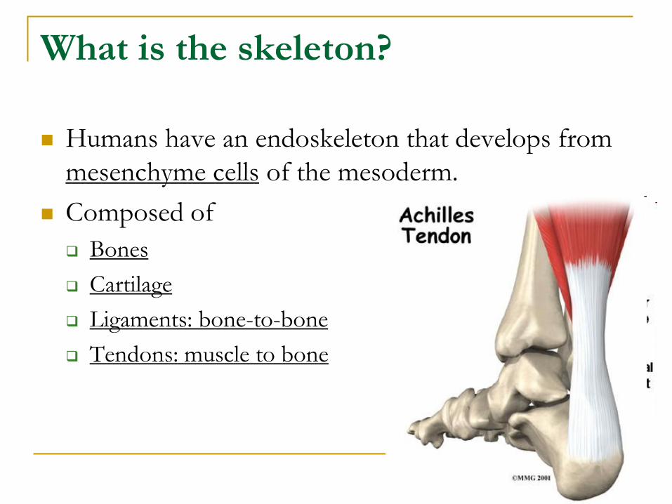
Humans have an endoskeleton that develops from
mesenchyme cells of the mesoderm.
Composed of
Bones
Cartilage
Ligaments: bone-to-bone
Tendons: muscle to bone
What does the skeleton do?
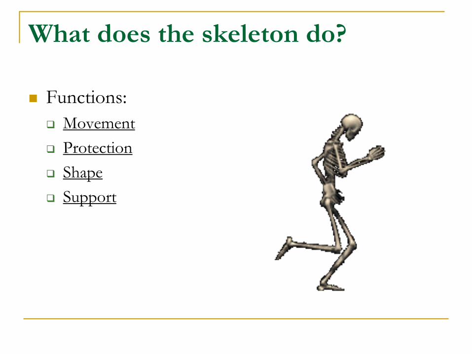
Functions:
Movement
Protection
Shape
Support
The Human Skeleton
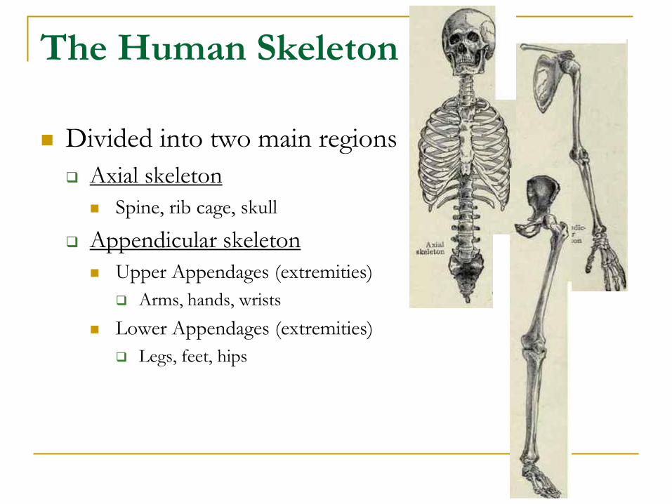
Divided into two main regions
Axial skeleton
Spine, rib cage, skull
Appendicular skeleton
Upper Appendages (extremities)
Arms, hands, wrists
Lower Appendages (extremities)
Legs, feet, hips
The Human Skeleton
Articulations: where two bones meet to make a
joint
Surface features: protrusions and edges on the bone
Sites of tendon and ligament attachment
Projections that help form joints
Depressions and openings allowing blood vessels and
nerve fibers to pass
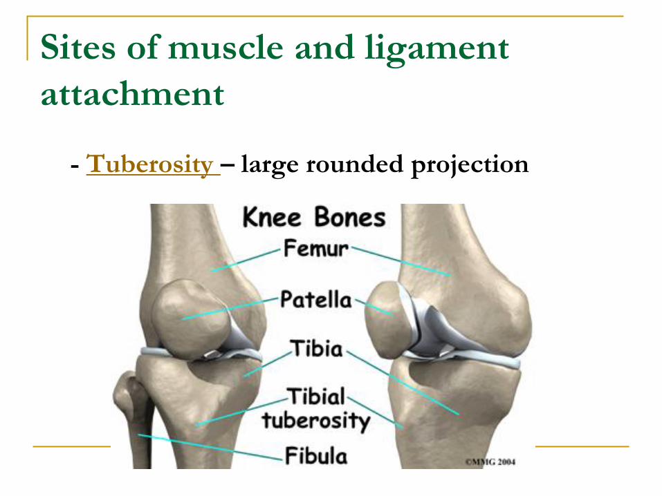
Sites of muscle and ligament
attachment
- Tuberosity – large rounded projection
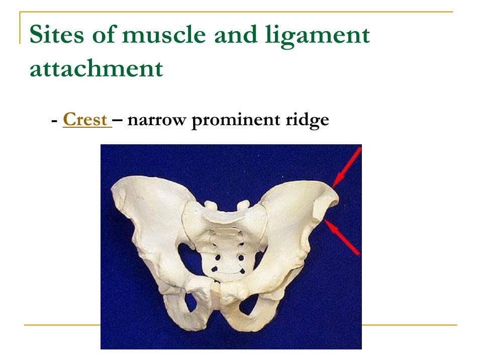
Sites of muscle and ligament
attachment
- Crest – narrow prominent ridge
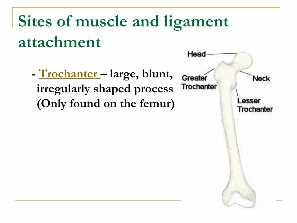
Sites of muscle and ligament
attachment
- Trochanter – large, blunt,
irregularly shaped process
(Only found on the femur)
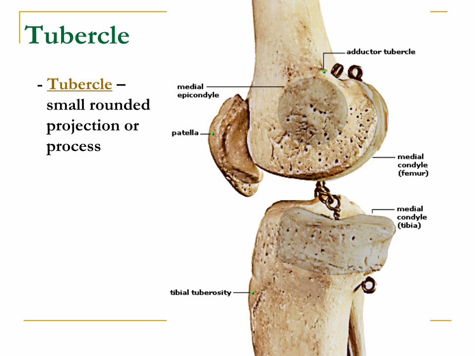
Tubercle
- Tubercle –
small rounded
projection or
process
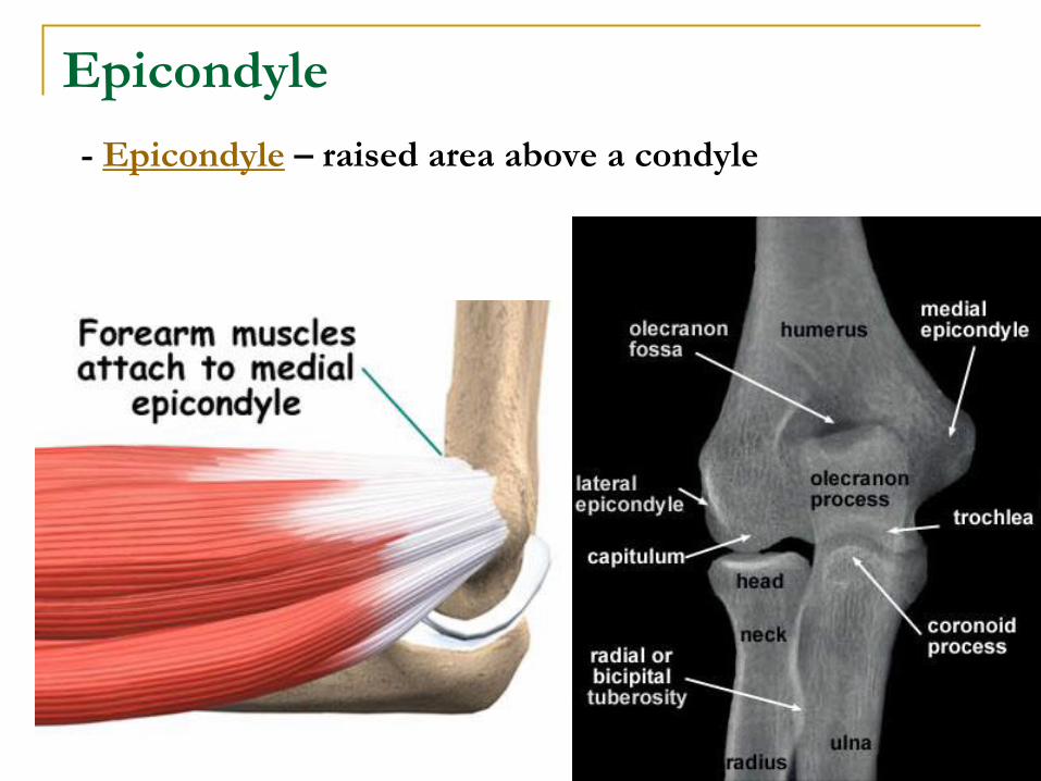
Epicondyle
- Epicondyle – raised area above a condyle
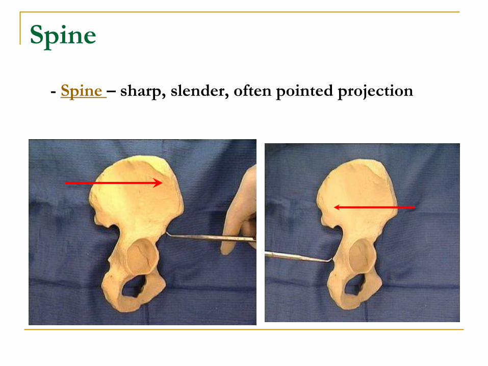
Spine
- Spine – sharp, slender, often pointed projection
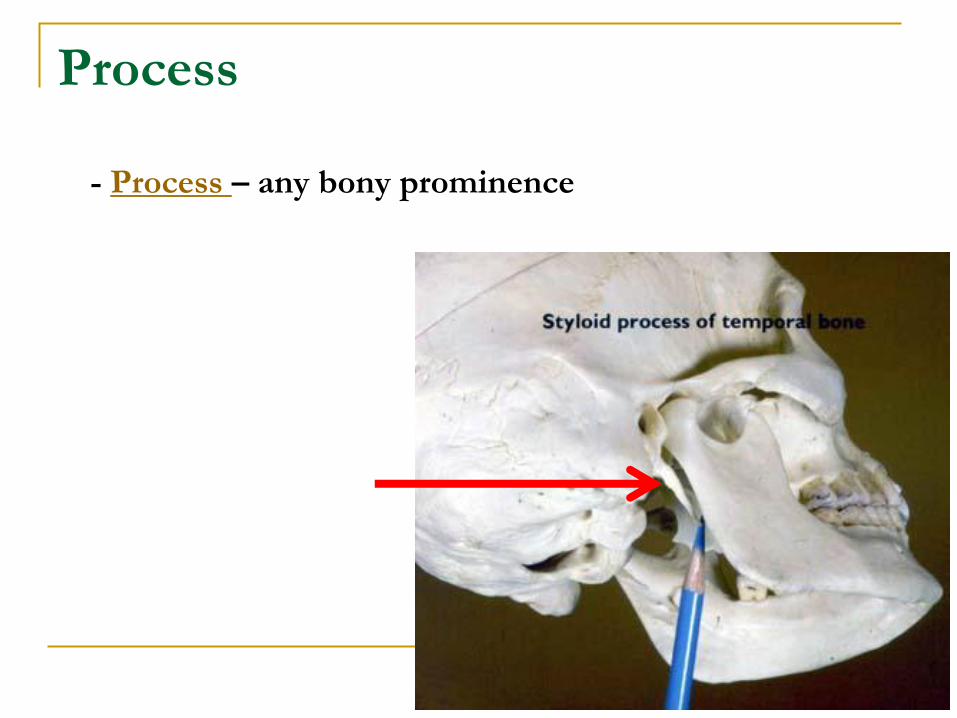
Process
- Process – any bony prominence
Projections that help
form joints
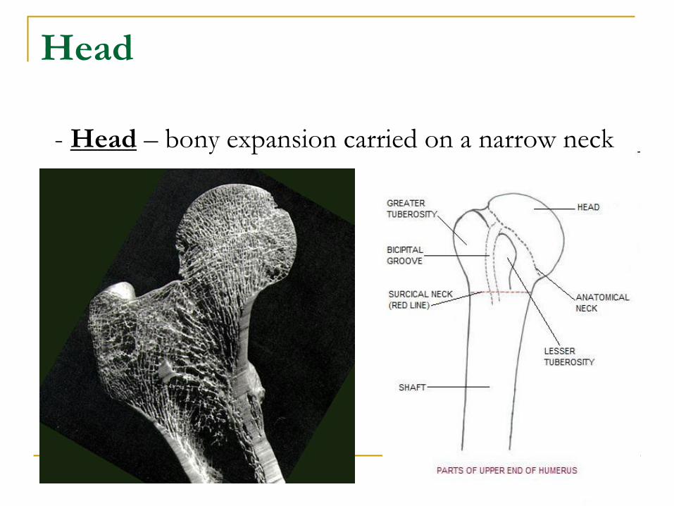
Head
- Head – bony expansion carried on a narrow neck
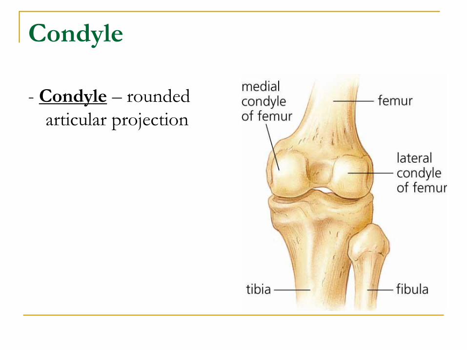
Condyle
- Condyle – rounded
articular projection
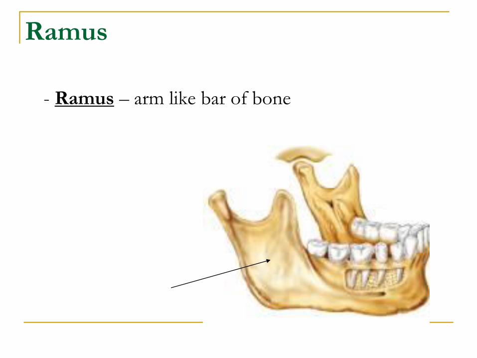
Ramus
- Ramus – arm like bar of bone
Depressions and
openings allowing blood
vessels and nerve fibers
to pass
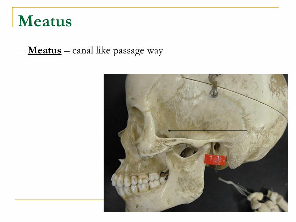
Meatus
- Meatus – canal like passage way
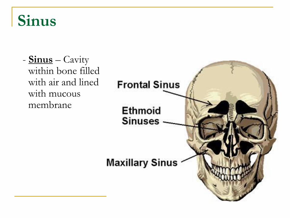
Sinus
- Sinus – Cavity within bone filled with air and lined with mucous membrane
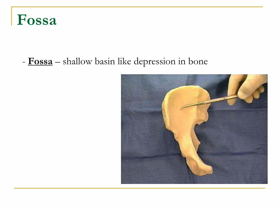
Fossa
- Fossa – shallow basin like depression in bone
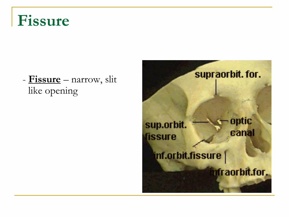
Fissure
- Fissure – narrow, slit like opening
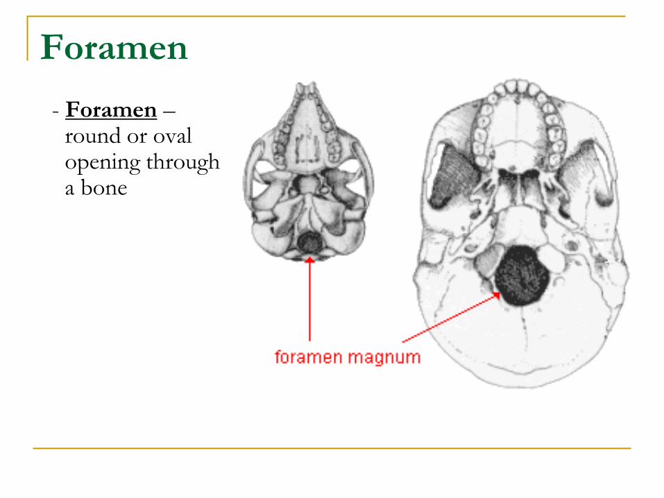
Foramen
- Foramen – round or oval opening through a bone
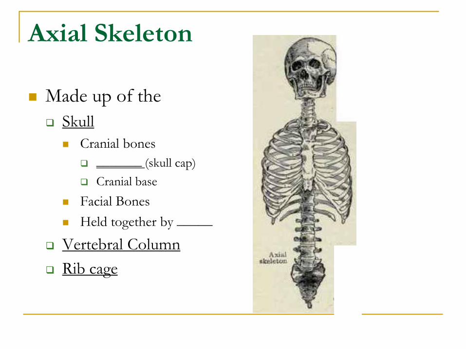
Axial Skeleton
Made up of the
Skull
Cranial bones
_______ (skull cap)
Cranial base
Facial Bones
Held together by _____
Vertebral Column
Rib cage
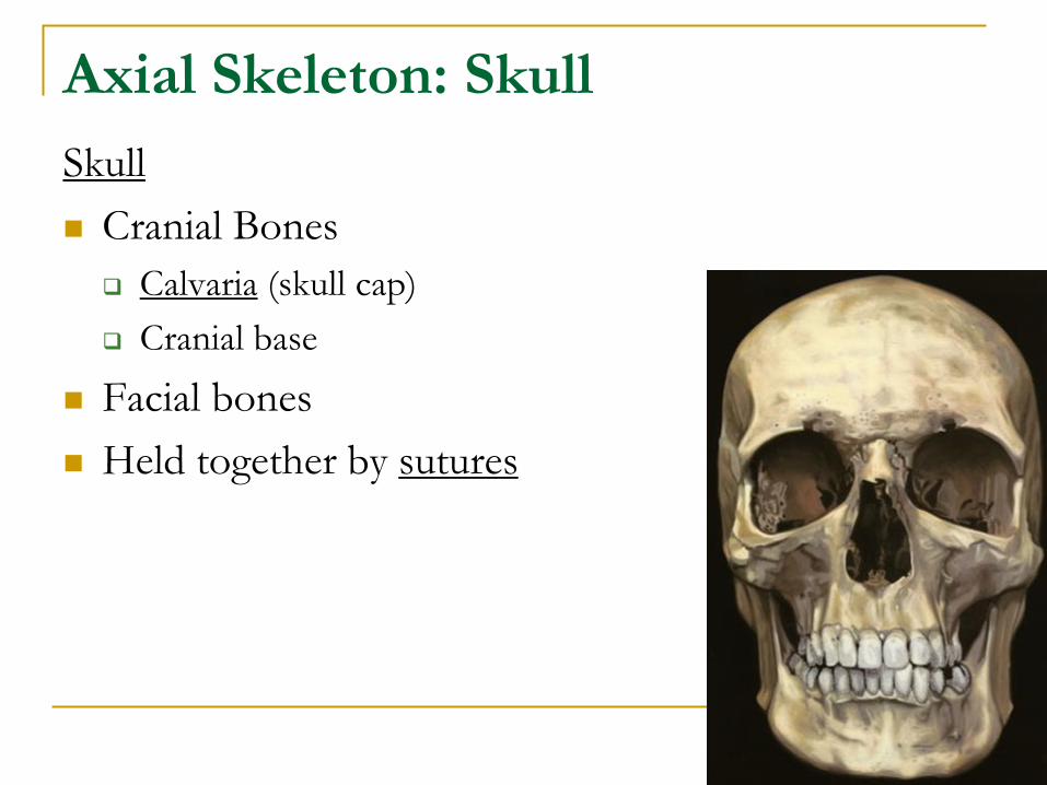
Axial Skeleton: Skull
Skull
Cranial Bones
Calvaria (skull cap)
Cranial base
Facial bones
Held together by sutures
Axial Skeleton: Skull
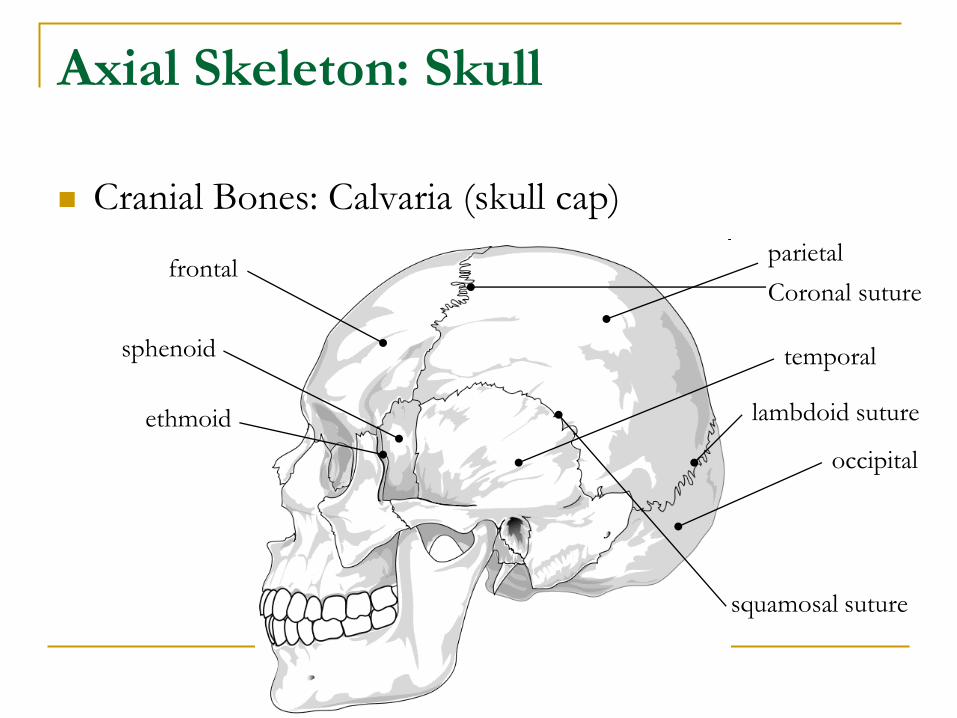
Cranial Bones: Calvaria (skull cap)
parietal
temporal
occipital
frontal
sphenoid
ethmoid
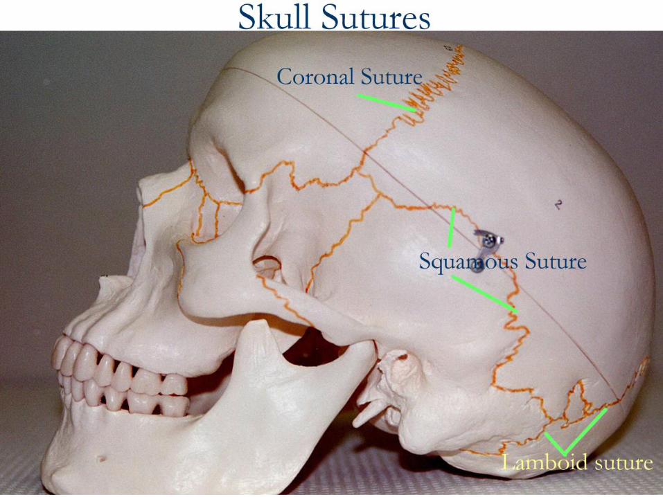
Coronal suture
squamosal suture
lambdoid suture
Axial Skeleton: Skull
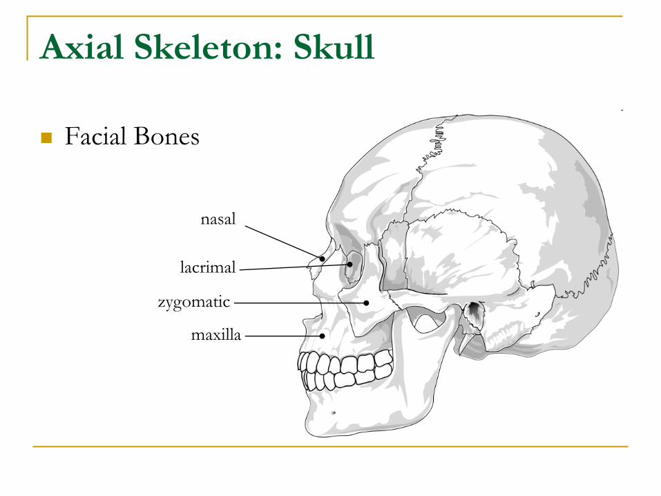
Facial Bones
nasal
lacrimal
zygomatic
maxilla
Axial Skeleton: Skull
Cranial Bones
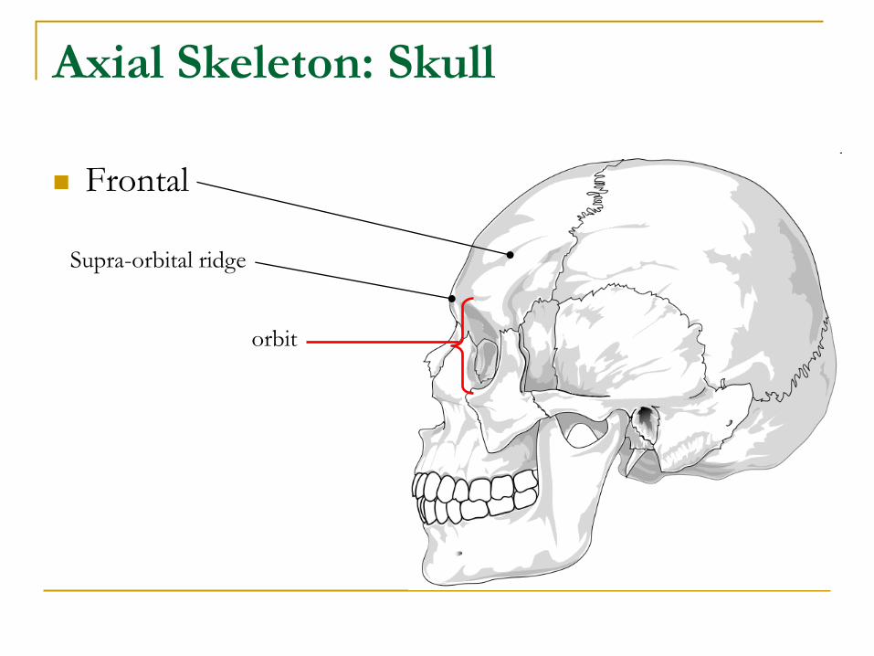
Frontal: makes up the forehead and eyebrow ridges
Upper part of the orbits (eye sockets)
Thicker area above the orbits is the supra-orbital ridge
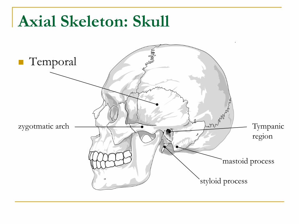
Temporal: has a flat portion, along with…
mastoid process: where neck muscles attach
styloid process: attachment for neck & throat muscles
tympanic region: contain the ear bones
zygomatic process (arch): forms the cheek bone and articulates with the mandible
Parietal: helps protect brain
Axial Skeleton: Skull
Frontal
Supra-orbital ridge
orbit
Axial Skeleton: Skull
Temporal
zygotmatic arch Tympanic
region
mastoid process
styloid process
Axial Skeleton: Skull
Cranial Bones
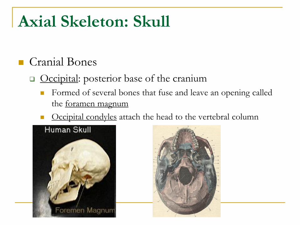
Occipital: posterior base of the cranium
Formed of several bones that fuse and leave an opening called
the foramen magnum
Occipital condyles attach the head to the vertebral column
Axial Skeleton: Skull
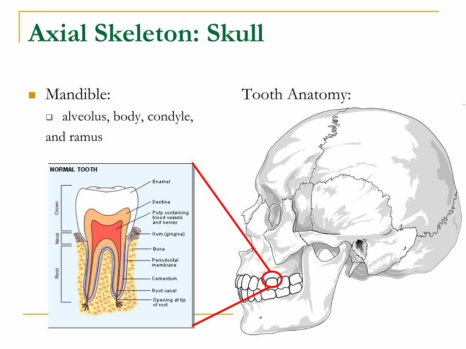
Mandible:
alveolus, body, condyle,
and ramus
Tooth Anatomy:
crown: covered by enamel
(made of calcium)
Axial Skeleton
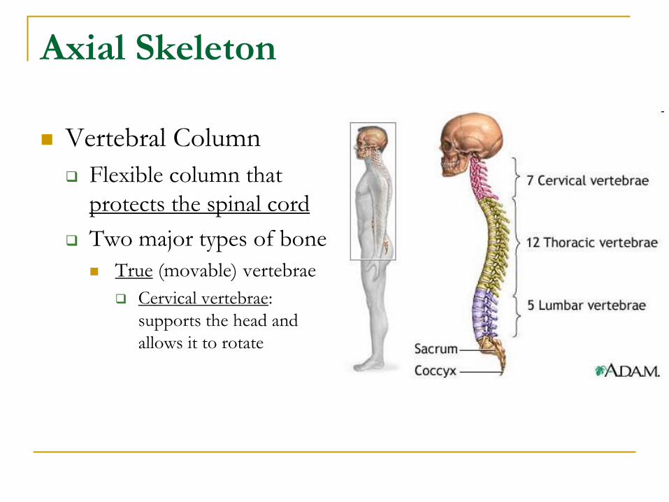
Vertebral Column
Flexible column that
protects the spinal cord
Two major types of bone
True (movable) vertebrae
Cervical vertebrae:
supports the head and
allows it to rotate
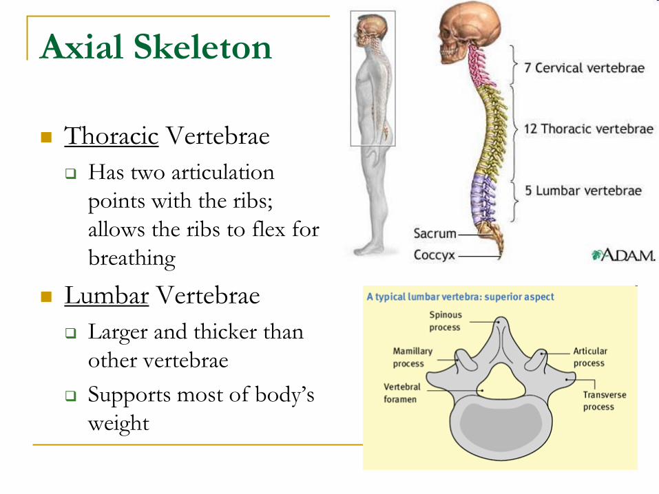
Axial Skeleton
Thoracic Vertebrae
Has two articulation
points with the ribs;
allows the ribs to flex for
breathing
Lumbar Vertebrae
Larger and thicker than
other vertebrae
Supports most of body’s
weight
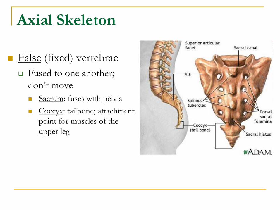
Axial Skeleton
False (fixed) vertebrae
Fused to one another;
don’t move
Sacrum: fuses with pelvis
Coccyx: tailbone; attachment
point for muscles of the
upper leg
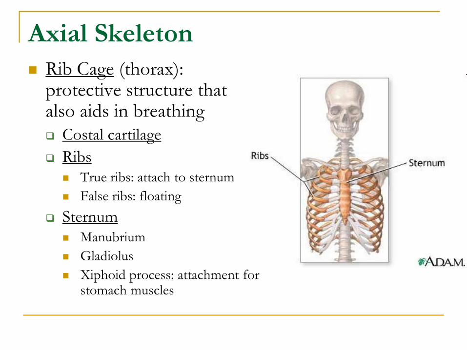
Axial Skeleton
Rib Cage (thorax): protective structure that also aids in breathing
Costal cartilage
Ribs
True ribs: attach to sternum
False ribs: floating
Sternum
Manubrium
Gladiolus
Xiphoid process: attachment for stomach muscles
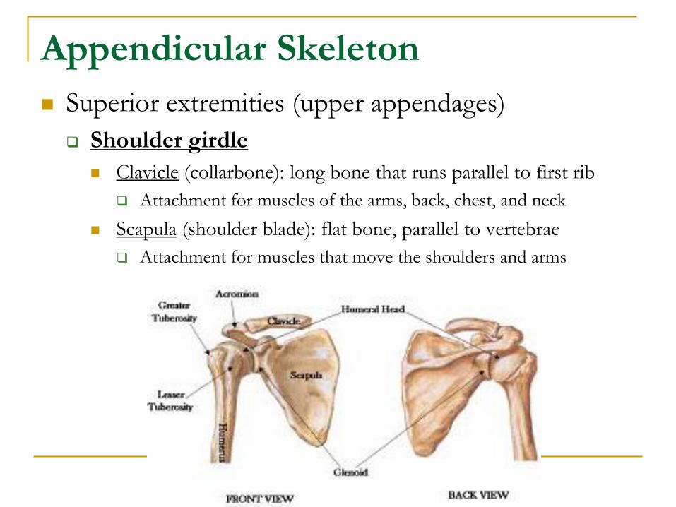
Appendicular Skeleton
Superior extremities (upper appendages)
Shoulder girdle
Clavicle (collarbone): long bone that runs parallel to first rib
Attachment for muscles of the arms, back, chest, and neck
Scapula (shoulder blade): flat bone, parallel to vertebrae
Attachment for muscles that move the shoulders and arms
Appendicular Skeleton
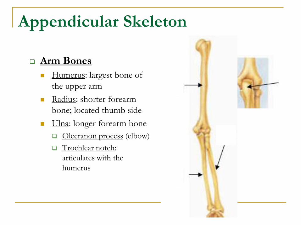
Arm Bones
Humerus: largest bone of
the upper arm
Radius: shorter forearm
bone; located thumb side
Ulna: longer forearm bone
Olecranon process (elbow)
Trochlear notch:
articulates with the
humerus
Appendicular Skeleton
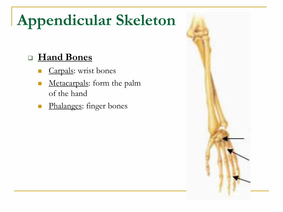
Hand Bones
Carpals: wrist bones
Metacarpals: form the palm
of the hand
Phalanges: finger bones
Appendicular Skeleton
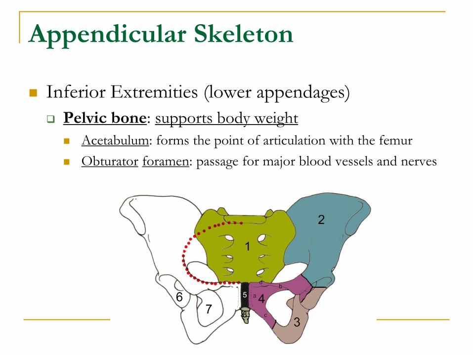
Inferior Extremities (lower appendages)
Pelvic bone: supports body weight
Acetabulum: forms the point of articulation with the femur
Obturator foramen: passage for major blood vessels and nerves
Appendicular Skeleton
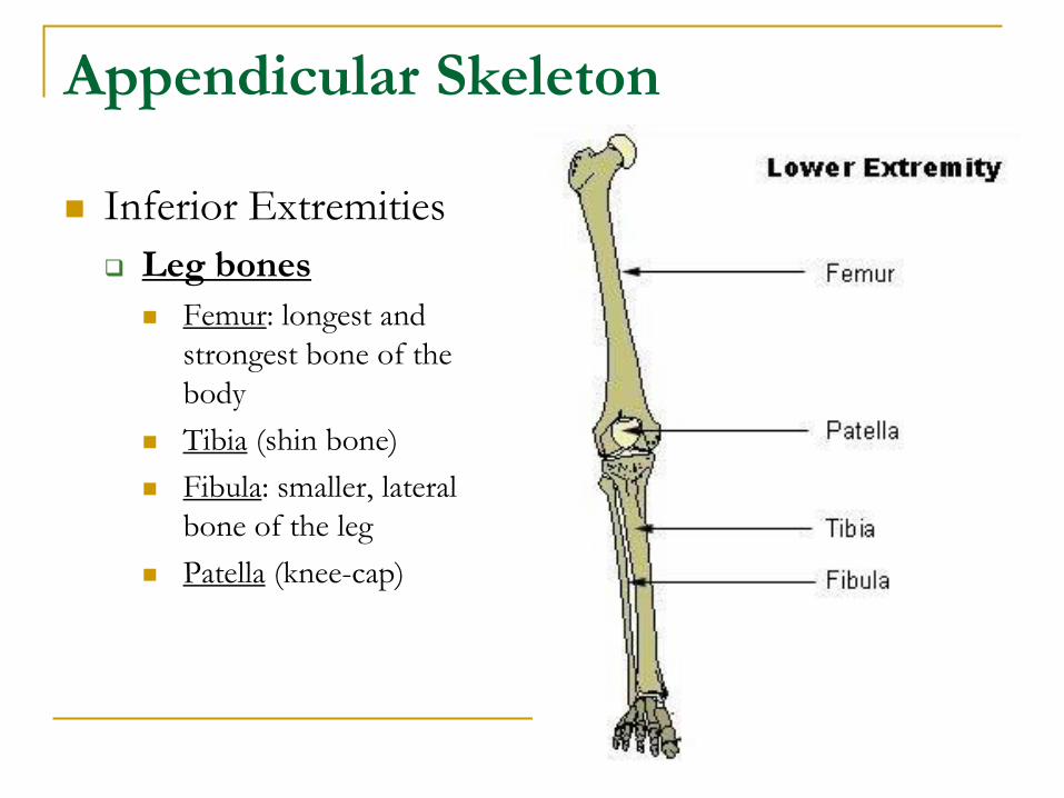
Inferior Extremities
Leg bones
Femur: longest and
strongest bone of the
body
Tibia (shin bone)
Fibula: smaller, lateral
bone of the leg
Patella (knee-cap)
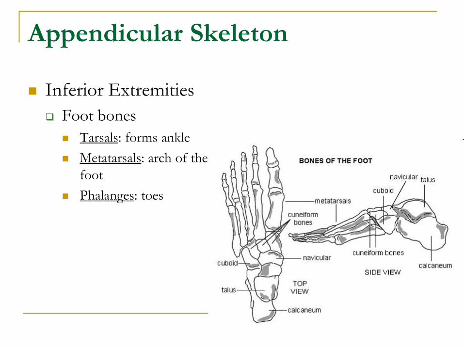
Appendicular Skeleton
Inferior Extremities
Foot bones
Tarsals: forms ankle
Metatarsals: arch of the
foot
Phalanges: toes
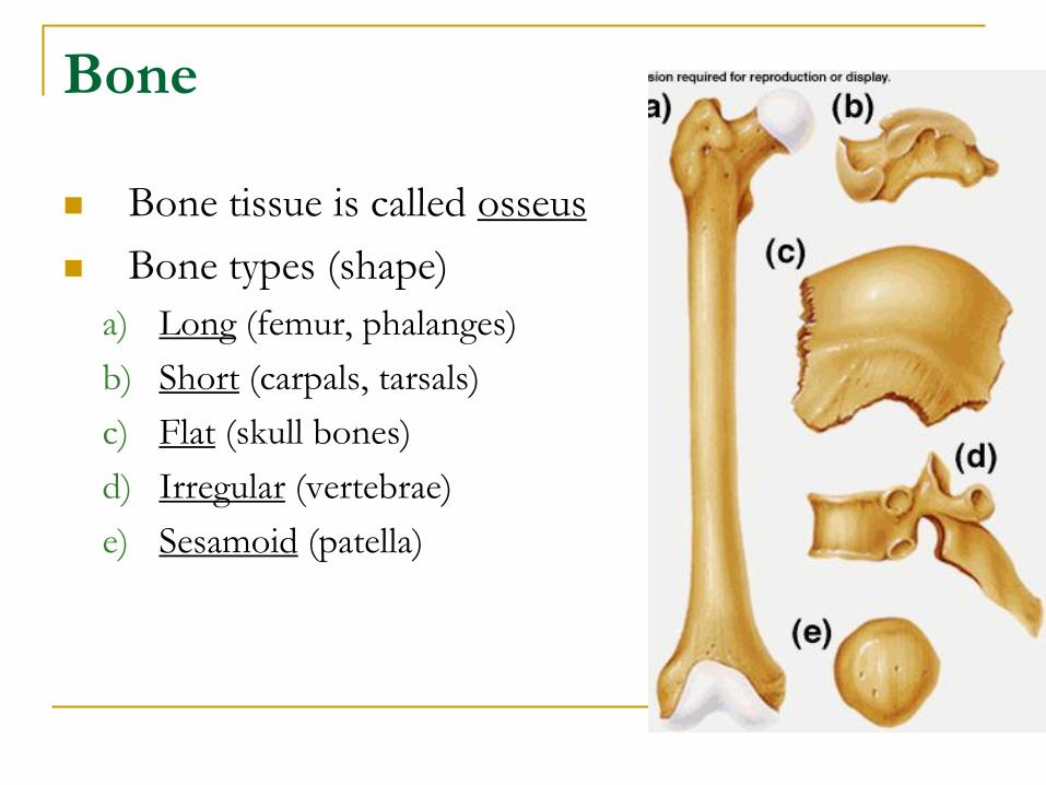
Bone
Bone tissue is called osseus
Bone types (shape)
a) Long (femur, phalanges)
b) Short (carpals, tarsals)
c) Flat (skull bones)
d) Irregular (vertebrae)
e) Sesamoid (patella)
Bone Structure
Bone Structure
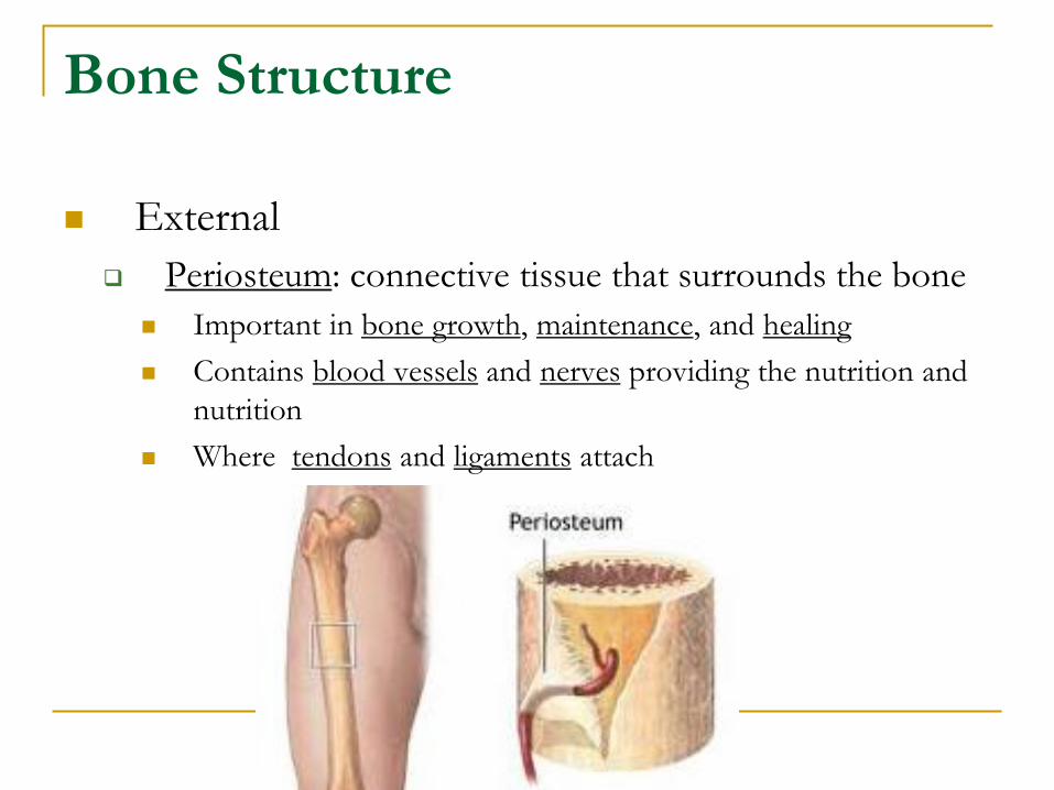
External
Periosteum: connective tissue that surrounds the bone
Important in bone growth, maintenance, and healing
Contains blood vessels and nerves providing the nutrition and
nutrition
Where tendons and ligaments attach
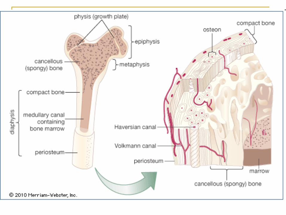
Bone Structure
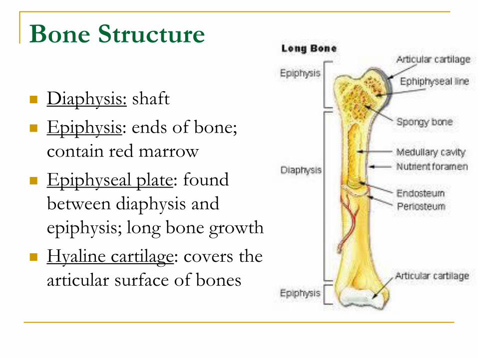
Diaphysis: shaft
Epiphysis: ends of bone;
contain red marrow
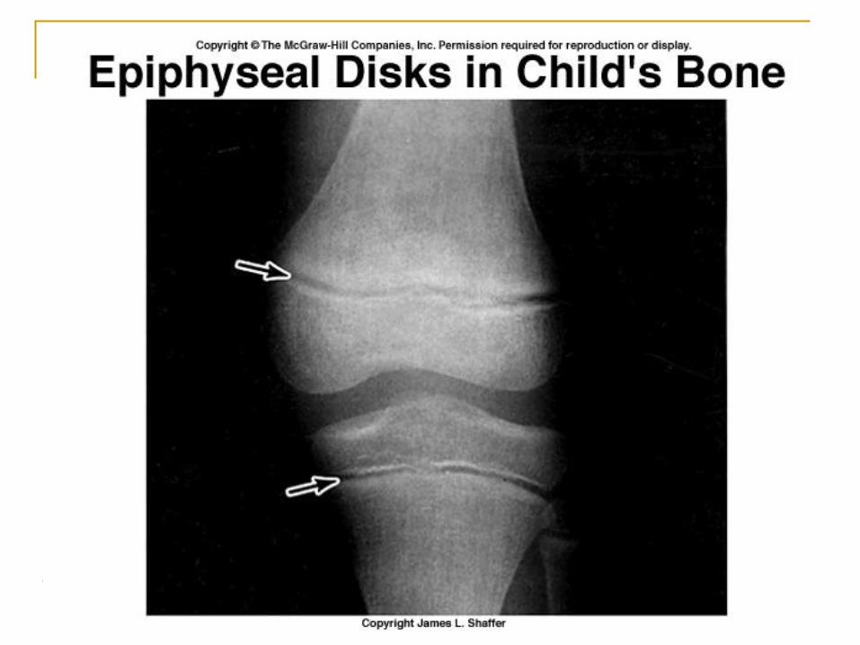
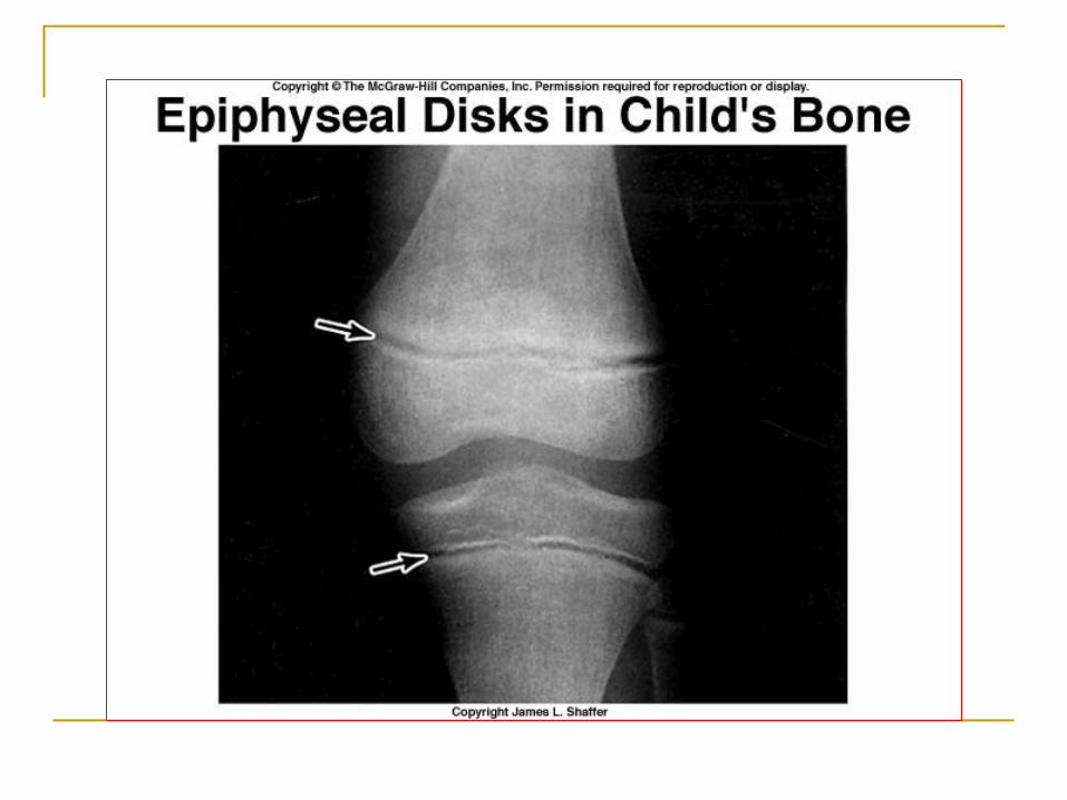
Epiphyseal plate: found
between diaphysis and
epiphysis; long bone growth
Hyaline cartilage: covers the
articular surface of bones
Internal Features
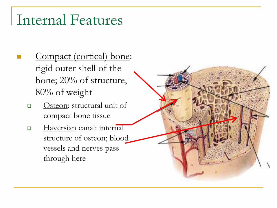
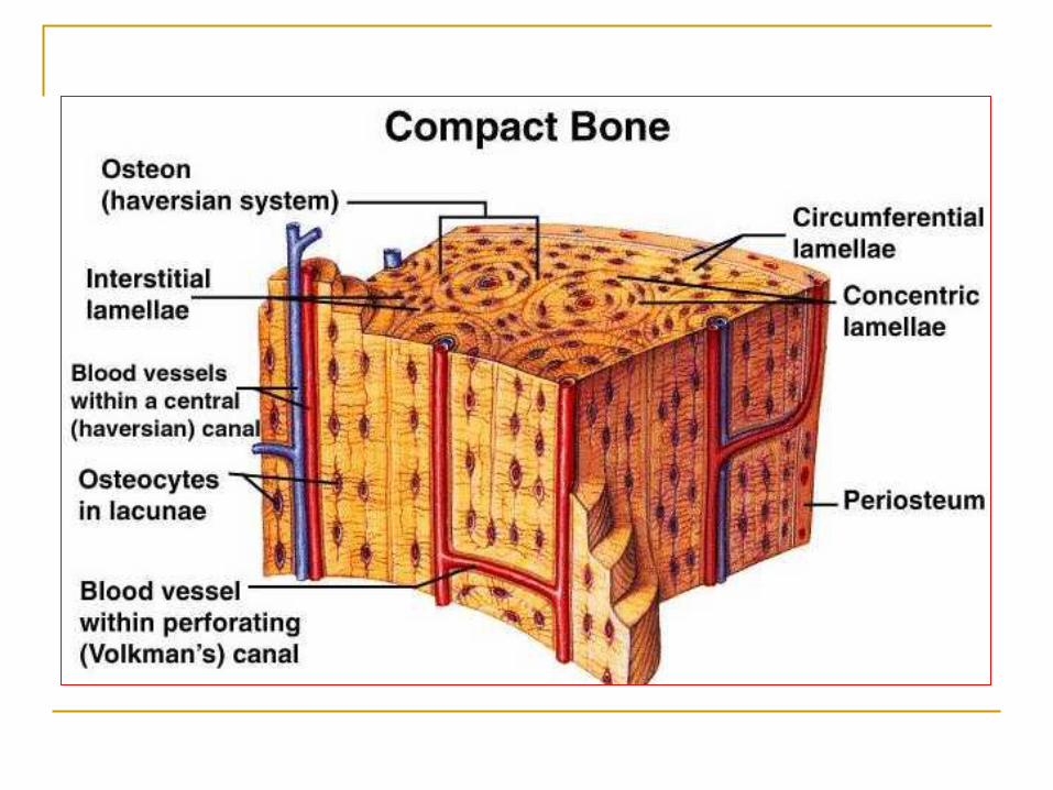
Compact (cortical) bone:
rigid outer shell of the
bone; 20% of structure,
80% of weight
Osteon: structural unit of
compact bone tissue
Haversian canal: internal
structure of osteon; blood
vessels and nerves pass
through here
Internal Features
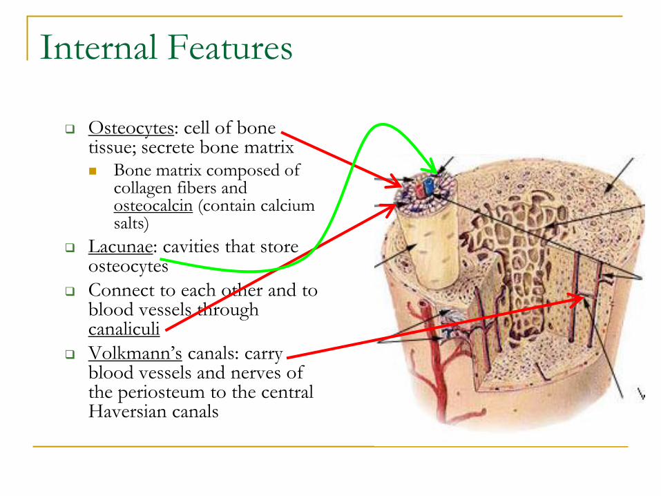
Osteocytes: cell of bone tissue; secrete bone matrix Bone matrix composed of
collagen fibers and osteocalcin (contain calcium salts)
Lacunae: cavities that store osteocytes
Connect to each other and to blood vessels through canaliculi
Volkmann’s canals: carry blood vessels and nerves of the periosteum to the central Haversian canals
Internal Features
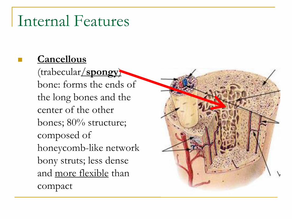
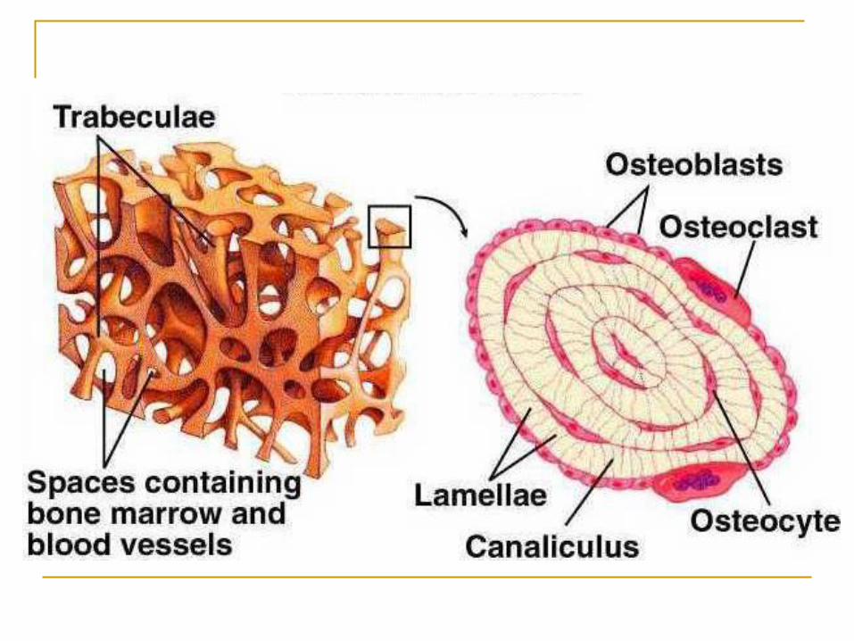
Cancellous
(trabecular/spongy)
bone: forms the ends of
the long bones and the
center of the other
bones; 80% structure;
composed of
honeycomb-like network
bony struts; less dense
and more flexible than
compact
Internal Features
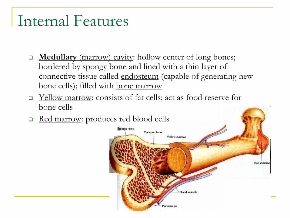
Medullary (marrow) cavity: hollow center of long bones; bordered by spongy bone and lined with a thin layer of connective tissue called endosteum (capable of generating new bone cells); filled with bone marrow
Yellow marrow: consists of fat cells; act as food reserve for bone cells
Red marrow: produces red blood cells
Joints
Joints: some accompanied by a bursa that contains
synovial fluid.
Type of movement depends on
The manner in which the bones fit together
The tightness of fit between the articular surfaces
The tension of the tissues forming the articulation
The position of the ligaments, muscles, and tendons
associated with the joints
Joints
Structural
Cartilaginous: cartilage covers the articulating bone surfaces;
example is the pubic bones
Fibrous: articulated bones are attached by fibrous
connective tissue; example is between the radius and
ulna
Synovial: articulating surfaceis coverd with a fluid-filled,
fibrous, connective-tissue sack called the synovial
capsule; examples are elbows and knees
Joints
Functional
Synarthrosis: permit no movement;
Gomphosis: formed by a conical process; it is held in a socket by a
ligament; teeth
Syndesmosis: formed when two bones are joined by one or more
ligaments; example is the joint that holds together the tibia and
fibula in the ankle region
Synchondrosis (symphysis) joints: two bones join together by a
piece of cartilage
Synostosis (sutures): formed by the fusion of two bones
Amphiarthrosis: slight movement
Joints
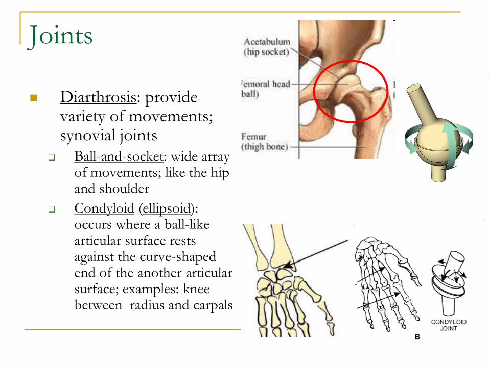
Diarthrosis: provide variety of movements; synovial joints
Ball-and-socket: wide array of movements; like the hip and shoulder
Condyloid (ellipsoid): occurs where a ball-like articular surface rests against the curve-shaped end of the another articular surface; examples: knee between radius and carpals
Joints
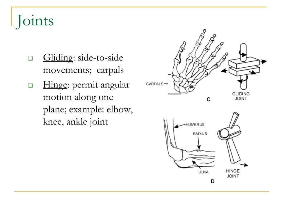
Gliding: side-to-side
movements; carpals
Hinge: permit angular
motion along one
plane; example: elbow,
knee, ankle joint
Joints
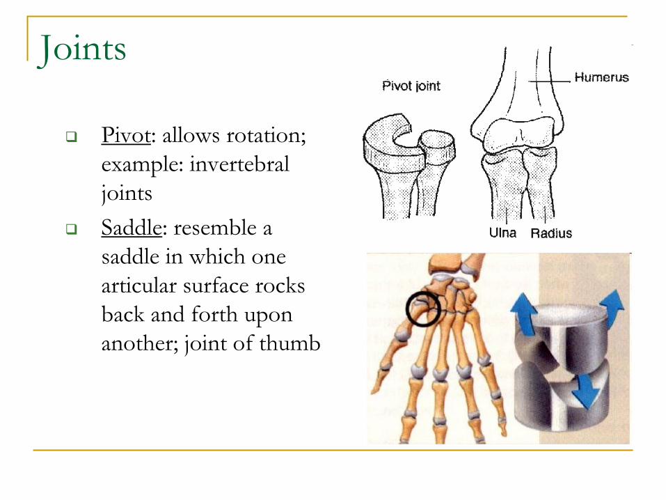
Pivot: allows rotation;
example: invertebral
joints
Saddle: resemble a
saddle in which one
articular surface rocks
back and forth upon
another; joint of thumb
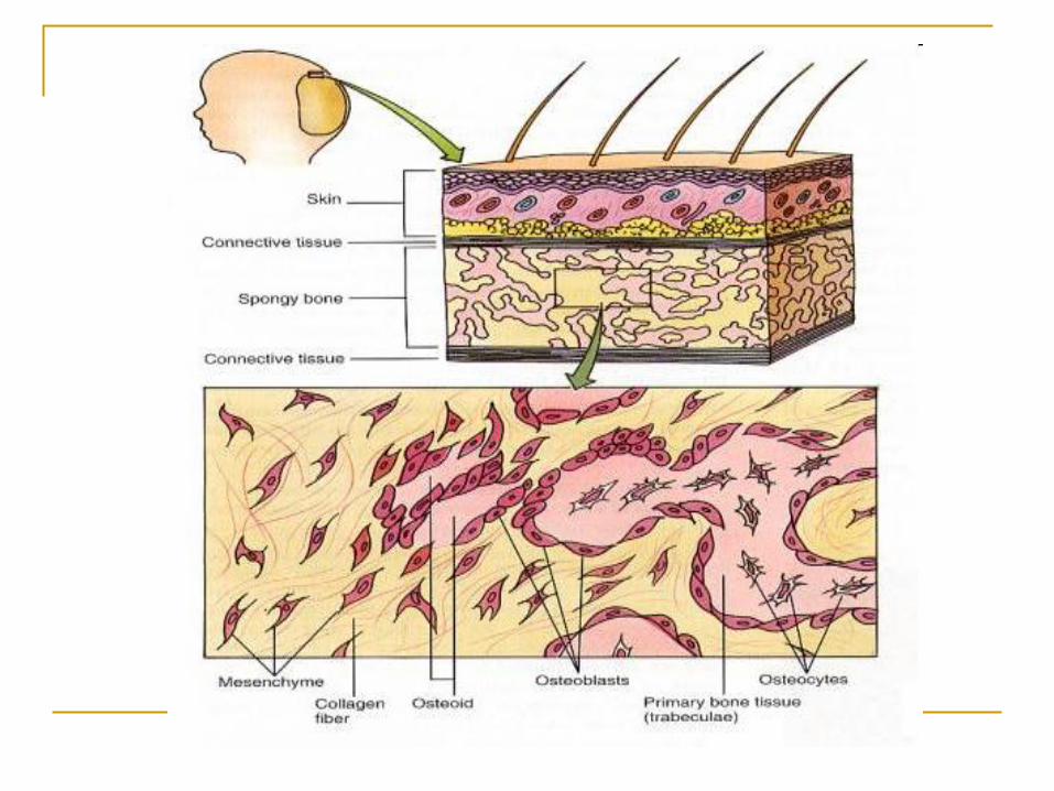
Bone Development and Healing
Bone Growth
Bone-forming cells (osteoblasts and osteoclasts)
move into the cartilage pegs helping to sculpt
growing bones
Osteoblasts build bone tissue
Osteoclasts break down bone and cartilage; carve &
sculpt bone
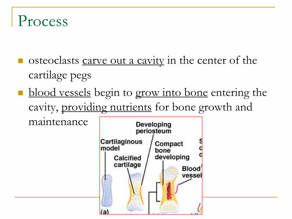
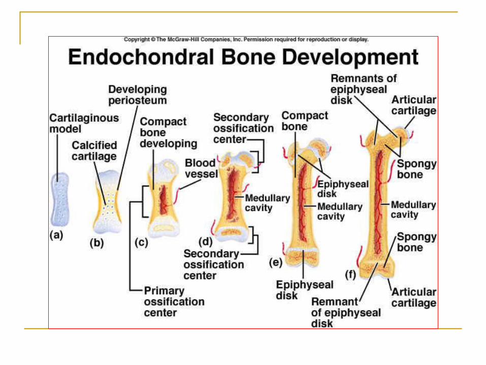
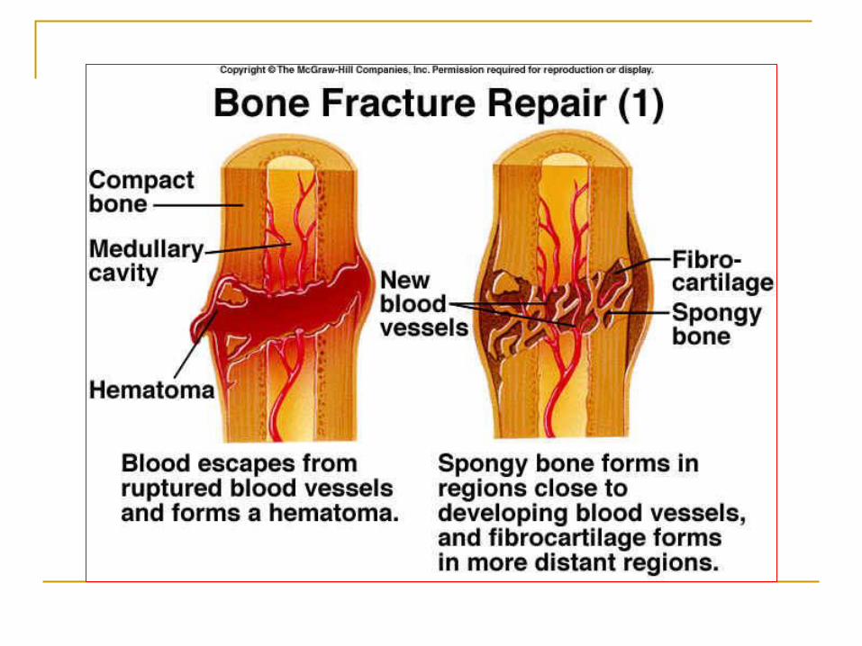
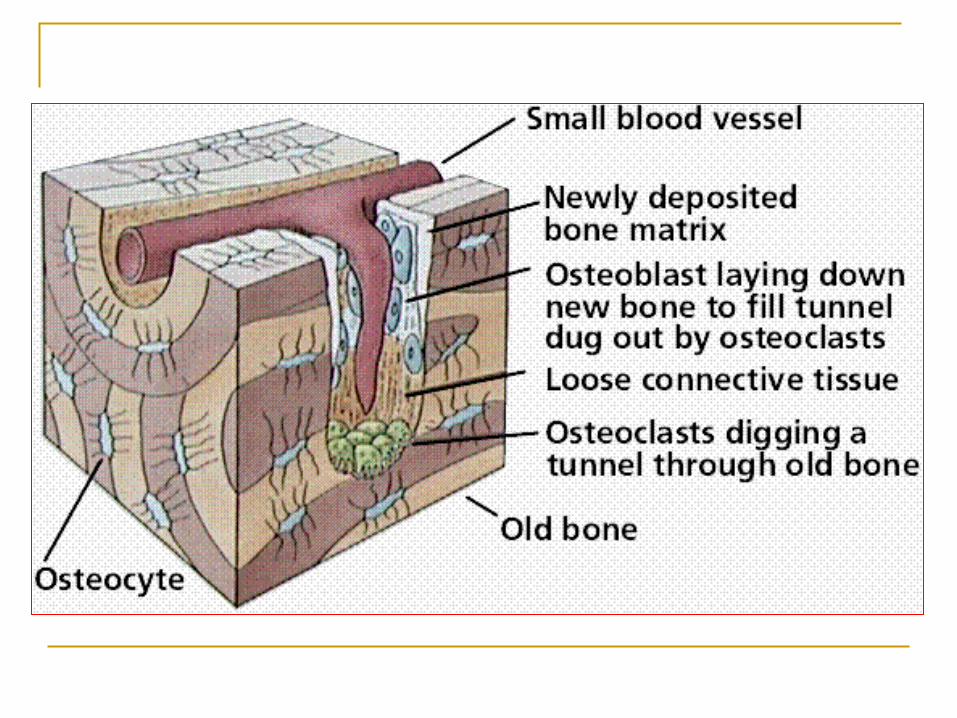
Process
osteoclasts carve out a cavity in the center of the
cartilage pegs
blood vessels begin to grow into bone entering the
cavity, providing nutrients for bone growth and
maintenance
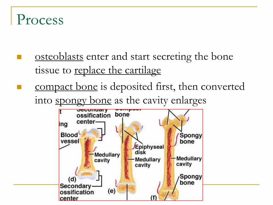
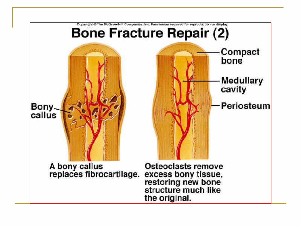
Process
osteoblasts enter and start secreting the bone
tissue to replace the cartilage
compact bone is deposited first, then converted
into spongy bone as the cavity enlarges
Homeostatic Imbalance
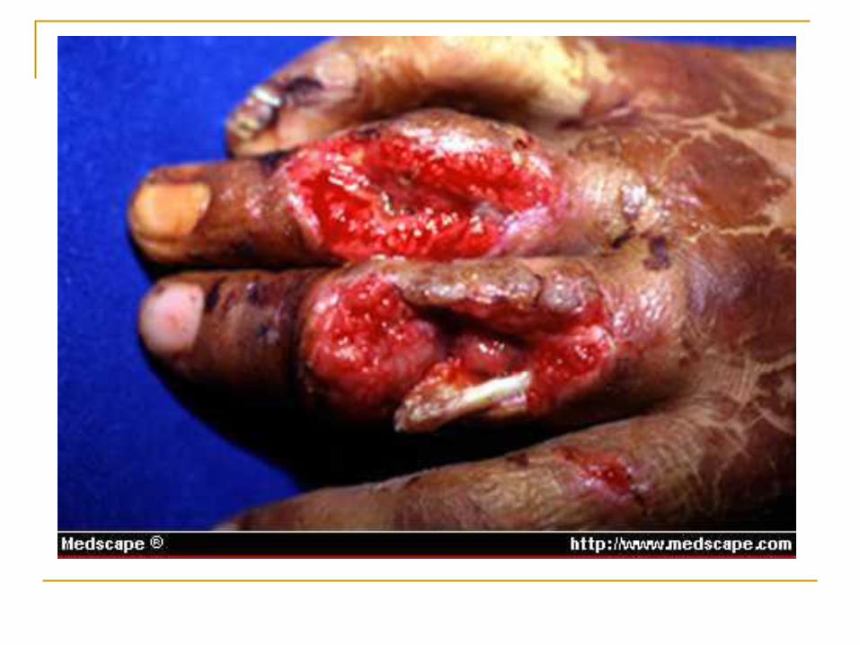
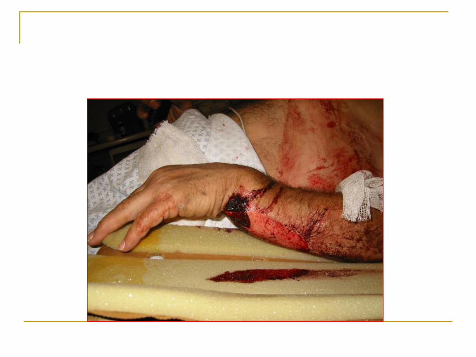
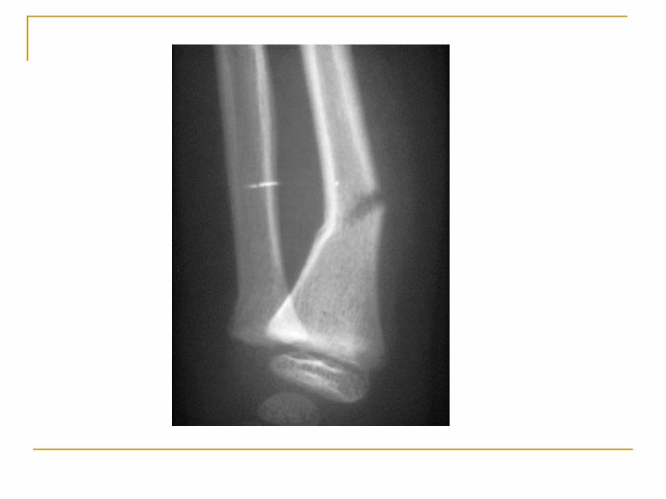
Fracture – a break in the bone
Open fracture – bone penetrates through the skin
Closed fracture – bone does not penetrate skin
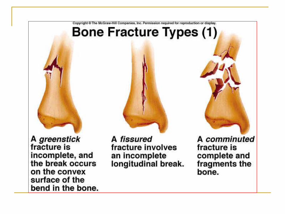
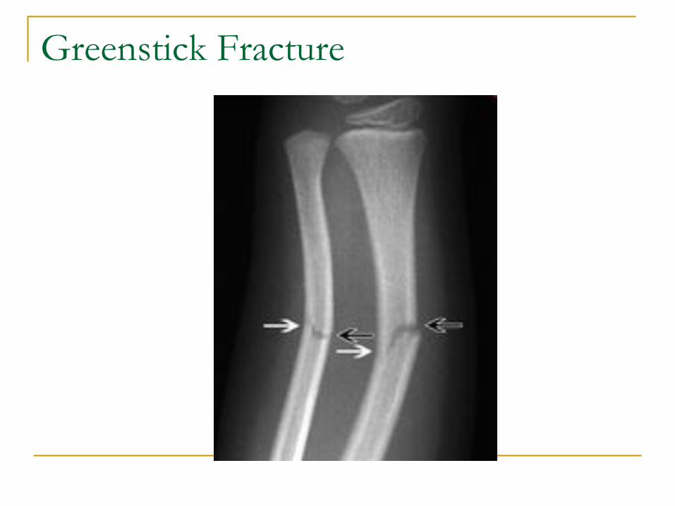
Greenstick Fracture
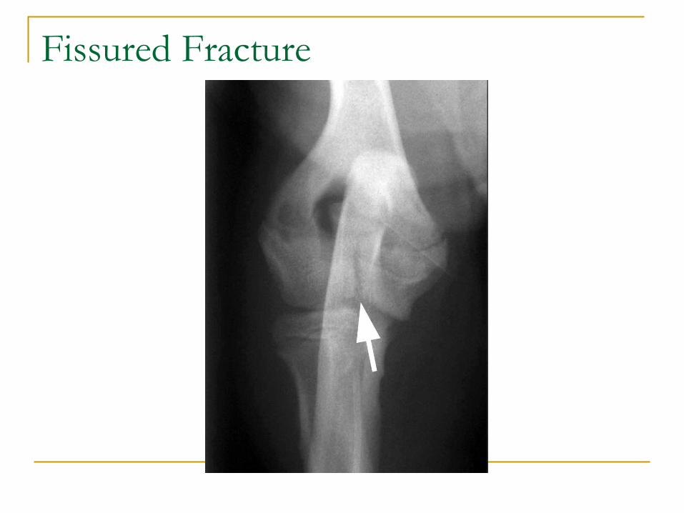
Fissured Fracture
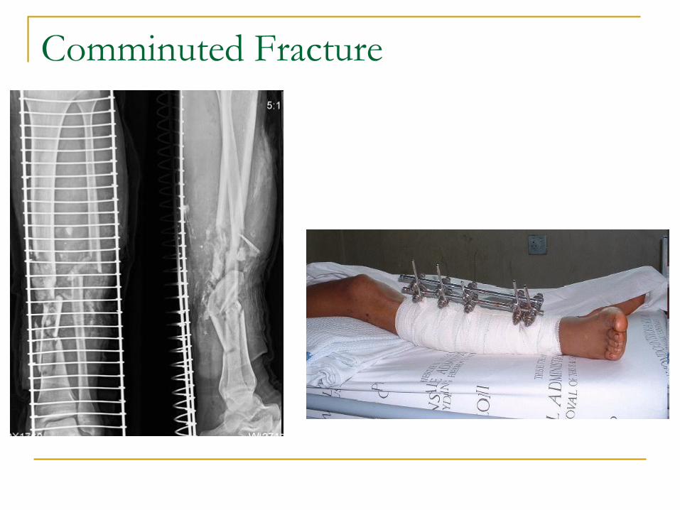
Comminuted Fracture
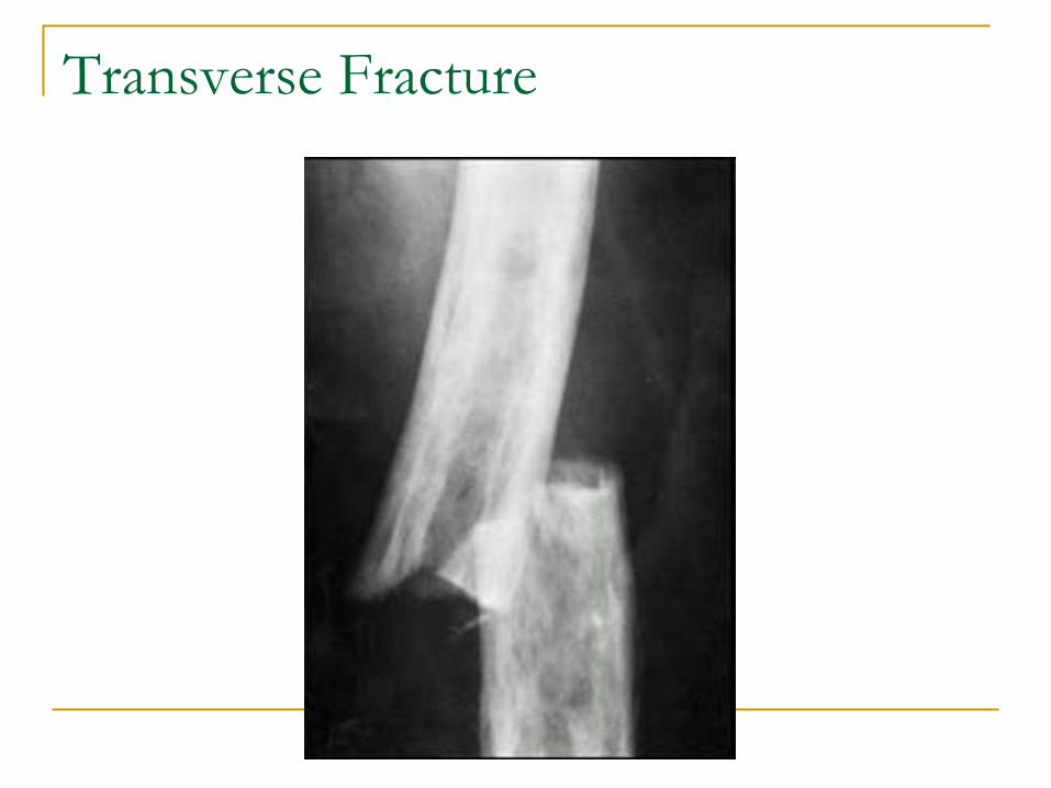
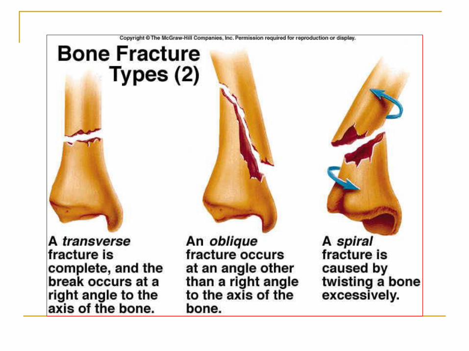
Transverse Fracture
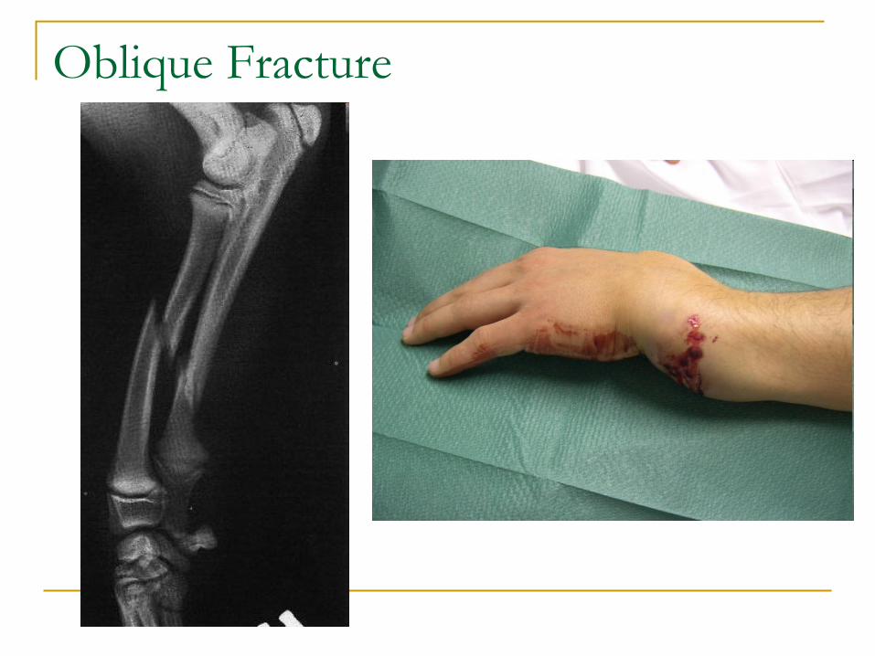
Oblique Fracture
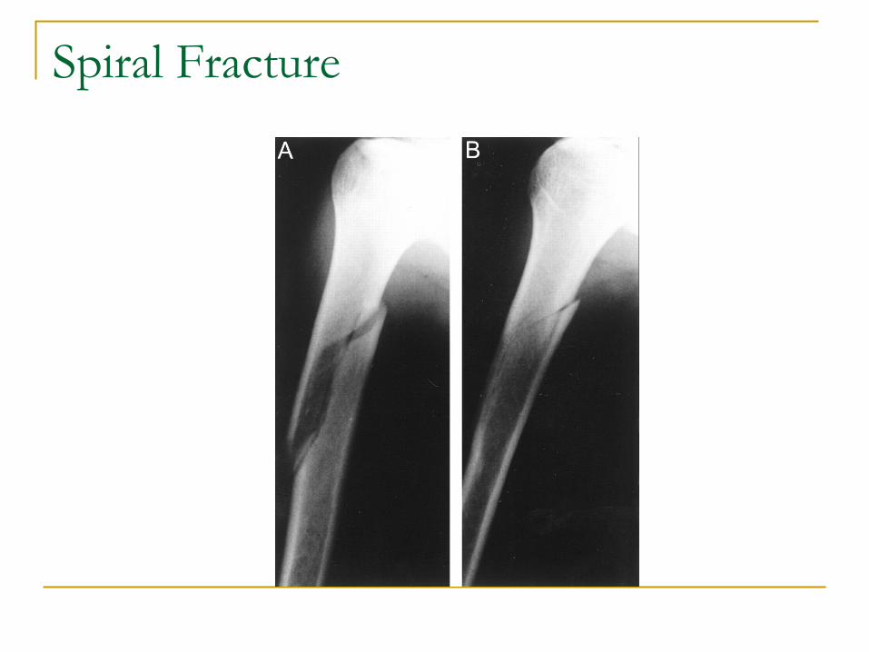
Spiral Fracture
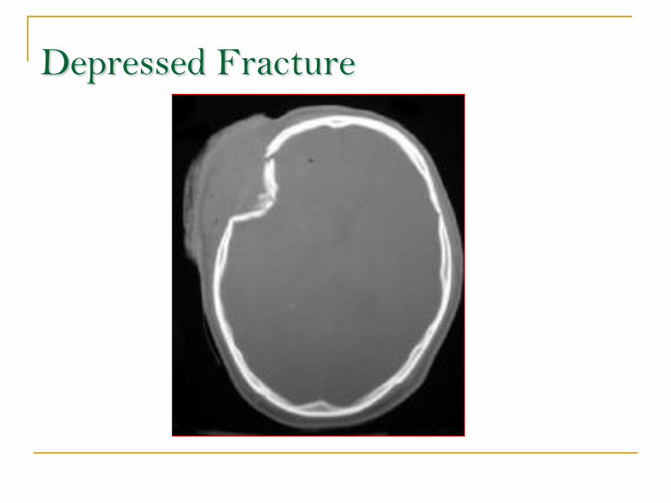
Depressed Fracture
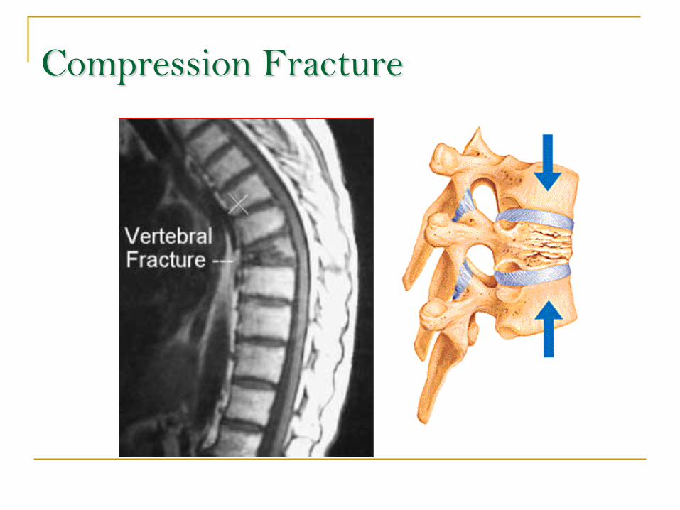
Compression Fracture
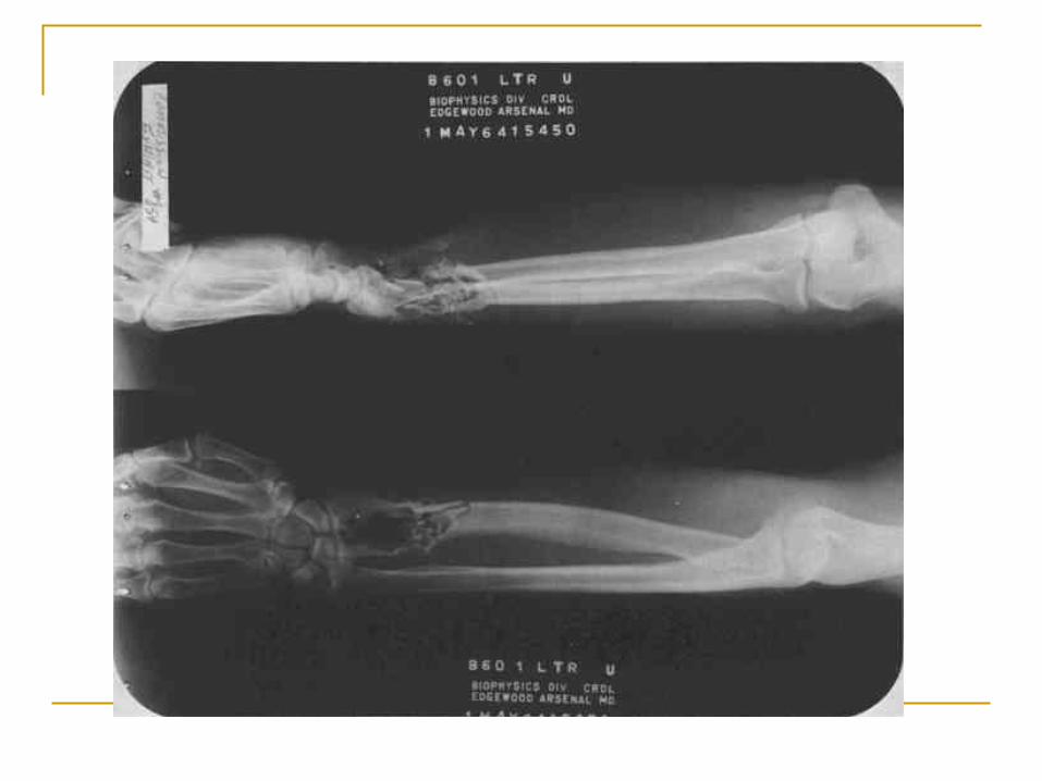
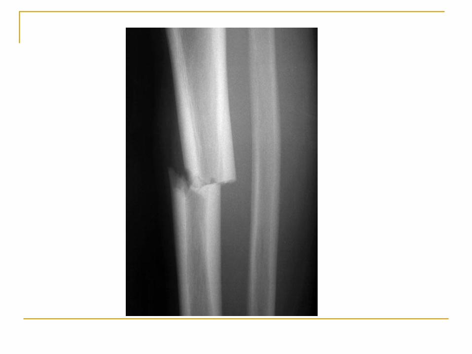
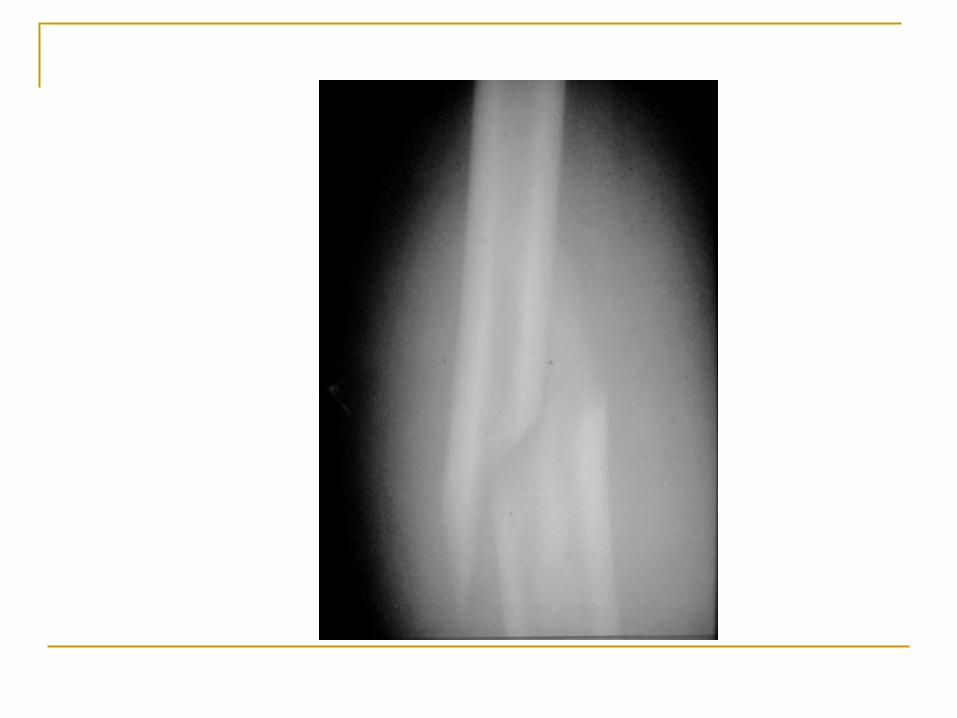
Fracture ID practice
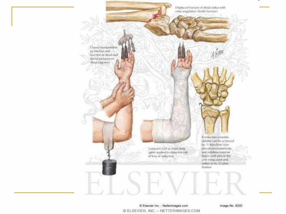
Fracture treatment
Closed reduction – physician pulls on bone to
realign bone ends
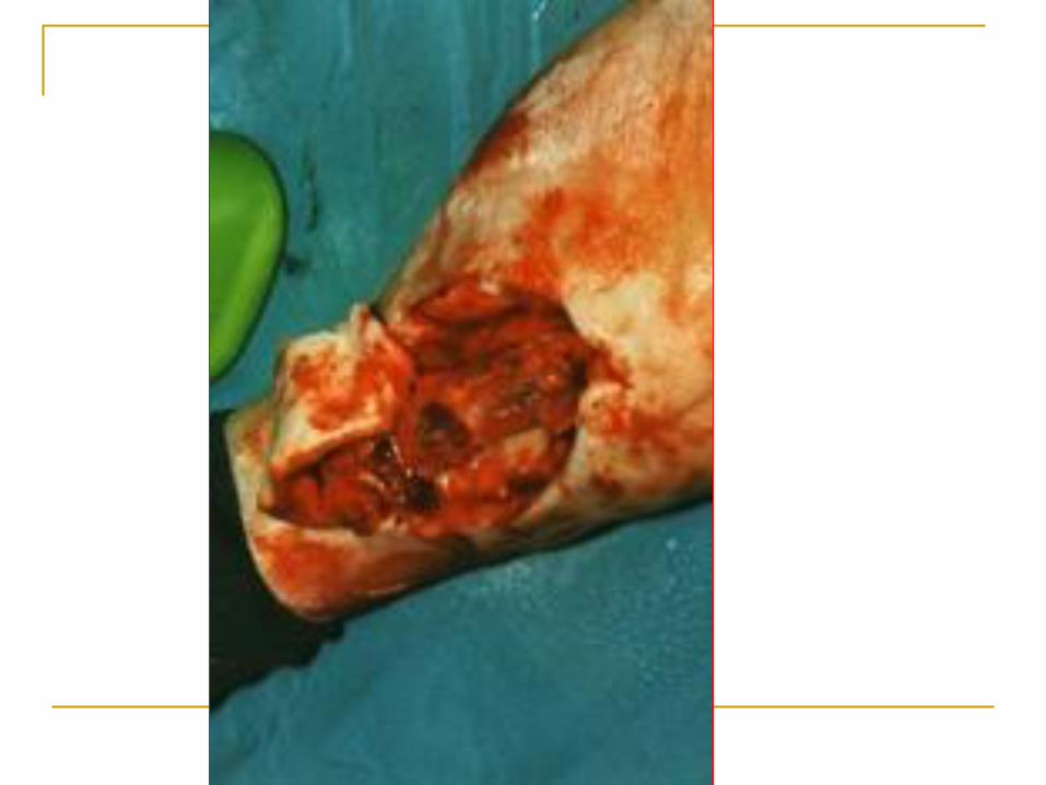
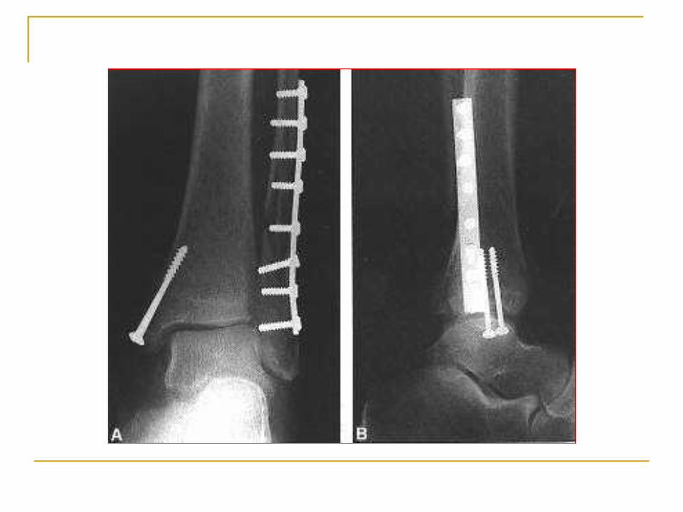
Open reduction – physician surgically inserts
pins or wires to realign bones
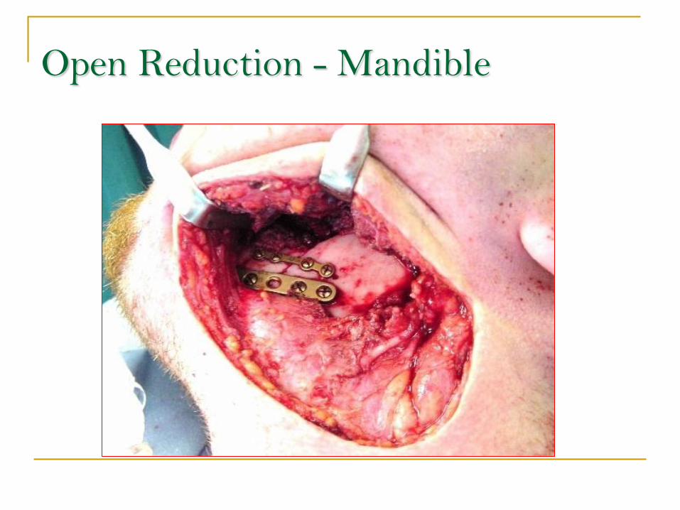
Open Reduction - Mandible
Skeletal System Diseases How would damage to the components affect function?
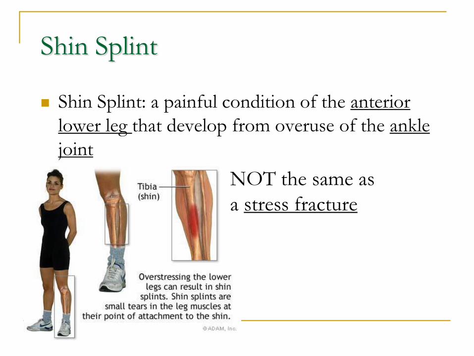
Shin Splint
Shin Splint: a painful condition of the anterior
lower leg that develop from overuse of the ankle
joint
NOT the same as
a stress fracture
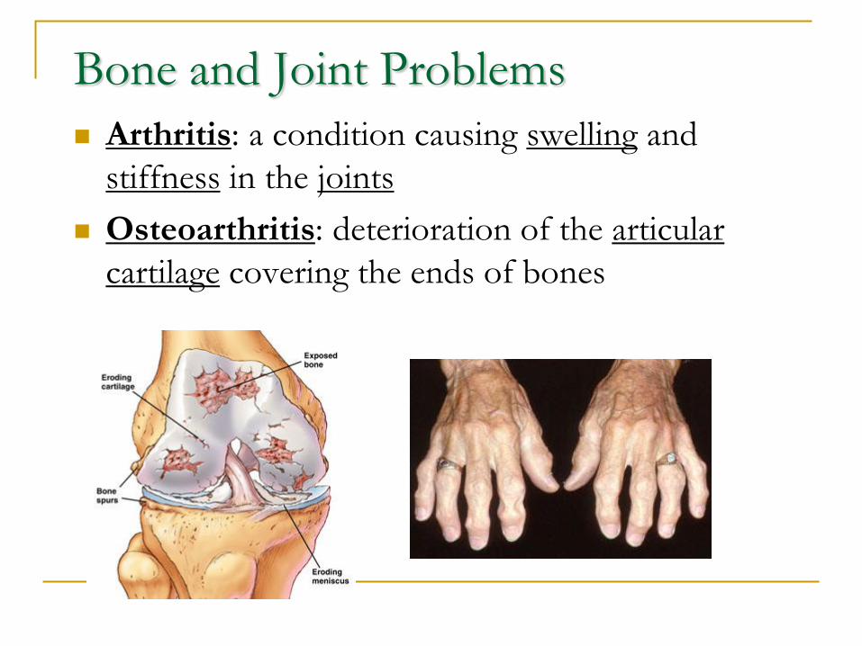
Bone and Joint Problems
Arthritis: a condition causing swelling and
stiffness in the joints
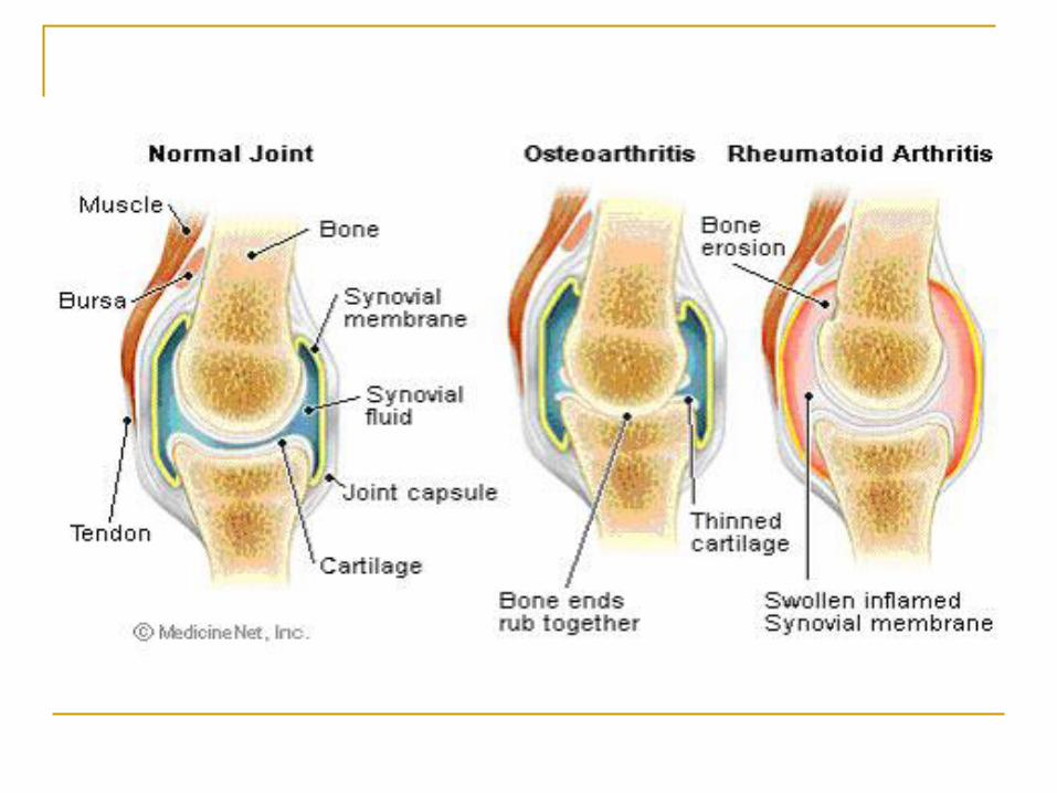
Osteoarthritis: deterioration of the articular
cartilage covering the ends of bones
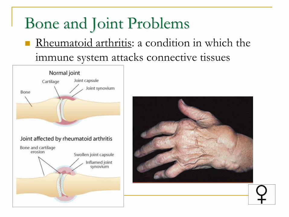
Bone and Joint Problems Rheumatoid arthritis: a condition in which the
immune system attacks connective tissues
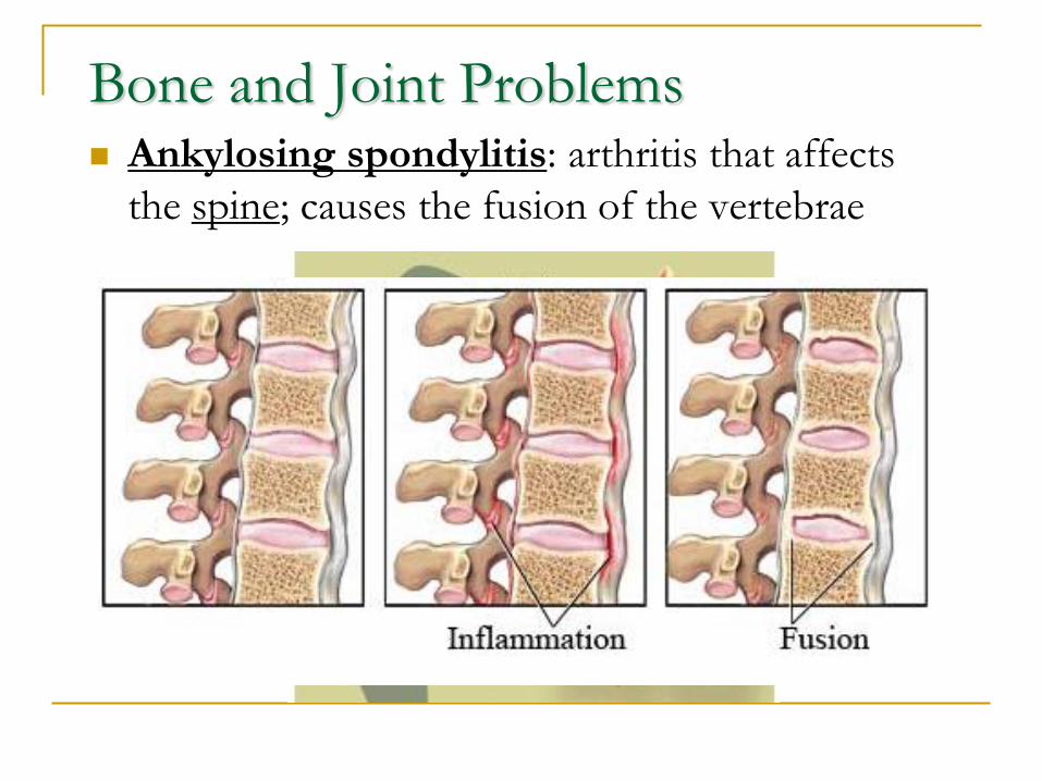
Bone and Joint Problems Ankylosing spondylitis: arthritis that affects
the spine; causes the fusion of the vertebrae
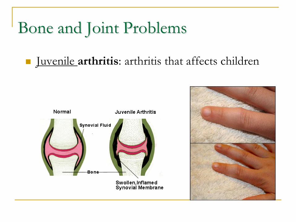
Bone and Joint Problems
Juvenile arthritis: arthritis that affects children
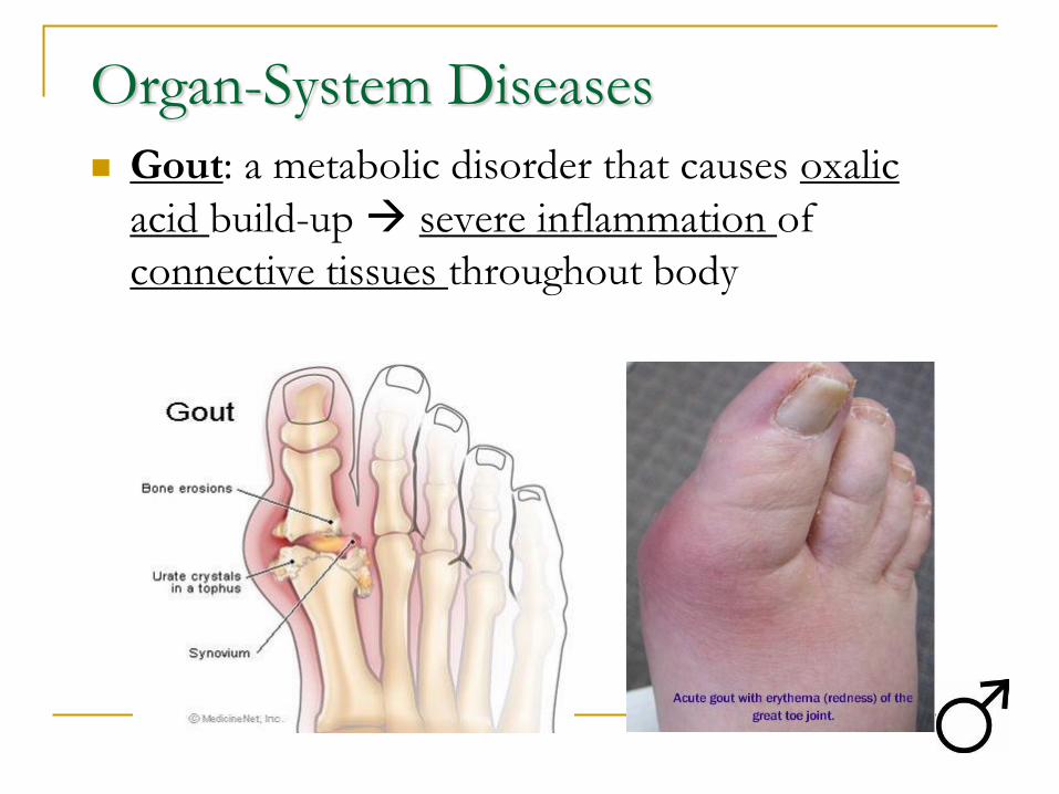
Organ-System Diseases
Gout: a metabolic disorder that causes oxalic
acid build-up severe inflammation of
connective tissues throughout body
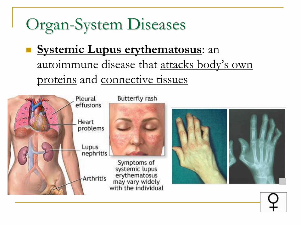
Organ-System Diseases
Systemic Lupus erythematosus: an
autoimmune disease that attacks body’s own
proteins and connective tissues
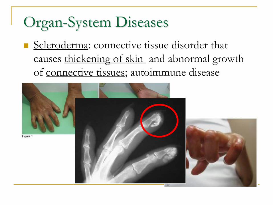
Organ-System Diseases
Scleroderma: connective tissue disorder that
causes thickening of skin and abnormal growth
of connective tissues; autoimmune disease
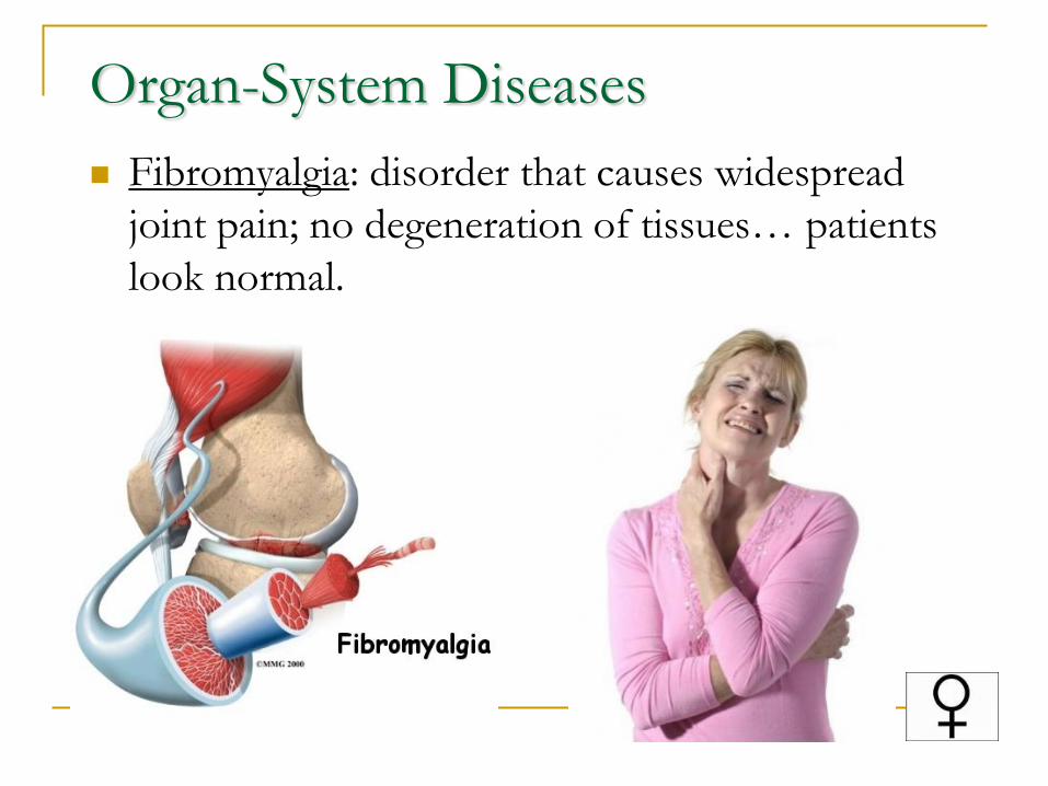
Organ-System Diseases
Fibromyalgia: disorder that causes widespread
joint pain; no degeneration of tissues… patients
look normal.
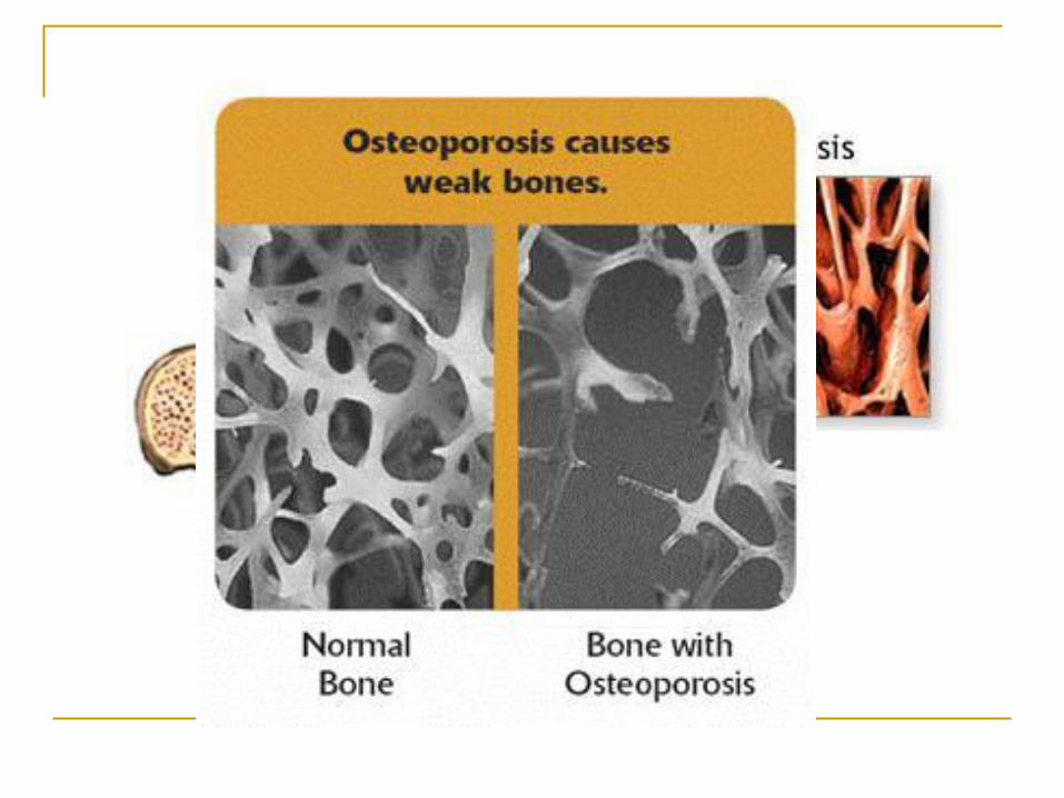
Osteoporosis
Osteoporosis: degenerative bone disorder; can
occur at any age
Causes:
Malnutrition or undernutrition (not enough calcium-
rich foods or vitamin D)
Decreased levels of sex hormones
Smoking
Excessive alcohol consumption
A less severe case is bone density loss.
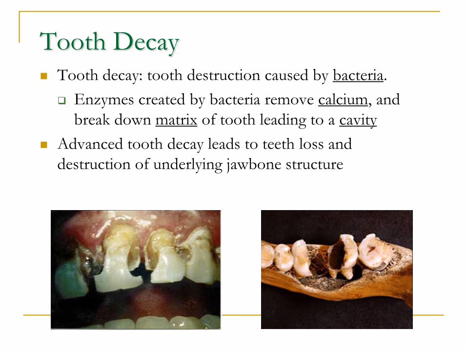
Tooth Decay Tooth decay: tooth destruction caused by bacteria.
Enzymes created by bacteria remove calcium, and
break down matrix of tooth leading to a cavity
Advanced tooth decay leads to teeth loss and
destruction of underlying jawbone structure
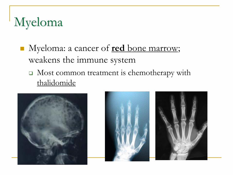
Myeloma
Myeloma: a cancer of red bone marrow;
weakens the immune system
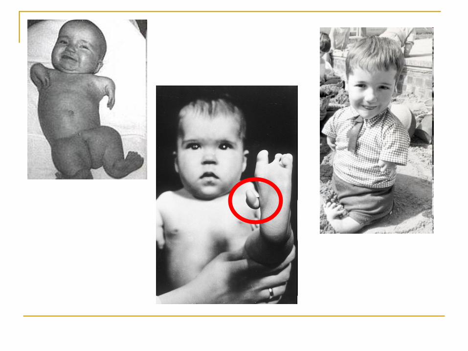
Most common treatment is chemotherapy with
thalidomide
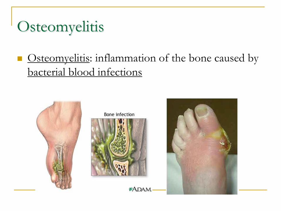
Osteomyelitis
Osteomyelitis: inflammation of the bone caused by
bacterial blood infections
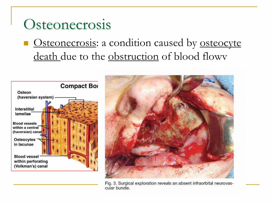
Osteonecrosis Osteonecrosis: a condition caused by osteocyte
death due to the obstruction of blood flowv
Aging of the Skeletal System
The Aging process
Bone is regularly broken down as new bone is
added
Rate of bone production is greater in young
people bones are denser, heavier, and larger
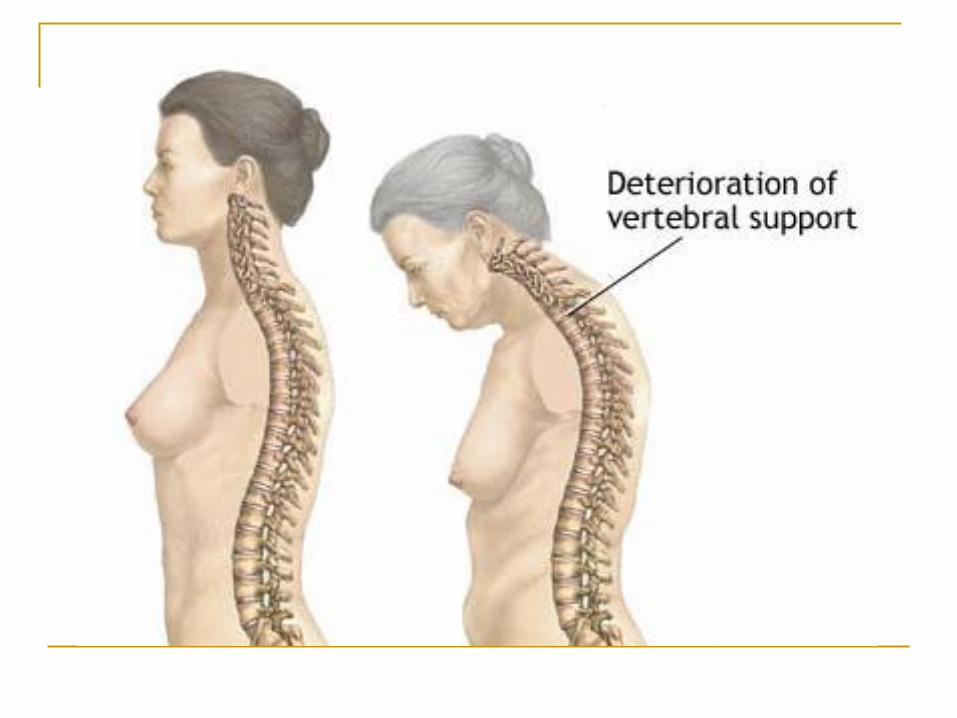
As we grow older, osteoclast activity outpaces
the bone rebuilding activity of the osteoblasts.
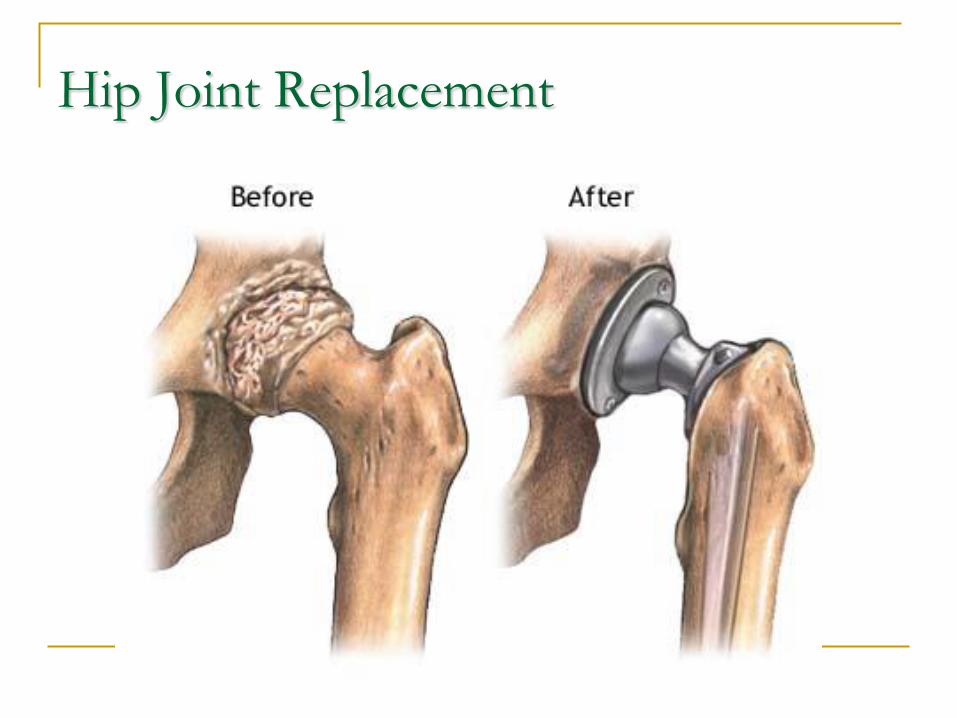
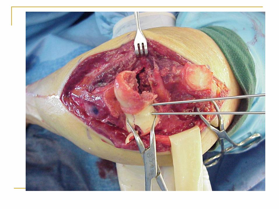
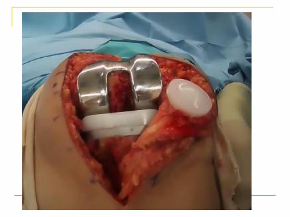
Hip Joint Replacement