Embed Size (px)
Citation preview
The Skeletal The Skeletal SystemSystem
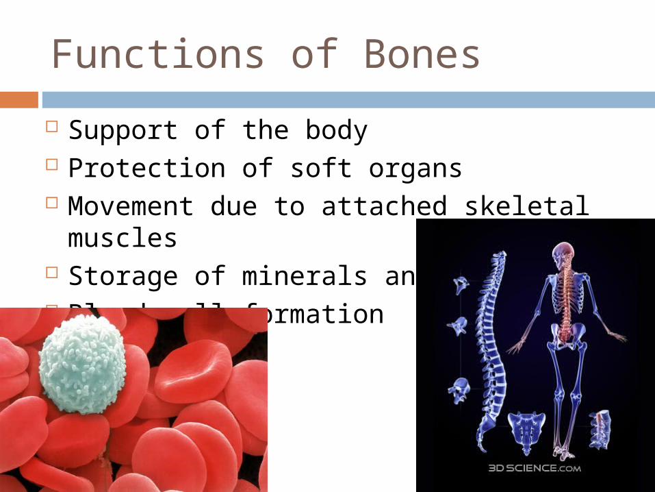
Functions of Bones
Support of the body Protection of soft organs Movement due to attached skeletal
muscles Storage of minerals and fats Blood cell formation
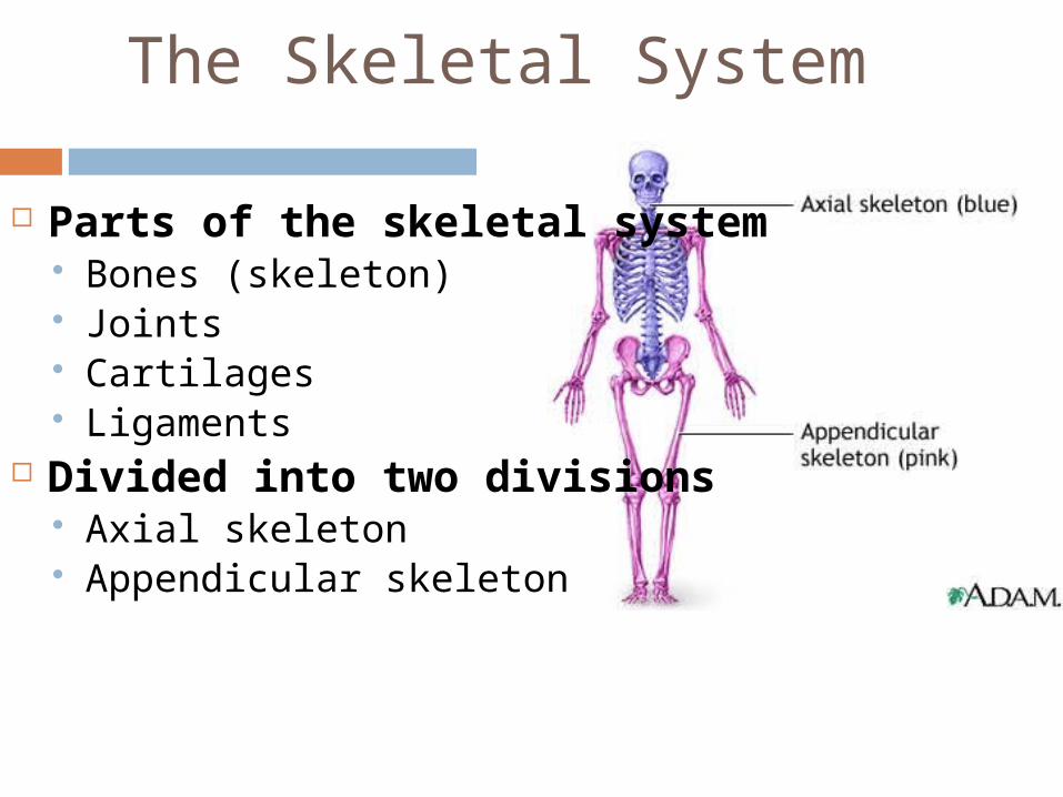
The Skeletal System
Parts of the skeletal system Bones (skeleton) Joints Cartilages Ligaments
Divided into two divisions Axial skeleton Appendicular skeleton
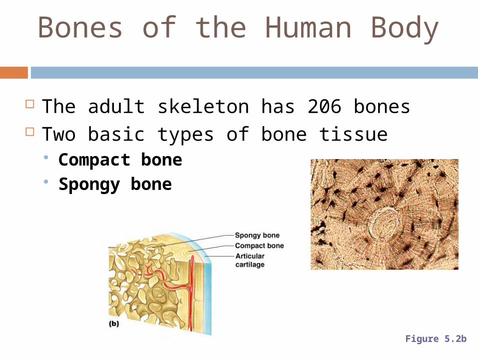
Bones of the Human Body
The adult skeleton has 206 bones Two basic types of bone tissue
Compact bone Spongy bone
Figure 5.2b
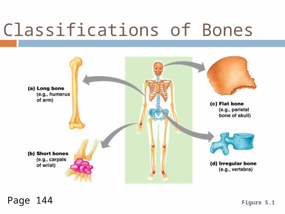
Classifications of Bones
Figure 5.1Page 144
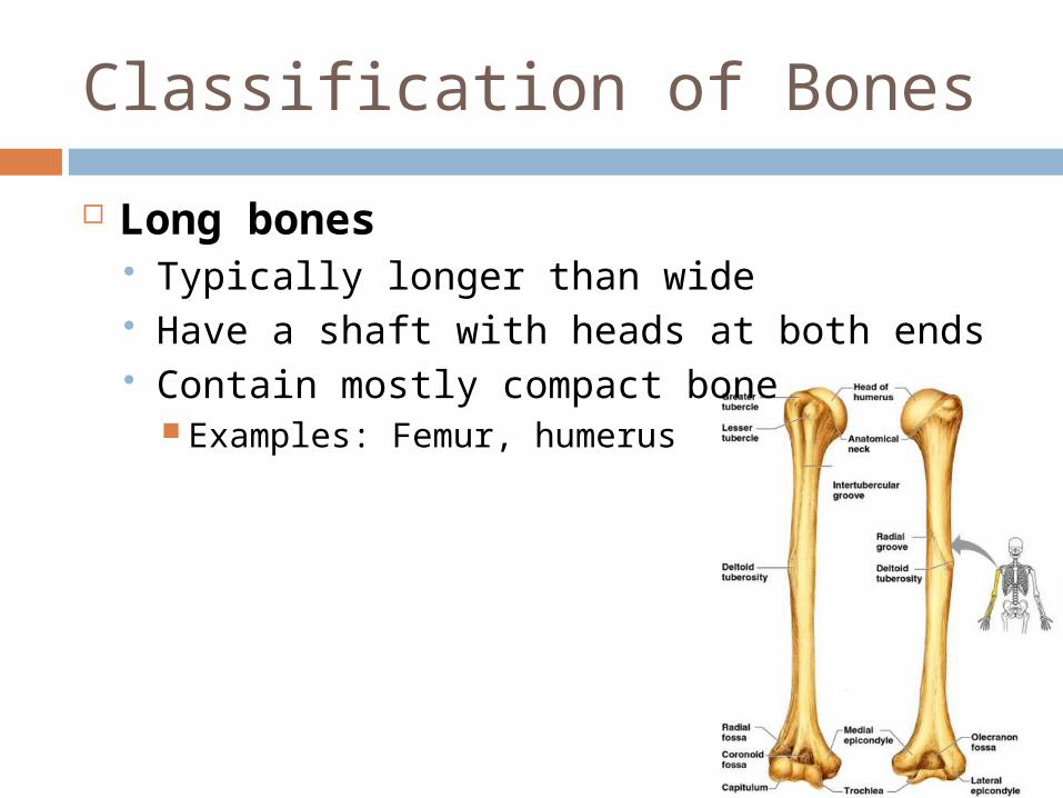
Classification of Bones
Long bones Typically longer than wide Have a shaft with heads at both ends Contain mostly compact bone
Examples: Femur, humerus
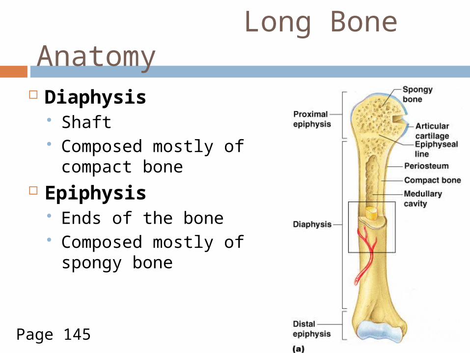
Long Bone Anatomy
Diaphysis Shaft Composed mostly of
compact bone Epiphysis
Ends of the bone Composed mostly of
spongy bone
Figure 5.2aPage 145
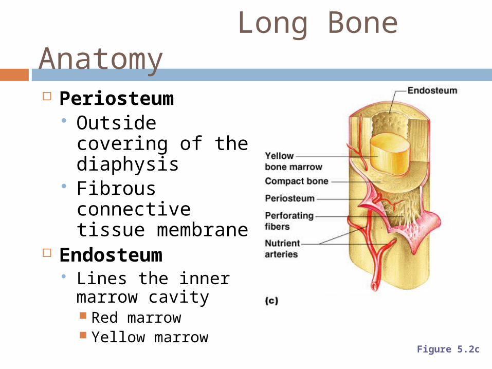
Long Bone Anatomy
Periosteum Outside covering
of the diaphysis Fibrous
connective tissue membrane
Endosteum Lines the inner
marrow cavity Red marrow Yellow marrow
Figure 5.2c
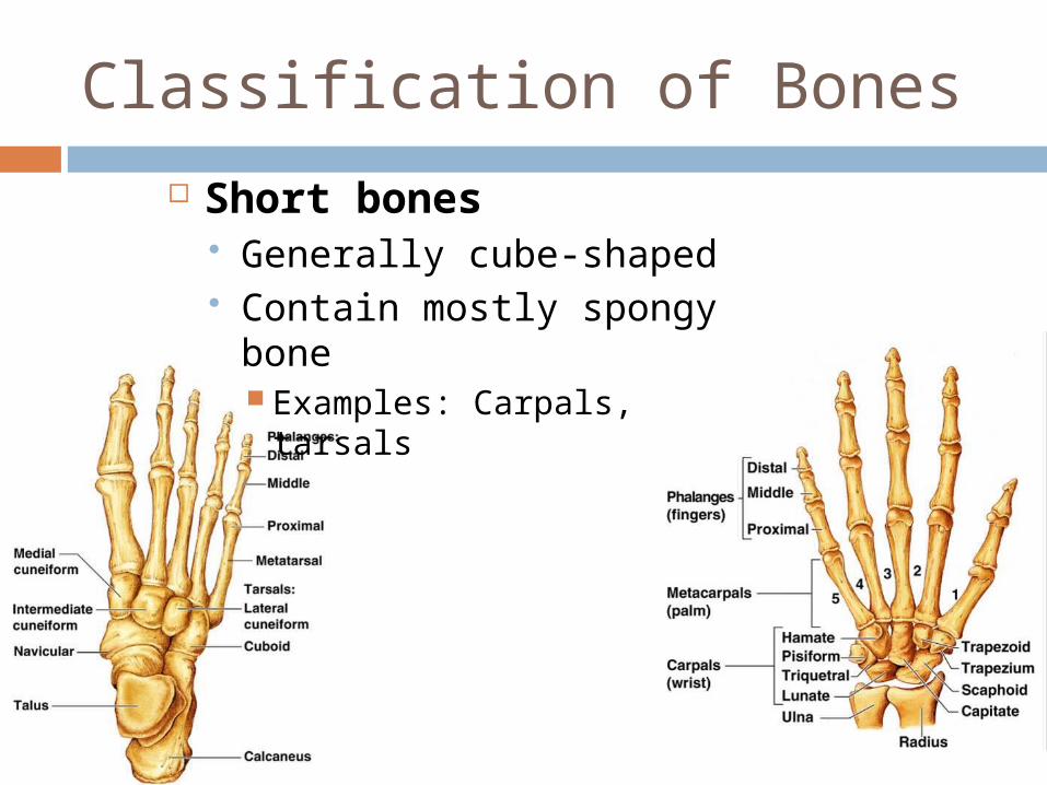
Classification of Bones
Short bones Generally cube-shaped Contain mostly spongy
bone Examples: Carpals, tarsals
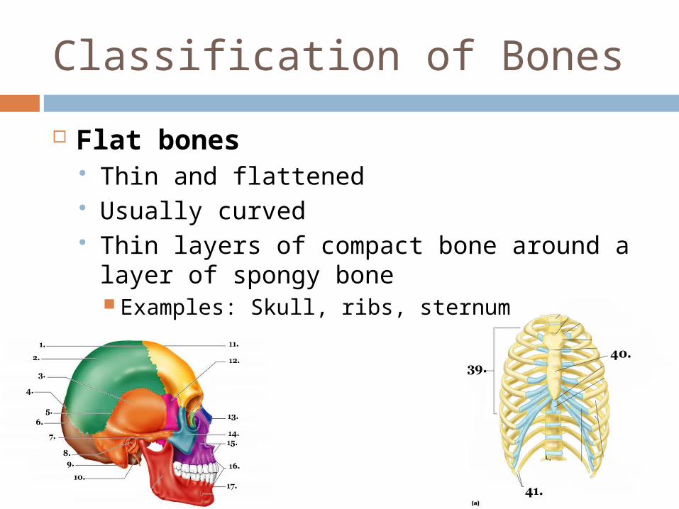
Classification of Bones
Flat bones Thin and flattened Usually curved Thin layers of compact bone around a layer
of spongy bone Examples: Skull, ribs, sternum
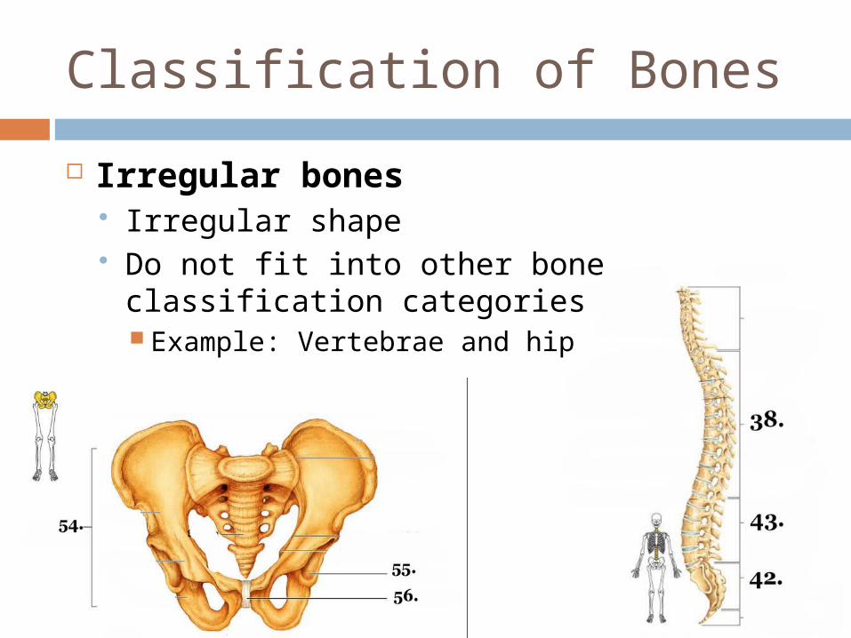
Classification of Bones
Irregular bones Irregular shape Do not fit into other bone classification
categories Example: Vertebrae and hip
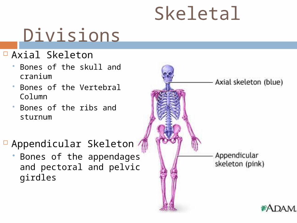
Skeletal Divisions Axial Skeleton
Bones of the skull and cranium
Bones of the Vertebral Column
Bones of the ribs and sturnum
Appendicular Skeleton Bones of the appendages
and pectoral and pelvic girdles
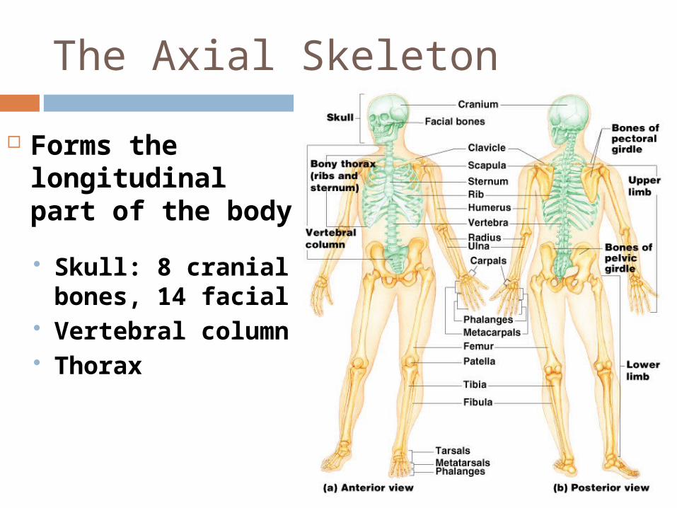
The Axial Skeleton
Forms the longitudinal part of the body
Skull: 8 cranial bones, 14 facial
Vertebral column Thorax
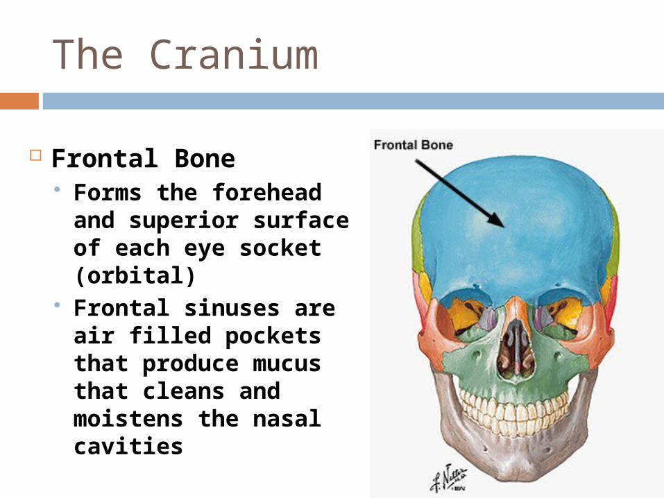
The Cranium
Frontal Bone Forms the forehead
and superior surface of each eye socket (orbital)
Frontal sinuses are air filled pockets that produce mucus that cleans and moistens the nasal cavities
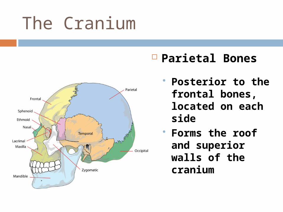
The Cranium
Parietal Bones
Posterior to the frontal bones, located on each side
Forms the roof and superior walls of the cranium
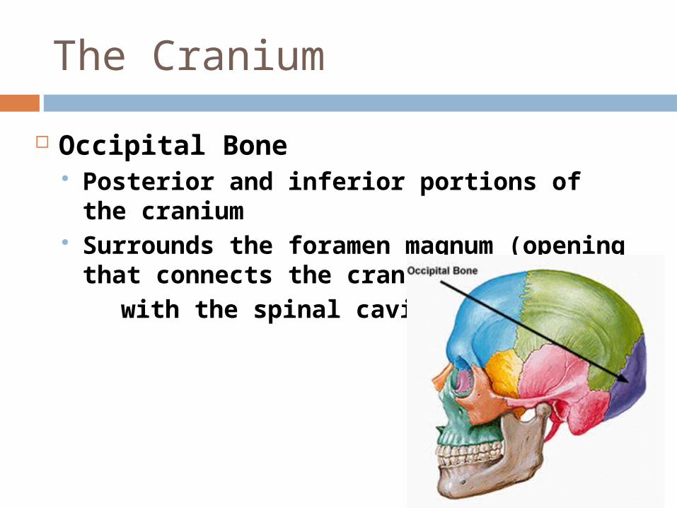
The Cranium
Occipital Bone Posterior and inferior portions of the
cranium Surrounds the foramen magnum
(opening that connects the cranial cavity
with the spinal cavity)
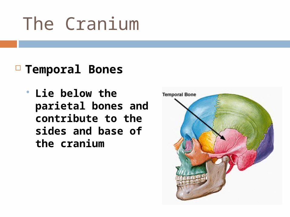
The Cranium
Temporal Bones
Lie below the parietal bones and contribute to the sides and base of the cranium
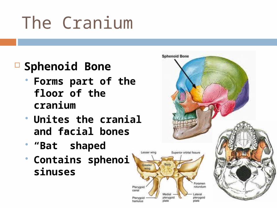
The Cranium
Sphenoid Bone Forms part of the
floor of the cranium Unites the cranial
and facial bones “Bat” shaped Contains sphenoidal
sinuses
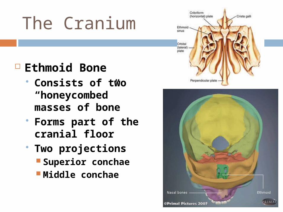
The Cranium
Ethmoid Bone Consists of two
“honeycombed” masses of bone
Forms part of the cranial floor
Two projections Superior conchae Middle conchae
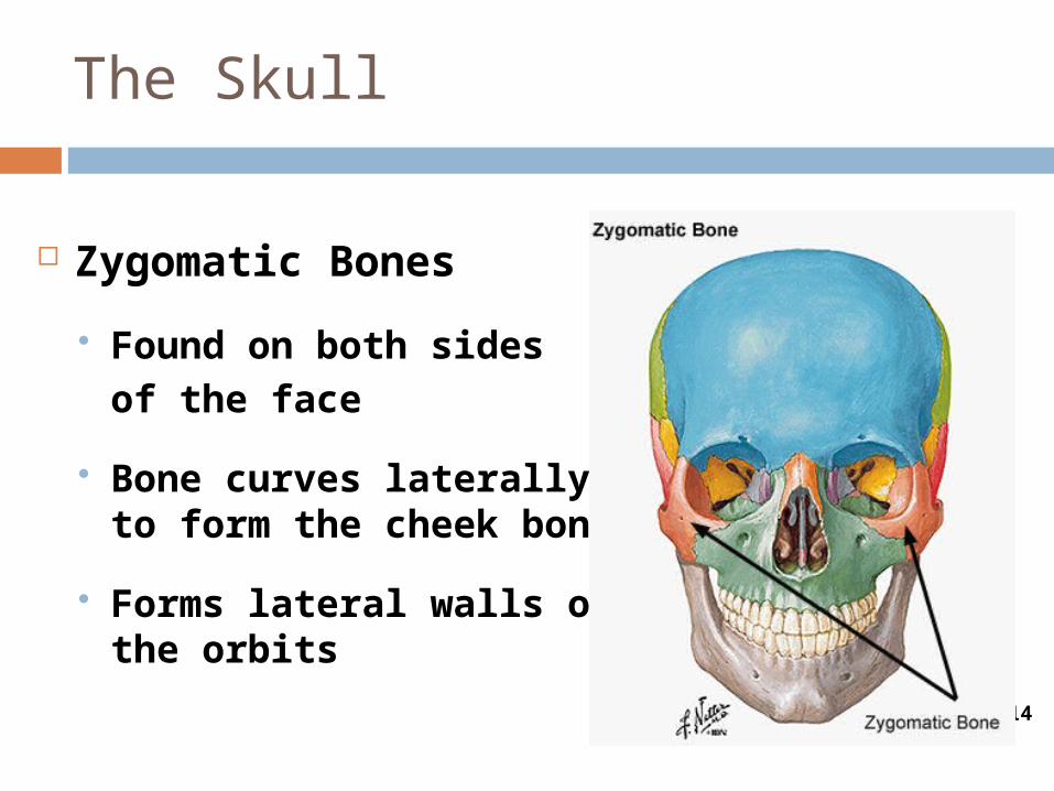
The Skull
Figure 6-14
Zygomatic Bones
Found on both sidesof the face
Bone curves laterally to form the cheek bone
Forms lateral walls of the orbits
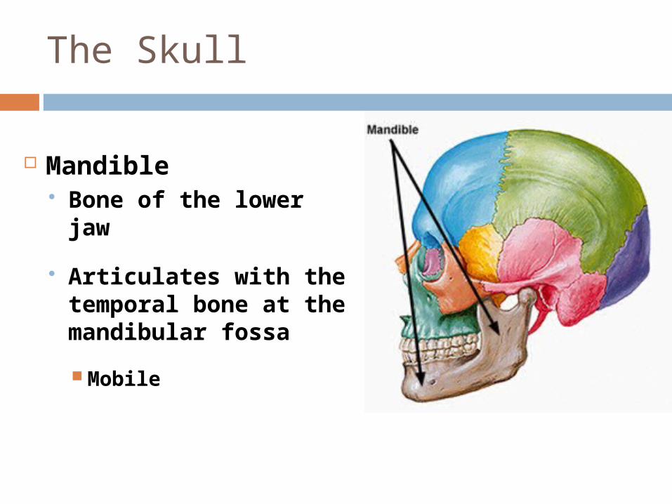
The Skull
Mandible Bone of the lower
jaw
Articulates with the temporal bone at the mandibular fossa
Mobile
The Skull
Figure 6-14
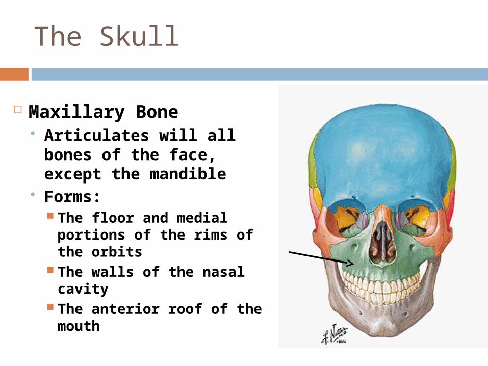
Maxillary Bone Articulates will all bones
of the face, except the mandible
Forms: The floor and medial
portions of the rims of the orbits
The walls of the nasal cavity
The anterior roof of the mouth
The Skull
Figure 6-14
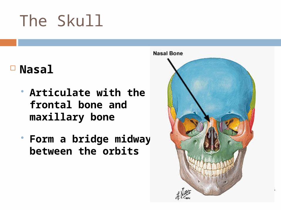
Nasal
Articulate with the frontal bone and maxillary bone
Form a bridge midway between the orbits
The Skull
Figure 6-14
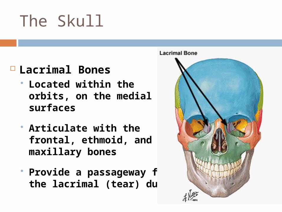
Lacrimal Bones Located within the orbits,
on the medial surfaces
Articulate with the frontal, ethmoid, and maxillary bones
Provide a passageway for the lacrimal (tear) ducts
The Skull
Figure 6-14
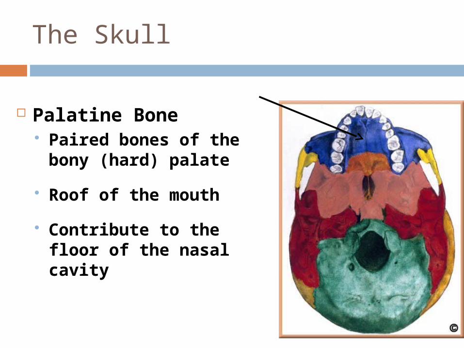
Palatine Bone Paired bones of the
bony (hard) palate
Roof of the mouth
Contribute to the floor of the nasal cavity
The Skull
Figure 6-14
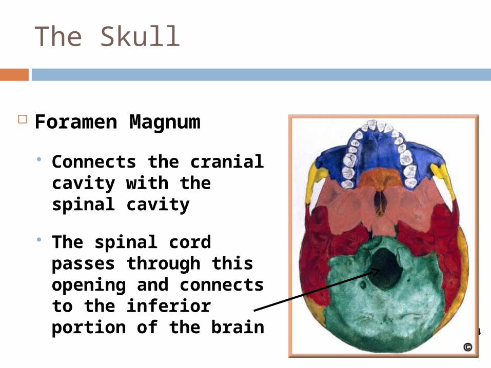
Foramen Magnum
Connects the cranial cavity with the spinal cavity
The spinal cord passes through this opening and connects to the inferior portion of the brain
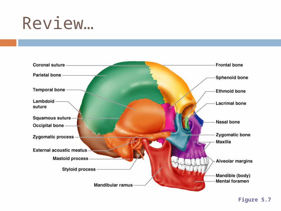
Review…
Figure 5.7
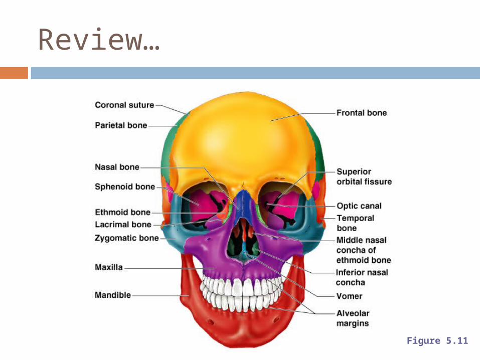
Review…
Figure 5.11
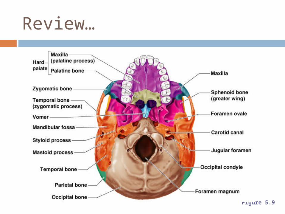
Review…
Figure 5.9