Embed Size (px)
Citation preview
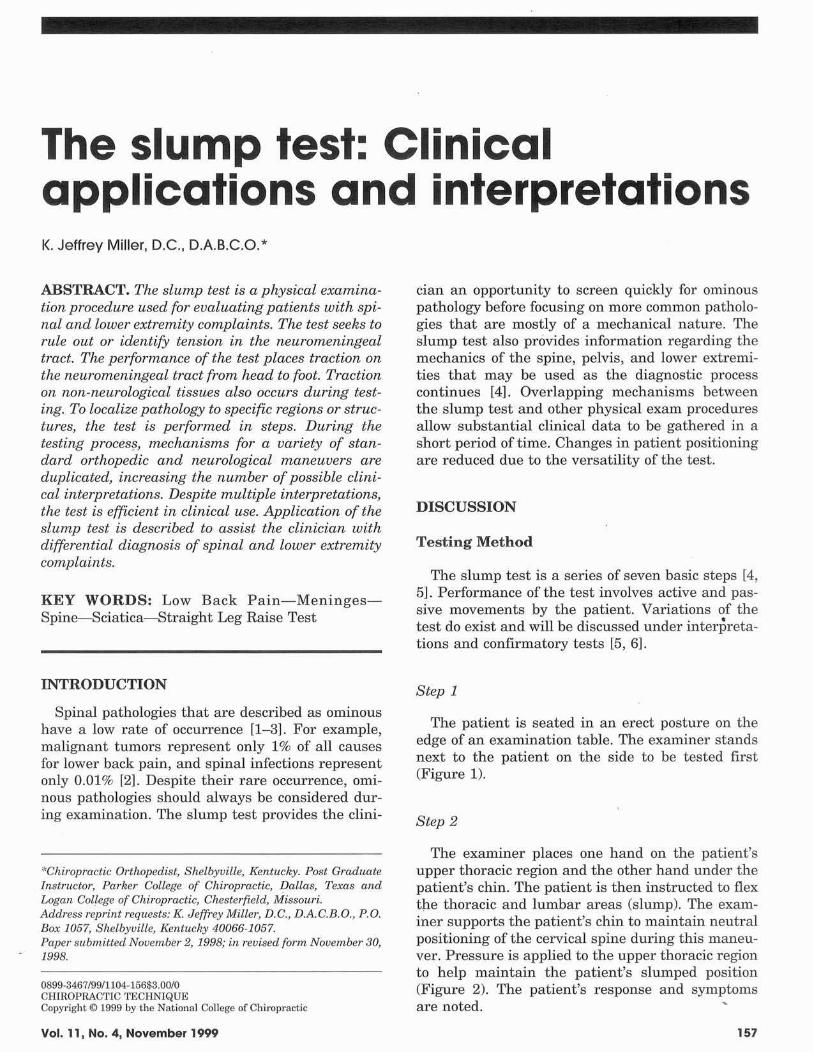
The slump test: Clinicalapplications and interpretationsK. Jeffrey Miller, D.C.. D.A.B.C.O.·
ABSTRACT. The slump test is a physical examination procedure used for evaluating patients with spinal and lower extremity complaints. The test seeks torule out or identifY tension in the neuromeningealtract. The performance of the test places traction onthe neuromeningeal tract from head to foot. Tractionon non-neurological tissues also occurs during testing. To localize pathology to specific regions or structures, the test is pelfarmed in steps. During thetesting proces§, mechanisms for a variety of standard orthopedic and neurological maneuvers areduplicated, increasing the number of possible clinical interpretations. Despite multiple interpretations,the test is efficient in clinical use. Application of theslump test is described to assist the clinician withdifferential diagnosis of spinal and lower extremitycomplaints.
KEY WORDS: Low Back Pain-MeningesSpine-Sciatica-8traight Leg Raise Test
INTRODUCTION
Spinal pathologies that are described as ominoushave a low rate of occurrence [1-3]. For example,malignant tumors represent only 1% of all causesfor lower back pain, and spinal infections representonly 0.01% [2]. Despite their rare occurrence, ominous pathologies should always be considered during examination. The slump test provides the clini-
·Chiropractic Orthopedist, Shelbyville, Kentucky. Post GraduateInstructor, Parker College of Chiropractic, pallas, Texas andLogoll Collcge o(Chiropractic, Chesterfield, Missouri.Address reprint requests: K. Jeffrey Miller, D.C., D.A.G.B.O., P.O.Box 1057, Shelbyville, Kentucky 40066·1057.Poper submitted Novcmber 2, 1998; ill revised {arm November 30,1998.
0899·346719911104·156$3.0010CHIROPRACTIC TECHNIQUECOllyright Q 1999 by the National College of Chiropractic
Vol. 11, No.4, November 1999
cian an opportunity to screen quickly for ominouspathology before focusing on more common pathologies that are mostly of a mechanical nature. Theslump test also provides information regarding themechanics of the spine, pelvis, and lower extremities that may be used as the diagnostic processcontinues [4]. Overlapping mechanisms betweenthe slump test and other physical exam proceduresallow substantial clinical data to be gathered in ashort period of time. Changes in patient positioningare reduced due to the versatility of the test.
DISCUSSION
Testing Method
The slump test is a series of seven basic steps [4,5]. Performance of the test involves active and passive movements by the patient. Variations of thetest do exist and will be discussed under interpretations and confirmatory tests [5, 6].
Step 1
The patient is seated in an erect posture on theedge of an examination table. The examiner standsnext to the patient on the side to be tested first(Figure 1).
Step 2
The examiner places one hand on the patient'supper thoracic region and the other hand under thepatient's chin. The patient is then instructed to flextpe thoracic and lumbar areas (slump). The examiner supports the patient's chin to maintain neutralpositioning of the cervical spine during this maneuver. Pressure is applied to the upper thoracic regionto help maintain the patient's slumped position(Figure 2). The patient's response and symptomsare noted.
157
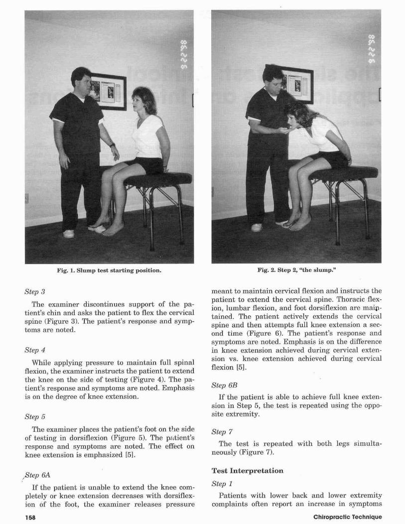
Fig. 1. Slump test starting position.
Step 3
The examiner discontinues support of the patient's chin and asks the patient to flex the cervicalspine (Figure 3). The patient's response and symptoms are noted.
Step 4
While applying pressure to maintain full spinalflexion, the examiner instructs the patient to extendthe knee on the side of testing (Figure 4). The patient's response and symptoms are noted. Emphasisis on the degree of knee extension.
Step 5
The examiner places the patient's foot on t!le sideof testing in dorsiflexion (Figure 5). The pfltient'sresponse and symptoms are noted. The effect onknee extension is emphasized [5].
/Step 6A
If the patient is unable to extend the knee completely or knee extension decreases with dorsiflexion of the foot, the examiner releases pressure
158
Fig. 2. Step 2, "the slump."
meant to maintain cervical flexion and instructs thepatient to extend the cervical spine. Thoracic flexion, lumbar flexion, and foot dorsiflexion are maiptained. The patient actively extends the cervicalspine and then attempts full knee extension a second time (Figure 6). The patient's response andsymptoms are noted. Emphasis is on the differencein knee extension achieved during cervical extension vs. knee extension achieved during cervicalflexion [5].
Step 6B
If the patient is able to achieve full knee extension in Step 5, the test is repeated using the opposite extremity.
Step 7
The test is repeated with both legs simultaneou.sly (Figure 7).
Test Interpretation
Step 1
Patients with lower back and lower extremitycomplaints often report an increase in symptoms
Chiropractic Technique
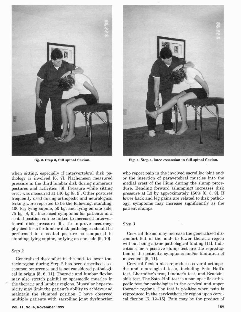
Fig. 3. Step 3, full spinal flexion.
when sitting, especially if intervertebral disk pathology is involved [6, 7]. Nachemson measuredpressure in the third lumbar disk during numerouspostures and activities [8). Pressure while sittingerect was measured at 140 kg [8, 9]. Other posturesfrequently used during orthopedic and neurologicaltesting were reported to be the following: standing,100 kg; lying supine, 50 kg; and lying on one side,75 kg [8, 9J. Increased symptoms for patients in aseated position can be linked to increased intervertebral disk pressure [9]. To improve accuracy,physical tests for lumbar disk pathologies should beperformed in a seated posture as compared tostanding, lying supine, or lying on one side [9, 10].
Step 2
Generalized discomfort in the mid- to lower thoracic region dLU'ing Step 2 has been described as acommon occurrence and is not considered pathological in origin [5,6, 11]. Thoracic and lumbar flexionmay also stretch painful or spasmodic muscles in
r the thoracic and lumbar regions. Muscular hypertonicity may limit the patient's ability to achieve andmaintain the slumped position. I have observedmultiple patients with sacroiliac joint dysfunction
Vol. 11, No.4, November 1999
Fig. 4. Step 4, knee extension in full spinal flexion.
who report pain in the involved sacroiliac joint and!or the insertion of paravetebral muscles into themedial crest of the ilium during the slump pFocedure. Bending forward (slumping) increases diskpressure at L3 by approximately 150% [6, 8, 9]. Iflower back and leg pains are related to disk pathology, symptoms may increase significantly as thepatient slumps.
Step 3
Cervical flexion may increase the generalized discomfort felt in the mid- to lower thoracic regionwithout being a true pathological finding [11]. Indications for a positive slump test are the reproduction of the patient's symptoms and/or limitation ofmovement [5, 11].
Cervical flexion also reproduces several orthopedic and neurological tests, including Soto-Hall'stest, Lhermitte's test, Lindner's test, and Brudzinski's test. The Soto-Hall test is a non~specific orthopedic test for pathologies in the cervical and upperthoracic regions. The test is positive when pain isreproduced in the cervicothol'acic region upon cervical flexion [6, 12-15]. Pain may be the product of
,.9
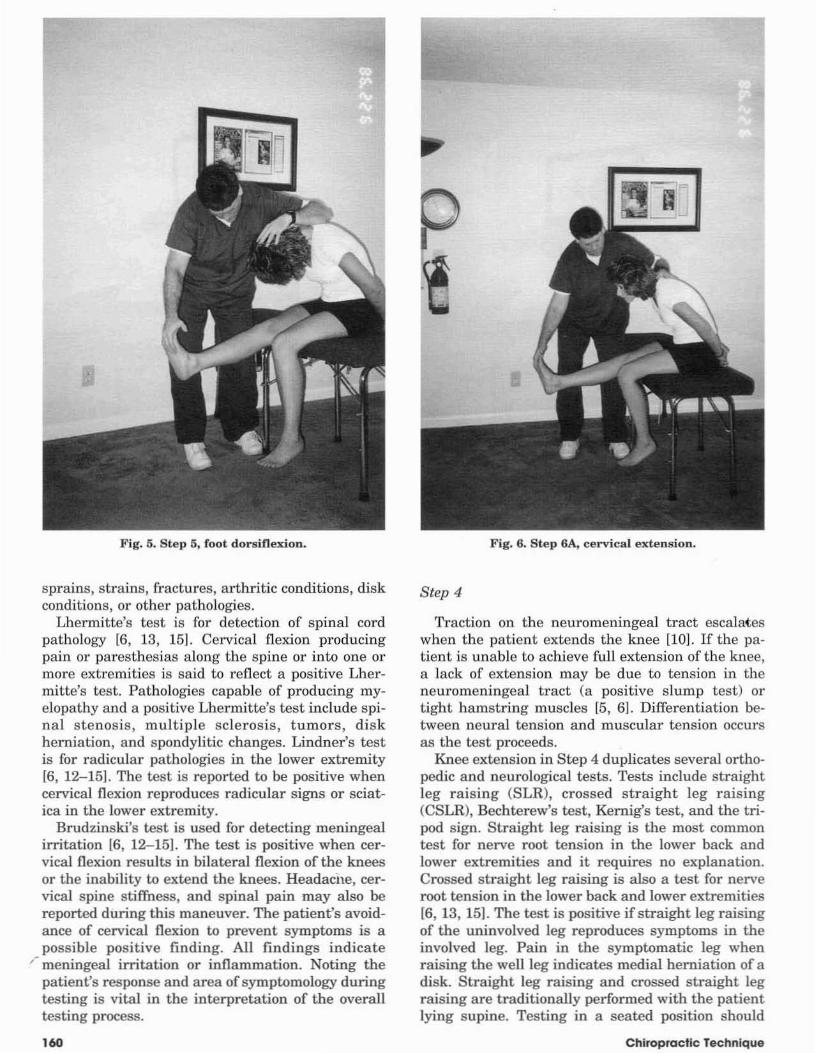
Fig. 5. Step 5, foot dorsiflexion.
sprains, strains, fractures, althritic conditions, diskconditions, or other pathologies.
Lhennitte's test is for detection of spinal cordpathology [6, 13, 151. Cervical flexion producingpain or paresthesias along the spine or into one ormore extremities is said to reflect a positive LherM
mitte's test. Pathologies capable of producing myelopathy and a positive Lhennitte's test include spinal stenosis, multiple sclerosis, tumors, diskherniation, and spondylitic changes. Lindner's testis for radicular pathologies in the lower extremity(6, 12-15J. The tesl is reported to be positive whencervical flexion reproduces radicular signs or sciatica in the lower extremity.
Brudzinski's test is used for detecting meningealirritation [6, 12-151. The test is positive when cerM
vical flexion results in bilateral flexion of the kneesor the inability to extend the knees. Headaclle, cervical spine stiffness, and spinal pain may also bereported during this maneuver. The patient's avoidance of cervical flexion to prevent symptoms is apossible positive finding. All findings indicate
,. meningeal irritation or inflammation. Noting thepatient's response and area of symptomology duringtesting is vital in the interpretation of the overalltesting process.
160
Fig. 6. Step GA, cervical extension.
Step 4
Traction on the neuromeningeal tract escalateswhen the patient extends the knee [10]. If the patient is unable to achieve full extension of the knee,a lack of extension may be due to tension in theneuromeningeal tract (a positive slump test) ortight hamstring muscles [5, 6]. Differentiation between neural tension and muscular tension occursas the test proceeds.
Knee extension in Step 4 duplicates several orthopedic and neurological tests. Tests include straightleg raising (SLR), crossed straight leg raising(CSLR), Bechterew's test, Kernig's test, and the tripod sign. Straight leg raising is the most commontest for nerve root tension in the lower back andlower extremities and it requires no explanation.Crossed straight leg raising is also a test for nerveroot tension in the lower back and lower extremities[6,13,151. The test is positive if straight leg raisingof the uninvolved leg reproduces symptoms in theinvolved leg. Pain in the symptomatic leg whenraising the well leg indicates medial herniation of adisk. Straight leg raising and crossed straight legraising are traditionally perfonned with the patientlying supine. Testing in a seated position should
Chiropractic Technique
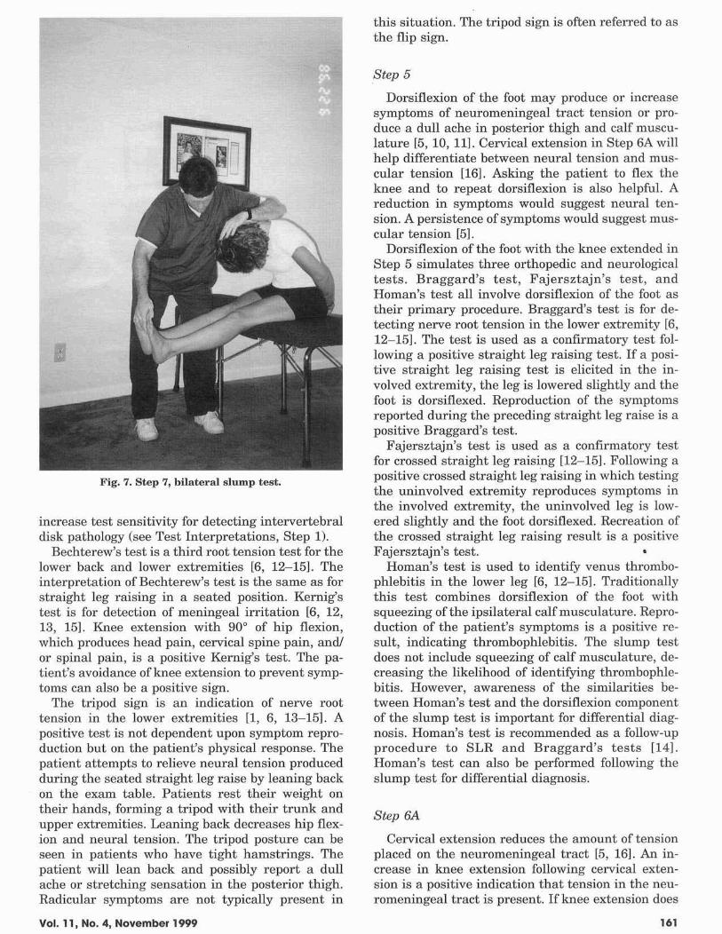
Fig. 7. Step 7, bilateral slump test.
increase test sensitivity for detecting intervertebraldisk pathology (see Test Interpretations, Step 1).
Bechterew's test is a third root tension test for thelower back and lower extremities [6, 12-151. Theinterpretation of Bechtel'ew's test is the same as forstraight leg raising in a seated position. Kernig'stest is for detection of meningeal irritation [6, 12,13, 151. Knee extension with 90° of hip flexion,which produces head pain, cervical spine pain, and!or spinal pain, is a positive Kernig's test. The patient's avoidance of knee extension to prevent symptoms can also be a positive sign.
The tripod sign is an indication of nerve roottension in the lower extremities fl, 6, 13-15]. Apositive test is not dependent upon symptom reproduction but on the patient's physical response. Thepatient attempts to relieve neural tension producedduring the seated straight leg raise by leaning backon the exam table. Patients rest their weight ontheir hands, forming a tripod with their trunk andupper extremities. Leaning back decreases hip flexion and neural tension. The tripod posture can beseen in patients who have tight hamstrings. Thepatient will lean back and possibly report a dullache or stretching sensation in the posterior thigh.Radicular symptoms are not typically present in
Vol. 11, No.4, November 1999
this situation. The tripod sign is often referred to asthe flip sign.
Step 5
Dorsiflexion of the foot may produce or increasesymptoms of neuromeningeal tract tension or produce a dull ache in posterior thigh and calf musculature [5, 10, 11J. Cervical extension in Step 6A willhelp differentiate between neural tension and muscular tension [16}. Asking the patient to flex theknee and to repeat dorsiflexion is also helpful. Areduction in symptoms would suggest neural tension. A persistence of symptoms would suggest muscular tension [5].
Dorsiflexion of the foot with the knee extended inStep 5 simulates three orthopedic and neurologicaltests. Braggard's test, Fajersztajn's test, andHoman's test all involve dorsiflexion of the foot astheir primary procedure. Braggard's test is for detecting nerve root tension in the lower extremity [6,12-15]. The test is used as a confirmatory test following a positive straight leg raising test. If a positive straight leg raising test is elicited in the involved extremity, the leg is lowered slightly and thefoot is dorsiflexed. Reproduction of the symptomsreported during the preceding straight leg raise is apositive Braggard's test.
Fajersztajn's test is used as a confirmatory testfor crossed sh·aight leg raisi.ng [12~151. Following apositive crossed straight leg raising in which testingthe uninvolved extremity reproduces symptoms inthe involved extremity, the uninvolved leg is lowered slightly and the foot dorsiflexed. Recreation ofthe crossed straight leg raising result is a positiveFajersztajo's test. •
Homan's test is used to identify venus thrombophlebitis in the lower leg [6, 12-15]. Traditionallythis test combines dorsiflexion of the foot withsqueezing of the ipsilateral calf musculature. Reproduction of the patient's symptoms is a positive result, indicating thrombophlebitis. The slump testdoes not include squeezing of calf musculatw·e, decreasing the likelihood of identifying thrombophlebitis. However, awareness of the similarities be·tween Homan's test and the dorsiflexion componentof the slump test is important for differential diagnosis. Homan's test is recommended as a follow-upprocedure to SLR and Braggard's tests [14].Homan's test can also be performed following theslump test for differential diagnosis.
Step 6A
Cervical extension reduces the amount of tensionplaced on the neuromeningeal tract [5, 16]. An increase in knee extension following cervical extension is a positive indication that tension in the neuromeningeal tract is present. If knee extension does
161
not increase, muscular tightness should be considered the primary cause for limited extension.
Step 6B
Both extremities are examined individually re·gardless of unilateral or bilateral presentation ofsymptoms. All diagnostic possibilities describedfrom Step 1 through Step 6A apply when testing thesecond extremi ty.
Step 7
Testing the lower extremities simultaneouslyplaces peak tension on the neuromeningeal tract [5,171. Bilateral testing is most important when symptoms are reproduced during testing of the unin·valved extremity. Patients experiencing only muscular discomfort in the full slumped position duringunilateral and/or bilateral testing have a negativeslump test. In this situation, Soto-Hall's, Lhermitte's, Lindner's, Brudzinski's, SLR, CSLR, Bechterew's, Kernig's, tripod sign, Braggard's, Fajersztajn's, and Homan's tests are negative. Performingthe slump test early during examination covers avariety of tests and can rule out several ominouspathologies. The e'xaminer can then proceed in testing for more common conditions.
Test Use
Patient history is the starting point for determining if the slump test is used. All patients describinglower back pain, leg pain, radicular symptoms,trauma, spinal cord symptoms, headaches, cervicalpain, and thoracic spine pain are candidates for theslump procedure [4-6]. Additional candidates areidentified by reports of symptom reproduction during daily activities similar to the slump test. Possibilities include extending the legs to operate footpedals while driving, sitting in a recliner and sittingin a bathtub [6, 18].
Positive results of individual orthopedic and neurological tests described under interpretations (Sota-Hall, SLR, Braggard's, etc.) warrant the use ofthe slump test for differential diagnosis [10]. Theslump test as a treatment procedure has been described [5, 11, 19]. Use as a therapeutic procedurewill not be discussed.
Slump Test Variations
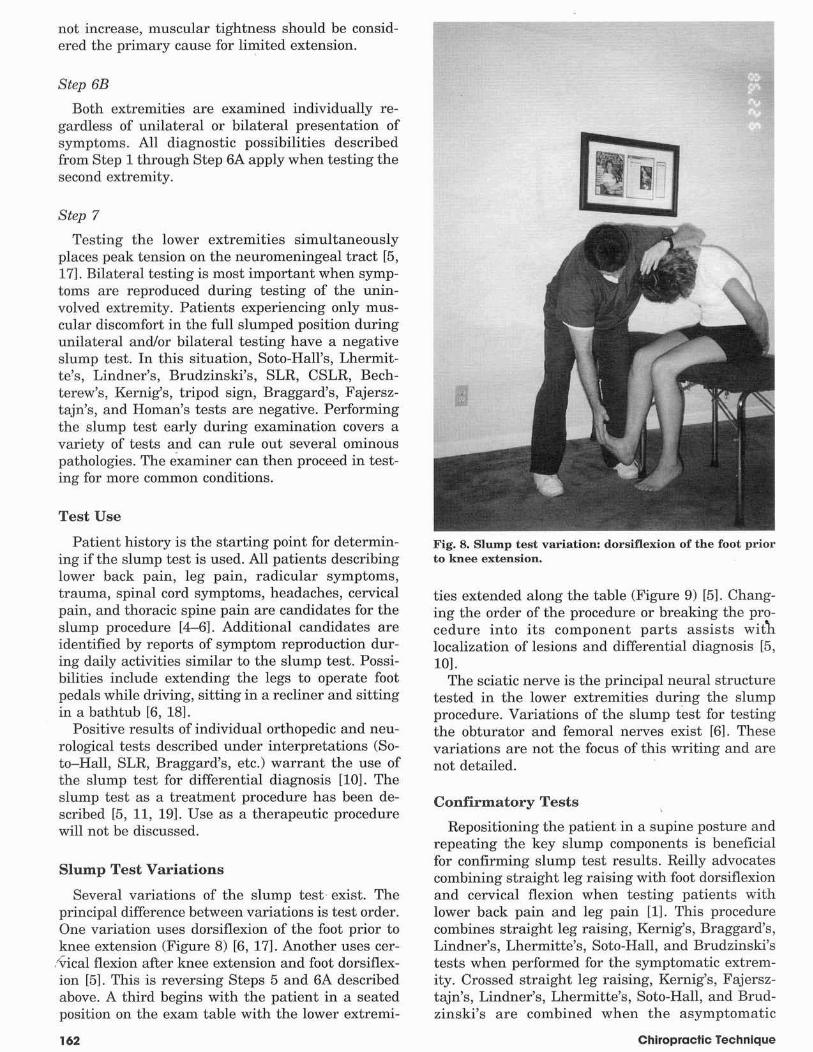
Several variations of the slump test· exist. Theprincipal difference between variations is test order.One variation uses dorsiflexion of the foot prior toknee extension (Figure 8) [6, 17]. Another uses eel'·
:'Vical flexion after knee extension and foot dorsiflexion [5]. This is reversing Steps 5 and 6A describedabove. A third begins with the patient in a seatedposition on the exam table with the lower extremi-
162
Fig. 8, Slump test variation: dorsinexion of the foot prim'to knee extension.
ties extended along the table (Figure 9) [5]. Changing the order of the procedure or breaking the procedure into its component parts assists wit11localization of lesions and differential diagnosis [5,1OJ.
The sciatic nerve is the principal neural structuretested in the lower extremities during the slumpprocedure. Variations of the slump test for testingthe obturator and femoral nerves exist [6]. Thesevariations are not the focus of this writing and arenot detailed.
Conf"rrmatory Tests
Repositioning the patient in a supine posture andrepeating the key slump components is beneficialfor confirming slump test results. Reilly advocatescombining straight leg raising with foot dorsiflexionand cervical flexion when testing patients withlower back pain and leg pain [1]. This procedurecombines straight leg raising, Kernig's, Braggard's,Lindner's, Lhermitte's, Soto-Hall, and Brudzinski'stests when performed for the symptomatic extremity. Crossed straight leg raising, Kernig's, Fajersztajn's, Lindner's, Lhermitte's, Soto-Hall, and Brudzinski's are combined when the asymptomatic
Chiropractic Technique
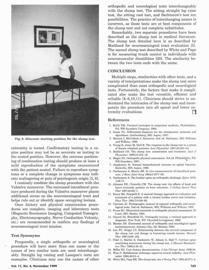
Fig. 9. Alternate starting position for the slump test.
extremity is tested. Confirmatory testing in a supine position may not be as accurate as testing inthe seated position. However, the extreme positioning of combination testing should produce at least amild reproduction of the symptoms encounteredwith the patient seated. Failure to reproduce symptoms or a complete change in symptoms may indicate malingering or pain of psychogenic origin [3, 6].
I routinely combine the slump procedure with theValsalva maneuver. The increased intrathecal pressure produced during the Valsalva maneuver placesadditional stress on the neuromeningeal tract andhelps rule out or identify space occupying lesions.
Once history and physical examination procedures are complete, imaging and laboratory tests(Magnetic Resonance Imaging, Computed Tomography, Electromyography, Nerve Conduction Velosity,etc.) are recommended to confirm any findings ofneuromeningeal tract tension.
Test Synonyms
Frequently, a single orthopedic or neurologicalprocedure will have more than one name or thenames of two similar tests are used interchangeably. Straight leg raising and Lasegue's tests areexamples. Clinicians may use the names of other
Vol. 11, No.4, November 1999
orthopedic and neurological tests interchangeablywith the slump test. The sitting straight leg raisetest, the sitting root test, and Bechterew's test arepossibilities. The practice of interchanging names isincorrect, as these tests are at best components ofthe slump test and not complete substitutes.
Remarkably, two separate procedures have beendescribed as the slump test in medical literature.The slump test detailed here is as described byMaitland for neuromeningeal tract evaluation [5].The second slump test described by White and Papeis for measuring trunk control in individuals withneuromuscular disabilities [201. The similarity between the two tests ends with the name.
CONCLUSION
Multiple steps, similarities with other tests, and avariety of interpretations make the slump test morecomplicated than most orthopedic and neurologicaltests. Fortunately, the factors that make it complicated also make the test versatile, efficient andreliable [4-6,10,11]. Clinicians should strive to understand the intricacies of the slump test and incorporate the procedure into all spinal and lower extremity evaluations. rlReferences
1. Reilly HM. Practical strategies in outpatient medicine. Philadelphin,PA: WB Saunders Company; 1984.
2. Souza TA. Differential diagnoais for the chiropmctor: protocols andalgorithms. Gaithersburg. MD: Aspen: 1997.
3. Macnab I. McCulloch J. Backache. 2nd ed. Bnltimore. MD: Williamsand Wilkins; 1990.
4. Yeung E. Jones M, Hall B. The response to the slump test in a groupof female whiplash patients. Alisf Physiother 1997:43:245-52.
5. Maitland GO. The slump test: elCamilllltion nnd trentment. AIlsl JPhysiothcr 1986:31:215-19.
6. Magee OJ. Orthopedic physical assessment. 3rd ed. Philadelphia, PA:WB Saunders; 1997. •
7. Junghanns H. Normal biomechanical stress.es on spinal runction.Rockville. MD: Aspen; 1990.
8. Nachemson A. Morris JM. In vivo meaaurements of intradiscal pres·sure. J 80lle Joint Surg 1964:5:1077-92.
9. Nachemson A. The lumbar spine an orthopedic challenge. Spi11e 1976;1:59·71.
10. Johnson EK, Chiarello CM. The slump lest: the effectB of hefld andlower extromity position on knee extension. J Orthop Sports PhysTher 1997;6:310-7.
11. Koury MJ, Scarpelli E. A manual therapy approach to evnlulltion andtreatment of a pntient with a chronic lumbar nerve root irritation.Phys Tlwr 1994;74:548-60.
12. Cipriano JJ. Photographic mnnual of regional orthopedic nnd neurologicsl tests. 2nd ed. Baltimore, MD:. Williams and Wilkins; 1991.
13. Evans RC. Illustrated essentinls in orthopedic physical asse5sment. 5tLouis, MO: Mosby; 1994.
14. Gerard JA, Kleinfield SL.. Orthopedic testing: 9. rational approach todingnosis. New York, NY: Churchill Livingstone; 1993.
15. Ma2:ion JM. Illustrated manual of orthopedic and neurological sign&'tests/maneuvcrs. Ariwna City, AZ: Mll2:ion; 1980.
16. Lew PC, Briggs CA. Relntionship between the cervicnl component ofthe slump leSt and change in hamstring muscle tension. ManllalThcr 1997;2:98-105.
17. Fidel C, Mnrtin E. Dankaerts W, Allison G. Hall T. Cervicnl spinesensitizing maneuvers during the slump test. J Monual Mallipula·tille Tllcr 1996;4:16--21.
18. Miller KJ. Case history documentntion. JAm Chiropr Assoc 1996:34.19. Wise P. Mobilization technique improves neural mobility. Allst Phys·
iother 1994;40:51-4.20. White MA, Pape KE. The slump test. Am JOcCllp Ther 1992;46:271-4.
163


![MA] OR SLUMP - Marxists](https://img.pdfslide.net/doc/110x75/620116a0fdba16249f074845/ma-or-slump-marxists.jpg)