Embed Size (px)
Citation preview
1
THE USE OF 320 DETECTOR COMPUTED TOMOGRAPHY CORONARY ANGIOGRAPHY TO DIAGNOSE CORONARY ARTERY DISEASE IN EMERGENCY
DEPARTMENT PATIENTS WITH CHEST PAIN
By
DAVID E. WINCHESTER
A THESIS PRESENTED TO THE GRADUATE SCHOOL OF THE UNIVERSITY OF FLORIDA IN PARTIAL FULFILLMENT
OF THE REQUIREMENTS FOR THE DEGREE OF MASTER OF SCIENCE
UNIVERSITY OF FLORIDA
2011

2
© 2011 David E. Winchester

3
To Mom and Dad, for their love and support

4
ACKNOWLEDGMENTS
My achievements would not have been possible without the loving support of my
mother, father, and especially my wife Erin, who is my advocate, mentor, and best
friend. I appreciate the time and resources provided for me by the Division of
Cardiovascular Medicine to seek this Master of Science degree. Further, I would like to
extend my gratitude and thanks to my many mentors and colleagues for this and other
research endeavors including Dr. James Hill, Dr. Anthony Bavry, Dr. Carl Pepine, Dr.
Steven Kraft, Dr. Marian Limacher, Dr. Nabih Asal, Dr. David Wymer, Dr. Preeti Jois,
Dr. John Petersen, Dr. Rhonda Cooper-DeHoff, Dr. Eileen Handberg, Dr. William
Brearley, and Dr. Ki Park.

5
TABLE OF CONTENTS page
ACKNOWLEDGMENTS .................................................................................................. 4
LIST OF TABLES ............................................................................................................ 6
LIST OF FIGURES .......................................................................................................... 7
LIST OF ABBREVIATIONS ............................................................................................. 8
ABSTRACT ..................................................................................................................... 9
CHAPTER
1 INTRODUCTION .................................................................................................... 12
2 METHODS .............................................................................................................. 16
Study Design and Setting ....................................................................................... 16 Patient Selection ..................................................................................................... 16 Data Collection ....................................................................................................... 17 Outcomes ............................................................................................................... 17 CTCA Acquisition .................................................................................................... 18 Statistical Methods and Data Analysis .................................................................... 19
3 RESULTS ............................................................................................................... 21
Baseline Characteristics ......................................................................................... 21 Duration of Stay ...................................................................................................... 21 Detection of CAD and Clinical Outcomes ............................................................... 22
4 DISCUSSION AND CONCLUSIONS ...................................................................... 28
Discussion .............................................................................................................. 28 Conclusions ............................................................................................................ 32
LIST OF REFERENCES ............................................................................................... 33
BIOGRAPHICAL SKETCH ............................................................................................ 36

6
LIST OF TABLES
Table page 3-1 Baseline Characteristics ..................................................................................... 25
3-2 Outcomes ........................................................................................................... 26
3-3 Recidivism Patients ............................................................................................ 27
7
LIST OF FIGURES
Figure page 2-1 Cohort Selection Process ................................................................................... 20
3-1 Emergency department duration of stay ............................................................. 23
3-2 Results of CTCA ................................................................................................. 24

8
LIST OF ABBREVIATIONS
ACS acute coronary syndrome
CAC coronary artery calcium
CAD coronary artery disease
CT computed tomography
CTCA computed tomography coronary angiography
CVD cardiovascular disease
ED emergency department
ETT exercise treadmill test
HR heart rate
IQR interquartile range
MI myocardial infarction
MPI myocardial perfusion imaging
9
Abstract of Thesis Presented to the Graduate School of the University of Florida in Partial Fulfillment of the Requirements for the Degree of Master of Science
THE USE OF 320 DETECTOR COMPUTED TOMOGRAPHY CORONARY
ANGIOGRAPHY TO DIAGNOSE CORONARY ARTERY DISEASE IN EMERGENCY DEPARTMENT PATIENTS WITH CHEST PAIN
By
David E. Winchester
May 2011
Chair: Marian Limacher Major: Medical Sciences – Clinical and Translational Science
Chest pain is a common problem with over 7 million emergency department (ED)
visits in the U.S. annually. ED physicians are charged with establishing the absence of
potentially serious conditions and with accurately diagnosing the source of chest pain
without unduly burdening the patient with an extended duration of stay or unnecessary
admission to the hospital. Two crucial diagnoses in the cardiac differential diagnosis of
chest pain include acute coronary syndrome (ACS) and obstructive coronary artery
disease (CAD).
The likelihood of ACS can be minimized with normal findings using a thorough
history and physical examination, serial measurements of cardiac serum biomarkers,
and serial electrocardiograms. Typically, however, the diagnosis of obstructive CAD
requires additional testing. This testing might include electrocardiographic treadmill
testing (ETT), myocardial perfusion imaging (MPI), or stress echocardiography and
frequently these tests are not available in an ED setting.
A newer technology, computed tomography coronary angiography (CTCA), has
sensitivity and specificity for CAD superior to ETT and similar to MPI for establishing the
10
diagnosis of CAD. CTCA can be reliably performed on a 64-detector computed
tomography (CT) scanner which is a tool available in most EDs throughout the day and
on every day of the week.
At the University of Florida, the ED faculty was concerned about poor follow-up
for patients discharged after chest pain evaluation. To address this, the ED recently
switched from a strategy of ordering outpatient stress tests for patients with chest pain,
to ordering CTCA in the ED and prior to discharge for these patients. We hypothesized
that this change in strategy would reduce the ED duration of stay and increase the
detection of CAD. Using two cohorts of patients (n = 50 in each, total n = 100) we
compared the duration of stay in the ED and the detection of CAD in patients before and
after this change in clinical care to determine the impact of CTCA.
The duration of stay was not significantly different between the cohorts (417.5
minutes for the CT cohort, 400.0 for the control cohort, p = 0.53). Substantially more
patients in the CT cohort completed the test ordered for them (96% versus 36% for
control cohort, p < 0.0001) resulting in more patients being diagnosed with CAD (28%
versus 2% in control cohort, p = 0.0004). More patients in the CT cohort were
diagnosed with obstructive CAD, (12% versus 2%, p = 0.11) although this difference
was not statistically significant. Within 3 months of the index ED visit, recidivism was the
same in both cohorts (n = 4, 8%) and no patients in either cohort suffered myocardial
infarction (MI) or death.
In conclusion, for patients who present to the ED with chest pain who need
additional testing for CAD, a strategy of using CTCA prior to ED discharge is more
effective than a strategy of outpatient follow-up testing. The CTCA based strategy

11
detected more CAD, primarily due to low likelihood of follow-up in the stress testing
cohort. Using CTCA did not significantly change the duration of stay in the ED or reduce
ED recidivism. No patients suffered MI or death within 3 months of their ED visit.
12
CHAPTER 1 INTRODUCTION
Cardiovascular disease (CVD) is the leading cause of mortality in the United
States for both men and women.1 The best estimate is that over 82 million Americans
(over one in three) suffers from some form of CVD. Fortunately, the rate of death from
CVD has decreased 27.8% from 1997 to 2007.2 Nearly half of this decrease has been
attributed to increased use of evidence-based medical therapies.3
An important technological component of evidence based medical therapies
includes advanced techniques for diagnosing CVD. Several modalities of functional and
anatomic testing for CVD are available for physicians to apply in patient care. Of course,
no test is completely accurate and each method has unique limitations and levels of
precision. One of the biggest challenges, therefore, lies in selecting the right test for the
right patient at the right time, ideally based on the patient’s pretest likelihood of CVD.4
For example, in evaluating a patient who complains of chest pain, considering
the pretest likelihood that the patient has CVD is helpful in selecting the optimal test.
Young patients without significant CVD risk factors are unlikely to have CVD. Ordering a
test with a low positive predictive value increases the chance of a false positive test,
potentially subjecting the patient to unnecessary testing. On the opposite end of the
spectrum, older patients with many CVD risk factors are likely to have CVD. In these
patients, a test with a low negative predictive value increases the chance of a false
negative test, potentially delaying an accurate diagnosis of CVD. Patients with
intermediate pretest likelihood of CVD are the most likely to receive valuable diagnostic
information from noninvasive testing and are therefore the ideal population for these
tests.5-7
13
While the symptoms of CVD are legion, the best known to both physicians and
the public is chest pain. Because this symptom is well known to be associated with
CVD, patients frequently consider chest pain to be an emergency and seek medical
attention in a nearby emergency department (ED). In fact, over 7 million ED visits
annually result from a complaint of chest pain.8 While ED physicians are primarily
responsible for identifying life-threatening conditions, they also find themselves in an
increasing role as the front line interface between the public and the medical
community. Therefore, they bear some responsibility to help patients become
established within the healthcare system and must also offer appropriate follow-up.
Making accurate diagnoses facilitates better follow-up, and the diagnosis of CVD is
important to make. Not only does CVD carry a high burden of mortality, as previously
discussed, but advances in evidence based medical therapies provide excellent
potential to reduce the burden of morbidity and mortality associated with CVD.
For patients with chest pain presenting to an ED, physicians are encumbered to
establish the presence or absence of two important diagnoses. The first is an acute
coronary syndrome (ACS). The second is obstructive coronary artery disease (CAD)
resulting in angina pectoris. ACS, such as acute myocardial infarction or unstable
angina, can be reliably ruled out using serial assessments of the electrocardiogram and
cardiac biomarkers, such as serum troponin.9 Diagnosing obstructive CAD typically
requires additional noninvasive testing, and such testing provides the best diagnostic
yield in patients with intermediate pretest likelihood of CAD.
ED physicians order tests and noninvasive imaging they consider to be best for
the patient, however most imaging tests are only available during business hours and
14
require travel to facilities outside the ED setting. This reality requires that the ED
physician discharge the patient with instructions to follow-up at another time and
location for further testing. This strategy frequently fails, with up to half of patients not
completing scheduled follow-up testing.10 Failure to follow up could be due to financial
constraints, misunderstandings leading patients to believe that they do not have a
medical problem requiring testing, or other reasons. As opposed to other testing
modalities, computed tomography coronary angiography (CTCA) is a noninvasive test
for CAD potentially available to ED physicians at any time of the day or night.
CTCA is a recently adopted imaging modality that can be performed on most
modern computed tomography (CT) scanners. The diagnostic accuracy of CTCA has
been shown to be similar to other noninvasive imaging tests for CAD including stress
echocardiography,11 single photon emission tomography,12 and rubidium based positron
emission tomography.13 Prognostic information can be gleaned from CTCA, and a
normal exam is associated with an exceedingly low risk of future cardiovascular events,
with a 10-year survival of 99.4%.14 This prognostic information can readily be obtained
in the ED setting.15 Other research in the ED setting has demonstrated that CTCA can
reduce duration of ED stay and reduce costs.16
In the past, ED physicians at the University of Florida typically evaluated chest
pain by first ruling out ACS and then discharging patients for further CAD evaluation in
the outpatient setting. Out of concern that few patients were completing follow-up, this
strategy was altered to incorporate CTCA during the ED visit as the primary strategy for
diagnosing CAD in intermediate risk patients. We designed this investigation to test the
hypothesis that a strategy of CAD testing based on CTCA would be superior to an

15
outpatient follow-up strategy as measured by the success rate for completion of CAD
testing, by the percentage of patients diagnosed with CAD, and by the duration of ED
stay.
16
CHAPTER 2 METHODS
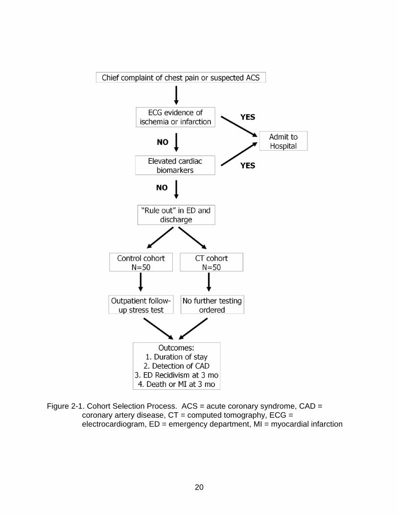
Study Design and Setting
This investigation was conducted in the ED of a large tertiary care medical
center. The design is a retrospective cohort study using a historical control group.
Patients were considered for inclusion if they presented to the ED with a chief complaint
of chest pain, or of other symptoms suggestive of CAD. Based on the patient’s history
and clinical presentation, the attending ED physician was responsible for determining
which patients were at low risk of ACS. These patients underwent serial testing with
ECGs and cardiac biomarkers. Once the diagnosis of ACS had been reliably excluded
to the satisfaction of the attending ED physician, and further CAD testing was
determined to be warranted, patients were nonrandomly assigned either to follow-up
outpatient stress testing or CTCA during their index ED visit as described below (Figure
2-1). Two cohorts were thus established, the CT cohort of patients tested for CAD by
CTCA, and the control cohort of patients tested for CAD using outpatient follow-up
stress testing referral. The Institutional Review Board at the University of Florida
approved this research protocol and waived the requirement to obtain consent for
access to existing medical records.
Patient Selection
The ED routinely documented all patients referred for outpatient stress testing as
a method of quality assurance. This quality assurance logbook was used to identify
patients for the control cohort. On June 1, 2009, the ED began the routine use of CTCA
for all chest pain patients without ACS who needed further testing for CAD. After this
date, all patients considered for CTCA were documented and the logbook was used to
17
identify patients for the CT cohort. Patients in each cohort were identified sequentially
and all patients were included, even if they failed to complete the assigned testing
methodology. The first 50 patients documented after June 1, 2009 comprised the CT
cohort and the last 50 prior to June 1, 2009 comprised the control cohort. Patients were
excluded from the study in either cohort if they had any contraindications for CTCA
including: acute or chronic kidney disease with glomerular filtration rate less than 60
mL/minute or allergy to iodinated contrast.
Data Collection
Patient information was collected regarding age, gender, height and weight, chief
complaint, medical history, history of tobacco and recreational drug use, prescription
medication use, ECGs, and laboratory tests. ED duration of stay was determined using
the time of ED arrival and discharge as documented in the medical record. In the control
cohort, adherence to referral for follow-up outpatient stress testing within 3 months of
ED discharge was determined.
Outcomes
The primary aim of this study was to determine the effect of a CTCA based-
strategy on the duration of stay in the ED. Secondary outcomes included, the rate of
detection of CAD, the success of each strategy at completing testing, and recidivism
(the rate of return ED visit for chest pain). Detection of CAD for the control cohort was
defined as patients who both completed follow-up testing as ordered and had a positive
test for CAD. Detection of CAD in the CT cohort was defined as discovery of any
coronary artery stenotic lesion. Patients with only coronary calcium and without stenosis
were not included in the definition of CAD. Obstructive CAD was defined by the
detection of any lesion of greater than or equal to 50% luminal stenosis. This definition
18
was used to maximize the sensitivity for patients who would be suitable candidates for
further testing, including invasive angiography.17 Recidivism was defined as a return
visit to the ED within 3 months with chest pain or symptoms suggestive of cardiac
ischemia. As a safety outcome, we determined the rate of myocardial infarction (MI) or
death within 3 months.
CTCA Acquisition
CTCA studies were acquired using the 320 detector Aquilion One CT Scanner
(Toshiba, Nasu, Japan). Beta-blocker use was encouraged for all patients with a heart
rate (HR) over 70 beats per minute; however, use was not required and was done at the
discretion of the ED physician. A weight-based protocol was used to determine the dose
of iodinated contrast (VisipaqueTM [iodixanol] 60-90 mL; Amersham Health, Princeton,
NJ), tube current (400-580 mA) and tube voltage (120-135 kV). After coronary artery
calcium (CAC) scoring was completed, contrast bolus tracking was used and the scan
was triggered when contrast density in the descending aorta reached 180 Hounsfield
units. Scans were performed with retrospective gating, prospective gating, or dose-
modulation based on patient suitability. CTCA studies were reconstructed at 70%, 75%,
and 80% of the R-R interval with additional reconstructions performed if necessary and
interpreted using a Vitrea® workstation (Vital Images, Minnesota). All studies were read
in a preliminary fashion by radiology housestaff with radiology faculty available for
oversight. Within 12 hours of any study, final interpretation was provided by both
cardiology and radiology faculty who are board certified in cardiovascular CT. Results
were communicated to ED faculty immediately upon reading. Abnormal findings were
communicated to the patient by the responsible ED faculty. All stenoses were classified
19
as < 50%, 50-75%, or > 75% stenotic by the consensus of the interpreting faculty
physicians, some of whom were investigators in this study.
Statistical Methods and Data Analysis
Using an estimated duration of stay of 480 minutes in the ED, we considered a
reduction or increase in length of stay by 60 minutes would be clinically relevant. We set
a beta level of 0.8 and determined that a sample size of 50 patients would be adequate
to detect a 60 minute change in the duration of stay. We selected two time frames for
secondary analysis of our duration of stay data. First we examined duration of stay if the
patient arrived during peak ancillary staff availability (8 AM to 5 PM) or not (5 PM to 8
AM). Second, we examined duration of stay if the patient arrived during peak ED patient
volume (4 PM to 12 AM) or not (12 AM to 4 PM). Power calculations were performed
using G*Power 3.1.18 Continuous variables were compared using the Student’s t-test
and Wilcoxon rank-sum test as appropriate for normal and skewed distributions.
Categorical data were compared by Fisher’s exact test and chi-square as appropriate.
Calculations were completed using MyStat version 12 (Systat Software; Chicago, IL).
We defined a p value < 0.05 to be statistically significant.
20
Figure 2-1. Cohort Selection Process. ACS = acute coronary syndrome, CAD = coronary artery disease, CT = computed tomography, ECG = electrocardiogram, ED = emergency department, MI = myocardial infarction
21
CHAPTER 3 RESULTS
Baseline Characteristics
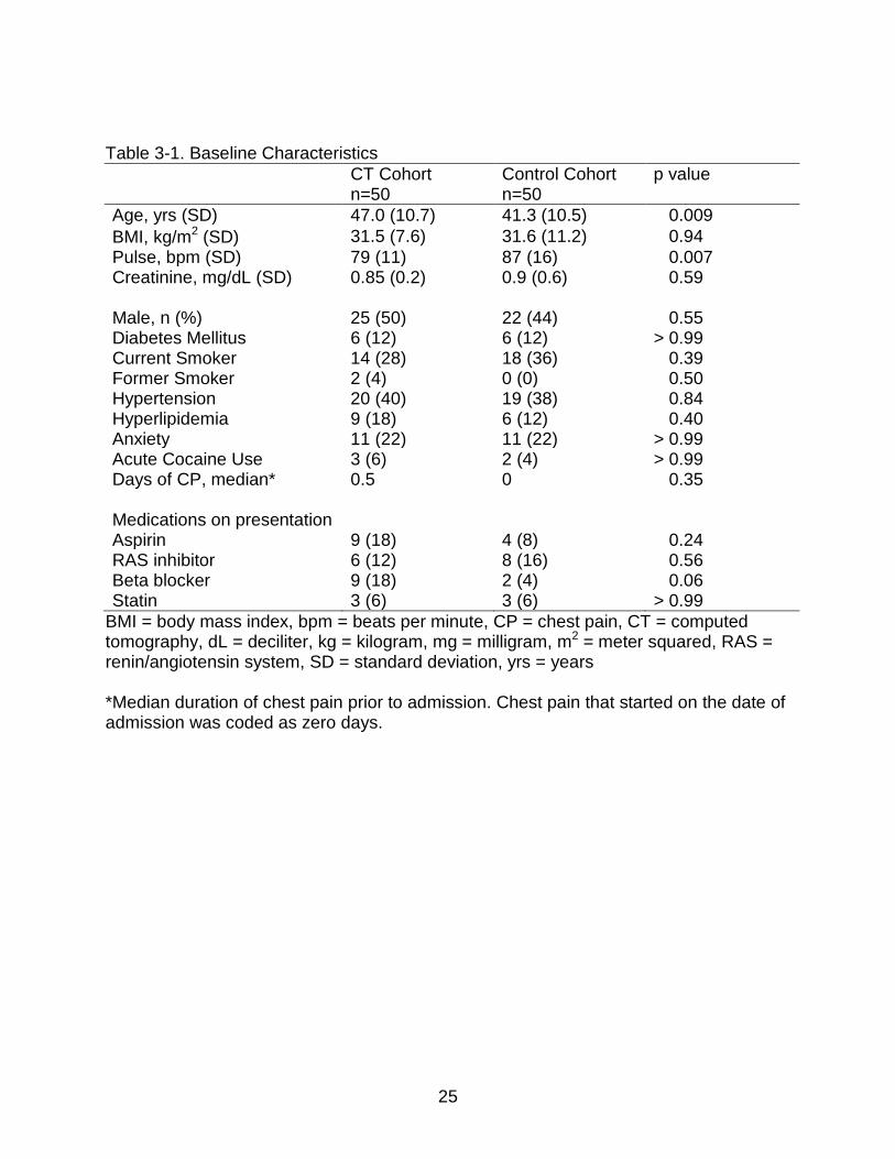
The mean age of patients in the CT cohort was 47.0 years compared to 41.3 in
the control group (p = 0.009). Male patients comprised 50% of the CT cohort versus
44% of controls (p = 0.55) Patients in the two cohorts did not have any significant
differences in their mean body mass index, medication use, medical history, or social
history (Table 3-1). Family history of CAD was inconsistently recorded and therefore
excluded from the investigation. Median duration of chest pain prior to presentation was
12 hours or less for both cohorts (p = 0.35).
Duration of Stay
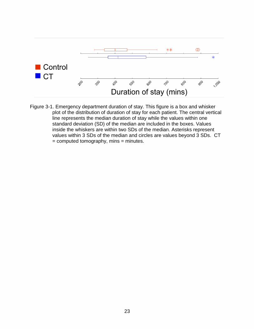
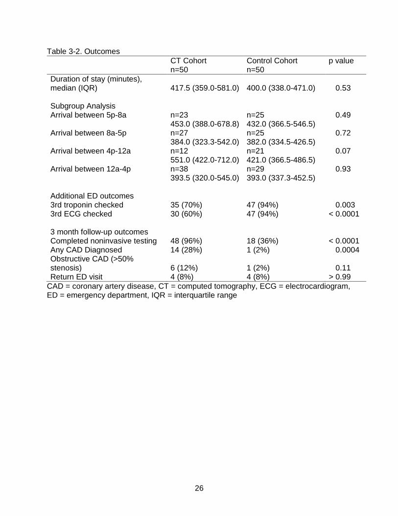
The median duration of stay in the ED was 417.5 minutes (359.0 – 581.0
interquartile range [IQR]) for the CT cohort and 400.0 minutes (338.0 – 471.0 IQR) for
the control cohort (p = 0.53) (Figure 3-1, Table 3-2). When patients arrived during peak
ancillary staff availability (8 AM to 5 PM), duration of stay was 384.0 minutes versus
382.0 minutes (p = 0.72), while arrival from 5 PM to 8 AM duration of stay was 453.0
minutes versus 432.0 minutes (p = 0.49). When patients arrived during peak ED patient
volume (4 PM to 12 AM), duration of stay was 551.0 minutes versus 421.0 minutes (p =
0.07), while arrival from 12 AM to 4 PM duration of stay was 393.5 versus 393.0) (p =
0.93). Fewer patients in the CT cohort (n = 35, 70%) had three sets of cardiac
biomarkers checked during the ED visit as compared to the control cohort (n = 47,
94%).
22
Detection of CAD and Clinical Outcomes
In the control cohort, only 18 patients (36%) completed outpatient stress testing
while all but two patients in the CT cohort completed CTCA (96%, p < 0.0001) (Table 3-
2). One patient assigned to the CT cohort was not scanned due to inability to establish
IV access and the second patient was unable to be scanned due to a scanner
malfunction. Neither of these patients (a 39 year old woman and a 58 year old man)
underwent further CAD testing over the following 3 months.
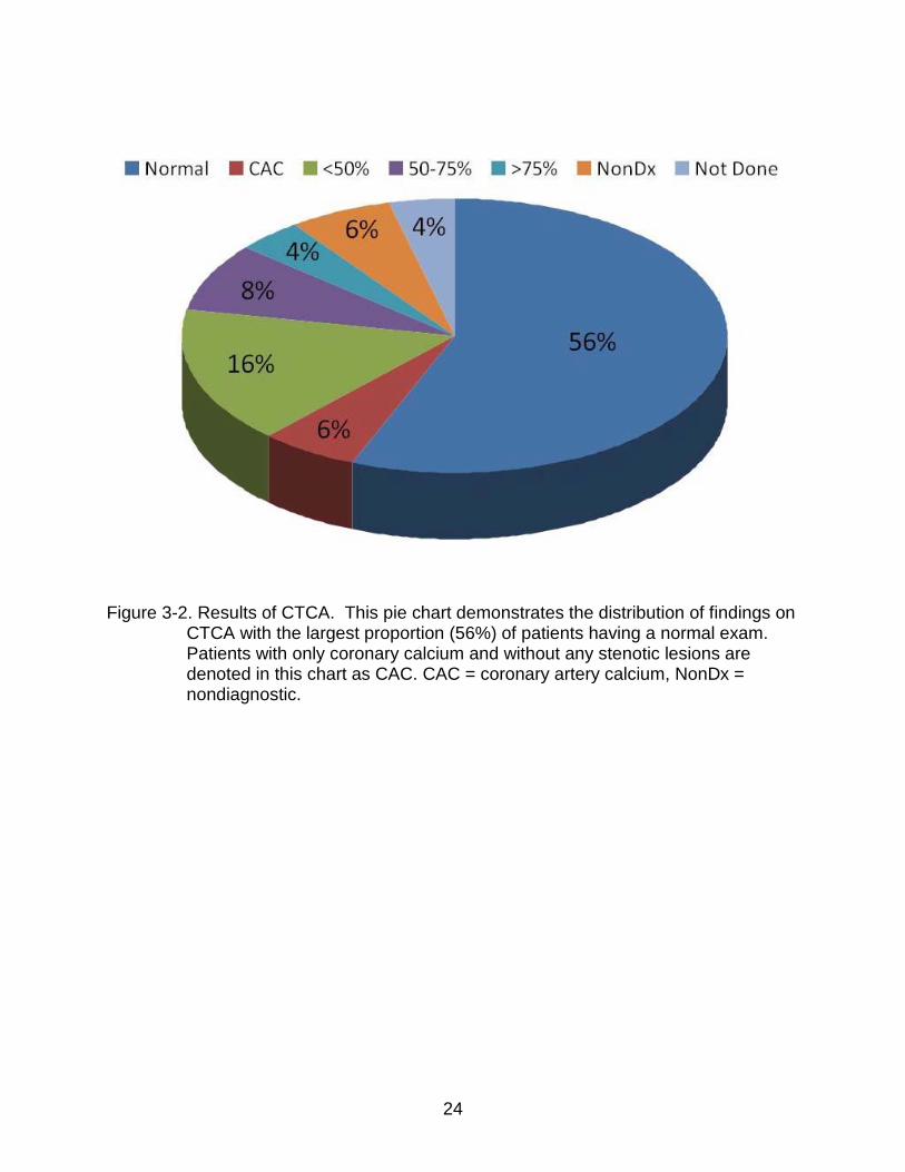
Of the 48 patients from the CT cohort who successfully completed CTCA, 31 had
CAC scores of zero, with median CAC for the cohort of zero. CTCA for three patients
was not evaluable due to incorrect bolus timing (n = 2) and arrhythmia (n = 1), however
all three patients had CAC of zero. Of 42 patients with data available for HR, the median
was 53.5 beats per minute. Radiation dose data were not available in the radiology
reports. CAD (defined as the presence of a stenotic lesion) was detected in 14 CT
cohort patients compared to 1 patient in the control cohort (p = 0.0004). Obstructive
CAD was detected in 6 CT cohort patients compared to 1 control patient (p = 0.11)
(Figure 3-2).
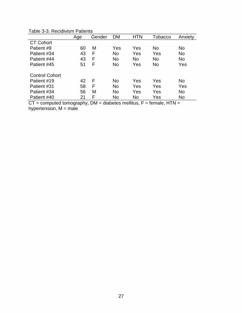
During three months of follow-up, 4 patients in each cohort sought repeat
evaluation in the ED (p > 0.99). (Table 3-3) No patients suffered death or MI during the
subsequent three months.
23
Figure 3-1. Emergency department duration of stay. This figure is a box and whisker
plot of the distribution of duration of stay for each patient. The central vertical line represents the median duration of stay while the values within one standard deviation (SD) of the median are included in the boxes. Values inside the whiskers are within two SDs of the median. Asterisks represent values within 3 SDs of the median and circles are values beyond 3 SDs. CT = computed tomography, mins = minutes.
24
Figure 3-2. Results of CTCA. This pie chart demonstrates the distribution of findings on
CTCA with the largest proportion (56%) of patients having a normal exam. Patients with only coronary calcium and without any stenotic lesions are denoted in this chart as CAC. CAC = coronary artery calcium, NonDx = nondiagnostic.
25
Table 3-1. Baseline Characteristics CT Cohort Control Cohort p value n=50 n=50 Age, yrs (SD) 47.0 (10.7) 41.3 (10.5) 0.009 BMI, kg/m2 (SD) 31.5 (7.6) 31.6 (11.2) 0.94 Pulse, bpm (SD) 79 (11) 87 (16) 0.007 Creatinine, mg/dL (SD) 0.85 (0.2) 0.9 (0.6) 0.59 Male, n (%) 25 (50) 22 (44) 0.55 Diabetes Mellitus 6 (12) 6 (12) > 0.99 Current Smoker 14 (28) 18 (36) 0.39 Former Smoker 2 (4) 0 (0) 0.50 Hypertension 20 (40) 19 (38) 0.84 Hyperlipidemia 9 (18) 6 (12) 0.40 Anxiety 11 (22) 11 (22) > 0.99 Acute Cocaine Use 3 (6) 2 (4) > 0.99 Days of CP, median* 0.5 0 0.35 Medications on presentation Aspirin 9 (18) 4 (8) 0.24 RAS inhibitor 6 (12) 8 (16) 0.56 Beta blocker 9 (18) 2 (4) 0.06 Statin 3 (6) 3 (6) > 0.99
BMI = body mass index, bpm = beats per minute, CP = chest pain, CT = computed tomography, dL = deciliter, kg = kilogram, mg = milligram, m2 = meter squared, RAS = renin/angiotensin system, SD = standard deviation, yrs = years *Median duration of chest pain prior to admission. Chest pain that started on the date of admission was coded as zero days.
26
Table 3-2. Outcomes CT Cohort Control Cohort p value n=50 n=50 Duration of stay (minutes), median (IQR) 417.5 (359.0-581.0) 400.0 (338.0-471.0) 0.53 Subgroup Analysis Arrival between 5p-8a n=23 n=25 0.49 453.0 (388.0-678.8) 432.0 (366.5-546.5) Arrival between 8a-5p n=27 n=25 0.72 384.0 (323.3-542.0) 382.0 (334.5-426.5) Arrival between 4p-12a n=12 n=21 0.07 551.0 (422.0-712.0) 421.0 (366.5-486.5) Arrival between 12a-4p n=38 n=29 0.93 393.5 (320.0-545.0) 393.0 (337.3-452.5) Additional ED outcomes 3rd troponin checked 35 (70%) 47 (94%) 0.003 3rd ECG checked 30 (60%) 47 (94%) < 0.0001 3 month follow-up outcomes Completed noninvasive testing 48 (96%) 18 (36%) < 0.0001 Any CAD Diagnosed 14 (28%) 1 (2%) 0.0004 Obstructive CAD (>50% stenosis) 6 (12%) 1 (2%) 0.11 Return ED visit 4 (8%) 4 (8%) > 0.99
CAD = coronary artery disease, CT = computed tomography, ECG = electrocardiogram, ED = emergency department, IQR = interquartile range
27
Table 3-3. Recidivism Patients Age Gender DM HTN Tobacco Anxiety
CT Cohort Patient #9 60 M Yes Yes No No Patient #34 43 F No Yes Yes No Patient #44 43 F No No No No Patient #45 51 F No Yes No Yes
Control Cohort Patient #19 42 F No Yes Yes No Patient #31 58 F No Yes Yes Yes Patient #34 56 M No Yes Yes No Patient #40 21 F No No Yes No
CT = computed tomography, DM = diabetes mellitus, F = female, HTN = hypertension, M = male
28
CHAPTER 4 DISCUSSION AND CONCLUSIONS
Discussion
Testing for CAD using a CTCA based strategy for ED patients with chest pain
was more effective than a strategy of outpatient stress testing. As compared to
outpatient stress testing, a CTCA based strategy had no significant effect on ED
duration of stay and detected a greater number of patients with both nonobstructive and
obstructive CAD. No patients in either cohort suffered MI or death and the same
number in each cohort returned to the ED for evaluation of chest pain during 3 months
of follow-up.
Our investigation has demonstrated that for ED patients with chest pain and
without ACS who warrant further testing for CAD, a CTCA based strategy detected both
nonobstructive and obstructive CAD in more patients as compared with a strategy of
outpatient stress testing. Prior studies have documented this phenomenon in stable
outpatients.19, 20 We recognize that several factors could contribute to these differences
including the greater ability of CTCA to diagnose nonobstructive CAD, low follow-up
rates in the control cohort, and the nonrandomized design of our investigation.
Because CTCA can detect nonobstructive lesions while ETT, MPI, and stress
echocardiography detect myocardial ischemia, greater detection of nonobstructive CAD
is an expected finding. A CTCA based strategy, therefore, is an opportunity to provide
unique and robust prognostic information. For patients without CAD, the prognostic
value of a zero calcium score has been well established and is a valuable tool in
reassuring patients about their risk of cardiovascular events.14, 21 Three patients had
nondiagnostic CTCA studies, however their CAC scores were zero and therefore they
29
could still be reassured of low cardiovascular risk. Patients with nonobstructive CAD can
be reassured of a similarly low cardiovascular event risk over the next 12 months.22
These two groups accounted for 84% of the patients in our CT cohort. When patients
are diagnosed with CAD, that knowledge provides physicians an opportunity to
intervene and potentially reduce the burden of cardiovascular events. Further study is
needed to establish if earlier diagnosis could alter future clinical outcomes.23 In addition,
future studies should address new strategies to effectively communicate low risk results
to patients to reduce return visits to the ED for the same complaints.
The increased detection of CAD in the CT cohort is also expected given the low
rate of follow-up observed in the control cohort. Failure to follow up has been linked to
many factors,24-26 some of which can be overcome by completing testing prior to ED
discharge. Because our investigation is a comparison of patient care strategies, the low
follow-up rate reinforces the limitations of delayed outpatient stress testing as a patient
care strategy. Because CTCA can be reliably performed on a CT scanner with at least
64 detectors,27 the strategy we have described could be used at many EDs. Few
hospitals have 24 hour, immediate reading of CTCA studies, but we have demonstrated
that a strategy of routine CTCA with prompt preliminary reading is safe.
Because our diagnostic strategy was not conducted by randomized assignment,
the true prevalence of CAD may not be similar between the cohorts. If we assume,
however, that the CT cohort is an accurate reflection of CAD prevalence, the disparity in
coronary findings compared to the control cohort is worrisome. Despite the fact that the
same population was used to construct the CT and control cohorts, the routine use of
CTCA detected obstructive CAD in 12% of patients as compared to 2% detected by
30
outpatient follow-up testing in the control cohort. This observation suggests that patients
with obstructive CAD may go undiagnosed when delayed outpatient stress testing is
employed. Prior reports have documented a similar magnitude of missed diagnoses.28
The rapid adoption of new CT imaging techniques and the theoretical risks
associated with medical radiation has raised appropriate concerns.29 We share
concerns about the potential overuse of diagnostic imaging studies; however, all
patients in our study were determined to warrant further CAD testing based on the
judgment of the treating physician. Previous work documents that CTCA is more
sensitive and specific at detecting CAD than ETT and a direct comparison of these two
testing modalities may not be a fair comparison. We have endeavored, however, to
compare two strategies: immediate CTCA versus delayed stress testing. While
outpatient stress testing is commonly used by EDs, we have demonstrated that this
strategy is not only incapable of detecting nonobstructive CAD, but may frequently fail to
detect obstructive CAD.
We observed no significant change in duration of stay in the ED. Because many
forces affect the duration of stay, our investigation may not have altered clinical care
enough to detect a difference. In the CT cohort, ED physicians less frequently ordered
three sets of cardiac biomarkers. This suggests that they found the CTCA clinically
useful in accelerating decisions about disposition and therefore CTCA has potential to
reduce ED duration of stay. Mixed results have been found with prior investigations on
how CTCA affects the duration of stay.16, 30 Because our investigation was conducted
using the first 50 patients in the ED evaluated using a CTCA strategy, the test may not
have been optimally used by technologists and physicians. The duration of stay in the
31
CT cohort was nonsignificantly higher than the control cohort, and waiting for a CTCA
scan and the results could be causing an increase that this investigation was not
powered to detect. We could not account for the time savings garnered by avoiding
outpatient stress testing and this could potentially confer benefit in a formal cost-benefit
analysis.
Based on 3 months of follow-up data, a CTCA based strategy appears to be
safe, given a cardiovascular event rate of 0%. Prior studies on CTCA have described
similar event rates with both short and long term follow-up.31-33 We suspect that the low
event rate is likely related to the purposeful selection of patients with low to intermediate
pretest likelihood of cardiovascular events. Our analysis was not powered to detect a
difference in recidivism, however we observed 4 patients in each cohort return to the ED
with chest pain within 3 months. Further study will be necessary to determine if
comprehensive evaluation of chest pain with CTCA is reassuring enough to convince
patients to not return to the ED with similar chest pain, but instead pursue less costly
care for their chest discomfort.
Our study has several limitations. As a retrospective cohort study, our study does
not have the benefits of a randomized trial which would have minimized differences
between the two patient populations, yet few differences were observed in the baseline
characteristics. The difference in pulse may be related to the nearly significant
difference in baseline beta blocker use. Age difference may reflect the nonrandom
selection of patients with the ED staff ordering tests on older patients they considered to
be at higher risk of CAD and could lead to a higher prevalence of CAD in the CT cohort.
Our study is limited by the fact that we only reviewed records for our own institution.
32
Conclusions
For symptomatic ED patients who warrant noninvasive testing for CAD, a
strategy of immediate CTCA is superior to delayed outpatient stress testing for detecting
CAD. A delayed outpatient stress testing strategy may fail to diagnose obstructive CAD
and such a strategy is limited by low follow-up rates. A CTCA based strategy does not
significantly affect on the ED duration of stay. Patients with chest pain and no evidence
of ACS can safely be discharged with an expectation of low cardiovascular event risk in
the ensuing 3 months.
33
LIST OF REFERENCES
1. Roger VL, Go AS, Lloyd-Jones DM, et al. Heart Disease and Stroke Statistics--2011 Update: A Report From the American Heart Association. Circulation. 2011;123:e18-e209.
2. Xu J, Kochanek KD, Murphy S, Tejada-Vera B. Deaths: Final Data for 2007. Natl Vital Stat Rep. 2010;58:1-135.
3. Ford ES, Ajani UA, Croft JB, et al. Explaining the decrease in U.S. deaths from coronary disease, 1980-2000. N Engl J Med. 2007;356:2388-2398.
4. Diamond GA, Forrester JS. Analysis of probability as an aid in the clinical diagnosis of coronary-artery disease. N Engl J Med. 1979;300:1350-1358.
5. Taylor AJ, Cerqueira M, Hodgson JM, et al. ACCF/SCCT/ACR/AHA/ASE/ASNC/NASCI/SCAI/SCMR 2010 Appropriate Use Criteria for Cardiac Computed Tomography. J Am Coll Cardiol. 2010;56:1864-1894.
6. Douglas PS, Khandheria B, Stainback RF, et al. ACCF/ASE/ACEP/AHA/ASNC/SCAI/SCCT/SCMR 2008 Appropriateness Criteria for Stress Echocardiography. Catheter Cardiovasc Interv. 2008;71:E1-19.
7. Hendel RC, Berman DS, Di Carli MF, et al. ACCF/ASNC/ACR/AHA/ASE/SCCT/SCMR/SNM 2009 Appropriate Use Criteria for Cardiac Radionuclide Imaging. Circulation. 2009;119:e561-587.
8. Nawar EW, Niska RW, Xu J. National Hospital Ambulatory Medical Care Survey: 2005 emergency department summary. Advance Data. 2007;386:1-32.
9. Farkouh ME, Aneja A, Reeder GS, et al. Clinical risk stratification in the emergency department predicts long-term cardiovascular outcomes in a population-based cohort presenting with acute chest pain: primary results of the Olmsted county chest pain study. Medicine. 2009;88:307-313.
10. Richards D, Meshkat N, Chu J, Eva K, Worster A. Emergency department patient compliance with follow-up for outpatient exercise stress testing: a randomized controlled trial. CJEM. 2007;9:435-440.
11. Nixdorff U, Kufner C, Achenbach S, et al. Head-to-head comparison of dobutamine stress echocardiography and cardiac computed tomography for the detection of significant coronary artery disease. Cardiology. 2008;110:81-86.
12. Budoff MJ, Rasouli ML, Shavelle DM, et al. Cardiac CT angiography (CTA) and nuclear myocardial perfusion imaging (MPI)-a comparison in detecting significant coronary artery disease. Academic Radiology. 2007;14:252-257.
34
13. Chow BJ, Dennie C, Hoffmann U, et al. Comparison of computed tomographic angiography versus rubidium-82 positron emission tomography for the detection of patients with anatomical coronary artery disease. Can J Cardiol. 2007;23:801-807.
14. Budoff MJ, Shaw LJ, Liu ST, et al. Long-term prognosis associated with coronary calcification: observations from a registry of 25,253 patients. J Am Coll Cardiol. 2007;49:1860-1870.
15. Gallagher MJ, Ross MA, Raff GL, Goldstein JA, O'Neill WW, O'Neil B. The diagnostic accuracy of 64-slice computed tomography coronary angiography compared with stress nuclear imaging in emergency department low-risk chest pain patients. Ann Emerg Med. 2007;49:125-136.
16. Goldstein JA, Gallagher MJ, O'Neill WW, Ross MA, O'Neil BJ, Raff GL. A randomized controlled trial of multi-slice coronary computed tomography for evaluation of acute chest pain. J Am Coll Cardiol. 2007;49:863-871.
17. Meijboom WB, Meijs MF, Schuijf JD, et al. Diagnostic accuracy of 64-slice computed tomography coronary angiography: a prospective, multicenter, multivendor study. J Am Coll Cardiol. 2008;52:2135-2144.
18. Faul F, Erdfelder E, Lang AG, Buchner A. G*Power 3: a flexible statistical power analysis program for the social, behavioral, and biomedical sciences. Behav Res Methods. 2007;39:175-191.
19. Ovrehus KA, Jensen JK, Mickley HF, et al. Comparison of usefulness of exercise testing versus coronary computed tomographic angiography for evaluation of patients suspected of having coronary artery disease. Am J Cardiol. 2010;105:773-779.
20. Weustink AC, Mollet NR, Neefjes LA, et al. Diagnostic accuracy and clinical utility of noninvasive testing for coronary artery disease. Ann Intern Med. 2010;152:630-639.
21. Nabi F, Chang SM, Pratt CM, et al. Coronary Artery Calcium Scoring in the Emergency Department: Identifying Which Patients With Chest Pain Can Be Safely Discharged Home. Ann Emerg Med. 2010;56:220-9.
22. Hollander JE, Chang AM, Shofer FS, et al. One-year outcomes following coronary computerized tomographic angiography for evaluation of emergency department patients with potential acute coronary syndrome. Acad Emerg Med. 2009;16:693-698.
23. Arad Y, Spadaro LA, Roth M, Newstein D, Guerci AD. Treatment of asymptomatic adults with elevated coronary calcium scores with atorvastatin, vitamin C, and vitamin E: the St. Francis Heart Study randomized clinical trial. J Am Coll Cardiol. 2005;46:166-172.
35
24. Engel KG, Heisler M, Smith DM, Robinson CH, Forman JH, Ubel PA. Patient comprehension of emergency department care and instructions: are patients aware of when they do not understand? Ann Emerg Med. 2009;53:454-461.
25. Wang NE, Gisondi MA, Golzari M, van der Vlugt TM, Tuuli M. Socioeconomic disparities are negatively associated with pediatric emergency department aftercare compliance. Acad Emerg Med. 2003;10:1278-1284.
26. Clarke C, Friedman SM, Shi K, Arenovich A, Culligan C. Emergency department discharge instructions comprehension and compliance study. CJEM. 2005;7:5-11.
27. Rubinshtein R, Halon DA, Gaspar T, et al. Usefulness of 64-slice cardiac computed tomographic angiography for diagnosing acute coronary syndromes and predicting clinical outcome in emergency department patients with chest pain of uncertain origin. Circulation. 2007;115:1762-1768.
28. Madsen T, Mallin M, Bledsoe J, et al. Utility of the emergency department observation unit in ensuring stress testing in low-risk chest pain patients. Crit Pathw Cardiol. 2009;8:122-124.
29. Einstein AJ, Henzlova MJ, Rajagopalan S. Estimating risk of cancer associated with radiation exposure from 64-slice computed tomography coronary angiography. JAMA. 2007;298:317-323.
30. Chang SA, Choi SI, Choi EK, et al. Usefulness of 64-slice multidetector computed tomography as an initial diagnostic approach in patients with acute chest pain. Am Heart J. 2008;156:375-383.
31. Gopal A, Nasir K, Ahmadi N, et al. Cardiac computed tomographic angiography in an outpatient setting: an analysis of clinical outcomes over a 40-month period. J Cardiovasc Comput Tomogr. 2009;3:90-95.
32. Beigel R, Oieru D, Goitein O, et al. Usefulness of routine use of multidetector coronary computed tomography in the "fast track" evaluation of patients with acute chest pain. Am J Cardiol. 2009;103:1481-1486.
33. Laudon DA, Behrenbeck TR, Wood CM, et al. Computed tomographic coronary artery calcium assessment for evaluating chest pain in the emergency department: long-term outcome of a prospective blind study. Mayo Clin Proc. 2010;85:314-322.

36
BIOGRAPHICAL SKETCH
David E. Winchester is a physician originally from Tallahassee, FL. He holds a
Bachelor of Science in microbiology and a Bachelor of Arts in sociology, both from the
University of Florida. He completed his medical degree at the University of South
Florida and training in internal medicine at the University of Virginia. Work on the Master
of Science degree was completed concurrent with fellowship training in cardiovascular
medicine.