Embed Size (px)
Citation preview
Acta path. microbiol. immunol. scand. Sect. C, 93: 43-48, 1985.
THE USE OF FROZEN MONOCYTES IN PHAGOCYTOSIS STUDIES
VESLEM0Y MYHRVOLD and BERIT M0RLAND
Department of Microbiology, Dental Faculty, University of Oslo, Oslo, Norway
Myhrvold, V. & Msrland, B. The use of frozen monocytes in phagocytosis studies. Acta path. microbiol. immunol. scand. Sect. C, 93: 43-48, 1985.
Human monocytes were isolated from peripheral blood by Lymphoprep density-gradient centrifugation and adherence to fibronectin. The cells were loosened by ethylene-diamino-tetra-acetate (EDTA), frozen by different freezing methods, thawed, washed and compared to unfrozen cells. After freezing, thawing and washing, cell recovery was calculated and found to vary with the freezing procedure. The best result was about 76% cell recovery. No morphological differences were observed between unfrozen and frozen cells. The experiments also showed that the percentage of cells that attached or phagocytized IgG-opsonized erythrocytes (E-IgG) via the Fc-receptor was unaltered after freezing. Neither was there any difference between unfrozen and frozen monocytes with respect to their ability to phagocytize latex particles. There was no significant difference in reactivity between monocytes frozen for one day and those frozen for six weeks.
Key words: Unfrozen monocytes; frozen monocytes; attachment; phagocytosis.
V. Myhrvold, Department of Microbiology, Dental Faculty, University of Oslo, P.b. 1052, Blindern, 0316 Oslo 3, Norway.
Accepted as submitted 28.ix.84 The study of mononuclear phagocytes has
emerged as a central theme in both immunology and biology. Since human monocytes isolated from different individuals show functional varia- tions, it would be advantageous to have a fixed and well-defined test batch of these cells. Cryopre- servation could provide this facility. Cryopreserva- tion is also time-saving, since thawing of mo- nocytes takes considerable less time than repeatedly isolating these cells from human blood. Van der Meulen el al. (1 8) have shown that mo- nocytes could be kept frozen for at least three months. Cell recovery was about 64%.
Before using frozen cells in various assays, it is of great importance to evaluate the effect of freez- ing procedures on different cell functions. Since freezing may affect the cell membrane primarily (10, 14, 16, 17), we started our studies by testing some monocyte membrane functions. The ability to adhere and spread on plastic surfaces is an often used method to evaluate monocyte membrane
morphology (26) . In addition, the effect of cryo- preservation on monocyte phagocytosis via the Fc-receptor and the foreign surface receptor were tested. The last functions are obviously also of great importance for the various monocyte/mac- rophage functions in vivo. Van der Meulen el al. (18) found in their study that the monocytes’ capacity to phagocytize via the C3b-receptor and their chemotactic response toward casein were diminished after freezing. Random migration re- mained, however, unaltered. Attachment to the Fc-receptor or the monocytes capacity to lyse E- IgG was not influenced by cryopreservation. Less free IgG, however, was needed to inhibit E-IgG adherence to monocytes after cryopreservation than before. Dean & Strong ( 7 ) reported a decrease in nondirected migration of human mononuclear cells after freezing. Thurman el al. (27) found that the migration ability of monocytes was affected very little by cryopreservation, although the re- sponse to the inhibitory activity of especially the
43
lymfokine human gamma interferon was de- creased.
Monocytes have a lower average density than other cells in human blood (13), and many me- thods of monocyte isolation based on density dif- ferences have been developed (1 , 2,4, 5, 8, 1 1, 12, 24, 25). However, owing to overlapping predomi- nantly by lymphocytes, efficient separation based on density gradient centrifugation alone cannot be acheived. In this work an additional method based on the fact that monocytes have a high affinity for fibronectin immobilized on a gelatin-coated sur- face was used to separate monocytes and lym- phocytes (3). By this method the cell preparations obtained were more than 90% monocytes (9), well suited for freezing experiments.
In the present study several methods of freezing human monocytes were tested and compared. Earlier experiments (2 1) have shown it profitable to use frozen IgG opsonized erythrocytes for at- tachment and phagocytosis studies, and these test particles were therefore routinely used in the Fc- receptor studies in the present report.
MATERIALS AND METHODS
Mononuclear cells. The mononuclear cells were isolated from citrated blood (450 ml) of healthy volunteers. After centrifugation (1 5 min, I500 x g at 4 "C) a buffy coat of 50 to 100 ml was collected. The buffy coat was obtained from The Blood Bank and Department of Immunology, Ulleval Hospital, Oslo. To the buffy coat was added RPMI medium 1640 (Gibco) buffered at pH 7.3 with NaHCO, (RPMI NaHCO,) to a volume of 210 mi. The mononuclear cells were isolated by gradient centrifu- gation on Lymphoprep (Nyegaard & Co., Oslo) accord- ing to the method of Boyurn (6). Mononuclear cells were resuspended (2-4 x lo4 cells/ml) in RPMI NaHCO, sup- plemented with 10% heat inactivated (56 "C for 30 min) foetal calf serum (FC).
Prepararion ofperri dishes. Plastic petri dishes (9 cm diameter) were coated with 5 ml gelatin (2% in water) (Oxoid), supplemented with 0.02% sodium azide (Sigma) and incubated for 2 h at 37 "C. Gelatin was then removed by suction, and the dishes were allowed to dry. Gelatin-treated dishes were stored at room temperature (they can be used after at least three months' storage).
Separafion ofrnonocyfes. Five ml human serum was added to each dish and incubated for a minimum of 30 min at 37 "C just before use. The serum was aspirated, and 10 ml of mononuclear cell suspension (2-4 x lo6 cells/ml) in RPMI NaHCO, 10% FC was added to each dish and incubated for 30 min in 5% C 0 2 in air, at 37 "C. Non-adherent cells were removed and the dishes were washed gently three times with RPMI NaHCO, pre-warmed at 37 "C. To loosen the adherent cells, 5 ml
44
10 mM ethylene-diamino-tetra-acetate (EDTA) in RPMI NaHCO, (37 "C) was added to each dish, and dishes were incubated as above for 5-10 min. The dishes were washed with fresh medium to obtain optimal cell detachment. The cells were centrifuged at 400 x g for 10 min, washed once, and resuspended in RPMI medium I640 buffered at pH 7.3 with N-2-Hydroxy-ethyl-pipe- razine-N'-2-ethanesulfonic acid (Sigma) (RPMI Hepes) for freezing procedures and in RPMI NaHCO, 10% heat-inactivated AB serum for cultivation of unfrozen cells (= control).
Freezing procedures. Before freezing, the monocyte suspension was put on melting ice and slowly diluted with an equal volume of ice-cold RPMI-Hepes contain- ing 40% heat-inactivated FC serum and most often 20% dimethyl sulfoxide (DMSO) (Sigma). The final cell con- centration varied from 1.6 to 2.0 x lo6 cells/ml. The monocyte suspension was put into 2 ml plastic serum test-tubes No. 1076 (Nunc) (1.8 ml per tube), and the following five freezing procedures were used:
In methods I, 11 and I11 the tubes were immersed in an ethanol bath at 0 "C. In method I the cooling of the tubes proceeded at a cooling rate of 5 "C per min from 0 "C to -75 "C. From -75 "C to -85 "C cooling was con- tinued at a rate of 1 "C per min. After 10 min at this temperature the tubes were transferred direct into liquid nitrogen in an LR-35 liquid nitrogen container (Linde Company) (cp. method IV). In methods I1 and 111 the cooling rate was 1.5 "C per min from 0 "C to -28 "C. In method I1 cooling was continued at a rate of 6-7 "C per min from -28 "C to -100 "C, when the tubes were trans- ferred direct into liquid nitrogen (18). In method 111 the tubes were stored at -28 "C for 15 min whereafter they were transferred direct into liquid nitrogen (cp. method V). The cooling system used in these methods (1-111) consisted of one Denver flask containing 200 ml etha- nol, another flask containing 1000 ml liquid nitrogen, and copper wires as thermal conductors between the vessels.
In method IV the tubes were placed in a -90 "C refrigerator for 35 min. The cooling rate was 5 "C per min until a temperature of -75 "C. From -75 "C to -85 "C the cooling rate was about I "C per min; thereafter no temperature change could be seen. In these experiments various DMSO concentrations were tested with final concentrations of 20, 15, 10 and 5%. In method V the tubes were placed in a -40 "C refrigerator for 35 min. The cooling rate was about 1.5 "C per min until a temperature of -28 " C thereafter no temperature change could be seen. From the refrigerators the tubes were plunged direct into liquid nitrogen.
The cooling rates were in all experiments monitored by a temperature recorder (Speedomax H, Leeds & Nor- thrup Company, Philadelphia, Pa., U.S.A.).
Thawing was performed by immediate immersion of the tubes in a water bath at 37 "C under continuous shaking. Once the ice nucleus had disappeared, the su- spension was diluted fivefold with cold (4 "C) RPMI- Hepes supplemented with 20% FC serum. The cells were washed and counted. Counting was assessed after stain- ing with trypan blue (0.59/0 in 0.1 5 M NaCI).
Ciiltivation of the monocytes. Monocytes frozen by method IV with a final concentration of 10% DMSO were chosen to be further compared to freshly isolated monocytes. The cells were seeded in Linbro plates (Lin- bro Chem. Co., New Haven, Connecticut, U.S.A.) with glass coverslips (14 mm 0) in a concentration of 0.75 x lo6 monocytes/ml RPMI NaHCO, 10% heat-inactivated AB serum in each well. Cultivation was performed in 5% CO, in air at 37 "C until experimentation on day 0, day 3 and day 9.
Cell morphology. On day 0, day 3 and day 9 the culture coverslips were washed in 0. I5 M NaCl (37 "C), and fixed at 4 "C for at least 15 rnin in 2% glutaralde- hyde in 0.1 M cacodylate buffer (pH 7.3) with 0.1 M sucrose (37 "C). The coverslips were washed in 0.15 M NaCl (4 "C) and mounted on a drop of Aquamount (Edward Gurr Ltd., London). The slides were sealed and examined in a Zeiss phase contrast microscope. In order to describe differences between unfrozen and
frozen-thawed cells, the following culture characteristics were evaluated. I . Cells with most of their circumference consisting of a
wide cytoplasmic veil (= veiled cells). 2. Cells with long cytoplasmic extensions a polar orien-
tation (= polar cells). 3. Cells free from cytoplasmic veil or extensions (=
rounded cells). The percentage of cells expressing the features was
calculated by counting 500 cells per culture. Opsonization and freezing of sheep red blood cells.
Sheep red blood cells (E) stored in Alsever's solution were obtained from The National Institute of Health, Oslo, and most often used within one week. Rabbit anti-sheep red cell immunoglobulin G (IgG) was ob- tained from Cordis Laboratories, Miami, U.S.A. A one per cent solution of erythrocytes was incubated with 0.5 pg/ml IgG for 15 min at 37 "C in serum-free RPMl Hepes giving E-IgG (22). The cells were washed three times in the same medium by centrifugation at 400 x g
for 10 min. For freezing, the E-lgG was suspended to a 5% concentration in gelatin-veronal-buffered saline-su- crose containing 0.15 mM-Ca and 1 mM-Mg (GVBSM++-sucrose) (20). One volume of 5% DMSO in NaCl was added to one volume of the 5% opsonized sheep erythrocyte suspension. The mixture was distrib- uted (1 ml) to 2 ml plastic serum test tubes NO. 1076 (Nunc) and plunged direct into liquid nitrogen in an LR-35 liquid nitrogen container (Linde Company). Thawing and washing were performed as described pre- viously (19), and the cells were diluted in RPMI Hepes for the monocyte tests.
Attachment and phagocytosis of E-lgG. For attach- ment of E-IgG to monocytes, the monocyte monolayers were incubated with I ml 0.25Yo frozen E-JgG in RPMl Hepes for 30 rnin at 4 "C (22). For the phagocytosis of E-IgG by monocytes, the monocyte monolayers were incubated with I ml 0.25Yo frozen E-IgG 'in RPMl Hepes for 60 rnin at 37 "C (22). At the end of this incubation period the medium was removed, the wells were gently washed twice with I ml RPMl Hepes pre- warmed at 37 "C, and I m10.14 M solution of NH,CI 37 "C was added for about 30 s to lyse attached, non-inter- nalized erythrocytes (lysis was observed in the micro- scope). The cultures were fixed, mounted and examined in a Zeiss phase contrast microscope as described for morphology studies. The percentage of monocytes that attached or internalized red cells was calculated by counting 500 cells per culture (23). In some experiments freshly opsonized (unfrozen) E-IgG was used in the pha- gocytosis studies to evaluate the effect of freezing on the test particle (21).
Phagocytosis oflarex particles. Latex particles 0.8 pm (Dow) were suspended in sterile saline at 0.2% v/v. and 50 p1 of latex suspension was added to each culture in I ml RPMI Hepes. After incubation for 60 rnin at 37 "C the cultures were washed twice with medium, fixed and mounted as described above.
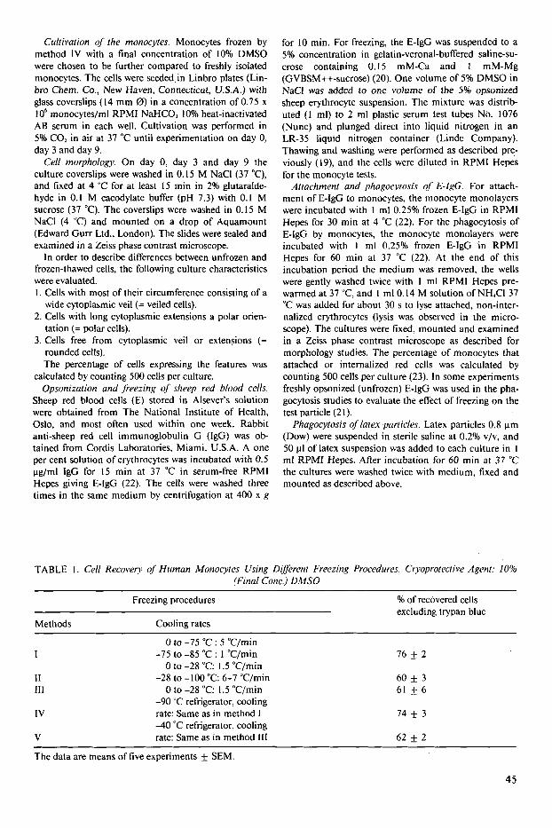
TABLE I . Cell Recovery of Human Monocytes Using Diflerent Freezing Procedures. Cryoprotective Agent: 10% (Final Conc.) DMSO
Freezing procedures
Methods Cooling rates
Yo of recovered cells excluding trypan blue
I
I I 111
I V
V
0 to -75 "C : 5 "C/min -75 to -85 "C : 1 "C/min
0 to -28 "C: I .5 "C/min -28 to - 100 "C: 6-7 "C/min
0 to -28 "C: 1.5 "C/min -90 "C refrigerator, cooling rate: Same as in method I -40 "C refrigerator, cooling rate: Same as in method 111
76 2
60 k 3 61 f 6
74 f 3
62 f 2
The data are means of five experiments f SEM.
45
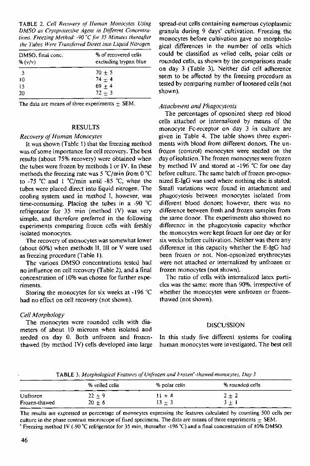
TABLE 2. Cell Recovery of Human Monocytes Using DMSO us Cryoprotective Agent in Different Concentra- tions. Freezing Method: -90 "C for 35 Minutes thereafler [he Tiibes Were Transferred Direct into Liquid Nitrogen
DMSO, final conc. % (VIV) excluding trypan blue
O/O of recovered cells
5 10 15 20
70 + 5 74 + 4 69 & 4 72 * 3
The data are means of three experiments f SEM.
RESULTS Recovery of Human Monocyces
It was shown (Table I ) that the freezing method was of some importance for cell recovery. The best results (about 75% recovery) were obtained when the tubes were frozen by methods I or IV. In these methods the freezing rate was 5 "C/min from 0 "C to -75 "C and 1 "C/min until -85 'C, when the tubes were placed direct into liquid nitrogen. The cooling system used in method I, however, was time-consuming. Placing the tubes in a -90 "C refrigerator for 35 min (method IV) was very simple, and therefore preferred in the following experiments comparing frozen cells with freshly isolated monocytes.
The recovery of monocytes was somewhat lower (about 60%) when methods 11, I11 or V were used as freezing procedure (Table I ) .
The various DMSO concentrations tested had no influence on cell recovery (Table 2), and a final concentration of 10% was chosen for further expe- riments.
Storing the monocytes for six weeks at -196 "C had no effect on cell recovery (not shown).
Cell Morphology The monocytes were rounded cells with dia-
meters of about 10 microns when isolated and seeded on day 0. Both unfrozen and frozen- thawed (by method IV) cells developed into large
spread-out cells containing numerous cytoplasmic granula during 9 days' cultivation. Freezing the monocytes before cultivation gave no morpholo- gical differences in the number of cells which could be classified as veiled cells, polar cells or rounded cells, as shown by the comparisons made on day 3 (Table 3). Neither did cell adherence seem to be affected by the freezing procedure as tested by comparing number of loosened cells (not shown).
Attachment and Phagocycosis The percentages of opsonized sheep red blood
cells attached or internalized by means of the monocyte Fc-receptor on day 3 in culture are given in Table 4. The table shows three experi- ments with blood from different donors. The un- frozen (control) monocytes were seeded on the day ofisolation. The frozen monocytes were frozen by method IV and stored at -196 "C for one day before culture. The same batch of frozen pre-opso- nized E-IgG was used where nothing else is stated. Small variations were found in attachment and phagocytosis between monocytes isolated from different blood donors; however, there was no difference between fresh and frozen samples from the same donor. The experiments also showed no difference in the phagocytosis capacity whether the monocytes were kept frozen for one day or for six weeks before cultivation. Neither was there any difference in this capacity whether the E-IgG had been frozen or not. Non-opsonized erythrocytes were not attached or internalized by unfrozen or frozen monocytes (not shown).
The ratio of cells with internalized latex parti- cles was the same: more than 9Oo/o, irrespective of whether the monocytes were unfrozen or frozen- thawed (not shown).
DISCUSSION
In this study five different systems for cooling human monocytes were investigated. The best cell
TABLE 3. Morphological Features of Unfrozen and Frozen"-thuwed-monocytes* Day 3
O/o veiled cells 'Yo polar cells 'Yo rounded cells ~~
Unfrozen 22 + 9 Frozen-thawed 20 + 6
I I * 4 13 k 3
2 f 2 3 + 1
The results are expressed as percentage of monocytes expressing the features calculated by counting 500 cells per culture in the phase contrast microscope of fixed specimens. The data are means of three experiments f SEM.
Freezing method IV (-90 "C refrigerator for 35 min, thereafter -196 "C) and a final concentration of 10% DMSO.
46
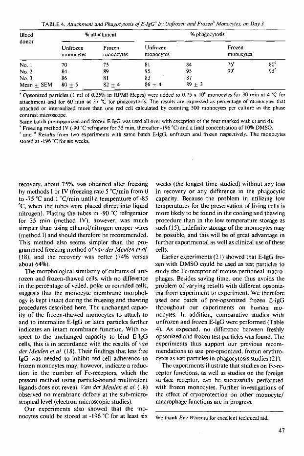
TABLE 4. Atfachrnent and Phagocytosis of E-lgC" by Unfrozen and Frozenh Monocytes, on Day 3 ~
Blood % attachment % phagocytosis donor
Unfrozen Frozen Unfrozen Frozen monocytes monocytes monocytes monocytes
No. I 70 75 81 84 7 6' 80d No. 2 84 89 95 95 99' 95d No. 3 86 81 83 87 Mean f SEM 80 f 5 82 f 4 86 f 4 89 f 3
a Opsonized particles ( 1 ml of 0.25% in RPMI Hepes) were added to 0.75 x lo6 monocytes for 30 min at 4 "C for attachment and for 60 min at 37 "C for phagocytosis. The results are expressed as percentage of monocytes that attached or internalized more than one red cell calculated by counting 500 monocytes per culture in the phase contrast microscope. Same batch pre-opsonized and frozen E-IgG was used all over with exception of the four marked with c) and d).
Freezing method IV (-90 "C refrigator for 35 min, thereafter -196 "C) and a final concentration of 10% DMSO. ' and stored at - I96 "C for six weeks.
Results from two experiments with same batch E-lgG, unfrozen and frozen respectively. The monocytes
recovery, about 75%, was obtained after freezing by methods I or IV (freezing rate 5 "C/min from 0 to -75 "C and 1 "C/min until a temperature of -85 'C, when the tubes were placed direct into liquid nitrogen). Placing the tubes in -90 "C refrigerator for 35 min (method IV), however, was much simpler than using ethanol/nitrogen copper wires (method I ) and should therefore be recommended. This method also seems simpler than the pro- grammed freezing method of van der Meulen et d. (IS), and the recovery was better (74% versus about 64%).
The morphological similarity of cultures of unf- rozen and frozen-thawed cells, with no difference in the percentage of veiled, polar or rounded cells, suggests that the monocyte membrane morphol- ogy is kept intact during the freezing and thawing procedures described here. The unchanged capac- ity of the frozen-thawed monocytes to attach to and to internalize E-IgG or latex particles further indicates an intact membrane function. With re- spect to the unchanged capacity to bind E-IgG cells, this is in accordance with the results of van der Mettlen et a/, ( 18). Their findings that less free IgG was needed to inhibit red-cell adherence to frozen monocytes may, however, indicate a reduc- tion in the number of Fc-receptors, which the present method using particle-bound multivalent ligands does not reveal. Van der Meulen et a/. ( 18) observed no membrane defects at the sub-micro- scopical level (electron microscopic studies).
Our experiments also showed that the mo- nocytes could be stored at - 196 "C for at least six
weeks (the longest time studied) without any loss in recovery or any difference in the phagocytic capacity. Because the problem in utilizing low temperatures for the preservation of living cells is more likely to be found in the cooling and thawing procedure than in the low temperature storage as such ( 1 3 , indefinite storage of the monocytes may be possible, and this will be of great advantage in further experimental as well as clinical use of these cells.
Earlier experiments (2 1) showed that E-lgG fro- zen with DMSO could be used as test particles to study the Fc-receptor of mouse peritoneal macro- phages. Besides saving time, one thus avoids the problem of varying results with different opsoniz- ing from experiment to experiment. We therefore used one batch of pre-opsonized frozen E-IgG throughout our experiments on human mo- nocytes. In addition, comparative studies with unfrozen and frozen E-IgG were performed (Table 4). As expected, no difference between freshly opsonized and frozen test particles was found. The experiments thus support our previous recom- mendations to use pre-opsonized, frozen erythro- cytes as test particles in phagocytosis studies (2 1 ) .
The experiments illustrate that studies on Fc-re- ceptor functions, as well as studies on the foreign surface receptor, can be successfully performed with frozen monocytes. Further investigations of the effect of cryoprotection on other monocyte/ macrophage functions are in progress.
We thank Evy Winsnes for excellent technical aid.
47
REFERENCES
I . Bennet. W. E. & Cohn, Z . A,: The isolation and selected properties of blood monocytes. J. Exp. Med. 123: 145-167, 1966.
2. Berthold. F.: Isolation of human monocytes by Fi- coll density gradient centrifugation. Blut 43: 367- 371, 1981.
3. Brvilacquc, M . P., Amrani, D., Mosesson, M . W. & Bianco. C.: Receptors for cold-insoluble globulin (plasma fibronectin) on human monocytes. J. Exp. Med. 153: 42-60, 1981.
4. de Boer, M.. Reijneke, R., van de Griend. R. J., Loos, J. A . & Roos, D.: Large-scale purification and cryopreservation of human monocytes. J. Immu- nol. Meth. 43: 225-239, 198 I .
5 . Brandshmd. J., Moller Rnsrnussen, J., Fisker, D. & Svehag, S.-E.: Separation of human peripheral blood monocytes on continuous density gradient of polyvinyl pyrrolidone-coated silica gel (Percoll). J. Immunol. Meth. 48: 199-21 1, 1982.
6. Boyurn. A,: Isolation of leukocytes from human blood. A two-phase system for removal of red cells with methylcellulose as erythrocyte-aggregating agent. Scand. J. Clin. Lab. Invest. 21: (Suppl. 97)
7. Dean. D. A. & Srrong, D. M.:, Improved assay for monocyte chemotaxis using frozen stored responder cells. J. Immunol. Meth. 4: 65-72, 1977.
8. Fluks, A. J.: Three-step isolation of human blood monocytes using discontinuous density gradient of Percoll. J. Immunol. Meth. 41: 225-233, 1981.
9. Freundlich. B. & Avdalovic, N.: Use of gelatin/pla- sma coated flasks for isolating human peripheral blood monocytes. J. Immunol. Meth. 62: 31-37, 1983.
10. Fujikawa, S.: Morphology evidence of membrane damage caused by intracellular ice crystals. Cryo- biology 15: 707, 1978.
1 I . Gmelig-Meyling. F. & Waldeman, T. A,: Separation of human blood monocytes and lymphocytes on a continuous Percoll gradient. J. Immunol. Meth. 33:
12. Hardin. J. A. & Downs, J. T.: Isolation of human monocytes on reorienting gradients of Percoll. J. Immunol. Meth. 40: 1-6, 1981.
13. Loos. H. , Blok-Shut. B., van Dorn, R., Hokshergeu, R. , de la Riviere, A . B. & Meerhof; L.: A method for the recognition and separation of human blood mo- nocytes on density gradients. Blood 48: 731-742, 1976.
14. Lovelock, J. E.: The haemolysis of human red blood-cells by freezing and thawing. Biochim. Bio- phys. Acta 10: 414-426, 1953.
9-29, 1968.
1-9. 1980.
15. Mazur, P.; Freezing and low-temperature storage of living cells. In: Miihlhock, 0. (Ed.): Proc. Workshop on Basic aspects of freeze preservation of mouse strains. Gustav Fischer Verlag, Stuttgart 1976, pp. 1-12.
16. Meryman, H. T.: Freezing injury and its prevention in living cells. Ann. Rev. Biophys. Bioeng. 3: 341- 363, 1974.
17. Merymnn, H. T., Williams, R. J. & Douglas, M. St. J.: Freezing injury from “solution effects” and its prevention by natural or artificial cryoprotection. Cryobiology 14: 287-302, 1977.
18. van der Meulen, F. W. , Reiss, M . , Stricker. E. A. M.. van Elven, E. & von dem Borne. A. E. G. Kr.: Cryopreservation of human monocytes. Cryobiol-
19. Myhrvold, V.: Cryopreservation of sheep red blood cells. I . Experiments with polyvinylpyrolidone and other protective agents. Acta Vet. Scand. 20: 525- 530, 1979.
20. Myhrvold, V.: Cryopreservation of sheep red blood cells. 3. Complement titrations with frozen sensi- tized cells. Acta Vet. Scand. 20: 537-545, 1979.
21. Myhrvold, V., Jonsen, J. & Modand, B.: The use of frozen erythrocytes in macrophage studies. 2. At- tachment and phagocytosis mediated by the two immunological receptors of the macrophages. Acta path. microbiol. immunol. scand. Sect. B, 91: 43- 47, 1983.
22. Morland, B. & Kaplan, C.: Macrophage activation in vivo and in vitro. Exp. Cell Res. 108: 279-288, 1977.
23. Mnrlnnd, B., Smievoll, A. I . & Midtvedt, T.: Com- parison of peritoneal macrophages from germfree and conventional mice. Infection and Immunity
24. Nathanson, S. D.. Zarnfisrescu, P. L., Drew, S. I . & Wilbur, S.: Two-step separation of human per- pheral blood monocytes on discontinuous density gradients of collodial silica-polyvinylpyrrolidinone. J. Immunol. Meth. 18: 225-234, 1977.
25. Pertoft, H., Johnsson, A. , Varmegrird, B. & Seijelid, R.: Separation of human monocytes on density gra- dients of Percoll. J. Immunol. Meth. 33: 221-229, 1980.
26. Sundsmo, J . S. & G ~ t z e . 0.: Human monocyte spreading induced by factor Bb of the alternative pathway of complement activation. J. Exp. Med.
27. Thurrnan, G. B.. Stull. H. B.. Miller, P. J.. Sreven- son, H. C. & Oldham, R. K.: Utilization of purified human monocytes in the agarose droplet assay for measuring migration inhibitory factors. J. Immu- nol. Meth. 65:41-53, 1983.
Ogy 18: 337-343, 1981.
26: 1129-1 136, 1979.
154: 763-777, 198 1.
48

![ReviewArticle Phagocytosis: A Fundamental Process in …downloads.hindawi.com/journals/bmri/2017/9042851.pdfresponses including phagocytosis [77]. Another molecule that negatively](https://img.pdfslide.net/doc/110x75/5f09f83a7e708231d429615f/reviewarticle-phagocytosis-a-fundamental-process-in-responses-including-phagocytosis.jpg)