Embed Size (px)
Citation preview
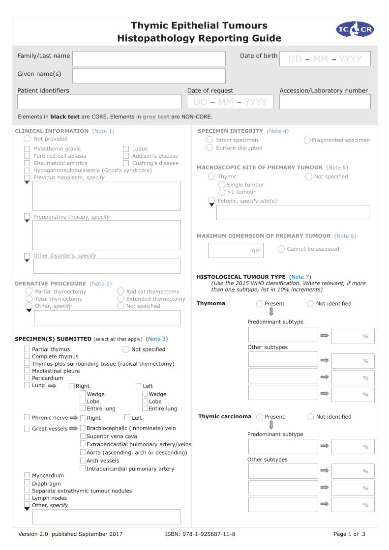
Thymic Epithelial Tumours Histopathology Reporting Guide
OPERATIVE PROCEDURE (Note 2) Partial thymectomy Total thymectomy Other, specify
Radical thymectomy Extended thymectomy Not specified
MACROSCOPIC SITE OF PRIMARY TUMOUR (Note 5)
ThymicSingle tumour>1 tumour
Ectopic, specify site(s)
Not specified
SPECIMEN(S) SUBMITTED (select all that apply) (Note 3)
Partial thymusComplete thymusThymus plus surrounding tissue (radical thymectomy)Mediastinal pleuraPericardiumLung
Phrenic nerve
Great vessels
MyocardiumDiaphragmSeparate extrathymic tumour nodulesLymph nodesOther, specify
Not specified
Lupus Addison’s diseaseCushing’s disease
Not provided SPECIMEN INTEGRITY (Note 4)
Intact specimenSurface disrupted
Fragmented specimen
Right LeftWedge WedgeLobe LobeEntire lung Entire lung
Right Left
Brachiocephalic (innominate) vein Superior vena cavaExtrapericardial pulmonary artery/veinsAorta (ascending, arch or descending) Arch vesselsIntrapericardial pulmonary artery
Version 2.0 published September 2017 ISBN: 978-1-925687-11-8 Page 1 of 3
Family/Last name
Given name(s)
Patient identifiers Date of request Accession/Laboratory number
Elements in black text are CORE. Elements in grey text are NON-CORE.
Date of birth DD – MM – YYYY
CLINICAL INFORMATION (Note 1)
Preoperative therapy, specify
Myasthenia gravisPure red cell aplasia Rheumatoid arthritisHypogammaglobulinemia (Good’s syndrome)Previous neoplasm, specify
Other disorders, specify
MAXIMUM DIMENSION OF PRIMARY TUMOUR (Note 6)
mm Cannot be assessed
HISTOLOGICAL TUMOUR TYPE (Note 7) (Use the 2015 WHO classification. Where relevant, if more
than one subtype, list in 10% increments)
Thymoma Present Not identified
Thymic carcinoma Present Not identified
%
Predominant subtype
%
Other subtypes
%
%
%
Predominant subtype
%
Other subtypes
%
%
DD – MM – YYYY
Version 2.0 published September 2017 ISBN: 978-1-925687-11-8 Page 2 of 3
SEPARATE EXTRATHYMIC TUMOUR NODULES/METASTASES (Note 9)
Pleural and/or pericardial Present Not identified
Specify location(s) Specify number/location
Thymic neuroendocrine tumours
%
%
Typical carcinoid tumour
Large cell neuroendocrine carcinoma
Present Not identified
% Small cell carcinoma
% Atypical carcinoid tumour
Final histological diagnosis (Use 2015 WHO classification for combined tumours)
EXTENT OF DIRECT INVASION (Note 8)
Mediastinal pleura
Not involvedInvolved
Cannot be assessedNot applicable
PericardiumNot involvedInvolved
Cannot be assessedNot applicable
Aorta (ascending, arch or descending)
Not involvedInvolved
Cannot be assessedNot applicable
Lung (pulmonary parenchyma, visceral pleura, or both)Not involvedInvolved
Cannot be assessedNot applicable
Brachiocephalic (innominate) veinNot involvedInvolved
Cannot be assessedNot applicable
Specify lobe(s) of the lung
GREAT VESSELS
Arch vessels Not involvedInvolved
Cannot be assessedNot applicable
Superior vena cava
Not involvedInvolved
Cannot be assessedNot applicable
Intrapericardial pulmonary artery Not involvedInvolved
Cannot be assessedNot applicable
Phrenic nerveNot involvedInvolved
Cannot be assessedNot applicable
Other involved organ(s)/site(s) by direct spread
Pulmonary intraparenchymal
Present Not identified
Distant organ
Present Not identified
Specify site(s)
Cannot be assessedPrior treatment not knownNo prior treatment No response Positive response
RESPONSE TO NEOADJUVANT THERAPY (Note 10)
No or minimal tumour response Partial tumour response Complete or near-complete response
COEXISTENT PATHOLOGY (Note 11)
Thymic hyperplasia FollicularEpithelial True
Other, specify
Cystic changesIn tumourIn adjacent thymus
MARGIN STATUS (Note 12)Cannot be assessedNot involved
Involved
Specify margin(s), if possible
Macroscopic
Specify margin(s), if possible
Microscopic
Tumour capsuleNo invasion beyond capsule or limit of the thymusInvasion beyond the mediastinum
Extrapericardial pulmonary artery or veins Not involvedInvolved
Cannot be assessedNot applicable
Version 2.0 published September 2017 ISBN: 978-1-925687-11-8 Page 3 of 3
LYMPH NODE STATUS (Note 13)
No nodes submitted or foundNot involvedInvolved
Anterior (perithymic) nodes (N1)
Number of lymph nodes examined
Number of positive lymph nodes
Number cannot be determined
Deep intrathoracic or cervical nodes (N2)
Number of lymph nodes examined
Number of positive lymph nodes
Number cannot be determined
Unspecified location within N 1 or 2
Positive markers
Negative markers
Equivocal markers
ANCILLARY STUDIES
Immunohistochemical markers (Note 14)
Interpretation and conclusions
Performed Not performed
Location(s) outside N 1 or 2 (M1b disease)
Number of lymph nodes examined
Number of positive lymph nodes
Number cannot be determined
Number of lymph nodes examined
Number of positive lymph nodes
Number cannot be determined
TNM 8TH EDITION PATHOLOGIC STAGING FOR THYMIC
Primary tumour (pT)
m - multiple primary tumorsy - post treatment
r - recurrent
TX Primary tumour cannot be assessed T0 No evidence of primary tumourT1 Tumour encapsulated or extending into the
mediastinal fat, may involve the mediastinal pleura.T1a No mediastinal pleural involvementT1b Direct invasion of the mediastinal pleuraT2 Tumour with direct involvement of the pericardium
(partial or full thickness).T3 Tumour with direct invasion into any of the
following; lung, brachiocephalic vein, superior vena cava, phrenic nerve, chest wall, or extrapericardial pulmonary artery or vein
T4 Tumour with direct invasion into any of the following; aorta (ascending, arch or descending), arch vessels, intrapericardial pulmonary artery, myocardium, trachea, or oesophagus
No nodes submitted or foundNX Regional lymph nodes cannot be assessed N0 No regional lymph node metastasisN1 Metastasis in anterior (perithymic) lymph nodesN2 Metastasis in deep intrathoracic or cervical lymph
Regional lymph nodes(pN)
Not applicableM0 No pleural, pericardial or distant metastasisM1 Distant metastasisM1a Separate pleural or pericardial nodule(s)*M1b Distant metastasis beyond the pleura or pericardium*
*listed as clinical M
Distant metastases (pM)
EPITHELIAL TUMOURS## (Note 16)
Molecular studies (Note 15)
Performed Not performed
Specify tests and results
## Reproduced with permission. Source:Brierley JD, Gospodarowicz MK and Wittekind C (eds) (2016). UICC TNM Classification of Malignant Tumours, 8th Edition, Wiley-Blackwell.
1
Note 1 - Clinical information (Non-core) Reason/Evidentiary Support
It is helpful to know whether the patient has myasthenia gravis or other conditions including neoplasms that can be associated with thymomas. Knowledge of any neoadjuvant treatment is also important as it may explain necrosis and scarring seen macroscopically and microscopically, and allows the pathologist to comment on histologic treatment response.
If clinical conditions other than those listed are provided, then these should be noted under ‘Other disorders’.
Back
Note 2 - Operative procedure (Non-core) Reason/Evidentiary Support
Documentation of the operative procedure is useful, as correlation of the type of procedure with the material received can be important for both pathological diagnosis and patient safety. Further, the type of surgical procedure is important in determining the assessment of surgical margins.1
The surgeon should inform the pathologist of the type of operation/procedure.
A thymectomy is an operation to remove the thymus. A partial thymectomy is the removal of less than the whole thymus. A total (standard) thymectomy is the removal of the thymus gland without surrounding fatty tissue. An extended thymectomy is the removal of the thymus gland including the fatty tissue of the mediastinum and neck. A radical (maximal) thymectomy is the removal of the thymus gland and wide resection of fatty tissue of the middle and anterior mediastinum and neck from the diaphragm to the thyroid gland and between both phrenic nerves; the technique includes visualization of recurrent laryngeal and phrenic nerves and wide opening of both pleural spaces.
Back
Note 3 – Specimen(s) submitted (Core) Reason/Evidentiary Support
Specimen type should indicate what was submitted.1 Specimen type varies according to the type of operation. If the specimen was obtained by a radical thymectomy, the specimen type is indicated as “Thymus plus surrounding tissue.”
Specimens obtained by combined resection with other organs or parts thereof, should be itemised, such as lung, pleura, pericardium, great vessels and myocardium. Other organs or tissues are reported as “Other” and details should be recorded.1-3
Separate extrathymic tumour nodules submitted should be recorded; these include pleural and pericardial seedings, pulmonary intraparenchymal nodules and distant organ metastases. The location, number and size of extrathymic nodules are described later in the dataset (see Note 10 - SEPARATE EXTRATHYMIC TUMOUR NODULES/METASTASES).
Submitted lymph nodes should also be recorded.4,5 These may be submitted separately or within a combined mediastinal specimen, so labelling or discussion with the surgeon may be required. Further details on lymph nodes are captured later in the dataset (see Note 14 – LYMPH NODE STATUS).
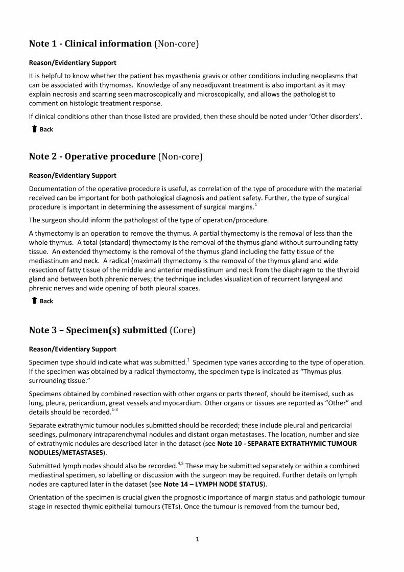
Orientation of the specimen is crucial given the prognostic importance of margin status and pathologic tumour stage in resected thymic epithelial tumours (TETs). Once the tumour is removed from the tumour bed,
2
orientation becomes difficult. Furthermore, the fatty tissue can become easily disrupted. Therefore, orientation of the specimen ideally should be started in situ by the surgeon and areas of concern need to be clearly communicated to the pathologist. Orientating the specimen on a mediastinal board is encouraged (Figure 1).1 Anterior, posterior, right and left surfaces should be clearly distinguished (e.g. inked with different colours or with a detailed block key). Furthermore, the surgeon should mark areas of concern and also representative areas adjacent to the pericardium, the innominate (brachiocephalic) vein and superior vena cava (or mark these structures if resected) and right/left mediastinal pleural surfaces (if resected).
Figure 1: Mediastinal board that could be used to orient the specimen1 Mediastinal board. A diagram on a soft board is useful in maintaining proper dimensions and orientation of specimens. Printing this figure as a full page corresponds roughly to the normal mediastinal dimensions and can be placed directly on a standard soft specimen board that is generally available in surgical pathology departments. (Reprinted from Detterbeck FC, Moran C, Huang J, Suster S, Walsh G, Kaiser L and Wick M (2011). Which way is up? Policies and procedures for surgeons and pathologists regarding resection specimens of thymic malignancy. J Thorac Oncol 6:S1730-1738 with permission from Elsevier)
Back
Note 4 - Specimen integrity1,3 (Non-core) Reason/Evidentiary Support
Although there are no studies specifically evaluating the prognosis of patients who underwent thymectomy where the capsule was disrupted intraoperatively or the lesion was resected in fragments, it is important to record these features because in these circumstances the pathologist cannot properly evaluate the presence of capsular invasion or completeness of resection. The latter are important prognostic features.
‘Intact specimen’ means that a TET is either completely surrounded by a fibrous capsule or is present in its entirety within the submitted specimen, without rupture of the tumour into surrounding tissues or on to the external surface of the specimen.
‘Surface disrupted’ means that a TET remains in one piece but shows exposure of the tumour onto the external surface of the specimen, secondary to disruption.
A fragmented specimen is when a TET is submitted in piecemeal form that precludes satisfactory identification of margins.
Back
3
Note 5 - Macroscopic site of primary tumour (Non-core)
Reason/Evidentiary Support
TETs usually arise as a single nodule or mass in the thymus in the anterior mediastinum. However, cases of multiple, synchronous TETs have been described.6-8 Although synchronous TETs generally occur in the thymus in the anterior mediastinum, these tumours can also occur at ectopic sites. Although rare, ectopic TETs have been described in the neck, posterior mediastinum, pretracheal fat, deep to phrenic nerves, posterior to brachiocephalic (innominate) vein, aortopulmonary window, aortocaval groove, anterior mediastinal fat, cardiophrenic fat and base of skull. Ectopic thymomas can also present in the lung, where they should be dealt with as primary pulmonary neoplasms. Importantly, ectopic TETs should be distinguished from pleural or pericardial implants and metastases because the latter will up-stage the tumour. Many reported synchronous TETs differ in tumour subtype and stage. In addition, a case of synchronous thymoma and thymic carcinoid tumour has been reported in a patient with multiple neuroendocrine neoplasia type I.9 Therefore, when synchronous TETs are identified, each tumour should be recorded, microscopically reviewed and staged. Back
Note 6 - Maximum dimension of primary tumour (Non-core) Reason/Evidentiary Support
A retrospective analysis of 5845 cases showed that size was not useful in predicting survival in relation to staging of TETs, so this is viewed as a non-core rather than as a core parameter.3
Identification of the primary tumour may be uncertain in cases with multiple foci and therefore the maximum dimension of the largest tumour should be recorded.
The maximum tumour size should still be recorded as the number of blocks sampled in a resected tumour is recommended to be 1 per centimetre of the maximum diameter. Inadequate sampling may lead to incorrect tumour classification.10
Back
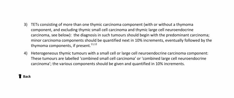
Note 7 - Histological tumour type (Core) Reason/Evidentiary Support
Tumours should be classified according to the World Health Organisation (WHO) 2015 classification system for thymic tumours (see below).11-13
In cases of TETs showing more than one morphological subtype the following should be applied:
1) TETs showing more than one histological thymoma subtype: The diagnosis in such tumours should list all the histological WHO types, starting with the predominant component and then minor components. All should be quantified in 10% increments. This rule does not apply to AB thymoma which is a distinct entity (this should be documented as type AB 100%).12,14
2) TETs consisting of a thymic carcinoma component together with one or more thymoma component: Irrespective of the size/percentage of the thymic carcinoma component the diagnosis in such tumours should begin with the label “thymic carcinoma” (specifying the histological type and percentage) followed by the thymoma component(s) (quantified in 10% increments).11,12
4
3) TETs consisting of more than one thymic carcinoma component (with or without a thymoma component, and excluding thymic small cell carcinoma and thymic large cell neuroendocrine carcinoma, see below): the diagnosis in such tumours should begin with the predominant carcinoma; minor carcinoma components should be quantified next in 10% increments, eventually followed by the thymoma components, if present.11,12
4) Heterogeneous thymic tumours with a small cell or large cell neuroendocrine carcinoma component: These tumours are labelled ‘combined small cell carcinoma’ or ‘combined large cell neuroendocrine carcinoma’; the various components should be given and quantified in 10% increments.
Back
5
WHO classification of tumours of the thymusa,b Descriptor ICD0 codes
Epithelial tumours
Thymoma
Type A thymoma, including atypical variant 8581/3*
Type AB thymoma 8582/3*
Type B1 thymoma 8583/3*
Type B2 thymoma 8584/3*
Type B3 thymoma 8585/3*
Micronodular thymoma with lymphoid stroma 8580/1*
Metaplastic thymoma 8580/3
Other rare thymomas
Microscopic thymoma 8580/0
Sclerosing thymoma 8580/3
Lipofibroadenoma 9010/0*
Thymic carcinoma
Squamous cell carcinoma 8070/3
Basaloid carcinoma 8123/3
Mucoepidermoid carcinoma 8430/3
Lymphoepithelioma-like carcinoma 8082/3
Clear cell carcinoma 8310/3
Sarcomatoid carcinoma 8033/3
Adenocarcinomas
Papillary adenocarcinoma 8260/3
Thymic carcinoma with adenoid cystic carcinoma-like features 8200/3
Mucinous adenocarcinoma 8480/3
Adenocarcinoma, NOS 8140/3
NUT carcinoma 8023/3*
Undifferentiated carcinoma 8020/3
Other rare thymic carcinomas
Adenosquamous carcinoma 8560/3
Hepatoid carcinoma 8576/3
Thymic carcinoma, NOS 8586/3
Thymic neuroendocrine tumours
Carcinoid tumours
Typical carcinoid 8240/3
Atypical carcinoid 8249/3
Large cell neuroendocrine carcinoma 8013/3
Combined large cell neuroendocrine carcinoma 8013/3
Small cell carcinoma 8041/3
Combined small cell carcinoma 8045/3
Combined thymic carcinomas
a The morphology codes are from the International Classification of Diseases for Oncology (ICD-O). Behaviour is coded /0 for benign tumours; /1 for unspecified, borderline, or uncertain behaviour; /2 for carcinoma in situ and grade III intraepithelial neoplasia; and /3 for malignant tumours. b The classification is modified from the previous WHO classification, taking into account changes in our understanding of these lesions. * These new codes were approved by the IARC/WHO Committee for ICD-O. © World Health Organisation/International Agency for Research on Cancer (IARC). Reproduced with permission.
Back
6
Note 8 - Extent of direct invasion (Core) Reason/Evidentiary Support
The Masaoka-Koga staging system has been the most frequently used for staging,15,16 with refinement of definitions for anatomic staging parameters proposed in 2011,17 but this staging system has now been superseded by a TNM-based classification based on data from the ITMIG retrospective database of over 8000 patients analysed by an International Association for the Study of Lung Cancer (IASLC), thymic domain, committee.3,5 The T category is dependent on extent of direct local invasion. Use of an elastic stain is strongly recommended in assessing involvement of mediastinal structures in relation to elastic layers within mediastinal and visceral pleura, fibrous layer of the pericardium and the adventitia and media of the great vessels.
In relation to the new TNM-based staging system, the presence of capsular invasion was not prognostically significant in data from the ITMIG retrospective database study and tumours are therefore categorised as pT1a, independent of whether the capsule is breached, if the tumour has not directly infiltrated the mediastinal pleura. Similar data were found in separate meta-analyses.3,18 Invasion through the mediastinal pleura was also not found to be of prognostic significance in the cases from the ITMIG database, although evidence from Japanese patients demonstrated that invasion of the mediastinal pleura was associated with the cumulative incidence of recurrence (CIR)19 so this parameter remains part of the dataset, to be collected for further review and is categorised as pT1b, although it is recognised that this anatomic margin may not be easily identifiable on histology.3 Discussion with the surgeon may facilitate its identification in specimens.1
In order to maintain consistency in data collection, the following definitions, agreed by expert consensus, were proposed by an ITMIG-based group:
Pericardial invasion - microscopic involvement of the pericardium (either partial in the fibrous layer or penetrating through the serosal layer);
Visceral pleura/lung - microscopically confirmed direct penetration through the outer elastin layer of the visceral pleura with or without invasion into the lung parenchyma.
In relation to the great vessels, opinions differed between involvement being defined as tumour cells being present within the adventitia, media or lumen. The consensus opinion, in the context of great vessels, was that tumour cells present within the media is the preferred histological compartment through which to define involvement, as it is easily seen compared to the adventitia on an elastic stain, and its involvement is likely relevant to surgical management in terms of need for partial resection and repair. In a similar fashion, involvement of the phrenic nerve is defined as tumour cells being present within the perineurium. ‘Other’ should be used if tumours infiltrate structures such as myocardium, trachea, oesophagus or chest wall. Involvement of muscle layers is viewed as the most reproducible parameter through which to collect data on positive involvement.
Back
Note 9 - Separate extrathymic tumour nodules/metastases (Core) Reason/Evidentiary Support
Separate extrathymic tumour nodules must be recorded as they form part of the TNM staging system. These are divided into two groups: first, those nodules that are limited to the pericardium and/or pleura (sometimes referred to as pericardial and pleural seeding), which constitute pM1a in TNM staging: second, nodules that are either within the lung parenchyma or distant organs, which constitute pM1b.1,4 The number of nodules in the pleura/pericardium should be recorded as there is some evidence that greater numbers portend an adverse prognosis.20
7
These synchronous metastatic foci will usually have the same morphology as the primary thymic neoplasm and need to be distinguished from the far rarer synchronous primary thymic epithelial tumours (see Note 5 - MACROSCOPIC SITE OF PRIMARY TUMOUR).7,8
Back
Note 10 - Response to neoadjuvant therapy (Non-core) Reason/Evidentiary Support
There is no recommended or agreed system for tumour regression grading (TRG) in TETs. There are sparse reports documenting the effects of neoadjuvant chemotherapy on TETs21 but there are no systematic studies on this subject. In other organ systems including carcinomas of the breast, stomach, oesophagus and colorectum, there is evidence that the response to neoadjuvant therapy provides prognostic information. Schemes for TRG for several of these organ systems have been published.22 Steroid therapy may also affect morphology by eliminating lymphocytes although this is not viewed as part of neoadjuvant therapy.
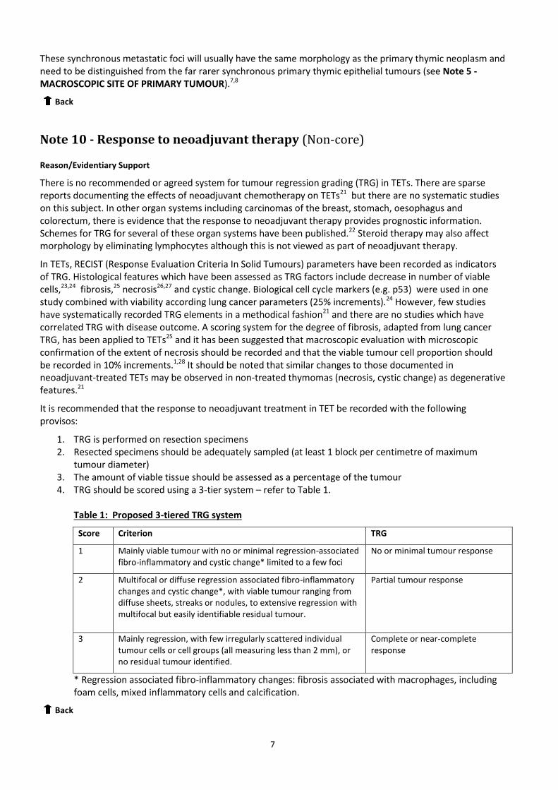
In TETs, RECIST (Response Evaluation Criteria In Solid Tumours) parameters have been recorded as indicators of TRG. Histological features which have been assessed as TRG factors include decrease in number of viable cells,23,24 fibrosis,25 necrosis26,27 and cystic change. Biological cell cycle markers (e.g. p53) were used in one study combined with viability according lung cancer parameters (25% increments).24 However, few studies have systematically recorded TRG elements in a methodical fashion21 and there are no studies which have correlated TRG with disease outcome. A scoring system for the degree of fibrosis, adapted from lung cancer TRG, has been applied to TETs25 and it has been suggested that macroscopic evaluation with microscopic confirmation of the extent of necrosis should be recorded and that the viable tumour cell proportion should be recorded in 10% increments.1,28 It should be noted that similar changes to those documented in neoadjuvant-treated TETs may be observed in non-treated thymomas (necrosis, cystic change) as degenerative features.21
It is recommended that the response to neoadjuvant treatment in TET be recorded with the following provisos:
1. TRG is performed on resection specimens 2. Resected specimens should be adequately sampled (at least 1 block per centimetre of maximum
tumour diameter) 3. The amount of viable tissue should be assessed as a percentage of the tumour 4. TRG should be scored using a 3-tier system – refer to Table 1.
Table 1: Proposed 3-tiered TRG system
Score Criterion TRG
1 Mainly viable tumour with no or minimal regression-associated fibro-inflammatory and cystic change* limited to a few foci
No or minimal tumour response
2 Multifocal or diffuse regression associated fibro-inflammatory changes and cystic change*, with viable tumour ranging from diffuse sheets, streaks or nodules, to extensive regression with multifocal but easily identifiable residual tumour.
Partial tumour response
3 Mainly regression, with few irregularly scattered individual tumour cells or cell groups (all measuring less than 2 mm), or no residual tumour identified.
Complete or near-complete response
* Regression associated fibro-inflammatory changes: fibrosis associated with macrophages, including foam cells, mixed inflammatory cells and calcification.
Back
8
Note 11 - Coexistent pathology (Non-core) Reason/Evidentiary Support
Thymectomy specimens from myasthenia gravis patients commonly demonstrate pathologic findings in the non-neoplastic thymus and the most common feature is thymic follicular hyperplasia. Thymic hyperplasia can be classified into three types: follicular, epithelial and true hyperplasia. Follicular hyperplasia is defined by the presence of B-cell follicles irrespective of the size or weight of the thymus. The standardised macroscopic and histopathological work-up of thymectomy specimens including the grading of thymic follicular hyperplasia has been reported by MGTXa.29,30 Epithelial hyperplasia (nodular epithelial hyperplasia, also called ‘microscopic thymoma’) is a thymic epithelial cell proliferation forming discrete microscopic islands and it is not infrequently observed in thymic tissue from myasthenia gravis patients.31,32 It should be differentiated from ‘microthymoma’ which represents microscopic-sized true thymoma.33 True thymic hyperplasia is an increase in volume of the thymus which maintains normal histology.34 Because of wide variations of sizes and weights of the thymus in the normal population, true thymic hyperplasia is difficult to define except for extreme cases. The presence of thymic hyperplasia adjacent to a thymoma, irrespective of the type, has no known clinical significance.
Cystic changes can involve both thymic epithelial tumours and adjacent thymus.35-39 The description of cystic changes, although not of prognostic significance, may be important for clinicopathological correlation.
Back
Note 12 - Margin status (Core) Reason/Evidentiary Support
Complete resection has been repeatedly shown to be a prognostic parameter in thymomas and thymic carcinomas.40-42 Therefore, the evaluation and recording of the margin status is important. To be able to assess the margins, orientation of the specimen is crucial. As discussed earlier (see Note 5 MACROSCOPIC SITE OF PRIMARY TUMOUR), once the tumour is removed from the tumour bed, orientation becomes difficult. Furthermore, the fatty tissue can become easily disrupted. Therefore, orientation of the specimen should ideally be started in situ by the surgeon and areas of concern need to be clearly communicated to the pathologist. Anterior, posterior, right and left surfaces should be clearly distinguished (e.g. inked with different colours or with a detailed block key). Furthermore, the surgeon should mark areas of concern and also representative areas adjacent to the pericardium, the large vessels (or mark these structures if resected) and right/left mediastinal pleural surfaces (if resected). If the resection specimen includes neighbouring organs such as lung, or large vessels, margins need to be evaluated on those organs as well.
R0 resection is defined as complete resection without macroscopic or microscopic involvement of the margin by the tumour. R1 (incomplete) resection indicates microscopic tumour at the resection margin. R2 (incomplete) resection is defined as macroscopic tumour present at the resection margin. If the specimen is disrupted at the time of gross evaluation and cannot be reconstructed, then the assessment of margins might not be possible.
Back
a Thymectomy and Myasthenia gravis multicentre, international clinical trial (MGTX)
9
Note 13 - Lymph node status (Core) Reason/Evidentiary Support
Involvement of lymph nodes by TETs is an adverse prognostic factor.4,43 Lymph node status should be recorded according to the recommended anatomic map in relation to the ITMIG & IASLC TNM system,4,5 namely anterior (perithymic) nodes (N 1) and deep intrathoracic or cervical nodes (N 2), whilst any positive lymph node was viewed as stage IVb within the Masaoka-Koga system. As the location of lymph nodes found during the gross inspection of a thymectomy specimen may be problematic, either the specimen needs to be properly oriented by the surgeon, or labelled specifically within separate pots. Lymph nodes outside N1 and N2 are regarded as distant metastasis (pM1b).4
Back
Note 14 - Immunohistochemical markers (Non-core) Reason/Evidentiary Support
Immunohistochemical analysis of thymic resection specimens may be performed for several reasons: 1. To exclude or confirm the presence of a tumour of thymic epithelial origin44 2. To aid in subtyping of thymomas45 3. To establish the origin of a thymic carcinoma as either a primary thymic carcinoma or a metastasis
The differential diagnostic spectrum of thymoma is related to either its epithelial component or to the lymphoid component. The lymphoid component of “B-type” thymoma and of thymic follicular hyperplasia may raise the suspicion of non-Hodgkin lymphoma, especially T-lymphoblastic leukaemia/lymphoma. Immunohistochemistry may be applied to type the lymphoid population [normally composed of immature, CD3/terminal deoxynucleotidyl transferase (TdT/CD1a/CD99+) lymphocytes], or to confirm the presence of an epithelial component, which may be highlighted by pan-cytokeratin and/or p63 stains. The epithelial component in thymic epithelial tumours with a sparse lymphoid component may raise the possibility of either a germ cell tumour or metastatic carcinoma.44,46 Germ cell tumours may be diagnosed by appropriate immunohistochemical stains including SALL4, OCT4, CD117, CD30, D2-40, human chorionic gonadotropin (hCG), placental alkaline phosphatase (PLAP), and α-fetoprotein (AFP).44
Subtyping of thymomas is primarily based on histology; immunohistochemical stains (cytokeratin and/or p63) may be helpful in the evaluation of the density of the epithelial cells in B-type thymoma thus aiding the diagnosis of B1/2/3 thymoma. Similarly, cytokeratin stains may be used to confirm the epithelial nature of the spindle cells in type A, type AB and in metaplastic thymoma. Epithelial expression of CD20 is reported to be more frequent among type A and AB thymomas.47 Neuroendocrine markers may be useful to rule out neuroendocrine tumours.45
Distinguishing thymoma (in particular type B3 thymoma) and thymic carcinoma may occasionally be problematic; there are no immunohistochemical markers that can reliably segregate these entities. However, CD5, CD117 and the recently described markers GLUT1 and MUC1 show a higher incidence of staining in thymic carcinoma (in particular, thymic squamous cell carcinoma) compared to thymoma.48,49 Ki-67 labelling index in epithelial tumour cells of ≥13.5% has been suggestive of thymic carcinoma.50
The diagnosis of thymic carcinoma essentially involves the exclusion of metastasis; immunohistochemical analysis may support a diagnosis of thymic carcinoma but cannot establish the diagnosis with certainty. Expression of CD5, particularly in combination with CD117 positivity, lends some support to a diagnosis of thymic carcinoma. Several new markers (FoxN1 and CD205) may further support a diagnosis of thymic carcinoma. Other markers may be applied to rule out thymic carcinoma by confirming a non-thymic origin, such as TTF-1. However, given the great diversity in histological subtypes of thymic carcinoma, the specificity
10
of markers routinely used to diagnose carcinoma of a particular origin may be considerably lower in this situation.12
Back
Note 15 – Molecular studies (Non-core)
Reason/Evidentiary Support
Molecular studies have not been applied routinely for the diagnosis of thymic epithelial tumours. A diagnosis of NUT carcinoma needs immunohistochemical and/or molecular genetic confirmation.51,52 The sensitivities of NUT immunohistochemical staining have been reported as 60% and 87%.51,52 There have been a few reports of primary mediastinal synovial sarcoma confirmed by FISH.
Back
Note 16 –TNM 8th edition Pathologic Staging for Thymic Epithelial Tumours (Core) Reason/Evidentiary Support
At least 15 different stage classification systems have been proposed, beginning as far back as 1978.53 Until 2016, the most widely used was the Masaoka system,15 modified and refined in 1994,16 with refinement of definitions for anatomic staging parameters proposed in 2011.17 This has now been replaced by a TNM-based classification based on data from the ITMIG retrospective database of over 8000 patients.5 In the new TNM 8th editions, both UICC54 and AJCC55, T stage is based on the extent of direct invasion of mediastinal structures (see above section),3 nodal disease is based on involvement of lymph nodes in anterior (perithymic) (N1) and deep/cervical (N2) compartments, and M stage based on the presence of separate pleural and pericardial nodules (M1a) and pulmonary intraparenchymal nodule or distant organ metastasis (M1b).4 The Masaoka-Koga system could still be used if part of ongoing studies but the TNM system should be used henceforth as the method of staging.56
Back
11
References 1 Detterbeck FC, Moran C, Huang J, Suster S, Walsh G, Kaiser L and Wick M (2011). Which way is up?
Policies and procedures for surgeons and pathologists regarding resection specimens of thymic malignancy. J Thorac Oncol 6:S1730-1738.
2 Detterbeck FC, Stratton K, Giroux D, Asamura H, Crowley J, Falkson C, Filosso PL, Frazier AA, Giaccone
G, Huang J, Kim J, Kondo K, Lucchi M, Marino M, Marom EM, Nicholson AG, Okumura M, Ruffini E and Van Schil P (2014). The IASLC/ITMIG Thymic Epithelial Tumors Staging Project: proposal for an evidence-based stage classification system for the forthcoming (8th) edition of the TNM classification of malignant tumors. J Thorac Oncol 9(9 Suppl 2):S65-72.
3 Nicholson AG, Detterbeck FC, Marino M, Kim J, Stratton K, Giroux D, Asamura H, Crowley J, Falkson C,
Filosso PL, Giaccone G, Huang J, Kondo K, Lucchi M, Marom EM, Okumura M, Ruffini E and Van Schil P (2014). The IASLC/ITMIG Thymic Epithelial Tumors Staging Project: proposals for the T Component for the forthcoming (8th) edition of the TNM classification of malignant tumors. J Thorac Oncol 9(9 Suppl 2):S73-80.
4 Kondo K, Van Schil P, Detterbeck FC, Okumura M, Stratton K, Giroux D, Asamura H, Crowley J, Falkson
C, Filosso PL, Giaccone G, Huang J, Kim J, Lucchi M, Marino M, Marom EM, Nicholson AG and Ruffini E (2014). The IASLC/ITMIG Thymic Epithelial Tumors Staging Project: proposals for the N and M components for the forthcoming (8th) edition of the TNM classification of malignant tumors. J Thorac Oncol 9(9 Suppl 2):S81-87.
5 Bhora FY, Chen DJ, Detterbeck FC, Asamura H, Falkson C, Filosso PL, Giaccone G, Huang J, Kim J, Kondo
K, Lucchi M, Marino M, Marom EM, Nicholson AG, Okumura M, Ruffini E and Van Schil P (2014). The ITMIG/IASLC Thymic Epithelial Tumors Staging Project: A Proposed Lymph Node Map for Thymic Epithelial Tumors in the Forthcoming 8th Edition of the TNM Classification of Malignant Tumors. J Thorac Oncol 9(9 Suppl 2):S88-96.
6 Suzuki H, Yoshida S, Hiroshima K, Nakatani Y and Yoshino I (2010). Synchronous multiple thymoma:
report of three cases. Surgery today 40:456-459. 7 Bernatz PE, Harrison EG and Clagett OT (1961). Thymoma: a clinicopathological study. J Thorac
Cardiovasc Surg 42:424-444. 8 Leuzzi G, Marino M, Alessandrini G, Sciuto R, Pescarmona E and Facciolo F (2015). Synchronous triple
thymoma and true thymic hyperplasia simultaneously detected by F FDG PET-CT. Rev Esp Med Nucl Imagen Mol 34(4):272-274.
9 Miller BS, Rusinko RY and Fowler L (2008). Synchronous thymoma and thymic carcinoid in a woman
with multiple endocrine neoplasia type 1: case report and review. Endocr Pract 14:713-716. 10 Moran CA and Suster S (2000). On the histologic heterogeneity of thymic epithelial neoplasms. Impact
of sampling in subtyping and classification of thymomas. Am J Clin Pathol 114(5):760-766. 11 WHO (World Health Organization) (2015). WHO Classification of Tumours of the Lung, Pleura, Thymus
and Heart. Fourth edition Travis WD, Brambilla E, Burke AP, Marx A and Nicholson AG. IARC Press, Lyon, France.
12
12 Marx A, Ströbel P, Badve SS, Chalabreysse L, Chan J, Chen G, de Leval L, Detterbeck F, Girard N, Huang J, Kurrer MO, Lauriola L, Marino M, Matsuno Y, Molina TJ, Mukai K, Nicholson AG, Nonaka D, Rieker R, Rosai J, Ruffini E and Travis WD (2014). ITMIG Consensus Statement on the Use of the WHO Histological Classification of Thymoma and Thymic Carcinoma: Refined Definitions, Histological Criteria and Reporting. J Thor Oncol 9:596-611.
13 Marx A, Chan JK, Coindre JM, Detterbeck F, Girard N, Harris NL, Jaffe ES, Kurrer MO, Marom EM,
Moreira AL, Mukai K, Orazi A and Strobel P (2015). The 2015 World Health Organization Classification of Tumors of the Thymus: Continuity and Changes. J Thorac Oncol 10(10):1383-1395.
14 Strobel P, Bauer A, Puppe B, Kraushaar T, Krein A, Toyka K, Gold R, Semik M, Kiefer R, Nix W, Schalke B,
Muller-Hermelink HK and Marx A (2004). Tumor recurrence and survival in patients treated for thymomas and thymic squamous cell carcinomas: a retrospective analysis. J Clin Oncol 22(8):1501-1509.
15 Masaoka A, Monden Y, Nakahara K and Tanioka T (1981). Follow-up study of thymomas with special
reference to their clinical stages. Cancer 48(11):2485-2492. 16 Koga K, Matsuno Y, Noguchi M, Mukai K, Asamura H, Goya T and Shimosato Y (1994). A review of 79
thymomas: modification of staging system and reappraisal of conventional division into invasive and non-invasive thymoma. Pathol Int 44(5):359-367.
17 Detterbeck FC, Nicholson AG, Kondo K, Van Schil P and Moran C (2011). The Masaoka-Koga stage
classification for thymic malignancies: clarification and definition of terms. J Thorac Oncol 6(7 Suppl 3):S1710-1716.
18 Gupta R, Marchevsky AM, McKenna RJ, Wick M, Moran C, Zakowski MF and Suster S (2008). Evidence-
based pathology and the pathologic evaluation of thymomas: transcapsular invasion is not a significant prognostic feature. Arch Pathol Lab Med 132(6):926-930.
19 Ogawa K, Uno T, Toita T, Onishi H, Yoshida H, Kakinohana Y, Adachi G, Itami J, Ito H and Murayama S
(2002). Postoperative radiotherapy for patients with completely resected thymoma: a multi-institutional, retrospective review of 103 patients. Cancer 94(5):1405-1413.
20 Okuda K, Yano M, Yoshino I, Okumura M, Higashiyama M, Suzuki K, Tsuchida M, Usuda J and Tateyama
H (2014). Thymoma patients with pleural dissemination: nationwide retrospective study of 136 cases in Japan. Ann Thorac Surg 97(5):1743-1748.
21 Weissferdt A and Moran CA (2013). The impact of neoadjuvant chemotherapy on the histopathological
assessment of thymomas: a clinicopathological correlation of 28 cases treated with a similar regimen. Lung 191(4):379-383.
22 McCluggage WG, Judge MJ, Clarke BA, Davidson B, Gilks CB, Hollema H, Ledermann J, Matias-Guiu X,
Mikami Y, Stewart CJR, Vang R and Hirschowitz L (2015). Dataset for reporting of ovary, fallopian tube and primary peritoneal carcinoma: Recommendations from the International Collaboration on Cancer Reporting (ICCR). Mod Path 28(8):1101-1122.
23 Korst R.J et al (2014). Neoadjuvant chemoradiotherapy for locally advanced thymic tumors: a phase II,
multi-institutional clinical trial. J Thorac Cardiovasc Surg 147(1):36-44, 46 e31. 24 Mineo TC et al (2010). New predictors of response to neoadjuvant chemotherapy and survival for
invasive thymoma: a retrospective analysis. Ann Surg Oncol 17(11):3022-3029.
13
25 Kawasaki H et al (2014). Weekly chemotherapy with cisplatin, vincristine, doxorubicin, and etoposide
followed by surgery for thymic carcinoma. Eur J Surg Oncol 40(9):1151-1155. 26 Wright CD et al (2008). Induction chemoradiotherapy followed by resection for locally advanced
Masaoka stage III and IVA thymic tumors. Ann Thorac Surg 85(2):385-389. 27 Kim ES et al (2004). Phase II study of a multidisciplinary approach with induction chemotherapy,
followed by surgical resection, radiation therapy, and consolidation chemotherapy for unresectable malignant thymomas: final report. Lung Cancer 44(3):369-379.
28 Huang J et al (2010). Standard outcome measures for thymic malignancies. J Thorac Oncol 5(12):2017-
2023. 29 Ströbel P, Moritz R, Leite MI, Willcox N, Chuang WY, Gold R, Nix W, Schalke B, Kiefer R, Müller-
Hermelink HK, Jaretzki III A, Newsom-Davis J and Marx A (2008). The ageing and myasthenic thymus: A morphometric study validating a standard procedure in the histological workup of thymic specimens. J Neuroimmunol 201-202:64-73.
30 Marx A, Pfister F, Schalke B, Nix W and Ströbel P (2012). Thymus pathology observed in the MGTX trial.
Ann NY Acad Sci 1275:92-100 31 Pescarmona E, Rosati S, Pisacane A, Rendina EA, Venuta F and Baroni CD (1992). Microscopic
thymoma: histological evidence of multifocal cortical and medullary origin. Histopathology 20:263-266. 32 Puglisi F, Finato N, Mariuzzi L, Marchini C, Floretti G and Beltrami CA (1995). Microscopic thymoma and
myasthenia gravis. J Clin Pathol 48:682-683. 33 Cheuk W, Tsang WY and Chan JK (2005). Microthymoma: definition of the entity and distinction from
nodular hyperplasia of thymic epithelium (so-called microscopic thymoma). Am J Sur Pathol 29:415-419
34 Hofmann WJ, Möller P and Otto HF (1987). Thymic hyperplasia. I. True thymic hyperplasia. Review of the literature. Klin Wochenschr 65:49-52.
35 Suster S and Rosai J (1991). Multilocular thymic cyst: an acquired reactive process. Study of 18 cases.
Am J Surg Pathol 15(4):388-398. 36 Moran CA and Suster S (2001). Thymoma with prominent cystic and hemorrhagic changes and areas of
necrosis and infarction: a clinicopathologic study of 25 cases. Am J Surg Pathol 25(8):1086-1090. 37 Weissferdt A and Moran CA (2011). Thymic carcinoma associated with multilocular thymic cyst: a
clinicopathologic study of 7 cases. Am J Surg Pathol 35(7):1074-1079. 38 Nakamura S, Tateyama H, Taniguchi T, Ishikawa Y, Kawaguchi K, Fukui T, Mizuno T, Ishiguro F and
Yokoi K (2012). Multilocular thymic cyst associated with thymoma. A clinicopathologic study of 20 cases with an emphasison the pathogenesis of cyst formation. Am J Surg Pathol 36:1857-1864.
39 Araki T, Sholl LM, Gerbaudo VH, Hatabu H and Nishino M (2014). Intrathymic cyst: clinical and
radiologic features in surgically resected cases. Clin Radiol 69(7):732-738 40 Kondo K and Monden Y (2003). Lymphogenous and hematogenous metastasis of thymic epithelial
tumors. Ann Thorac Surg 76(6):1859-1864; discussion 1864-1855.
14
41 Ruffini E, Detterbeck F, Van Raemdonck D, Rocco G, Thomas P, Weder W, Brunelli A, Evangelista A, Venuta F and European Association of Thoracic Surgeons (ESTS) Thymic Working Group (2014). Tumours of the thymus: a cohort study of prognostic factors from the European Society of Thoracic Surgeons database. Eur J Cardiothorac Surg 46(3):361-368.
42 Moser B, Scharitzer M, Hacker S, Ankersmit J, Matilla JR, Lang G, Aigner C, Taghavi S and Klepetko W
(2014). Thymomas and thymic carcinomas: prognostic factors and multimodal management. Thorac Cardiovasc Surg. 62(2):153-160.
43 Viti A, Bertolaccini L and Terzi A (2014). What is the role of lymph nodal metastases and
lymphadenectomy in the surgical treatment and prognosis of thymic carcinomas and carcinoids? Interact Cardiovasc Thorac Surg 19(6):1054-1058.
44 den Bakker MA and Oosterhuis JW (2009). Tumours and tumour-like conditions of the thymus other
than thymoma; a practical approach. Histopathology 54(1):69-89. 45 den Bakker MA, Roden AC, Marx A and Marino M (2014). Histologic classification of thymoma: a
practical guide for routine cases. J Thorac Oncol 9(9 Suppl 2):S125-130. 46 Marchevsky A, Marx A, Strobel P, Suster S, Venuta F, Marino M, Yousem S and Zakowski M (2011).
Policies and reporting guidelines for small biopsy specimens of mediastinal masses. J Thorac Oncol 6(7 Suppl 3):S1724-1729.
47 Chilosi M, Castelli P, Martignoni G, Pizzolo G, Montresor E, Facchetti F, Truini M, Mombello A, Lestani
M, Scarpa A and et al. (1992). Neoplastic epithelial cells in a subset of human thymomas express the B cell-associated CD20 antigen. Am J Surg Pathol 16(10):988-997.
48 Kaira K, Murakami H, Serizawa M, Koh Y, Abe M, Ohde Y, Takahashi T, Kondo H, Nakajima T and
Yamamoto N (2011). MUC1 expression in thymic epithelial tumors: MUC1 may be useful marker as differential diagnosis between type B3 thymoma and thymic carcinoma. Virchows Arch 458(5): 615-620.
49 Kojika M, Ishii G, Yoshida J, Nishimura M, Hishida T, Ota SJ, Murata Y, Nagai K and Ochiai A (2009).
Immunohistochemical differential diagnosis between thymic carcinoma and type B3 thymoma: diagnostic utility of hypoxic marker, GLUT-1, in thymic epithelial neoplasms. Mod Pathol 22(10):1341-1350.
50 Roden AC, Yi ES, Jenkins SM, Donovan JL, Cassivi SD, Garces YI, Marks RS and Aubry MC (2015).
Diagnostic significance of cell kinetic parameters in World Health Organization type A and B3 thymomas and thymic carcinomas. Hum Pathol 46(1):17-25.
51 French CA (2010). Demystified molecular pathology of NUT midline carcinomas. J Clin Pathol
63(6):492-496. 52 Haack H, Johnson LA, Fry CJ, Crosby K, Polakiewicz RD, Stelow EB, Hong SM, Schwartz BE, Cameron MJ,
Rubin MA, Chang MC, Aster JC and French CA (2009). Diagnosis of NUT midline carcinoma using a NUT-specific monoclonal antibody. Am J Surg Pathol 33(7):984-991.
53 Filosso PL, Ruffini E, Lausi PO, Lucchi M, Oliaro A and Detterbeck F (2014). Historical perspectives: The
evolution of the thymic epithelial tumors staging system. Lung Cancer 83(2):126-132.
15
54 Brierley JD, Gospodarowicz MK and Wittekind C (eds) (2016). UICC TNM Classification of Malignant Tumours, 8th Edition, Wiley-Blackwell.
55 Amin MB, Edge SB and Greene FL et al (eds) (2017). AJCC Cancer Staging Manual. 8th ed., Springer,
New York. 56 Rami-Porta R (ed) (2016). Staging Manual in Thoracic Oncology, 2nd edition: An International
Association for the Study of Lung Cancer Publication, Developed in collaboration with AJCC and UICC, Editorial Rx Press, North Fort Myers, FL, US.