Embed Size (px)
Citation preview
J. Embryol. exp. Morph. Vol. 24, 3, pp. 615-623, 1970 6 1 5
Printed in Great Britain
Thymus dysgenesis in nude (nu nu) mice
ByE. M. P A N T E L O U R I S ^ N D JANET HAIR*
From the Biology Department, University of Strathclyde
SUMMARYThe thymic rudiment in mice homozygous for the recessive mutation nude (nu) has been
compared to the normal at two stages: the 14- to 15-day foetus and the 1- to 2-day-old young.In the former, the normal thymus already comprises two adjacent lobes but in the nude it isrepresented by a pair of thin strands of tissue, each with a narrow central lumen. In furtherdevelopment these strands become thicker and vesiculated. They progress no further and arefound in the 1- to 2-day young as small vestiges that are never populated with lymphoid cells.
The thyroid, parathyroids and mandible of homozygous nude mice are normal, indicatingthat the complex of structures derived from the third branchial arch is not affected as awhole.
INTRODUCTION
Research over the last ten years or so has amply demonstrated the centralrole of the thymus in the establishment of immunological competence, par-ticularly in reactions to grafts of foreign tissue. The pioneering findings of Miller(1961) as well as much subsequent work were based on the use of laboratoryanimals, usually mice, thymectomized at birth (see Metcalf, 1966).
It would be obviously desirable to be able to thymectomize the foetus or,better still, to have animals born without a thymus. Hence the interest of themouse mutant nude (nu), described in the first instance by Flanagan (1966).Recently, the homozygous nu nu animals have been described as thymusless,in the sense that no thymus can be found in them by dissection and macroscopicexamination at any stage after birth (Pantelouris, 1968). It may be noted thatthere are in man at least three types of congenital thymus aplasia or deficiency,all leading to syndromes of the utmost gravity.
MATERIAL AND METHODS
Foetuses at the 14 to 15th day of pregnancy (timed by the vaginal plug method)and 1- to 2-day-old animals were obtained by crossing heterozygous +nu,phenotypically normal, mice. The homozygous nu nu foetuses were identifiedon dissection from the uterus by the piliferous colliculi which are much lessprominent than in the normal. Homozygous nu nu young were identified by theirdistorted, irregularly curled vibrissae. The histological findings confirmed these
1 Authors' address: Department of Biology, University of Strathclyde, George Street,Glasgow C. 1, Scotland.
39 E M 13 24
616 E. M. PANTELOURIS AND J. HAIR
identifications. Five foetuses, from three pregnant females, were identified ashomozygous nu nu and, together with an equal number of normal litter-mates,were fixed in Bouin's solution, serially sectioned at 5 /i and stained with Dela-field's haematoxylin. Four 1- to 2-day-old nu nu and four normal litter-mateswere also fixed, cut in two to facilitate processing.
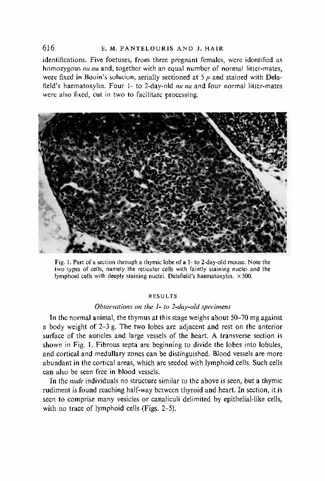
Fig. 1. Part of a section through a thymic lobe of a 1- to 2-day-old mouse. Note thetwo types of cells, namely the reticular cells with faintly staining nuclei and thelymphoid cells with deeply staining nuclei. Delafield's haematoxylin. x 500.
RESULTS
Observations on the 1- to 2-day-old specimens
In the normal animal, the thymus at this stage weighs about 50-70 mg againsta body weight of 2-3 g. The two lobes are adjacent and rest on the anteriorsurface of the auricles and large vessels of the heart. A transverse section isshown in Fig. 1. Fibrous septa are beginning to divide the lobes into lobules,and cortical and medullary zones can be distinguished. Blood vessels are moreabundant in the cortical areas, which are seeded with lymphoid cells. Such cellscan also be seen free in blood vessels.
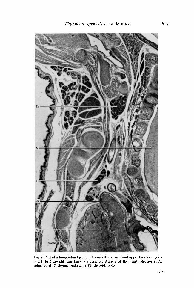
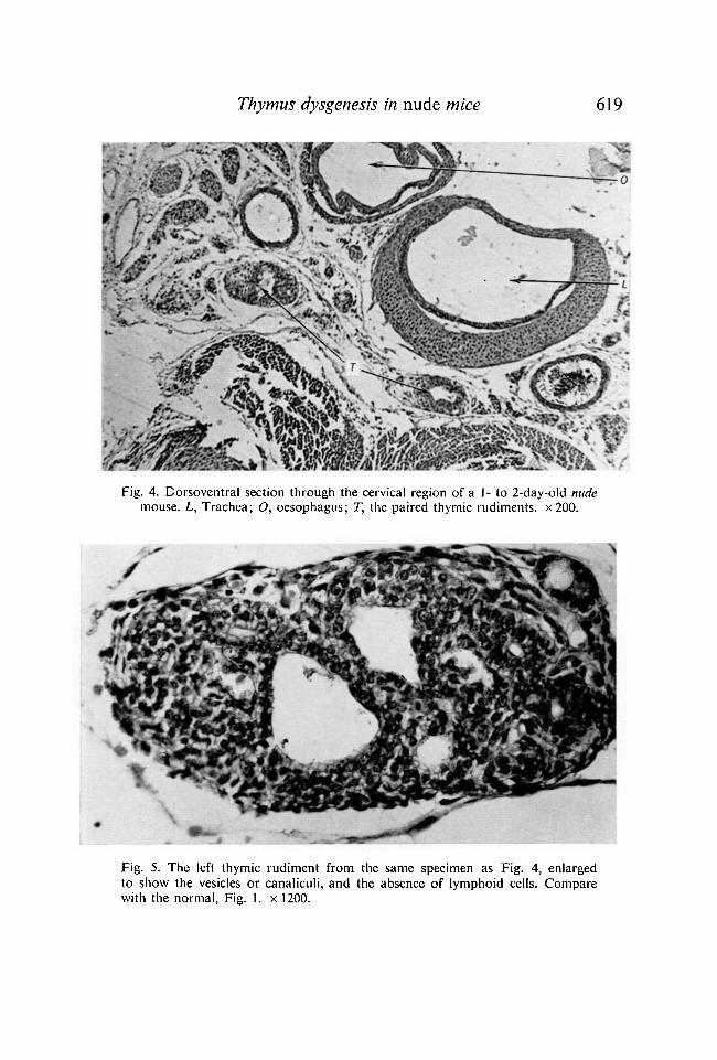
In the nude individuals no structure similar to the above is seen, but a thymicrudiment is found reaching half-way between thyroid and heart. In section, it isseen to comprise many vesicles or canaliculi delimited by epithelial-like cells,with no trace of lymphoid cells (Figs. 2-5).
Thymus dysgenesis in nude mice 617
Fig. 2. Part of a longitudinal section through the cervical and upper thoracic regionof a 1- to 2-day-old nude (nu nu) mouse. A, Auricle of the heart; Ao, aorta; N,spinal cord; T, thymus rudiment; Th, thyroid. x40.
39-2

618 E. M. PANTELOURIS AND J. HAIR
Fig. 3. Part of a section similar to that of Fig. 2 to show at a higher magnification(x 160) the thymic rudiment in the 1- to 2-day-old nude mouse. T, Thymic rudiment.
Thymus dysgenesis in nude mice 619
Fig. 4. Dorsoventral section through the cervical region of a 1- to 2-day-old nudemouse. L, Trachea; O, oesophagus; T, the paired thymic rudiments. x200.
Fig. 5. The left thymic rudiment from the same specimen as Fig. 4, enlargedto show the vesicles or canaliculi, and the absence of lymphoid cells. Comparewith the normal, Fig. 1. x 1200.
620 E. M. PANTELOURIS AND J. HAIR
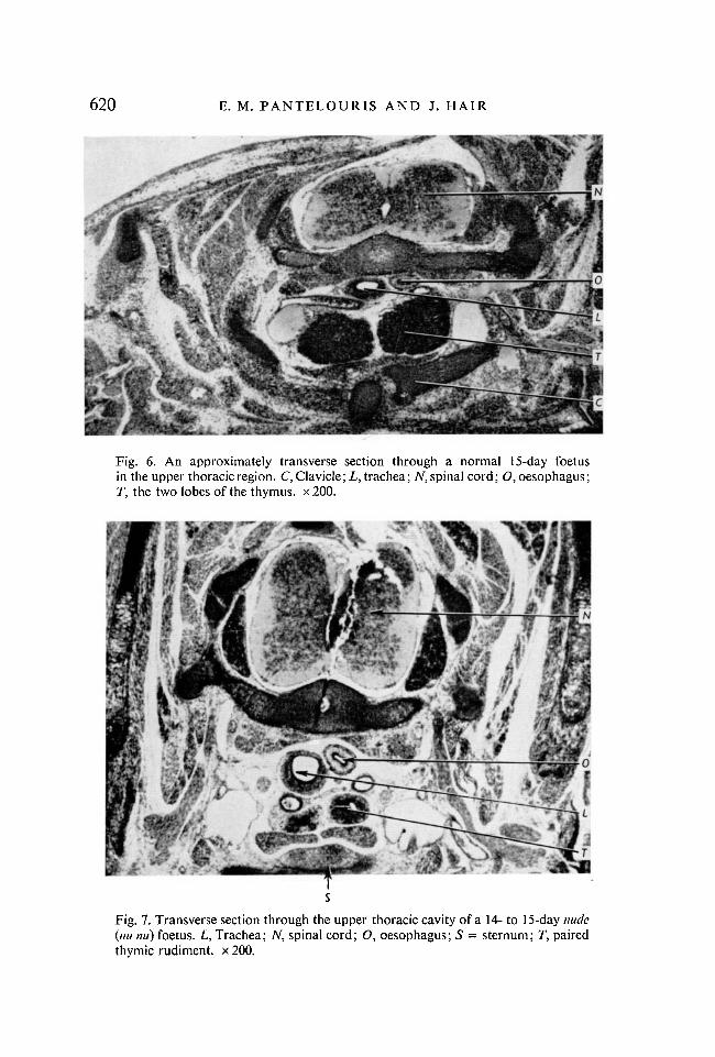
Fig. 6. An approximately transverse section through a normal 15-day foetusin the upper thoracic region. C, Clavicle; L, trachea; iV, spinal cord; O, oesophagus;T, the two lobes of the thymus. x 200.
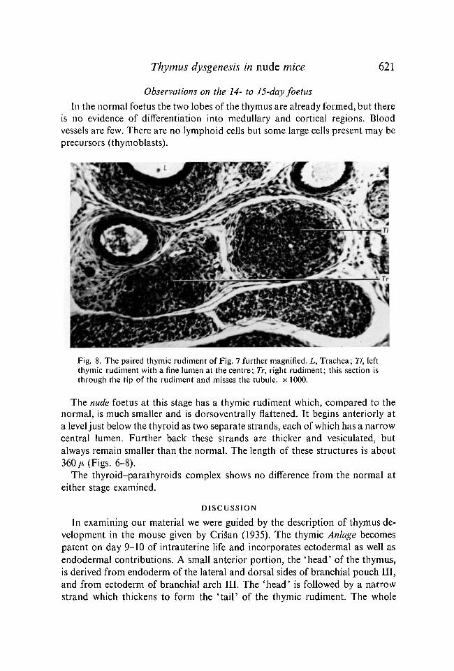
Fig. 7. Transverse section through the upper thoracic cavity of a 14- to 15-day nude(nu mi) foetus. L, Trachea; TV, spinal cord; O, oesophagus; 5 = sternum; T, pairedthymic rudiment, x 200.
Thymus dysgenesis in nude mice 621
Observations on the 14- to 15-day foetus
In the normal foetus the two lobes of the thymus are already formed, but thereis no evidence of differentiation into medullary and cortical regions. Bloodvessels are few. There are no lymphoid cells but some large cells present may beprecursors (thymoblasts).
Fig. 8. The paired thymic rudiment of Fig. 7 further magnified. L, Trachea; 77, leftthymic rudiment with a fine lumen at the centre; Tr, right rudiment; this section isthrough the tip of the rudiment and misses the tubule, x 1000.
The nude foetus at this stage has a thymic rudiment which, compared to thenormal, is much smaller and is dorsoventrally flattened. It begins anteriorly ata level just below the thyroid as two separate strands, each of which has a narrowcentral lumen. Further back these strands are thicker and vesiculated, butalways remain smaller than the normal. The length of these structures is about360/fc (Figs. 6-8).
The thyroid-parathyroids complex shows no difference from the normal ateither stage examined.
DISCUSSION
In examining our material we were guided by the description of thymus de-velopment in the mouse given by Cris>an (1935). The thymic Anlage becomespatent on day 9-10 of intrauterine life and incorporates ectodermal as well asendodermal contributions. A small anterior portion, the 'head' of the thymus,is derived from endoderm of the lateral and dorsal sides of branchial pouch III,and from ectoderm of branchial arch III. The 'head' is followed by a narrowstrand which thickens to form the 'tail' of the thymic rudiment. The whole
622 E. M. PANTELOURIS AND J. HAIR
rudiment is paired. Subsequently the thymus 'slides away' from the cervicalregion to the thoracic cavity where it grows into the definitive organ.
The thyroid begins as a thickening of the ventral side of the pharyngeal endo-derm at the level of branchial arch II. The mesobranchial plate so formed (be-coming later the discus thyroideus) incorporates also three pairs of diverticulafrom aortic arches, and becomes detached from the pharynx. Whilst it remainsthin at the midline (mesobranchial groove) it thickens at the lateral lobes. It alsoincorporates the paired ultimobranchial body, a product of branchial pouch V,and the paired epithelial body from pouch III. It is because of these shared con-nexions to the branchial arches, especially the third, that thymus, thyroid andparathyroids are viewed developmental^ as one complex.
From our observations we conclude that at about day 14 or 15 of intra-uterine development the thymic rudiment in homozygous nu nu mice is alreadyarrested at a stage more primitive than the corresponding normal rudiment, buthas passed the stage of' sliding' down into the thoracic cavity. The exact time, andnature of divergence from normal ontogenesis remains to be determined byfurther work. As a result of this developmental arrest, the animal is born, asdescribed, with no effective thymus as a lymphoid organ.
The situation in nude mice differs from all three forms of thymic aplasiadescribed in man so far. In the 'Swiss type' agammaglobulinemia it is not thethymus alone but all lymphoid tissues that fail to develop normally. In nude,the dysgenesis is restricted to the thymus, although the thymus-dependent areasof the lymph nodes are also affected secondarily (de Sousa, Parrott, & Pante-louris, 1969). The DiGiorge syndrome involves aplasia of the thymus as wellas of the parathyroids, thyroid and mandible. There is no such generalized defectof derivatives of branchial arch III in our mouse mutation; thyroid, para-thyroids and mandible remain normal. It may therefore be suggested that thebasic defect in the nude occurs later than in DiGiorge's syndrome. The thirdsyndrome described in man combines aplasia of the thymus with generalizedlymphopenia and imperfections of the lymph nodes (Nezelof et al. 1964). Infact, the three children described by Fulginiti et al. (1966) had rudimentarylymph nodes, quite difficult to find. Their vestigial thymus was described as a'small alveolar gland-like structure', a description that would also fit the 1- to2-day-old nude. In the latter, however, lymph nodes—although devoid oflymphocytes in the thymus-dependent areas—are normal as far as their corticaland medullary regions are concerned.
RESUME
La dysgenese du thymus chez des souris nudes (nu nu)
L'ebauche du thymus, chez des souris homozygotes pour la mutation recessive, nude (nu)a ete comparee a celle d'animaux normaux temoins. Deux stades ont ete etudies: le stadecorrespondant aux 14 a 15e jours du developpement foetal et le stade du 1 au 2e jour apres lanaissance. Pour le premier stade considere, il apparait que le thymus est deja constitue par
Thymus dysgenesis in nude mice 623deux lobes adjacents chez Ies temoins, alors que chez le mutant nude il est represents parune paire de fins cordons tissulaires presentant chacun une etroite lumiere centrale. Au coursdu developpement ulterieur ces cordons s'epaississent et deviennent vesiculeux. Us neprogressent pas davantage et, chez le jeune age de 1 a 2 jours, ils apparaissent sous une formevestigiale. A ce stade ils ne sont jamais peuples de cellules lymphoides.
La thyroide, Ies parathyroides et la mandibule des souris homozygotes pour la mutationnude sont normales, ce qui indique que le complexe de structures qui derive du troisieme arcbranchial n'a pas ete affecte dans sa totalite.
REFERENCES
CRISAN, C. (1935). Die Entwicklung des thyreo-parathyreothymischen Systems der weissenMaus. Z. Anat. EntwGesch. 104, 327-359.
DE SOUSA, M. A. B., PARROTT, D. M. V. & PANTELOURIS, E. M. (1969). The lymphoid tissuesin mice with congenital aplasia of the thymus. Clin. exp. Immunol. 4, 637-644.
FLANAGAN, S. P. (1966). 'Nude' , a new hairless gene with pleiotropic effects in the mouse.Genet. Res. 8, 295-309.
FULGINITI, V. A., PEARLMAN, D. S., REIQUAM, C. W., CLAMAN, H. N., HATHAWAY, W. E.,
BLACKBURN, W. R., GITHENS, J. H. & KEMPE, C. H. (1966). Dissociation of delayed hyper-sensitivity and antibody synthesising capacities in man. Report of two sibships withthymic dysplasia, lymphoid tissue depletion and normal immunoglobulins. Lancet ii, 5-8.
METCALF, D. (1966). The Thymus: Its Role in Immunological Responses, Leukemia Develop-ment and Carcinogenesis. Berlin: Springer-Verlag.
MILLER, J. F. A. P. (1961). Immunological function of the thymus. Lancet ii, 748-749.NEZELOF, C , JAMMET, M. L., LORTHOLARY, P., LABRUNE, B. & LAMY, M. (1964). L'hypo-
plasie hereditaire du thymus. Archs fr. Pediat. 21, 897-920.PANTELOURIS, E. M. (1968). Absence of thymus in a mouse mutant. Nature, Loncl. 217,
370-371.
{Manuscript received 25 March 1970)


![The Effect of Dibenzo[a,l]pyrene on the Thymus of Fetal Mice](https://img.pdfslide.net/doc/110x75/56812c68550346895d910088/the-effect-of-dibenzoalpyrene-on-the-thymus-of-fetal-mice.jpg)