Embed Size (px)
DESCRIPTION
This e book discusses the varying management modalities of thyroid nodules.
Citation preview
www.drtbalu.com Page 1
Thyroid nodules and their management
No More dilemma
Dr. T. Balasubramanian
2
Thyroid Nodules & their management
Introduction:
Thyroid nodule is a common occurrence. Majority of these nodules may be present
without clinical evidence of thyroid disease. Statistics reveal that the incidence of
palpable thyroid nodule is about 5%. Ultrasound neck is very sensitive in picking up
thyroid nodules. Ultrasound can diagnose thyroid nodules even as small as ½ cm.
Studies have shown that only 3-5% of thyroid nodules show malignant
transformation.
Causes of thyroid nodules include:
Benign causes:
1. Goiter
2. Hashimoto's thyroiditis
3. Simple / hemorrhagic cysts
4. Follicular adenoma
5. Subacute thyroiditis
Malignant causes:
1. Papillary carcinoma
2. Follicular carcinoma
3. Hurthle cell carcinoma
4. Medullary carcinoma
5. Anaplastic carcinoma
6. Primary thyroid lymphoma
7. Metastatic malignant lesion
Tools of diagnosis as far as these thyroid nodules include:
1. Ultrasound neck
2. FNAC
3. Molecular / genetic marker analysis of fine needle aspiration biopsies
Clinical features:
Majority of thyroid nodules are incidental in nature. These nodules are commonly
3
picked up during routine ultrasound of neck performed for some other problem. At
this juncture it should be pointed out that carcinoma is common in single nodule than
in multi nodular thyroid disease. A palpable neck node along with a firm thyroid
nodule should always lead to suspicion of thyroid malignancy with nodal metastasis.
Palpation:
It is very difficult to pick out thyroid nodules less than 1.5 cm by palpation. The
sensitivity of palpation varies from individual to individual. Moreover it is very
difficult to palpate any nodule in a patient with thick and short neck. Nodules which
are more than 4 cm are sinister. If these nodules are fixed to the skin and adjacent
structures then extra glandular spread of malignancy will have to be suspected.
Features that point towards malignancy:
1. History of prior neck irradiation
2. Male gender
3. Extremes of age (20 & 70)
4. Family h/o medullary carcinoma thyroid / Multiple endocrine neoplasia
5. Growing nodule
6. Firm / hard nodule
7. Nodule with ill defined edges
8. Nodule that is fixed
9. Nodules associated with dysphonia / dysphagia / cough
Features in physical examination that could cause concerns of malignant
transformation:
1. Thyroid nodules larger than 4 cms (20% risk of malignant transformation)
2. Firmness on palpation
3. Fixation of nodule to underlying structures
4. Cervical adenopathy
5. Vocal cord paralysis
Among the features listed above cervical adenopathy and vocal cord paralysis have
the maximum predictive value for malignant transformation (up to 100%).
In patients with rapid enlargement of thyroid nodule a diagnosis of anaplastic
carcinoma or primary lymphoma of thyroid should always be considered.
Growth of nodular goitre into the superior mediastinum can easily be identified by
the presence of Pemberton's sign. This sign gets manifested because of partial
occlusion of venous return from the thorax due to obstruction by the enlarging mass.
This will be seen as enlargement of jugular vein and flushing of the face when the
4
patient is asked to lift both the hands above the head. This maneuver further narrows
the mediastinal outlet which is already compromised by the presence of enlarging
thyroid mass.
Hormone status in patients with thyroid nodule:
Patients with thyroid nodule are usually euthyroid. It is always worthwhile to
perform TSH estimation in these patients as a routine. If TSH values are reduced
then T3 and T4 estimation should be performed since hyper functioning nodules are
10% common in patients with single nodule thyroid.
Estimation of serum calcitonin levels should be performed in all patients with solitary
nodule thyroid with family history of medullary carcinoma thyroid / MEN types 2 a
or b.
Role of ultrasound imaging in the diagnosis of thyroid nodule:
This is the most preferred imaging modality in evaluating a patient with thyroid
nodule. It identifies even small nodules that cannot be palpated.
Advantages of ultrasound imaging:
1. It is very sensitive test which picks up even small nodules which cannot be
palpated
2. Presence of multiple nodules can easily be identified.
3. Ultrasound can be used to assess the size of these nodules accurately and hence
periodical scanning will pick up rapid increase in the size of these nodules
4. Certain features seen in ultrasound imaging points towards malignant
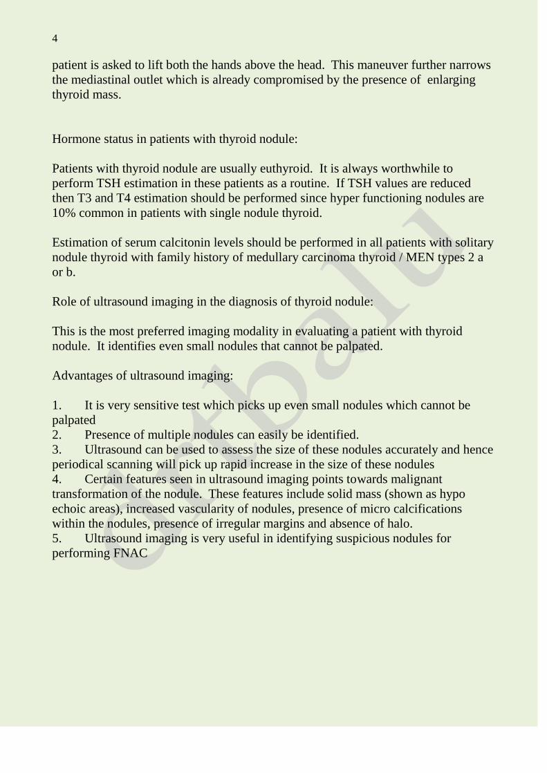
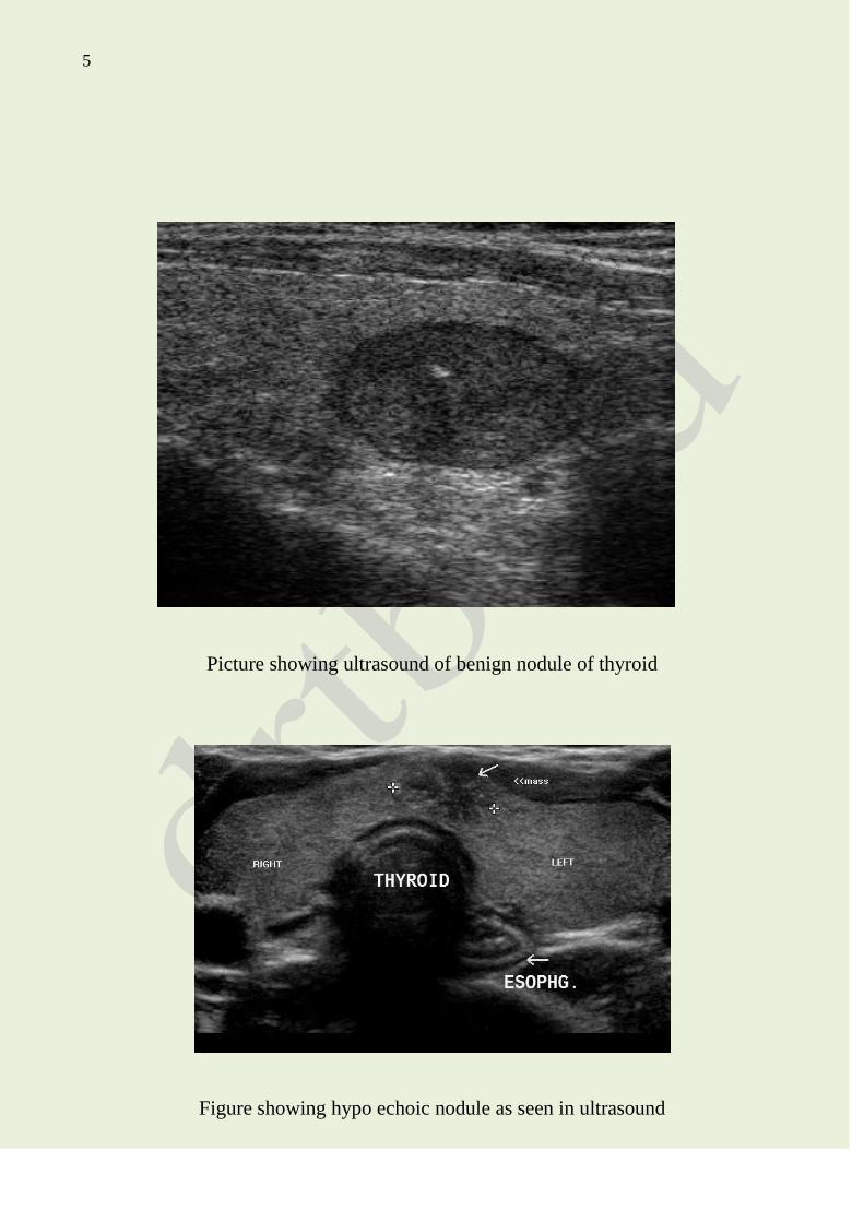
transformation of the nodule. These features include solid mass (shown as hypo
echoic areas), increased vascularity of nodules, presence of micro calcifications
within the nodules, presence of irregular margins and absence of halo.
5. Ultrasound imaging is very useful in identifying suspicious nodules for
performing FNAC
5
Picture showing ultrasound of benign nodule of thyroid
Figure showing hypo echoic nodule as seen in ultrasound
6
Picture showing increased vascularity of thyroid nodule suggestive of malignancy in
ultrasound
Radioisotope imaging:
This helps to ascertain whether a nodule is a functioning one or not. It does not
provide an accurate estimate of the size of the nodule. Commonly used radioisotopes
for this purpose include Technitium 99, I 123 and I 131. It has been estimated that
about 90% of thyroid nodules are cold and non functioning. About 10% of these non
functioning nodules show malignant transformation. The only use for this procedure
is that it warns that the nodule is hot and functioning so that FNAC need not be
performed on a hot nodule. Performing FNAC in hot nodules causes abnormally
7
high increase of thyroxin in the blood causing thyrotoxicity which may have
deleterious effects.
I 123 is more physiologic than technitium 99. Technitium 99 gets washed out of the gland
and hence allows for shorter scanning time (20-30 minutes). Scanning can be
immediately started after administering Technitium 99. If I123 is used then scanning
can be started only after 24 hours after administration of the isotope. The scanning
time lasts atleast 4 - 6 hours. Radiation exposure is more or less similar for both
isotopes. Imaging resolution is better with Technitium 99 than radioiodine. Nodules
below 1cm cannot be reliably detected by either of this scanning modality.
Radioactive iodine scans can identify hot and cold nodules. The incidence of
malignancy is more in cold nodule than hot nodule. Hot nodule is hyperfunctioning
thyroid nodule, where as a cold nodule is a hypofunctioning nodule.
Indications for radioisotope imaging:
1. Identification of a functional solitary nodule when initial serum TSH is
decreased.
2. If FNAC is reported as follicular neoplasm or suspicious lesion then the finding
of Hot nodule in a scan may decrease the suspicion of malignancy
3. For detecting neck node metastasis.
Thallium 201 scan: is a very useful diagnostic tool to differential benign and
malignant thyroid nodules. Three mCi of Thallium - 201 is used to image the thyroid
gland. The uptake of the isotope is categorized into low uptake, intermediate uptake
and high uptake. The risk of malignancy is more in high uptake lesions, and low in
low uptake lesions.
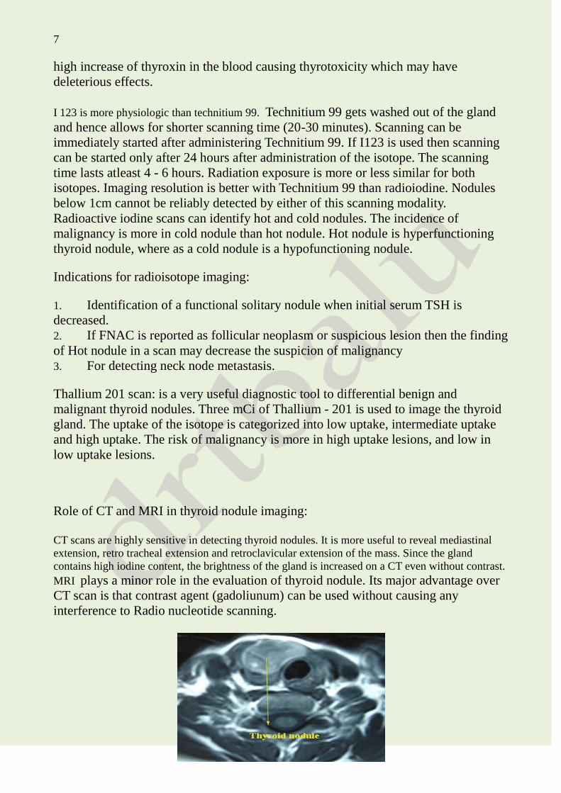
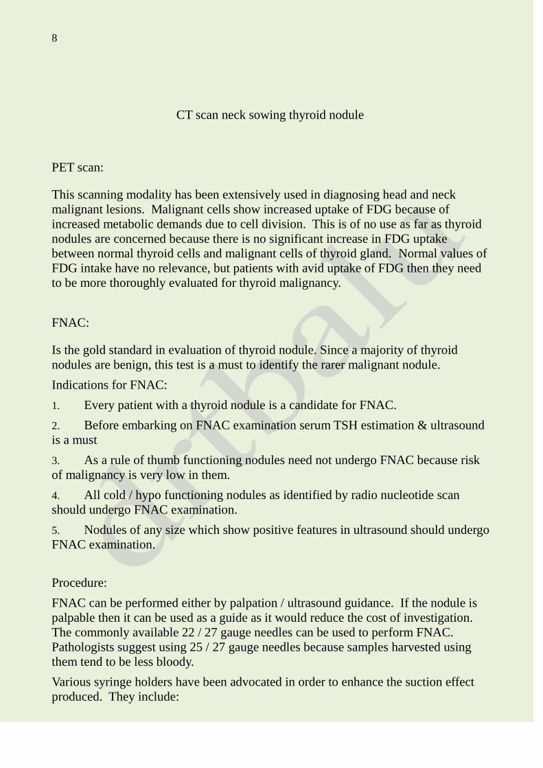
Role of CT and MRI in thyroid nodule imaging:
CT scans are highly sensitive in detecting thyroid nodules. It is more useful to reveal mediastinal
extension, retro tracheal extension and retroclavicular extension of the mass. Since the gland
contains high iodine content, the brightness of the gland is increased on a CT even without contrast. MRI plays a minor role in the evaluation of thyroid nodule. Its major advantage over
CT scan is that contrast agent (gadoliunum) can be used without causing any
interference to Radio nucleotide scanning.
8
CT scan neck sowing thyroid nodule
PET scan:
This scanning modality has been extensively used in diagnosing head and neck
malignant lesions. Malignant cells show increased uptake of FDG because of
increased metabolic demands due to cell division. This is of no use as far as thyroid
nodules are concerned because there is no significant increase in FDG uptake
between normal thyroid cells and malignant cells of thyroid gland. Normal values of
FDG intake have no relevance, but patients with avid uptake of FDG then they need
to be more thoroughly evaluated for thyroid malignancy.
FNAC:
Is the gold standard in evaluation of thyroid nodule. Since a majority of thyroid
nodules are benign, this test is a must to identify the rarer malignant nodule.
Indications for FNAC:
1. Every patient with a thyroid nodule is a candidate for FNAC.
2. Before embarking on FNAC examination serum TSH estimation & ultrasound
is a must
3. As a rule of thumb functioning nodules need not undergo FNAC because risk
of malignancy is very low in them.
4. All cold / hypo functioning nodules as identified by radio nucleotide scan
should undergo FNAC examination.
5. Nodules of any size which show positive features in ultrasound should undergo
FNAC examination.
Procedure:
FNAC can be performed either by palpation / ultrasound guidance. If the nodule is
palpable then it can be used as a guide as it would reduce the cost of investigation.
The commonly available 22 / 27 gauge needles can be used to perform FNAC.
Pathologists suggest using 25 / 27 gauge needles because samples harvested using
them tend to be less bloody.
Various syringe holders have been advocated in order to enhance the suction effect
produced. They include:
9
1. Cameco syringe pistol
2. Tao instrument
3. Inrad aspiration biopsy syringe / gun
It should also be borne in mind that the intrinsic suction effect provided by surface
tension which these smaller diameter needles produce makes these fancy equipments
redundant.
If FNAC is performed under ultrasound guidance then sampling should be done in
different areas of the nodule including its wall, solid elements within and even
calcified areas should not be ignored. Sampling should avoid cystic areas as yield
from them invariably contains less cellular elements.
On insertion of the needle into the thyroid nodule a dwelling time of 2 – 5 seconds
should be allowed. The needle should stay within the nodule during this dwelling
interval. Then 3 forward and backward oscillations are performed in order to
enhance the quality and quantity of yield. This maneuver also reduces blood
contamination of the specimen.
Local anesthesia should be administered for all deep seated thyroid nodules. This
will greatly reduce patient's discomfort and also enhance their active co operation.
For a thyroid FNAC to be reported as benign at least 6 groups of benign looking
follicular cells should be present in a smear. Each of these groups should contain not
less than 10 cells. It should also be stressed that any specimen containing abundant
colloid should be considered benign even if the mandatory 6 groups of cells are not
present in the smear. A sparsely cellular specimen with abundant colloid should
always indicate a macro follicular node and hence certainly benign.
Two types of smears are prepared. Air dried and wet smears.
Dry smear: Two methods can be used. Diff - Quick method and May Grunwald -
Giemsa methods. In quick dry method, the aspirate is expelled on to a glass slide, and
is air dried. This method is best for immediate reading by the pathologist. The dried
smear highlights the background colloid, cell architecture and cytoplasmic details.
This technique is useful in the diagnosis of medullary and lymphoid tumors.
The wet smear (papanicolaou) is a wet smear that requires immediate fixation with
95% alcohol. This method is best suited for detecting papillary cancer.
FNAC will be reported as:
1. Unsatisfactory
2. Intermediate
3. Malignant
Intermediate group can be further subclassified into:
10
1. Follicular lesion of undetermined significance (FLUS)
2. Follicular neoplasm
3. Suspicious for malignancy as per Bathesda thyroid cytology classification
Ancillary procedures can be used to improve the accuracy of FNAC. These include:
1. Immunohistochemistry
2. Ploidy studies
3. Molecular markers
4. Reverse polymerase chain reaction
FNAC is the most important method in the diagnosis of malignant nodule. Other
procedures that increases the accuracy of FNAC include studies of mutation
involving BRAF, RAS, RET / PTC genes. Majority of papillary carcinomas will
have mutations of the above mentioned genes. Mutations involving Ras proto
oncogenes have been implicated in the pathogenesis of follicular carcinoma of
thyroid gland.
It has been shown that sampling errors in FNAC increases as the size of the thyroid
nodule increases. When the size of the nodule increases to 4 cms the false negative
results also increase by 30%.
Ultrasound guided FNAC can be performed if the nodules are small (1cm / less in
size). It increases the accuracy of the procedure.
Frozen section analysis:
Thyroid tissue suspicious of malignancy can be subjected for frozen section analysis
while the patient is still on the operation table. This will reduce the rate of patients
who need to come back to the operation theater for completion thryoidectomy. The
hemithyroidectomy specimen is sent for frozen section evaluation while the surgeon
waits in the operation room. The report usually is expected within 15 mins. If it
turns out to be mitotic lesion then total thyroidectomy is resorted to. If the report
comes as benign lesion then the wound is closed and the patient is extubated.
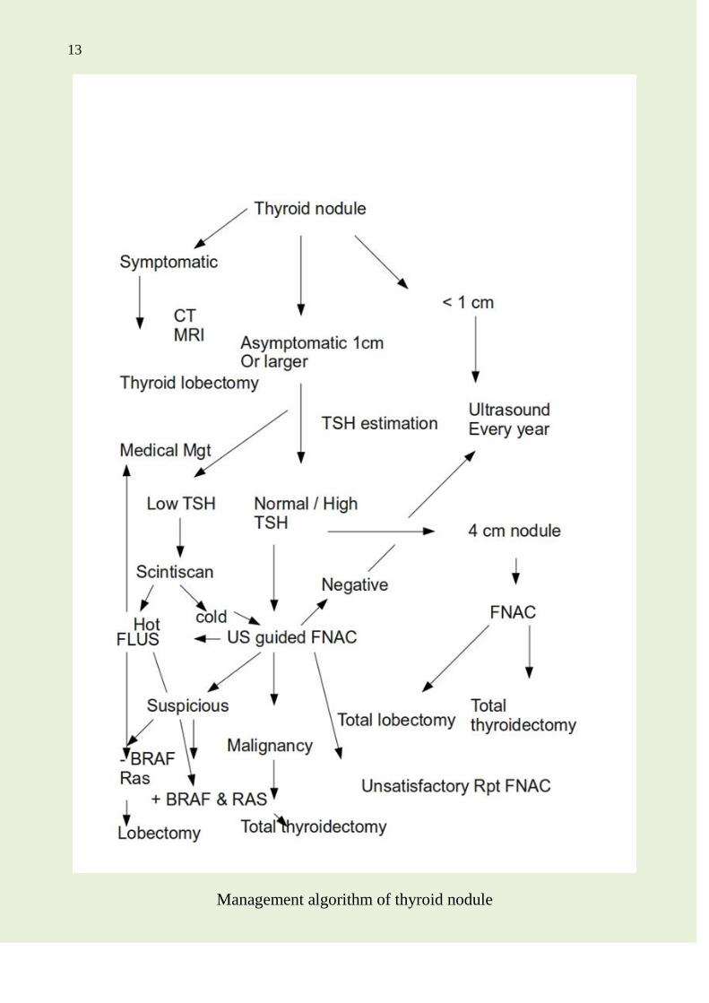
American thyroid association guidelines regarding management of thyroid nodule:
American thyroid association has come out with detailed guidelines for managing
patients with thyroid nodule. The salient feature of these guidelines is the
incorporation of mutational analysis. These guidelines include:
1. All patients with asymptomatic thyroid nodule under 1 cm size should undergo
11
repeated ultrasound examination every year. FNAC is indicated in these patients only
when there is a change in the growth pattern as evidenced by a rapid increase in the
size of the nodule. Biopsy of nodules under 1 cm is indicated only when there is a
history of radiation exposure or family history of thyroid malignancy.
2. Patients with asymptomatic thyroid nodule over 1 cm in size should undergo
TSH estimation. Reduced TSH values in these patients should arouse suspicion of
hot nodule (hyper functioning nodule). These patients should undergo thyroid uptake
scan. Since patients with hot nodules have less than 1% risk of malignant
transformation they are better managed medically by a medical endocrinologist.
3. If the nodule is asymptomatic and is 4 cm or more in size, FNAC should be
done. This is because that these patients require surgery and a pre operative
diagnosis of malignancy or otherwise will help in deciding whether the patient is
going to undergo total thryoidectomy or hemithyroidectomy. If FNAC report turns
out to be unsatisfactory then it should be repeated again. If FNAC is reported as
follicular lesion of undetermined significance then it should be submitted for
mutational studies. If FLUS (follicular lesion of undetermined significance) turns out
to be positive for BRAF and RAS mutations then the patient should be submitted for
total thryoidectomy. If mutational studies are not available then these patients should
be offered diagnostic lobectomy. If FNAC reports the lesion as benign then these
patients should be followed periodically by performing ultrasound examinations for
any abnormal increase in size of the nodule. Attempts at suppressing the lesion by
suppressing TSH levels are not effective. If FNAC report turns out to be malignancy
then total thyroidectomy should be resorted to. Before embarking on total
thyroidectomy ultrasound examination of neck should be performed to pick up any
suspicious nodes which are too small to be palpated.
4. Patients with symptomatic thyroid nodules (symptoms caused due to
obstruction) should be surgically managed. Symptoms could be airway obstruction
or difficult in swallowing or change in voice. Change in voice occurs commonly in
malignancy.
12
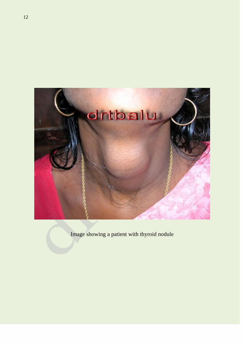
Image showing a patient with thyroid nodule
13
Management algorithm of thyroid nodule

14