Embed Size (px)
Citation preview
Page 1/39
Diagnosis and Surgical Management of SolitaryThyroid NoduleBurkan Nasr ( [email protected] )
Consultant General And Laparoscopic surgery Al Thowra Modern General/ Teaching HospitalSanàa AndSaudi Hospital At HajjahBurkan Nasr
Thamar UniversityMohammed Qubati
Taiz UniversitySultan Qubati
Consultant General And Laparoscopic surgery Al Thowra Modern General/ Teaching HospitalSanàa AndSaudi Hospital At HajjahAbd alhakim Al Tamimi
University of AdenYasser Abd Rabo
21 university YemenAbd alfatah Al Tam
21 university YemenMohamed Al Shujaa
Thamar UniversityMohammed Al Shehari
Sana'a UniversityAnwar Aljunaeed
Saudi Hospital at Hajjah
Research Article
Keywords: solitary thyroid nodule, malignancy, surgical management
Posted Date: August 5th, 2021
DOI: https://doi.org/10.21203/rs.3.rs-773309/v1
License: This work is licensed under a Creative Commons Attribution 4.0 International License. Read Full License
Page 2/39
Abstract
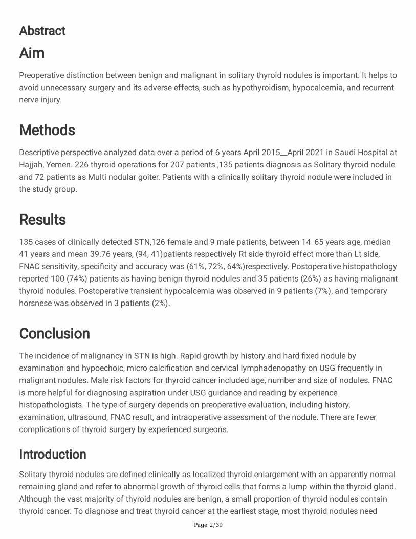
AimPreoperative distinction between benign and malignant in solitary thyroid nodules is important. It helps toavoid unnecessary surgery and its adverse effects, such as hypothyroidism, hypocalcemia, and recurrentnerve injury.
MethodsDescriptive perspective analyzed data over a period of 6 years April 2015__April 2021 in Saudi Hospital atHajjah, Yemen. 226 thyroid operations for 207 patients ,135 patients diagnosis as Solitary thyroid noduleand 72 patients as Multi nodular goiter. Patients with a clinically solitary thyroid nodule were included inthe study group.
Results135 cases of clinically detected STN,126 female and 9 male patients, between 14_65 years age, median41 years and mean 39.76 years, (94, 41)patients respectively Rt side thyroid effect more than Lt side,FNAC sensitivity, speci�city and accuracy was (61%, 72%, 64%)respectively. Postoperative histopathologyreported 100 (74%) patients as having benign thyroid nodules and 35 patients (26%) as having malignantthyroid nodules. Postoperative transient hypocalcemia was observed in 9 patients (7%), and temporaryhorsnese was observed in 3 patients (2%).
ConclusionThe incidence of malignancy in STN is high. Rapid growth by history and hard �xed nodule byexamination and hypoechoic, micro calci�cation and cervical lymphadenopathy on USG frequently inmalignant nodules. Male risk factors for thyroid cancer included age, number and size of nodules. FNACis more helpful for diagnosing aspiration under USG guidance and reading by experiencehistopathologists. The type of surgery depends on preoperative evaluation, including history,examination, ultrasound, FNAC result, and intraoperative assessment of the nodule. There are fewercomplications of thyroid surgery by experienced surgeons.
IntroductionSolitary thyroid nodules are de�ned clinically as localized thyroid enlargement with an apparently normalremaining gland and refer to abnormal growth of thyroid cells that forms a lump within the thyroid gland.Although the vast majority of thyroid nodules are benign, a small proportion of thyroid nodules containthyroid cancer. To diagnose and treat thyroid cancer at the earliest stage, most thyroid nodules need
Page 3/39
some type of evaluation. Often, these abnormal growths of thyroid tissue are located at the edge of thethyroid gland, so they can be felt as a lump in front of the neck. When they are large or when they occur invery thin individuals, they can sometimes even be seen as a lump in front of the neck[1]. Thyroid nodulesare common. The prevalence and incidence increase with age, with spontaneous nodules occurring at arate of 0.08% per year beginning early in life and extending into the eighth decade. Palpable thyroidnodules are found in 5% of persons aged an average of 60 years. With the use of imaging techniques,particularly ultrasound, the chance of detection of thyroid nodules has increased many-fold,approximately 20%_60%. [2,3,4,5,6,7].
Thyroid nodules are more common in women than in men. [3,4,5], its incidence in femalesis approximately one in 12–15 young women who have a thyroid nodule, but in males,it is approximately one in 40 young men who have a thyroid nodule. More than 95% of all thyroid nodulesare benign (noncancerous growths). [4,5].
However, the reported incidence of thyroid cancer in the general population islow, at only approximately 1%. Thyroid cancers occur in approximately 5_15% of all thyroid nodulesindependent of their size. [3,8]
Recent data suggest that the incidence of thyroid malignancy has increased over theyears. [2,3] the worldwide increase in the incidence of thyroid cancer is partly due to increased detectionby US and other imaging studies but also to a true increase in the incidence of papillary thyroidcarcinoma (PTC). [9]
The occurrence of malignancy is more common in solitary thyroid nodules (STNs) thanin multinodular goiters. [2,10,11].
The preoperative evaluation of thyroid nodules to distinguish between benign and malignant nodules isvery important. It helps to avoid unnecessary extensive surgery and potential surgery-related adverseeffects, such as hypothyroidism, hypocalcemia, and recurrent laryngeal nerve injury. [2]
Preoperative diagnoses were classi�ed as benign, suspicious or malignant based on history, clinicalexamination �ndings (i.e. cervical lymphadenopathy, hoarseness of voice, presence ofmetastasis), thyroid function test, ultrasonographic features[12] and FNAC (The Bethesda system forreporting thyroid cytopathology). [13]
Ultrasound of the thyroid gland is used to differentiate true solitary thyroid nodules from those withmultinodular glands. Additionally, it classi�es the nodule into solid, cystic, or mixed.However, it provides little help in determining the pathological types of the nodule[14].
Fine-needle aspiration (FNA) cytology is the �rst step that is performed to differentiate malignantnodules; however, 5–15% of FNA revealed inadequate nondiagnostic samples, and 15–30% ofFNA results in indeterminate cytology �ndings category III (atypia or follicular neoplasm of undetermined
Page 4/39
signi�cance) and category IV (suspicious for follicular neoplasm) according to the Bethesda system [15,16].
Fine-needle aspiration cytology (FNA) is regarded as the �rst diagnostic step to differentiate malignantfrom benign nodules. FNA has served with high accuracy to diagnose papillary thyroid carcinoma, whichaccounts for 80%–90% of all thyroid cancers because papillary thyroid carcinoma has several speci�ccytological nuclear features, such as optically clear elongated nuclei with nuclear grooves andintranuclear cytoplasmic pseudoinclusions [17,18,19]
Fine-needle aspiration cytology (FNAC) has become the cornerstone investigation. Unfortunately, on thebasis of cytological characteristics alone, pathologists cannot reliably distinguish benign from malignantfollicular thyroid lesions, ∼20% of �ne-needle aspiration cytology (FNAC) will be given a �nal diagnosisof follicular malignancy. [20].
For benign solitary nodules, hemithyroidectomy of the involved lobe is recommended, not totalthyroidectomy, but in treating suspicious and false-negative (FN) cases, �ne-needle aspiration cytology(FNAC) reports could be overcome by total thyroidectomy. Hemithyroidectomy with or withoutisthmusectomy is performed as the initial operation for patients with an indeterminate cytologicaldiagnosis and no clinical evidence of regional or distant metastatic disease or any other concurrentindication for total thyroidectomy. If gross extrathyroidal tumor extension or lymph node metastasis isfound at the time of operation, a total thyroidectomy is then carried out[21].
The aim of the present study was to evaluate patients with clinically detected solitary thyroid nodules forthe presence of malignancy in relation to various factors, such as age, gender family history, rapid growthand hard clinical examination, �xed nodules and ultrasonography (USG) �ndings, such as the size of thenodule, echogenicity, microcalci�cation, and presence of lymphadenopathy, as well as �ne-needleaspiration cytology (FNAC) results. We also planned to compare the prevalence of malignancy in bothsolitary and multiple thyroid nodules detected by ultrasonography (USG).
Materials And MethodsThis is a descriptive perspective analyzed our departmental data over a period of 6 years April2015__April 2021. In Saudi hospital at Hajjah,Yemen. Approximately 226 thyroid operations wereperformed for 207 patients; 135 patients were diagnosed with solitary thyroid nodules, and 72 patientswere diagnosed with multinodular goiters. All patients who underwent surgery in the surgical departmentwith a clinically detected solitary thyroid nodule were included in the study group. Our approach wasindividualized as single team. Preoperative history, examination, thyroid function test, ultrasonography(USG) and �ne-needle aspiration cytology were planned in all these patients. Hemithyroidectomy andtotal thyroidectomy with and without neck dissection were performed wherever appropriate. The patientsand their relatives gave consent to use the information for publication purposes. The study was approvedby the institutional ethics committee.
Page 5/39
For all patients, the following data were recorded: age, sex, history of radiation exposure, family history ofthyroid disease, symptoms and growth rate of nodules, and thyroid hormone pro�le. The operativeprocedure was based on different parameters, such as the age of the patients, clinicalexamination, ultrasound interpretation, �ne-needle aspiration cytology (FNAC) �ndings and indirectlaryngoscopy. The decision for surgery was based on individual patient examination and investigation�ndings.
In most of the patients, the plan of surgery was decided beforehand. If it was a solitary thyroid nodulediagnosed clinically , ultrasonographically and �ne-needle aspiration cytology (FNAC) as malignancy orhigh suspicion for malignancy proceeded with total thyroidectomy. For others, lower gradehemithyroidectomy of the involved side was performed, and the specimen was sent for routinehistopathological examination (HPE). Because of inconclusive results and the lack of frozen section use,we preferred to wait until the �nal histopathology report. If the histopathological result was positive formalignancy, completion thyroidectomy was performed in 4_6 weeks. The decision for other procedures,total thyroidectomy with central neck dissection, total thyroidectomy with selective neckdissection, and total thyroidectomy with modi�ed radical neck dissection, was based on clinical,radiological, �ne-needle aspiration cytology (FNAC) and histopathology �ndings.
During surgery, the site and type of incision were decided. Hemostasis, safeguarding of the recurrentlaryngeal nerve, parathyroid, and other vital structures, was taken care of during the dissection.Appropriate measures were taken to correct postoperative hypocalcemia, and care of the drain was taken.Further treatment plans were decided based on the �nal histopathology report. If the report was benign,the patient was managed by regular monitoring of hormone levels, with or without thyroid hormonesupplementation. Hypocalcemia features were managed with supplementation with calciumand vitamin D.
If the �nal histopathology report was either follicular or papillary carcinoma, the patients were advised toundergo an I-131 whole body scan, preferably within 4–6 weeks after surgery, and radioactive iodineablation was advised for residual tissue in the thyroid bed. All the patients were advised regular follow-up for one week, one month, 6 months, one year, and 2 years.
Statistical analysis was performed using Statistical Packages for Social Sciences (SPSS), version 20.0software (SPSS Inc; Chicago, IL, USA). Comparison of proportions between groups was performed by theχ2 test, taking P < 0.05 as signi�cant.
ResultsDuring our study period from April 2015-April 2020 in the Surgical Department of Saudi Hospital at HajjahYemen, approximately 226 thyroid operations were performed for 207 patients, 135 (65%) patients werediagnosed with solitary thyroid nodules and 72 (35%) patients were diagnosed with multinodular goiters.The present study included all patients with clinically detected solitary thyroid nodules.
Page 6/39
135 patients was diagnosed clinical as Solitary thyroid nodule underwent for 154 thyroid operations.incidence Solitary thyroid nodule according to the Sex to 126/135 (93%)female and 9/135(7%)male. (Table 1), There were 135 patients with solitary thyroid nodule b/n age 14-65 years, median age 41years, mode 45 years, average mean age 39.76 years, ring 51 years and Stander deviation 13.98.
The histopathology results showed that 100/135 patients (74%) developed benign thyroid nodules frompatients diagnosed clinically with solitary thyroid nodules. 95/135 was female(70%) and 5/135male(4%),95 /100 was female(95%) and 5/100 was male(5%).
All patients were diagnosed clinically with solitary thyroid nodules. Histopathology showed benignthyroid nodules at age b/n 14_65 years, median age 31.5 years, mode 30 years, average mean age 34.72years and standard deviation 12.40.
The histopathology results showed that 35/135 patients (26%) developed thyroid cancer from patientsdiagnosed clinically with solitary thyroid nodules. A total of 31/135 were female (23%), 4/135were male (3%), 31/35 were female (89%), and 4/35 were male (11%).
The incidence of thyroid cancer in females with solitary thyroid nodules was 31/126 (25%).
The incidence of thyroid cancer in males with solitary thyroid nodules was 4/9 (44.44%). This indicatesa high incidence of thyroid cancer in male patients.
All patients were diagnosed clinically with solitary thyroid nodules. Histopathology showed thyroid cancerby age range b/n 15_62 years, median age 35 years, mode 23 years, average mean age 35.97 years,and standard deviation 11.91.
(Table 2) show Solitary thyroid nodule more Common in age group between 21-30 years old are about 50patients and the next age group between 41-50 years old are about 29 patients.
(Table 3) show distribution according to the side that 135 Patients with Solitary Thyroid Nodule There is94 Patients Rt Side Solitary Thyroid Nodule and 41 Patients Lt Side Solitary Thyroid Nodule ,Our result,Solitary thyroid nodule appear (70%) in Rt Side of thyroid and (30%) in Lt Side of thyroid that indicated Rtside effects more than Lt side thyroid.
Benign solitary thyroid nodule distribute according to side as following Rt side 72/100(72%), and Lt side28/100(28%),Rt side benign solitary thyroid nodule appear in 72 patients 69 female and 3 male patients,clinical diagnosis as Rt side true Solitary thyroid nodule 49 patients, cystic Rt side solitary thyroid nodule7 patients, 13 patients prominent Rt side solitary thyroid nodule and 3 patients Rt side solitary thyroidnodule Toxic adenoma .
Lt side benign solitary thyroid nodule appear 28 patients 26 female and 2 male patients, clinicaldiagnosis as Lt side true Solitary thyroid nodule 17patients, 2 patients with huge nodule 7-8 cm, cystic Ltsolitary thyroid nodule 3 patients, prominent Lt Solitary thyroid nodule in 7 patients.
Page 7/39
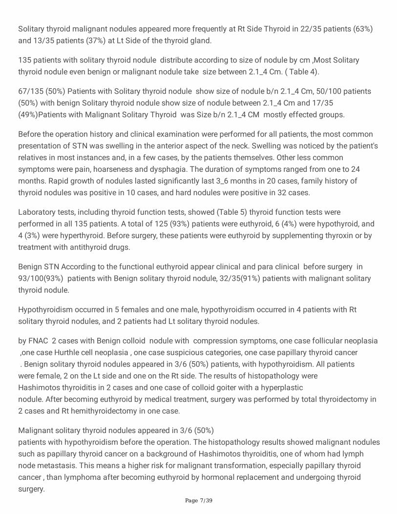
Solitary thyroid malignant nodules appeared more frequently at Rt Side Thyroid in 22/35 patients (63%)and 13/35 patients (37%) at Lt Side of the thyroid gland.
135 patients with solitary thyroid nodule distribute according to size of nodule by cm ,Most Solitarythyroid nodule even benign or malignant nodule take size between 2.1_4 Cm. ( Table 4).
67/135 (50%) Patients with Solitary thyroid nodule show size of nodule b/n 2.1_4 Cm, 50/100 patients(50%) with benign Solitary thyroid nodule show size of nodule between 2.1_4 Cm and 17/35(49%)Patients with Malignant Solitary Thyroid was Size b/n 2.1_4 CM mostly effected groups.
Before the operation history and clinical examination were performed for all patients, the most commonpresentation of STN was swelling in the anterior aspect of the neck. Swelling was noticed by the patient'srelatives in most instances and, in a few cases, by the patients themselves. Other less commonsymptoms were pain, hoarseness and dysphagia. The duration of symptoms ranged from one to 24months. Rapid growth of nodules lasted signi�cantly last 3_6 months in 20 cases, family history ofthyroid nodules was positive in 10 cases, and hard nodules were positive in 32 cases.
Laboratory tests, including thyroid function tests, showed (Table 5) thyroid function tests wereperformed in all 135 patients. A total of 125 (93%) patients were euthyroid, 6 (4%) were hypothyroid, and4 (3%) were hyperthyroid. Before surgery, these patients were euthyroid by supplementing thyroxin or bytreatment with antithyroid drugs.
Benign STN According to the functional euthyroid appear clinical and para clinical before surgery in93/100(93%) patients with Benign solitary thyroid nodule, 32/35(91%) patients with malignant solitarythyroid nodule.
Hypothyroidism occurred in 5 females and one male, hypothyroidism occurred in 4 patients with Rtsolitary thyroid nodules, and 2 patients had Lt solitary thyroid nodules.
by FNAC 2 cases with Benign colloid nodule with compression symptoms, one case follicular neoplasia ,one case Hurthle cell neoplasia , one case suspicious categories, one case papillary thyroid cancer . Benign solitary thyroid nodules appeared in 3/6 (50%) patients, with hypothyroidism. All patientswere female, 2 on the Lt side and one on the Rt side. The results of histopathology wereHashimotos thyroiditis in 2 cases and one case of colloid goiter with a hyperplasticnodule. After becoming euthyroid by medical treatment, surgery was performed by total thyroidectomy in2 cases and Rt hemithyroidectomy in one case.
Malignant solitary thyroid nodules appeared in 3/6 (50%)patients with hypothyroidism before the operation. The histopathology results showed malignant nodulessuch as papillary thyroid cancer on a background of Hashimotos thyroiditis, one of whom had lymphnode metastasis. This means a higher risk for malignant transformation, especially papillary thyroidcancer , than lymphoma after becoming euthyroid by hormonal replacement and undergoing thyroidsurgery.
Page 8/39
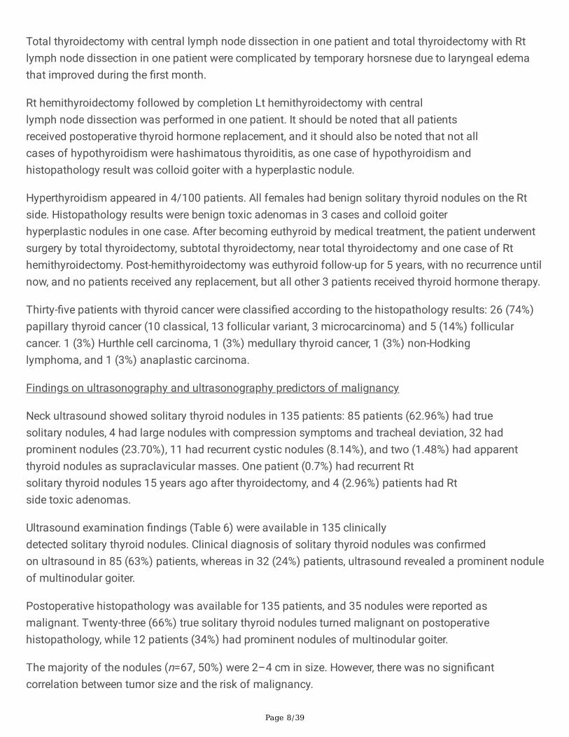
Total thyroidectomy with central lymph node dissection in one patient and total thyroidectomy with Rtlymph node dissection in one patient were complicated by temporary horsnese due to laryngeal edemathat improved during the �rst month.
Rt hemithyroidectomy followed by completion Lt hemithyroidectomy with centrallymph node dissection was performed in one patient. It should be noted that all patientsreceived postoperative thyroid hormone replacement, and it should also be noted that not allcases of hypothyroidism were hashimatous thyroiditis, as one case of hypothyroidism andhistopathology result was colloid goiter with a hyperplastic nodule.
Hyperthyroidism appeared in 4/100 patients. All females had benign solitary thyroid nodules on the Rtside. Histopathology results were benign toxic adenomas in 3 cases and colloid goiterhyperplastic nodules in one case. After becoming euthyroid by medical treatment, the patient underwentsurgery by total thyroidectomy, subtotal thyroidectomy, near total thyroidectomy and one case of Rthemithyroidectomy. Post-hemithyroidectomy was euthyroid follow-up for 5 years, with no recurrence untilnow, and no patients received any replacement, but all other 3 patients received thyroid hormone therapy.
Thirty-�ve patients with thyroid cancer were classi�ed according to the histopathology results: 26 (74%)papillary thyroid cancer (10 classical, 13 follicular variant, 3 microcarcinoma) and 5 (14%) follicularcancer. 1 (3%) Hurthle cell carcinoma, 1 (3%) medullary thyroid cancer, 1 (3%) non-Hodkinglymphoma, and 1 (3%) anaplastic carcinoma.
Findings on ultrasonography and ultrasonography predictors of malignancy
Neck ultrasound showed solitary thyroid nodules in 135 patients: 85 patients (62.96%) had truesolitary nodules, 4 had large nodules with compression symptoms and tracheal deviation, 32 hadprominent nodules (23.70%), 11 had recurrent cystic nodules (8.14%), and two (1.48%) had apparentthyroid nodules as supraclavicular masses. One patient (0.7%) had recurrent Rtsolitary thyroid nodules 15 years ago after thyroidectomy, and 4 (2.96%) patients had Rtside toxic adenomas.
Ultrasound examination �ndings (Table 6) were available in 135 clinicallydetected solitary thyroid nodules. Clinical diagnosis of solitary thyroid nodules was con�rmedon ultrasound in 85 (63%) patients, whereas in 32 (24%) patients, ultrasound revealed a prominent noduleof multinodular goiter.
Postoperative histopathology was available for 135 patients, and 35 nodules were reported asmalignant. Twenty-three (66%) true solitary thyroid nodules turned malignant on postoperativehistopathology, while 12 patients (34%) had prominent nodules of multinodular goiter.
The majority of the nodules (n=67, 50%) were 2–4 cm in size. However, there was no signi�cantcorrelation between tumor size and the risk of malignancy.
Page 9/39
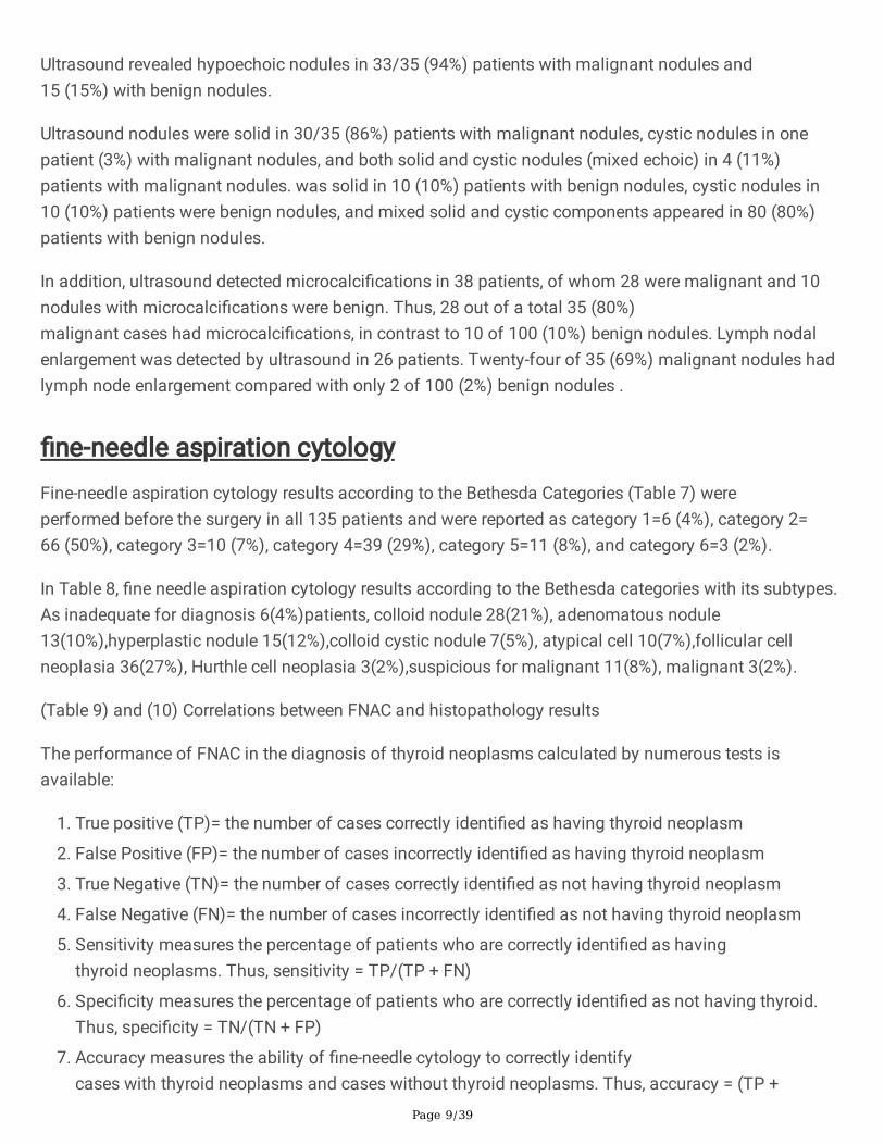
Ultrasound revealed hypoechoic nodules in 33/35 (94%) patients with malignant nodules and15 (15%) with benign nodules.
Ultrasound nodules were solid in 30/35 (86%) patients with malignant nodules, cystic nodules in onepatient (3%) with malignant nodules, and both solid and cystic nodules (mixed echoic) in 4 (11%)patients with malignant nodules. was solid in 10 (10%) patients with benign nodules, cystic nodules in10 (10%) patients were benign nodules, and mixed solid and cystic components appeared in 80 (80%)patients with benign nodules.
In addition, ultrasound detected microcalci�cations in 38 patients, of whom 28 were malignant and 10nodules with microcalci�cations were benign. Thus, 28 out of a total 35 (80%)malignant cases had microcalci�cations, in contrast to 10 of 100 (10%) benign nodules. Lymph nodalenlargement was detected by ultrasound in 26 patients. Twenty-four of 35 (69%) malignant nodules hadlymph node enlargement compared with only 2 of 100 (2%) benign nodules .
�ne-needle aspiration cytologyFine-needle aspiration cytology results according to the Bethesda Categories (Table 7) wereperformed before the surgery in all 135 patients and were reported as category 1=6 (4%), category 2=66 (50%), category 3=10 (7%), category 4=39 (29%), category 5=11 (8%), and category 6=3 (2%).
In Table 8, �ne needle aspiration cytology results according to the Bethesda categories with its subtypes.As inadequate for diagnosis 6(4%)patients, colloid nodule 28(21%), adenomatous nodule13(10%),hyperplastic nodule 15(12%),colloid cystic nodule 7(5%), atypical cell 10(7%),follicular cellneoplasia 36(27%), Hurthle cell neoplasia 3(2%),suspicious for malignant 11(8%), malignant 3(2%).
(Table 9) and (10) Correlations between FNAC and histopathology results
The performance of FNAC in the diagnosis of thyroid neoplasms calculated by numerous tests isavailable:
1. True positive (TP)= the number of cases correctly identi�ed as having thyroid neoplasm
2. False Positive (FP)= the number of cases incorrectly identi�ed as having thyroid neoplasm
3. True Negative (TN)= the number of cases correctly identi�ed as not having thyroid neoplasm
4. False Negative (FN)= the number of cases incorrectly identi�ed as not having thyroid neoplasm
5. Sensitivity measures the percentage of patients who are correctly identi�ed as havingthyroid neoplasms. Thus, sensitivity = TP/(TP + FN)
�. Speci�city measures the percentage of patients who are correctly identi�ed as not having thyroid.Thus, speci�city = TN/(TN + FP)
7. Accuracy measures the ability of �ne-needle cytology to correctly identifycases with thyroid neoplasms and cases without thyroid neoplasms. Thus, accuracy = (TP +
Page 10/39
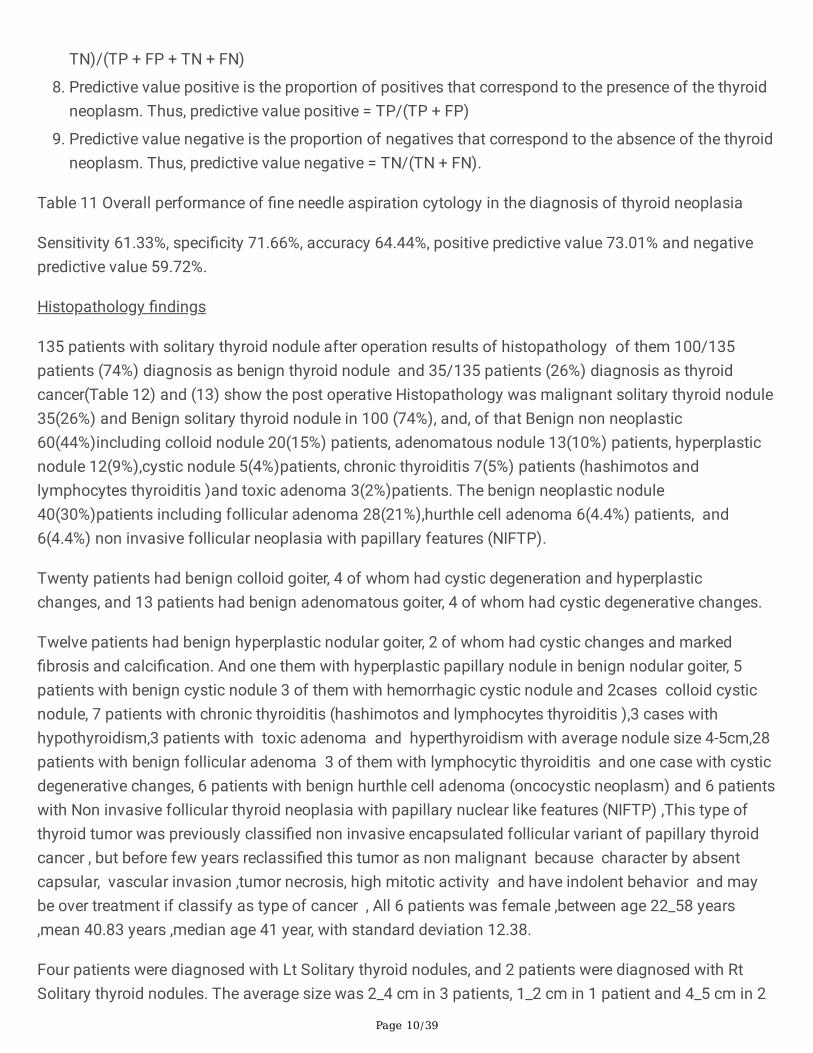
TN)/(TP + FP + TN + FN)
�. Predictive value positive is the proportion of positives that correspond to the presence of the thyroidneoplasm. Thus, predictive value positive = TP/(TP + FP)
9. Predictive value negative is the proportion of negatives that correspond to the absence of the thyroidneoplasm. Thus, predictive value negative = TN/(TN + FN).
Table 11 Overall performance of �ne needle aspiration cytology in the diagnosis of thyroid neoplasia
Sensitivity 61.33%, speci�city 71.66%, accuracy 64.44%, positive predictive value 73.01% and negativepredictive value 59.72%.
Histopathology �ndings
135 patients with solitary thyroid nodule after operation results of histopathology of them 100/135patients (74%) diagnosis as benign thyroid nodule and 35/135 patients (26%) diagnosis as thyroidcancer(Table 12) and (13) show the post operative Histopathology was malignant solitary thyroid nodule35(26%) and Benign solitary thyroid nodule in 100 (74%), and, of that Benign non neoplastic60(44%)including colloid nodule 20(15%) patients, adenomatous nodule 13(10%) patients, hyperplasticnodule 12(9%),cystic nodule 5(4%)patients, chronic thyroiditis 7(5%) patients (hashimotos andlymphocytes thyroiditis )and toxic adenoma 3(2%)patients. The benign neoplastic nodule40(30%)patients including follicular adenoma 28(21%),hurthle cell adenoma 6(4.4%) patients, and6(4.4%) non invasive follicular neoplasia with papillary features (NIFTP).
Twenty patients had benign colloid goiter, 4 of whom had cystic degeneration and hyperplasticchanges, and 13 patients had benign adenomatous goiter, 4 of whom had cystic degenerative changes.
Twelve patients had benign hyperplastic nodular goiter, 2 of whom had cystic changes and marked�brosis and calci�cation. And one them with hyperplastic papillary nodule in benign nodular goiter, 5patients with benign cystic nodule 3 of them with hemorrhagic cystic nodule and 2cases colloid cysticnodule, 7 patients with chronic thyroiditis (hashimotos and lymphocytes thyroiditis ),3 cases withhypothyroidism,3 patients with toxic adenoma and hyperthyroidism with average nodule size 4-5cm,28patients with benign follicular adenoma 3 of them with lymphocytic thyroiditis and one case with cysticdegenerative changes, 6 patients with benign hurthle cell adenoma (oncocystic neoplasm) and 6 patientswith Non invasive follicular thyroid neoplasia with papillary nuclear like features (NIFTP) ,This type ofthyroid tumor was previously classi�ed non invasive encapsulated follicular variant of papillary thyroidcancer , but before few years reclassi�ed this tumor as non malignant because character by absentcapsular, vascular invasion ,tumor necrosis, high mitotic activity and have indolent behavior and maybe over treatment if classify as type of cancer , All 6 patients was female ,between age 22_58 years,mean 40.83 years ,median age 41 year, with standard deviation 12.38.
Four patients were diagnosed with Lt Solitary thyroid nodules, and 2 patients were diagnosed with RtSolitary thyroid nodules. The average size was 2_4 cm in 3 patients, 1_2 cm in 1 patient and 4_5 cm in 2
Page 11/39
patients. Fine needle aspiration cytology showed benign cytology in 3 patients, follicular neoplasia in 2patients and suspicious nodules in 1 patientAll 6 patients were euthyroid before the operation, 3patientsunderwent Lt hemithyroidectomy, and 2 patients underwent Rt hemithyroidectomy.
Considering this term benign and not followed by total thyroidectomy, only follow-up is needed.
One patient underwent total thyroidectomy because it was in the suspicious category. Onepatient developed postoperative temporary horses that improved after a few weeks.
In (Table 14) Histopathology subtype results was malignant solitary thyroid nodule in 35(26%) patients,26(74%) papillary thyroid cancer(classical papillary thyroid cancer 10cases, papillary micro carcinoma 3case and 13 were reported as the follicular variant of papillary carcinoma (FVPTC) ) , 5(14%) Follicularcancer . 1 (3%) Hurthle cell carcinoma, 1 (3%) medullary thyroid cancer, 1 (3%) non-Hodking lymphoma,and 1 (3%) anaplastic carcinoma.
Management
Depending on the interpretation of the FNAB cytological specimen, management consists of observation,levothyroxine suppression therapy, or surgery.
Patients with benign solitary thyroid nodules may undergo observation or levothyroxine suppressiontherapy as the initial treatment modality. Levothyroxine is typically administered for 6-12 months todetermine if the solitary thyroid nodule decreases in size. If the nodule decreases in size after treatmentwith levothyroxine, this medication is discontinued, with follow-up examination of the thyroid nodule in 3-6 months. However, if a benign solitary thyroid nodule increases in size, a repeat trial of levothyroxine andrepeat FNAB may be indicated. Additionally, growth of a thyroid nodule during levothyroxine therapy is astrong indication for surgery.
No consensus exists regarding the degree of thyroid suppression or the e�cacy of levothyroxine therapy.In fact, many endocrinologists no longer recommend thyroid suppression because of potential long-termadverse effects, such as osteoporosis and cardiac arrhythmias. Still others maintain a thyroid-stimulatinghormone (TSH) level ranging from 0.1-0.3 mU/L rather than suppressing to the lowest limits ofdetectability to avoid immediate toxicity and long-term side effects.
Solitary thyroid nodules that are malignant, suspicious, or indeterminate on FNAB require excisionalbiopsy in the form of thyroidectomy. Considerable controversy exists regarding the extent of surgery formalignant, suspicious, or indeterminate solitary thyroid nodules.
Type of Surgery and operative �ndings
Patients with solitary thyroid nodules who underwent surgical procedures
In Table 15, there were 154 operations for 135 patients with solitary thyroid nodule, 126patients (93%) female and 9 (7%) male patients aged 14-65 years.
Page 12/39
One hundred thyroid operations were performed for 100 patients with benign solitary thyroid nodules, and54 thyroid operations were performed for 35 patients with malignant solitary thyroid nodules.
Rt hemithyroidectomy for 70 patients with Rt Solitary thyroid nodule, 60 cases was benign Solitarythyroid nodule, 9 cases followed by completion thyroidectomy after results of histopathology con�rmmalignant cancer and one case result of histopathology was papillary micro carcinoma 1_2cm wasenough treat by Rt hemithyroidectomy.
Lt hemithyroidectomy for 32 patients with Lt Solitary thyroid nodule, of 22 cases was benign Solitarythyroid nodule and 10 cases followed by completion thyroidectomy when results of histopathologycon�rm malignant cancer.
19 Completion thyroidectomy after hemithyroidectomy when the result of the histopathology wascancer, 10 patients completed Rt hemithyroidectomy and 9 patients completed Lt hemithyroidectomy.
Completion thyroidectomy with central lymph node dissection was performed in 17 patients (16 papillarythyroid cancer and one medullary thyroid cancer), and completion thyroidectomy with out central necklymph node dissection was performed in two patients (follicular thyroid cancer and hurthle cell cancer).
Total thyroidectomy for 22 patients, 16 patients Rt Solitary thyroid nodule and 6 patients Lt Solitarythyroid nodule, 14 patients with benign nodule and 8 patients with malignant solitary thyroid nodule (2patients Lt Solitary thyroid nodule and 6 patients Rt Solitary thyroid nodule ) treat by total thyroidectomyand results of histopathology was 3 patients papillary thyroid cancer,4 patients follicular cancer, oneanaplastic cancer . Here, papillary thyroid cancer was not followed by any type of neck dissectionbecause total thyroidectomy depending on the FNAC result was false negative for malignancy.
Near total thyroidectomy for one patient Rt Solitary thyroid nodule Toxic adenoma.
Subtotal thyroidectomy for one patient Rt Solitary prominent thyroid nodule Toxic adenoma.
Total thyroidectomy with central lymph nodes dissection 2 patients after FNAC results was malignantcategory 6 (papillary thyroid cancer) one of them underwent Total thyroidectomy with central lymphnodes dissection with resection underlying soft tissue in�ltrated and part of strap muscle involved inpatient with papillary thyroid cancer in�ltrated underlying soft tissue and muscle was complicated byTemporary hypocalcemia.
Total thyroidectomy With Modi�ed Neck dissection 4 patients ,3 cases Total thyroidectomy with Rtmodi�ed Neck dissection one case complicated by Temporary hypocalcemia,1 case Total thyroidectomywith Lt modi�ed Neck dissection complicated by Temporary hypocalcemia and Total thyroidectomy withSelective Rt lymph nodes dissection in 3 patient one patient with papillary thyroid cancer and level 2lymph nodes positive and 2 patients was suspicious category with Lymph nodes at level 3 by FNAC butResult of histopathology was benign hashimotos goiter .
Page 13/39
Neck dissection was performed in 26 patients, 24 of whom had malignant nodules, 6 of whom showedmetastatic deposits in the lymph nodes. Five patients had papillary thyroid cancer, one patient had non-Hodgkin lymphoma in the background of hashimatous thyroiditis, and two patients had benignthyroid nodules and underwent selective lymph node dissection because FNAC gave us falsepositive results. The patient underwent total thyroidectomy with Rt selective lymph node dissection level3, but the result of histopathology was a hyperplastic nodule with marked �brosis andcalci�cation. Another case result of the histopathology was Hashimotos thyroiditis,100 and thyroidoperation for 100 patients with benign solitary thyroid nodules was distributed as follows.
60 Rt hemithyroidectomy, 22 Lt hemithyroidectomy, 14 total thyroidectomy and 2 total thyroidectomywith selective Rt neck lymph node dissection (for these 2 cases, FNAC was suspicious category withclinical lymph nodes). One case result of the histopathology was benign hyperplastic nodule with marked�brosis and calci�cation also this case complicated by Temporary hypocalcemia, Other case result ofthe histopathology was hashimotos thyroiditis ),One case subtotal thyroidectomy for Solitary toxicadenoma and One case near total thyroidectomy for Solitary toxic adenoma .
Additionally, cancer was distributed according to surgical operation (Table 15), and 17patients underwent complete thyroidectomy with central lymph node dissection. Sixteen papillarythyroid cancer, 1 medullary thyroid cancer, 9 Rt solitary thyroid nodules proved thyroid cancer after Rthemithyroidectomy, and 8 Lt solitary thyroid nodules proved thyroid cancer after Lt hemithyroidectomy.all followed by completion thyroidectomy with central dissection in 17 cases.
Two patients underwent completion thyroidectomy with out central neck lymph nodes dissection after Lthemithyroidectomy with results of histopathology one case follicular thyroid cancer and other casehurthle cell cancer.
Eight patients with solitary thyroid nodules (2 patients had Solitary thyroid nodules, and 6patients had Solitary thyroid nodules) were treated by total thyroidectomy. 3 papillary thyroid cancer, 4follicular cancer, and 1 anaplastic cancer. Here, papillary thyroid cancer was not followed by any type ofneck dissection because total thyroidectomy depending on the FNAC result was false negativefor malignancy.
Two patients with solitary thyroid nodules (Rt Solitary thyroid nodules) were treated by totalthyroidectomy and central lymph node dissection. After FNAC was true positive for malignancy, one casewas the result of papillary thyroid cancer histopathology on the background of hashimatousthyroiditis, and the other case was papillary thyroid cancer with soft tissue in�ltration that was resectedwith part of the strap muscle involved and positive lymph nodes.
One patient with a solitary thyroid nodule (Rt Solitary thyroid nodule) was treated by total thyroidectomyand selective Rt Neck lymph node dissection level 2. The results of histopathology were papillary thyroidcancer of one lobe and free of other lobes with capsular and lymphovascular invasion. MetastaticDeposit Of Tumor In Two Cervical Lymph Nodes(2/4). AJCC TNM STAGING [pT3, N1,Mx].
Page 14/39
Four patients underwent thyroidectomy with modi�ed neck lymph node dissection (one Ltapparently thyroid nodule) FNAC metastasis papillary thyroid cancer by total thyroidectomy withfunctional Lt modi�ed neck dissection, resulting in histopathology papillary thyroid cancer with positivelymph nodes. (3 cases Rt Solitary thyroid nodule one of them Rt apparent thyroid nodule FNACadenocarcinoma thyroid origin and one case Recurrence papillary thyroid cancer 20years after thyroidsurgery with positive lymph node Result of histopathology was these cases ( in�ltrating Papillary thyroidcancer with positve lymph nod), and third one high suspicious vs follicular thyroid neoplasia Result ofhistopathology was (Lymphoma non hodgkin larg cell on back ground Hashimatouse thyroiditis).
One patient with a solitary thyroid nodule (Rt solitary thyroid nodule) treated by Rt hemithyroidectomyhistopathology was diagnosed with papillary thyroid cancer. Intrathyroid encapsulated follicular variant for 15 years female no family history and nodule size 1_2cm was not follow by completionthyroidectomy because low risk. And follow up for 5years no recurrent until now.
In Table 16, types of neck lymph node surgical dissection and metastatic deposits in the lymph nodeswere seen in 6 of the 24 patients who had undergone lymph node dissection. Central node dissectionwas performed in 19 (1 positive) patients, right side modi�ed neck dissection (MND) in 3 (3 positive)patients, Lt side modi�ed neck dissection in one patient (1 positive) and Rt selective necklymph node dissection in 1 patient (1 positive).
Complications
In Table 17, complications after thyroidectomy for solitary thyroid nodules appear.
12/135 patients (8.88%) ,all female patients, 7 cases post Lt Solitary thyroid nodule and 5 cases post RtSolitary thyroid nodule .6/135(4%) and 6 /100(6%)patients with benign nodule , 6/135(4%) and 6/35(17%)patients with malignant nodule .
Temporary hypocalcemia 9/135 patients (7%),5/135(4%) and 5/100 (5%)patients with benign solitarythyroid nodule ,4/135(3%) and 4/35(11%)patients with malignant thyroid nodule ,4 RT Solitary thyroidnodule and 5 Lt Solitary thyroid nodule ,All patients hypocalcemia symptoms appear 24_48 hours afteroperation with patients still at admission with Upper limb pain and numbness most common follow bycarbopedal spasm ,All response to oral calcium supplement But some time start by i.v infusion,Completely resolved symptoms and stop treatment 1 to 8 weeks But mostly second week .
1 patient 26 years female Post total thyroidectomy diagnosed clinical as Rt Solitary thyroid nodule (Toxicadenoma) Result FNAC was suspicious category with Rt clinical lymph nodes level 3 Patient underwentTotal thyroidectomy with Rt selective lymph nodes dissection level 3 but Result of histopathology washyperplastic nodule with marked �brosis and calci�cation developed temporary hypocalcemia wasimproved after few weeks.
Three patients after total thyroidectomy with Hashimotos thyroiditis and lymphocytic thyroiditis
Page 15/39
One case post total thyroidectomy with follicular adenoma ,3 cases post Total thyroidectomy withcentral lymph nodes dissection with papillary thyroid cancer in�ltrated ,One case post Totalthyroidectomy with Selective Rt lymph nodes dissection with papillary thyroid cancer with lymph nodemetastasis and One case post Total thyroidectomy with modi�ed Rt and Lt neck dissection.
All females were aged 20-62 years. All nodules were hardly �xed, with an average nodule size of 2_5 cm.
Five patients had Lt Solitary thyroid nodules, and 4 patients had Rt Solitary thyroid nodules.
Temporary horsnese 3 patients(2.22%),2/35(6%) patients with malignant nodule and One/100(1%)patient with benign nodule, One RT Solitary thyroid nodule ,2 Lt Solitary thyroid nodule ,3 cases allfemale with age 60,40,29 mostly due to the laryngeal edema post thyroidectomy .
One case after total thyroidectomy for anaplastic thyroid cancer older age in�ltrated tumor big tumorsize 6_9 cm and 1 case after total with selective Rt Neck dissection for patients with papillary thyroidcancer with lymph node metastasis and 1 case post Lt hemithyroidectomy for patient with Noninvasivefollicular neoplasia with papillary like features was 29 years old female developed temporary horsnesewas improved after few weeks. All patients appeared horsnese directly after the operation; they received awarm slin nebulizer and dexamethasone for 24_48 hours, and the hospital stay was notcompletely improved by 3_6 months.
The postoperative hospital stay ranged from one to 3 days, and the mean hospital stay was 2 days.
Follow-up ranged from one to 48 months with a mean follow-up of 12.1 ± 14.2 months.
DiscussionThyroid nodules refer to localized lesions within the thyroid gland that are palpably or radiologicallydistinct from the surrounding thyroid parenchyma. [22].
Because of the high risk for malignancy , surgeons tend to treat them with a high degree of suspicion andplan treatment in a systematic manner. Clinically, STNs are common and are present in up to 50% of theelderly population. The majority of STNs are malignant. [ 2, 10 , 11] .
Therefore, it is recommended that all thyroid nodules >1 cm in size should undergo evaluation. Thisincludes both palpable and nonpalpable nodules or nodules detected by imaging. [ 22].
Benign causes of thyroid nodules include colloid nodules, hyperplastic nodules,and adenomatous nodules . Occasionally, nodularity is noticed in patients with Hashimoto's thyroiditisand toxic adenoma . Malignant causes of nodules include thyroid cancer, lymphoma and metastasis tothe thyroid gland. [ 22].
In our country, a different study was performed on thyroid cancer Al-Hureibi, Abdulmughni, Y. ThyroidFNAC . (2003)[66], Abdulmughni, Yasser A., et al. thyroid cancer (2004)[67]. Al-Jaradi, Mansour, et al.
Page 16/39
Prevalence of thyroid cancer(2005)[34],. Al-Shara�, Butheinah A., et al.thyroid cancer (2020)[68].
During our study period, 135 patients had solitary thyroid nodules; there were 126 (93%) femaleswith STNs and 9 (7%) male patients with solitary thyroid nodules.
Thyroid nodules are more common in females similar as noted in the previous study.[ 2, 6].
Solitary thyroid nodules were 10–11times more common in females as compared to males,[ 2, 10], Ourstudy showed that solitary thyroid nodules were 14 times more common in female than male.
In our study 135 Patients with Solitary Thyroid Nodule b/n age 14-65 years, median age 41 years, mode45 years , average mean age 39.76 years, range 51 years and Stander deviation 13.98 . The age rangeand mean slightly wide, and higher compared with previous study by (Gupta ).[ 10].
In our study Solitary thyroid nodule more Common in age group between 21-30 years old are about 50patients and the next age group between 41-50 years old are about 29 patients. That mean seconddecade involved by majority of the patients (37%) this is lower than previous study by Gupta[[10] andDorairajan and Jayashree in that third decade of life majority of the patients involved (44%).
Evaluation of solitary thyroid nodules requires the collaboration of the primary care physician,endocrinologist, pathologist, radiologist, and head and neck surgeon to provide comprehensive andappropriate management of this clinical entity.[ 42].
Preliminary investigation should include careful history and thorough clinical examination and thyroidfunction tests.combination with thyroid ultrasound and FNAC becoming relevant in the management ofthyroid nodules.[ 22] [ 23].
Further investigation should be considered if the following factors are present in addition to the thyroidnodule like male gender, extremes of age (<20 or >70 years), history of neck irradiation, nodule >4 cm insize or the presence of any pressure symptoms.[ 22] None of our patients in the study group had historyof radiation exposure.
Patients under the age of 20 or over 70 years with thyroid nodules have an increased risk of malignancy,as do men. A history of persistent hoarseness, dysphagia, or dyspnea also increases the risk, althoughthese symptoms may also occur with benign nodules. Rapid painless growth of a solid nodule isconcerning and raises the suspicion for thyroid cancer. [25].
Numerous studies have documented that the risk of malignancy in patients with thyroid nodules is 5%–17%, whether detected by palpation or ultrasonography.
There were 135 cases of clinically detected STNs with available ultrasound �ndings in the study group.Thirty-�ve (26%) (3:1) clinically detected STNs were reported as malignant in the �nal HPE. This highincidence of malignancy reported in our study is similar to that of Tai et al.[ 2]. A total of 36.6% (97) of the265 patients also reported a 20% and 42.27% incidence in the papers. [ 10, 11] were proven to be
Page 17/39
malignant, which was higher than the general incidence of malignancy of 5%. It seems that STN has ahigher risk of malignancy, so in this condition, we should focus on the potential danger to all thesepatients.
A retrospective study by Keh et al of 61 patients found that 75.4% of solitary thyroid nodules had aneoplastic pathology and 34.4% were malignant. [ 51] .
The rise in incidence seems to be attributable both to the growing use of diagnostic imaging and �ne-needle aspiration biopsy, which has led to enhanced detection and diagnosis of subclinical nodules [52]and early diagnosis of low‐risk lesions [53].
The fact that the malignant percentages obtained in this study were higher is partly due to the pattern weused for selecting patients. In other words, we selected the cases from surgery wards, whereas otherstudies included in their experiments all the cases that were subjected to FNAC. As noted above, the riskof malignancy in this group has been reported to be 26%; however, a higher rate has also beenreported. [ 38, 39, 40].
In our study, 35 patients were diagnosed with malignant solitary thyroid nodules; 31patients were female, and 4 patients were male.
Among female patients, 31/126 (25%) were reported as malignant in histopathology results .Additionally, malignancy was found in 4 (44%) out of 9 male patients with solitary thyroid nodules .Hence, thyroid nodules predominate in females . [ 2, 6] and the increased incidence of malignant thyroidnodules in males noted in our study are similar to those of Tai et al (36%). [ 2,] [ 26].
The age of patients with malignant tumors ranged from b/n 15_62 years, median age 35 years, mode 23years, average mean age 35.97 years and standard deviation 11.91.
In our study, malignant solitary thyroid nodules were more common in the age group between 21-30 yearsold, with approximately 12 patients (34%).
Different studies have shown different results regarding the role of age as a risk factor for thyroidmalignancy. Pinchot et al (54) and Muratli et al.,[ 38] reported that thyroid carcinoma prevalence washigher in the elderly than in others, while Rosario et al. did not observe a signi�cant difference betweenthe ages of the patients (55). Nevertheless, some studies, including ours, revealed that the prevalence ofthyroid carcinoma is higher in younger patients[ 56, 57].
In our study, most solitary thyroid nodules, even benign or malignant nodules, were between 2.1_4Cm. The size of the nodule had no relation with malignancy in our study, which was also reported by Taiet al.[ 2] A study by Kamran et al. opined that the risk of follicular carcinomas and other rare thyroidmalignancies increases as nodules enlarge. [ 27] However, no such association with size was seen in ourcases.
Page 18/39
Usually, the size of the thyroid nodule does not predict the likelihood of thyroid cancer. Only 8% ofincidentally found thyroid nodules measuring <5 mm, 15% of nodules measuring 5–10 mm, and 13% ofnodules measuring 10–15 mm are malignant [24].
The results of this study revealed that the size of thyroid nodules is not reliable at predicting malignancyand should not be applied in medical decision making. [58 ][59] was similar to the our study .
A study by Valderrabano et al indicated that regardless of size, most solitary cytologically indeterminatethyroid nodules can be successfully treated with thyroid lobectomy. Comparing indeterminate tumors ofless than 4 cm with those 4 cm or greater, size was not seen as a categorical or continuous variable inrelation to cancer rate. Moreover, the prevalence of extrathyroidal extension, positive margins,lymphovascular invasion, lymph node metastasis, and distant metastasis did not differ by size. Theinvestigators also found the majority of malignant tumors in both size groups to be low-risk lesions. [45].
In our study (72%, 63%), the Rt-side thyroid was more affected by either benign or malignantsolitary thyroid nodules. A similar study by Liechty et al 9 noticed that there was a predilection for benignand malignant nodules to occur in the right lobe, and Robinson et al 1 also found that in40% of cases, the nodules were located in the right lobe. [60].
In our study Most common results of histopathology was Benign solitary thyroid nodule in 100 (74%), ofthat Benign non neoplastic 60(44%)including colloid nodule 20(15%) patients, adenomatous nodule13(10%)patients, hyperplastic nodule 12(9%),cystic nodule 5(4%)patients, chronic thyroiditis 7(5%)patients (hashimotos and lymphocytes thyroiditis )and toxic adenoma 3(2%)patients. The benignneoplastic nodule 40(30%)patients including follicular adenoma 28(21%),hurthle cell adenoma 6(4.4%)patients, and 6(4.4%) non invasive follicular neoplasia with papillary features (NIFTP).
Malignant solitary thyroid nodules appeared in 35 (26%) patients, and papillary thyroid cancer (74%) wasthe most common, followed by follicular thyroid cancer (14%), followed by equal frequency (3%), hurthle,medullary, lymphoma and anaplastic thyroid cancer.
Malignant solitary thyroid nodules appeared in 3/6 (50%)patients with hypothyroidism before the operation. The histopathology results showed malignant nodulessuch as papillary thyroid cancer on a background of Hashimotos thyroiditis, one of whom had lymphnode metastasis. This means a higher risk for malignant transformation, especially papillary thyroidcancer, than lymphoma[ 50].
Ultrasonography is the most cost-effective imaging procedure and is highly sensitive in assessing nodulesize and number. There are ultrasound patterns that suggest malignancy such as irregular shape, ill-de�ned borders, hypoechogenicity, solid texture, heterogeneous internal echoes, microcalci�cation,absence of a halo, an anteroposterior to transverse diameter ratio (A/T) >1, in�ltration into regionalstructures, and suspicious regional lymph nodes. [ 22].
Page 19/39
Thyroid ultrasonography can be helpful in certain cases when it is used to guide FNAB. Data havesuggested that ultrasonography-guided FNAB may be preferable to palpation-guided FNAB. [61]
Ultrasound may aid in the localization and examination of nodules, but FNA or excisional biopsy isnecessary to de�nitively determine the presence of malignancy[62].
addition, high-resolution ultrasound and ancillary testing in the form of molecular genetics andimmunocytochemistry can improve diagnostic accuracy. [ 41][63].
The likelihood that the increased incidence of thyroid cancer is largely related to early detection by high-resolution ultrasound and discovery of subclinical thyroid nodules. [63 ][64] is supported by evidencesuggesting that survival rates for thyroid cancer have remained fairly stable. [65].
In our study, 28 patients out of a total 35 (80%) malignant cases had microcalci�cations by thyroidultrasound, in contrast to 10 of 100 (10%) benign nodules. This �nding suggests that in the presence ofmicrocalci�cations, the incidence of malignancy is more similar to that in a study by Kuo et al, whichindicated that on ultrasonographic examination, the presence of calci�cations within a thyroid lesion andnodule-like solid masses are independent factors for thyroid cancer, especially follicular thyroidcarcinoma instead of follicular adenoma. [47]. Additionally, an article by Rago et al. suggested that atypia at cytology and spot microcalci�cation at ultrasound were predictive of malignancy[29].
The presence of solid echogenicity contributes to an increased incidence of malignancy in comparison toeither cystic or mixed echogenicity of the nodule. [ 2] Our study showed that similar results were solid in30/35 (86%) patients with malignant nodules. The �ndings of our study also suggest that the presence ofcervical lymphadenopathy is high in the presence of malignant thyroid nodules. Twenty-four of 35 (69%)malignant nodules had lymph node enlargement compared with only 2 of 100 (2%) benign nodules.
Noted male gander, solid nodule, hypoechoic, irregular borders, microcalci�cation, increased vascularity,and cervical lymphadenopathy are malignancy risk factors for solitary thyroid nodules study by Uyar etal( [44] ) and ( 2)( 47)( 29)( 30)( 62).
In our study, ultrasound revealed hypoechoic nodules in 33/35 (94%) patients withmalignant nodules and 15 (15%) with benign nodules. The presence of hypoechoic nodules was highin the presence of malignant thyroid nodules, similar to the study by DS Cooper - Thyroid, 2009 – Malignant lesions were found to be hypoechogenicity on ultrasound in almost 80% of cases. When the�nding of hypoechogenicity lesion is combined with microcalci�cations, irregular borders, and taller thanwide shape, the sensitivity for malignancy increases. Simple cysts, hyperechogenic solid nodules, andspongiform architecture are all associated with benign lesions. [ 62].
Papini et al. opined that ultrasound-guided FNAC should be performed on all 8–15 mm hypoechoicnodules with irregular margins, intranodular vascular spots or microcalci�cations. [ 30]
According to the literature, STNs have a higher risk of malignancy than multiple nodules. [ 2] .
Page 20/39
In our study group, (26%)(35/135) STNs were malignant(3:1) compared to that of multinodulargoiter(24/72) (22.5%). In a study from Nigeria, the authors described malignancy in 1 out of the 13 casesof STN (7.6%) and twenty-four out of 160 cases of MNG (15%). [28] Hence, multinodularity does notnecessarily exclude malignancy, as seen by our study group.
Male sex, normal thyroid volume, single nodularity, nodule hypoechogenicity, and blurred margins werealso associated with malignancy, but size was not signi�cantly associated with malignancy [29].
We noted that male sex, microcalci�cation, solid echogenicity of the nodule, and the presence of cervicallymphadenopathy were signi�cantly associated with malignancy, as noted by Tai et al.[ 2].
study by Yuan et al, however, indicated that the patterns of enhancement differ signi�cantly betweenbenign and malignant solitary thyroid nodules examined with real-time, contrast-enhancedultrasonography, with most malignant lesions in the report demonstrating an irregular shape, an unclearboundary, and inhomogeneous and incomplete enhancement. The study involved 78 patients, including41 with benign lesions and 37 with malignant nodules. [46]
Desjardins et al found that half of their patients with thyroid carcinoma had a cystic component in thetumor. [49].
�ne-needle aspiration biopsy (FNAB) has become the most important tool in the assessment of solitarythyroid nodules. [43].
Fine-needle aspiration cytology is recommended to be a cost-effective procedure in the initial assessmentand management of thyroid nodules. [2,11] It is recommended that every patient with a palpable thyroidnodule should undergo FNAC. USG-guided FNAC can lower the occurrence of nondiagnostic smears.Whenever we had problems in preoperative diagnosis by FNAC due to inadequate material or di�culty inaspiration by conventional methods, we repeated FNAC by USG guidance. In our study and previousstudy experience, noted a better yield of diagnostic cytological material with the help of USG-guidedaspirations compared to blind FNAC. [ 31, 32].
All our patients underwent FNAC by ultrasound guidance before surgery, as it helped us to decide the typeof surgery to be undertaken. When the FNAC report was malignant or suspicious, total thyroidectomywas performed. In all other cases, hemithyroidectomy was performed, and a subsequent plan wasdecided based on a conclusive para�n section report.
In a recent article, the authors emphasized the role of USG by suggesting that nodules with anondiagnostic FNAC result in the setting of low-risk demographics and benign appearance at ultrasoundcan be followed with serial ultrasound examinations, thereby avoiding repeat FNAC. [ 33] These �ndingsare in contrast to the recommended current guidelines to repeat FNAC after a nondiagnostic result. [ 62].
Determining the nature of STNs is very important, as aggressive surgery may be regarded as an excessivemode of treatment. [ 2] We opted for surgery in all patients, as there is a high incidence of malignancy in
Page 21/39
STN patients, as reported in the literature. [ 2] The postoperative histopathology reports corroborated our�ndings, as ~1/3 of STNs were reported to be malignant.
study by Arul and Masilamani indicated that in cases of solitary thyroid nodules, �ne-needle aspirationcytology reports using the Bethesda System for Reporting Thyroid Cytopathology correlate well withhistopathologic diagnosis of these nodules, having a sensitivity, speci�city, accuracy, positive predictivevalue, and negative predictive value of 94.4%, 97.6%, 95.8%, 98.1%, and 93.2%, respectively. [48].
Al_ hureibi et al study 2003 on 196 patients with nodular goiter �ne needle aspiration
having a sensitivity, speci�city, accuracy, positive predictive value, and negative predictive value of 38%,89.9%, 72%, 66.7%, and 79.2%, respectively. [66].
In our study, thyroid �ne needle aspiration had a sensitivity, speci�city, accuracy, positive predictive value,and negative predictive value of 61.33%, 71.66%, 64.44%, 73.1%, and 59.72%, respectively.
The sensitivity of FNA cytology in this study is low compared to published studies fromoutside countries where the sensitivity, speci�city and accuracy of FNA cytology are morethan 94%. which adversely affected the surgical decision making and the outcome. We should realisethat negative FNA cytology does not exclude malignancy, and we have to seriously evaluate the situationand to rethink on how to raise the scale of sensitivity in FNA cytology in the diagnosis of thyroid nodulesand to improve the level of expertise in cytology.
Yemen, as any developing country, lacks an accepted level of expertise in this�eld, something that makes it mandatory to continuouslymonitor and evaluate how valid this procedure is.
whose study reported . However, this high rate of malignancy is not surprising if we know that FNACis currently routinely performed for most cases of thyroid nodules. This has led to a reduction in thenumber of unnecessary surgeries and consequently to a rise in the percentage reported formalignancy. [ 39].
The respective risk of malignancy associated with each diagnostic category is as follows:
Non diagnosed ,Benign - < 1%, Atypia (AUS) - 5-10%, Follicular neoplasm - 20-30%, Suspicious formalignancy - 50-75%, Malignant - 100% [39].
In our study, the risk of malignancy for each Bethesda category was as follows:
Non diagnosed _17%, Benign - 21%, Atypia (AUS) - 50%, Follicular neoplasm - 33%, Suspicious formalignancy – 45%, Malignant - 100%.
The correlation between FNAC and histopathological diagnoses in our study shows the accuracy withwhich FNAC diagnosed follicular neoplasia. There were 14 cases of false negatives that had been
Page 22/39
reported as benign nodules by FNAC examination, and histopathological analysis showed follicularadenoma in 12 cases and hurthel adenoma in 2 cases and 8 cases of false positives (FPs), diagnosed asfollicular neoplasms by FNAC examination. Histopathological analysis showed that two ofthem were colloid nodular goiters, one adenomatous nodule, one hyperplastic nodule, one toxic adenomaand three Hashimoto's thyroiditis (chronic lymphocytic thyroiditis). There were 31 cases True positive(TP) cases, all case were follicular neoplasm by FNAC examination, by histopathological analysis, 15cases were follicular adenoma ,3 cases were hurthel adenoma , non invasive follicular thyroid neoplasiawith Papillary features and 5 papillary carcinoma,4 cases follicular carcinoma, hurthel cell carcinomaone case and one case lymphoma
The risk of malignancy for each Bethesda category ranged from 6.9% (the “benign and nonneoplastic”category) to 100% (the “malignant” category). This wide range shows the power of the Bethesda systemto differentiate and determine the probability of malignancy. The percentages obtained in our researchwere rather close to the �gures reported in other studies: 6.9% versus 0-3% (the “benign and non-neoplastic” category), 50% versus 5-15% (AUS/FLUS), 37% versus 15-30% (FN/SFN), 81.2% versus 60-75% (the “suspicious for malignancy” category), and 100% versus 97-99% (the “malignant”category). [ 39].
Surgical management
154 thyroid operation for 135 patients with solitary thyroid nodule, 100 thyroid operation for 100patients with benign solitary thyroid nodule and 54 thyroid operation for 35 patients with malignantsolitary thyroid nodule, 102/154 (66%) hemithyroidectomy either Rt or Lt side thyroid for benign ormalignant solitary thyroid nodules but 19 patients of hemithyroidectomy followed by completionthyroidectomy when results of histopathology was malignant thyroid nodules.
Wagana and colleagues agreed that hemithyroidectomy is the most common operation performed insolitary thyroid nodules (81 operations were performed for solitary thyroid nodules, and the mostcommon operations were lobectomy and isthmectomy). They performed a retrospective review of allsolitary thyroid nodules excised over a 3-year period from 1st January 1999 to 31st December 2001. Asimple protocol was used to manage this condition involving history, clinical examination, �ne-needleaspiration of the lesion, and excision. Clinical diagnosis and operation were performed for patientswho had solitary thyroid nodules over a 3-year period at Kijabe Hospital[35].
We performed hemithyroidectomy in benign nodules as reported by FNAC. In those cases wherepostoperative HPE was reported as malignant by para�n section, completion thyroidectomy of theremaining lobe was performed. Total thyroidectomy was performed in those cases where FNAC wasreported to be suspicious of malignancy or malignancy.
Total thyroidectomy for 22 patients, 14 patients with benign nodule and 8 patients with malignantsolitary thyroid nodule (2 patients Lt Solitary thyroid nodule and 6 patients Rt Solitary thyroid nodule )treat by total thyroidectomy and results of histopathology was 3 patients papillary thyroid cancer,4
Page 23/39
patients follicular cancer, one anaplastic cancer . Here, papillary thyroid cancer was not followed by anytype of neck dissection because total thyroidectomy depending on the FNAC result was false negativefor malignancy.
Near total or subtotal thyroidectomy for 2 patients Rt Solitary thyroid nodule Toxic adenoma.
Neck dissection was performed in 26 patients, 24 of whom had malignant nodules, 6 of whom showedmetastatic deposits in the lymph nodes. Five patients had papillary thyroid cancer, one patient had non-Hodgkin lymphoma in the background of hashimatous thyroiditis, and two patients had benignthyroid nodules and underwent selective lymph node dissection because FNAC gave us falsepositive results. This patient underwent total thyroidectomy with Rt selective lymph node dissection level3, but the result of histopathology was a hyperplastic nodule with marked �brosis andcalci�cation. Another case result of histopathology was Hashimotos thyroiditis.
Central node dissection was performed in 19 (1 positive) patients, right side modi�ed neck dissection(MND) in 3 (3 positive) patients, Lt side modi�ed neck dissection in one patient (1 positive) and Rtselective neck lymph node dissection in 1 patient (1 positive).
Decision of neck dissection was made in those cases with either palpable lymph nodes in the neck orUSG �ndings suggestive of lymphadenopathy. In some cases, the decision of lymph node dissectionwas made intraoperatively mainly for central nodes (level VI). Central node dissection was performed inall malignant cases with USG showing lymph node enlargement and in cases with enlargedintraoperative nodes.
Prophylactic central neck dissection in clinically node-negative patients remains controversial.
Calò, Pietro Giorgio, et al. found no statistically signi�cant difference in the rates of locoregionalrecurrence between the three modalities of treatment. Total thyroidectomy appears to be an adequatetreatment for clinically node-negative differentiated thyroid cancer. Prophylactic central neck dissectionmight be considered for differentiated thyroid cancer patients with large tumor sizes or extrathyroidalextension. [36].
In a study by Chen, Lawrence, et al., compared with no prophylactic central neckdissection, prophylactic central neck dissection signi�cantly reduced locoregnal recurrencebut was accompanied by numerous adverse effects.
Patients who underwent prophylactic central neck dissection had signi�cantly lower locoregnalrecurrence and locoregnal recurrence (odds ratio [OR] 0.65; 95% con�dence interval [CI] 0.48–0.88) butsigni�cantly higher incidence rates of transient recurrent laryngeal nerve injury (OR 2.03; 95% CI 1.32–3.13), transient hypocalcemia (OR 2.23; 95% CI 1.84–2.70), and permanent hypocalcemia (OR 2.22; 95%CI 1.58–3.13) than those in the no prophylactic central neck dissection group. [37].
intraoperative assessment
Page 24/39
During my study, we noted that intraoperative assessment for solitary thyroid nodules was FNACbefore surgery was benign, or follicular neoplasia should be assessed for hardnessand �xation of nodules if hard and �xed nodules are best intraoperatively to make decisionsto perform total thyroidectomy instead of hemithyroidectomy. We found hard and �xed nodulesintraoperatively in 32/35 (91%) patients diagnosed after the operation as thyroid cancer.
This means that intraoperative assessment of the hardness and �xation of the nodule and totalthyroidectomy at that time are reduced require a second operation, completion thyroidectomyand complications.
Complication of surgery in solitary thyroid nodules
Complications postoperatively were temporary hypocalcaemia and hoarseness of voice in 12patients12/135 patients 9%,all female patients,; out of them 9 (7%) patient, with temporaryhypocalcaemia and 3(2.2%) patient with temporary unilateral recurrent laryngeal nerve injury 7 cases post Lt Solitary thyroid nodule and 5 cases post Rt Solitary thyroid nodule .6/135(4%) and 6/100(6%)patients with benign nodule and 6/135(4%) and 6/35 (17%)patients with malignant nodule .
Temporary hypocalcemia 9/135 patients (7%),5/135(4%) and 5/100 (5%)patients with benign solitarythyroid nodule ,4/135(3%) and 4/35(11%)patients with malignant thyroid nodule.
Three patients (2.22%) had temporary unilateral recurrent laryngeal nerve injury and laryngeal edema,2/35 (6%) patients had malignant nodules, and one/100 (1%) patient had benign nodules.
ConclusionThe incidence of malignancy in STNs is indeed high. Clinically detected solitary nodules should betreated with a high degree of suspicion. Male patient and rapid growth by history and hard �xed nodulesby clinical examination and hypoechoic, microcalci�cation and cervical lymphadenopathy on USG wereseen more frequently in malignant nodules. FNAC is more accurate and helpful for diagnosing solitarythyroid nodules if aspiration under USG guidance and reading by experience histopathology. The type ofsurgery depends on preoperative evaluation, including history, clinical examination, ultrasound, FNACresult, and intraoperative assessment of the nodule. Male sex was identi�ed as a risk factor for thyroidcancer, while age, number and size of nodules were not. The most common indication for surgery was adiagnosis of malignant disease when preoperative FNAC and US were inconclusive. There were fewercomplications of thyroid surgery by experienced surgeons.
Intraoperative assessment for hardness and �xedity of nodules and decision for total thyroidectomy atthat time reduced the need for a second operation, such as completion thyroidectomy and itscomplications.
References
Page 25/39
1. Gharib H, Papini E. Thyroid nodules: clinical importance, assessment, andtreatment. Endocrinol Metab Clin North Am. 2007;36(3):707-vi. doi:10.1016/j.ecl.2007.04.009
2 Tai, Jun D., Jin L. Yang, Si C. Wu, Bin W. Wang, and Cong J. Chang. "Risk factors formalignancy in patients with solitary thyroid nodules and their impact on themanagement." Journal of cancer research and therapeutics 8, no. 3 (2012): 379.
[PubMed] [Google Scholar]
3 Yeung, Meei J., and Jonathan W. Serpell. "Management of the solitary thyroid nodule." Theoncologist 13.2 (2008): 105-112.
[PubMed] [Google Scholar]
4 Spanheimer, P.M., Sugg, S.L., Lal, G. et al. Surveillance and Intervention After ThyroidLobectomy. Ann Surg Oncol 18, 1729–1733 (2011).
https://doi.org/10.1245/s10434-010-1544-
5 Zdon MJ, Fredland AJ, Zaret PH. Follicular neoplasms of the thyroid: predictors ofmalignancy?. Am Surg 2001; 67:880–884. .
6 Mazzaferri, E L. “Management of a solitary thyroid nodule.” The New England journal ofmedicine vol. 328,8 (1993): 553-9.
doi:10.1056/NEJM19930225328080
7 Tan GH, Gharib H. Thyroid incidentalomas: management approaches to nonpalpable nodulesdiscovered incidentally on thyroid imaging. Annals of internal medicine. 1997 Feb1;126(3):226-31
8 Bongiovanni, Massimo, Alessandra Spitale, William C. Faquin, Luca Mazzucchelli, and ZubairW. Baloch. "The Bethesda system for reporting thyroid cytopathology: a meta-analysis." Actacytologica 56, no. 4 (2012): 333-339
9 La Vecchia, Carlo, et al. "Thyroid cancer mortality and incidence: a globaloverview." International journal of cancer 136.9 (2015): 2187-2195
10 Gupta, Manoj, Savita Gupta, and Ved Bhushan Gupta. "Correlation of �ne needle aspirationcytology with histopathology in the diagnosis of solitary thyroid nodule." Journal of thyroidresearch 2010 (2010). [PMC free article] [PubMed] [Google Scholar
11 Iqbal M, Mehmood Z, Rasul S, Inamullah, H Shah SS, Bokhari I. Carcinoma thyroid in multi anduninodular goiter. J Coll Physicians Surg Pak. 2010 May;20(5):310-2. PMID:20642922. [PubMed] [Google Scholar]
12 Russ, Gilles. "Risk strati�cation of thyroid nodules on ultrasonography with the French TI-RADS:description and re�ections." Ultrasonography 35.1 (2016): 25. 10.14366/usg.15027
Google Scholar
13 Renuka, I. V., et al. "The Bethesda system for reporting thyroid cytopathology: interpretation andguidelines in surgical treatment." Indian Journal of Otolaryngology and Head & NeckSurgery 64.4 (2012): 305-311.
10.1007/s12070-011-0289-4
Google Scholar
Page 26/39
14. Stacul, F., et al. "The radiologist and the cytologist in diagnosing thyroid nodules: results ofcooperation." La radiologia medica 112.4 (2007): 597-602
15 Cibas, Edmund S., and Syed Z. Ali. "The 2017 Bethesda system for reporting thyroidcytopathology." Thyroid 27.11 (2017): 1341-1346
16 Alexander, Erik K. "Approach to the patient with a cytologically indeterminate thyroidnodule." The Journal of Clinical Endocrinology & Metabolism 93.11 (2008): 4175-4182
17 Robbins, Kumar V. "Cotran Pathologic basis of Disease 9th edition/Kumar V., Abbas AK, AsterJC-Canada." (2015)
18 Lloyd, R. V. "OR, Klöppel G and Rosai J: Who classi�cation of tumours of endocrine organs."(2017)
19 Ali, S. Z., & Cibas, E. S. (2010). The Bethesda system for reporting thyroid cytopathology (Vol.11). New York: Springer
20. Theoharis, Constantine GA, et al. "The Bethesda thyroid �ne-needle aspiration classi�cationsystem: year 1 at an academic institution." Thyroid 19.11 (2009): 1215-1223
21. Freitas, John E. "Therapeutic options in the management of toxic and nontoxic nodulargoiter." Seminars in nuclear medicine. Vol. 30. No. 2. WB Saunders, 2000
Page 27/39
22 Unnikrishnan, A. G., et al. "Endocrine Society of India management guidelines for patients withthyroid nodules: A position statement." Indian journal of endocrinology and metabolism 15.1(2011): 2. [PMC free article] [PubMed] [Google Scholar]
23 Delbridge L. Solitary thyroid nodule: Current management. ANZ J Surg. 2006;76:381–6. [PubMed] [Google Scholar]
24 Nam‐Goong, I.S., Kim, H.Y., Gong, .G., Lee, H.K., Hong, S.J., Kim, W.B. and Shong, Y.K. (2004),Ultrasonography‐guided �ne‐needle aspiration of thyroid incidentaloma: correlation withpathological �ndings. Clinical Endocrinology, 60: 21-28. Google Scholar
25 Boelaert, K., et al. "Serum thyrotropin concentration as a novel predictor of malignancy inthyroid nodules investigated by �ne-needle aspiration." The Journal of Clinical Endocrinology &Metabolism 91.11 (2006): 4295-4301
26 Kuru, Bekir, et al. "Predictive index for carcinoma of thyroid nodules and its integration with �ne‐needle aspiration cytology." Head & Neck: Journal for the Sciences and Specialties of the Headand Neck 31.7 (2009): 856-866. [PubMed] [Google Scholar]
27 Kamran, Sophia C., et al. "Thyroid nodule size and prediction of cancer." The Journal of ClinicalEndocrinology & Metabolism 98.2 (2013): 564-570.[PubMed] [Google Scholar]
28 Edino, S. T., et al. "Thyroid cancers in nodular goiters in Kano, Nigeria." Nigerian journal ofclinical practice 13.3 (2010).
29 Rago, T., et al. "Combined clinical, thyroid ultrasound and cytological features help to predictthyroid malignancy in follicular and Hϋrthle cell thyroid lesions: results from a series of 505consecutive patients." Clinical endocrinology 66.1 (2007): 13-20.[ PubMed] [Google Scholar]
30 Papini, Enrico, et al. "Risk of malignancy in nonpalpable thyroid nodules: predictive value ofultrasound and color-Doppler features." The Journal of Clinical Endocrinology &Metabolism 87.5 (2002): 1941-1946. [PubMed] [Google Scholar]
31 Patnayak, Rashmi, et al. "Seeking help from shadows." American journal of clinicalpathology 137.3 (2012): 501-502. [PubMed] [Google Scholar]
32 Patnayak, Rashmi, Amitabh Jena, and Vijaylaxmi Bodagala. "Better cytological evaluation ofthyroid lesions is possible with imageological �ndings." Thyroid Research and Practice 9.3(2012): 107. [Google Scholar]
33 Anderson, Thomas JT, et al. "Management of nodules with initially nondiagnostic results ofthyroid �ne-needle aspiration: can we avoid repeat biopsy?." Radiology 272.3 (2014): 777-784. [PubMed] [Google Scholar
34 Al-Jaradi, Mansour, et al. "Prevalence of differentiated thyroid cancer in 810 cases of surgicallytreated goiter in Yemen." Annals of Saudi medicine 25.5 (2005): 394-397
35 Wagana, L. N., et al. "Management of solitary thyroid nodules in rural Africa." East Africanmedical journal 79.11 (2002): 584-587
36 Calò, Pietro Giorgio, et al. "Role of prophylactic central neck dissection in clinically node-negative differentiated thyroid cancer: assessment of the risk of regional recurrence." Updates insurgery 69.2 (2017): 241-248.
37 Chen, Lawrence, et al. "Prophylactic central neck dissection for papillary thyroid carcinoma withclinically uninvolved central neck lymph nodes: a systematic review and meta-analysis." WorldJournal of Surgery 42.9 (2018): 2846-2857.
Page 28/39
38 Muratli, Asli, et al. "Diagnostic e�cacy and importance of �ne-needle aspiration cytology ofthyroid nodules." Journal of Cytology/Indian Academy of Cytologists 31.2 (2014): 73..
[PMC free article] [PubMed] [Google Scholar
39 Cibas ES, Ali SZ; NCI Thyroid FNA State of the Science Conference. The Bethesda System ForReporting Thyroid Cytopathology. Am J Clin Pathol. 2009 Nov;132(5):658-65. doi:10.1309/AJCPPHLWMI3JV4LA. PMID: 19846805. [PubMed] [Google Scholar]
40 Mondal SK, Sinha S, Basak B, Roy DN, Sinha SK. The Bethesda system for reporting thyroid �neneedle aspirates: A cytologic study with histologic follow-up. J Cytol. 2013 Apr;30(2):94-9. doi:10.4103/0970-9371.112650. PMID: 23833397; PMCID: PMC3701345.PMC freearticle] [PubMed] [Google Scholar]
41 Bagga PK, Mahajan NC. Fine needle aspiration cytology of thyroid swellings: how useful andaccurate is it? Indian J Cancer. 2010 Oct-Dec;47(4):437-42. doi: 10.4103/0019-509X.73564.PMID: 21131759. . [PubMed] [Google Scholar]
Page 29/39
42 Zamora EA, Khare S, Cassaro S. Thyroid Nodule. StatPearls. 2020 Jan. [Medline]. [Full Text].
43 Tafti D, Schultz D. Thyroid Nodule Biopsy. StatPearls. 2020 Jan. [Medline]. [Full Text]
44 Uyar O, Cetin B, Aksel B, et al. Malignancy in Solitary Thyroid Nodules: Evaluation of RiskFactors. Oncol Res Treat. 2017. 40 (6):360-3. [Medline]
45 Valderrabano P, Khazai L, Thompson ZJ, et al. Association of Tumor Size With Histologic andClinical Outcomes Among Patients With Cytologically Indeterminate Thyroid Nodules. JAMAOtolaryngol Head Neck Surg. 2018 Sep 1. 144 (9):788-95. [Medline].
46 Yuan Z, Quan J, Yunxiao Z, Jian C, Zhu H. Contrast-enhanced ultrasound in the diagnosis ofsolitary thyroid nodules. J Cancer Res Ther. 2015 Jan-Mar. 11 (1):41-5. [Medline].
47 Kuo TC, Wu MH, Chen KY, Hsieh MS, Chen A, Chen CN. Ultrasonographic features fordifferentiating follicular thyroid carcinoma and follicular adenoma. Asian J Surg. 2020 Jan. 43(1):339-46. [Medline]. [Full Text]
48 Arul P, Masilamani S. A correlative study of solitary thyroid nodules using the Bethesda Systemfor Reporting Thyroid Cytopathology. J Cancer Res Ther. 2015 Jul-Sep. 11 (3):617-22. [Medline]. [Full Text].
49 Desjardins JG, Khan AH, Montupet P, et al. Management of thyroid nodules in children: a 20-year experience. J Pediatr Surg. 1987 Aug. 22(8):736-9. [Medline]
50 Sclafani AP, Valdes M, Cho H. Hashimoto's thyroiditis and carcinoma of the thyroid: optimalmanagement. Laryngoscope. 1993 Aug. 103(8):845-9. [Medline]
51 Keh, S. M., et al. "Incidence of malignancy in solitary thyroid nodules." The Journal ofLaryngology & Otology 129.7 (2015): 677-681 [Medline]
52 Kitahara, Cari M., and Julie A. Sosa. "The changing incidence of thyroid cancer." Nature ReviewsEndocrinology 12.11 (2016): 646-653.
53 Sanabria, Alvaro, et al. "Growing incidence of thyroid carcinoma in recent years: factorsunderlying overdiagnosis." Head & neck 40.4 (2018): 855-866.
54 Pinchot SN, Al-Wagih H, Schaefer S, Sippel R, Chen H. Accuracy of �ne-needle aspiration biopsyfor predicting neoplasm or carcinoma in thyroid nodules 4 cm or larger. Arch Surg. 2009Jul;144(7):649-55. doi: 10.1001/archsurg.2009.116. PMID: 19620545; PMCID: PMC2910711.
55 Rosario, P. W., G. C. Penna, and M. R. Calsolari. "Predictive factors of malignancy in thyroidnodules with repeatedly nondiagnostic cytology (Bethesda category I): value ofultrasonography." Hormone and Metabolic Research 46.04 (2014): 294-298.
PubMed] [Google Scholar
56 Lima, Priscila Carneiro Moreira, et al. "Risk factors associated with benign and malignantthyroid nodules in autoimmune thyroid diseases." ISRN endocrinology 2013 (2013).
PMC free article] [PubMed] [Google Scholar
Page 30/39
57 Rahmani, Nasrin, et al. "Clinical management and outcomes of papillary, follicular andmedullary thy-roid cancer surgery." Medicinski Glasnik 10.1 (2013): 164-167.
PubMed] [Google Scholar]
58 Cavallo, Allison, et al. "Thyroid nodule size at ultrasound as a predictor of malignancy and �nalpathologic size." Thyroid 27.5 (2017): 641-650.
59 Godazandeh, Gholamali, et al. "Evaluation the relationship between thyroid nodule size withmalignancy and accuracy of �ne needle aspiration biopsy (FNAB)." Acta informaticamedica 24.5 (2016): 347.
60 Khadilkar, Urmila N., and P. Maji. "Histopathological study of solitary nodules ofthyroid." Kathmandu University Medical Journal 6.4 (2008): 486-49
61 55 Can AS. Cost-effectiveness comparison between palpation- and ultrasound-guided thyroid�ne-needle aspiration biopsies. BMC Endocr Disord. 2009 May 16. 9:14. [Medline]. [Full Text].
..
62 Cooper DS, Doherty GM, Haugen BR, Kloos RT, Lee SL, et al. American Thyroid Association(ATA) Guidelines Taskforce on Thyroid Nodules and Differentiated Thyroid Cancer. RevisedAmerican Thyroid Association management guidelines for patients with thyroid nodules anddifferentiated thyroid cancer. Thyroid. 2009;19:1167–214. [PubMed] [Google Scholar]
63 Haugen BR, Alexander EK, Bible KC, Doherty GM, Mandel SJ, Nikiforov YE, et al. 2015 AmericanThyroid Association Management Guidelines for Adult Patients with Thyroid Nodules andDifferentiated Thyroid Cancer: The American Thyroid Association Guidelines Task Force onThyroid Nodules and Differentiated Thyroid Cancer. Thyroid. 2016 Jan. 26 (1):1-133. [Medline].
64 Wiltshire JJ, Drake TM, Uttley L, Balasubramanian SP. Systematic Review of Trends in theIncidence Rates of Thyroid Cancer. Thyroid. 2016 Nov. 26 (11):1541-1552. [Medline].
65 Davies L, Welch HG. Current thyroid cancer trends in the United States. JAMA Otolaryngol HeadNeck Surg. 2014 Apr. 140 (4):317-22. [Medline]
66 Al-Hureibi, K. A., Al-Hureibi, A. A., Abdulmughni, Y. A., Aulaqi, S. M., Salman, M. S., & Al-Zooba, E.M. (2003). The diagnostic value of �ne needle aspiration cytology in thyroid swellings in auniversity hospital, Yemen. Saudi medical journal, 24(5), 499-50
67 Abdulmughni, Yasser A., et al. "Thyroid cancer in Yemen." Saudi medical journal 25.1 (2004): 55-59.
68 Al-Shara�, Butheinah A., et al. "Thyroid cancer among patients with thyroid nodules in Yemen: athree-year retrospective study in a tertiary center and a specialty clinic." Thyroid Research 13.1(2020): 1-8
Tables

Page 31/39
Table 1:Solitary thyroid nodule according to the sex Sex Total
(N=135)% Benign
(N=100) % Malignant
(N=35)%
Female 126 93% 95 95% 31 89%Male 9 7% 5 5% 4 11%Total 135 100% 100 100% 35 100%
Table 2: Solitary thyroid nodule according to the age group Agegroup
Total(N=135)
% Benign(N=100)
% Malignant(N=35)
%
<20 13 10% 11 11% 2 6%21_30 50 37% 38 38% 12 34%31_40 25 19% 17 17% 8 23%41_50 29 21% 21 21% 8 23%51_60 16 12% 12 12% 4 11%>60 2 1% 1 1% 1 3%Total 135 100 35
Table 3:Solitary thyroid nodule according to Side Side Total
(N=135)% Benign
(N=100) % Malignant
(N=35)%
Rt side SolitaryThyroid Nodule
94 70% 72 72% 22 63%
Lt side SolitaryThyroid Nodule
41 30% 28 28% 13 37%
Table 4
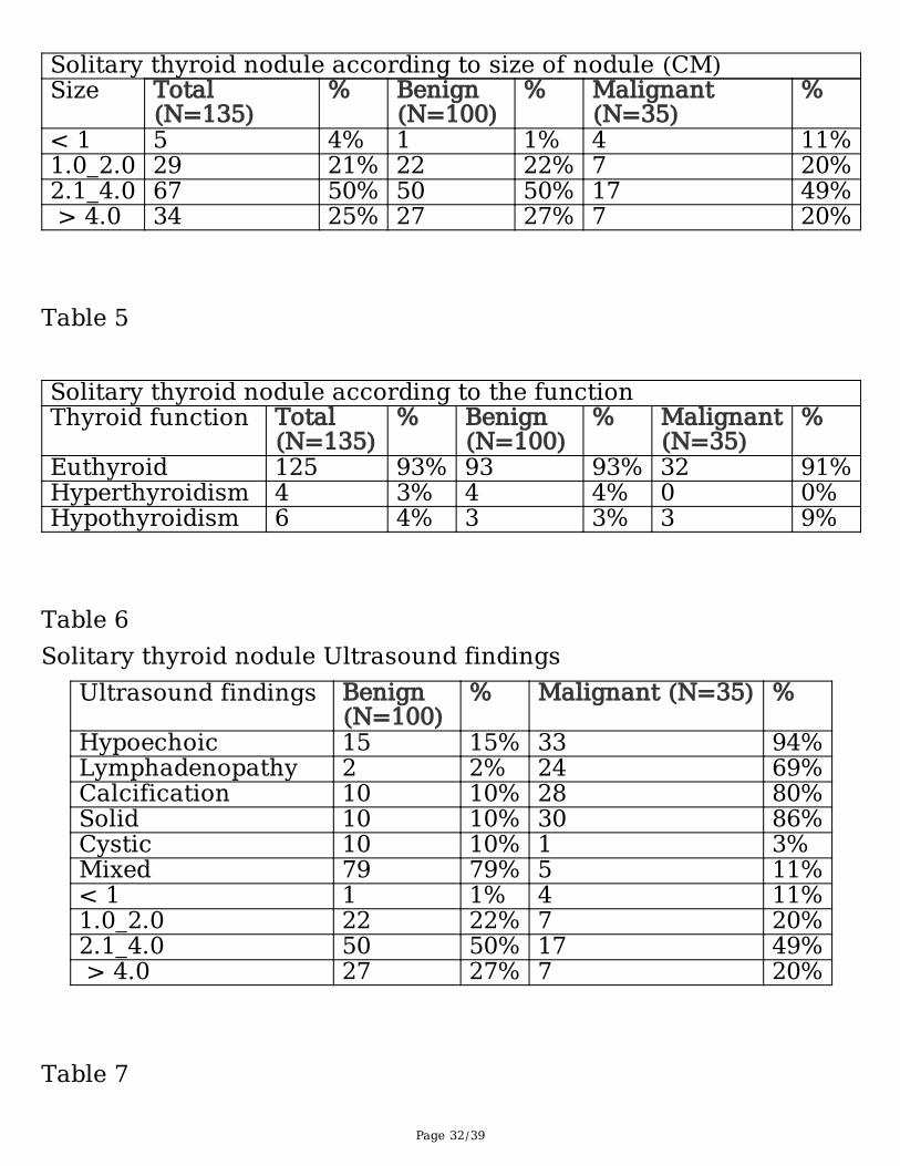
Page 32/39
Solitary thyroid nodule according to size of nodule (CM)Size Total
(N=135)% Benign
(N=100) % Malignant
(N=35)%
< 1 5 4% 1 1% 4 11%1.0_2.0 29 21% 22 22% 7 20%2.1_4.0 67 50% 50 50% 17 49% > 4.0 34 25% 27 27% 7 20%
Table 5 Solitary thyroid nodule according to the function Thyroid function Total
(N=135)% Benign
(N=100) % Malignant
(N=35)%
Euthyroid 125 93% 93 93% 32 91%Hyperthyroidism 4 3% 4 4% 0 0%Hypothyroidism 6 4% 3 3% 3 9%
Table 6Solitary thyroid nodule Ultrasound findings
Ultrasound findings Benign(N=100)
% Malignant (N=35) %
Hypoechoic 15 15% 33 94%Lymphadenopathy 2 2% 24 69%Calcification 10 10% 28 80%Solid 10 10% 30 86%Cystic 10 10% 1 3%Mixed 79 79% 5 11%< 1 1 1% 4 11%1.0_2.0 22 22% 7 20%2.1_4.0 50 50% 17 49% > 4.0 27 27% 7 20%
Table 7
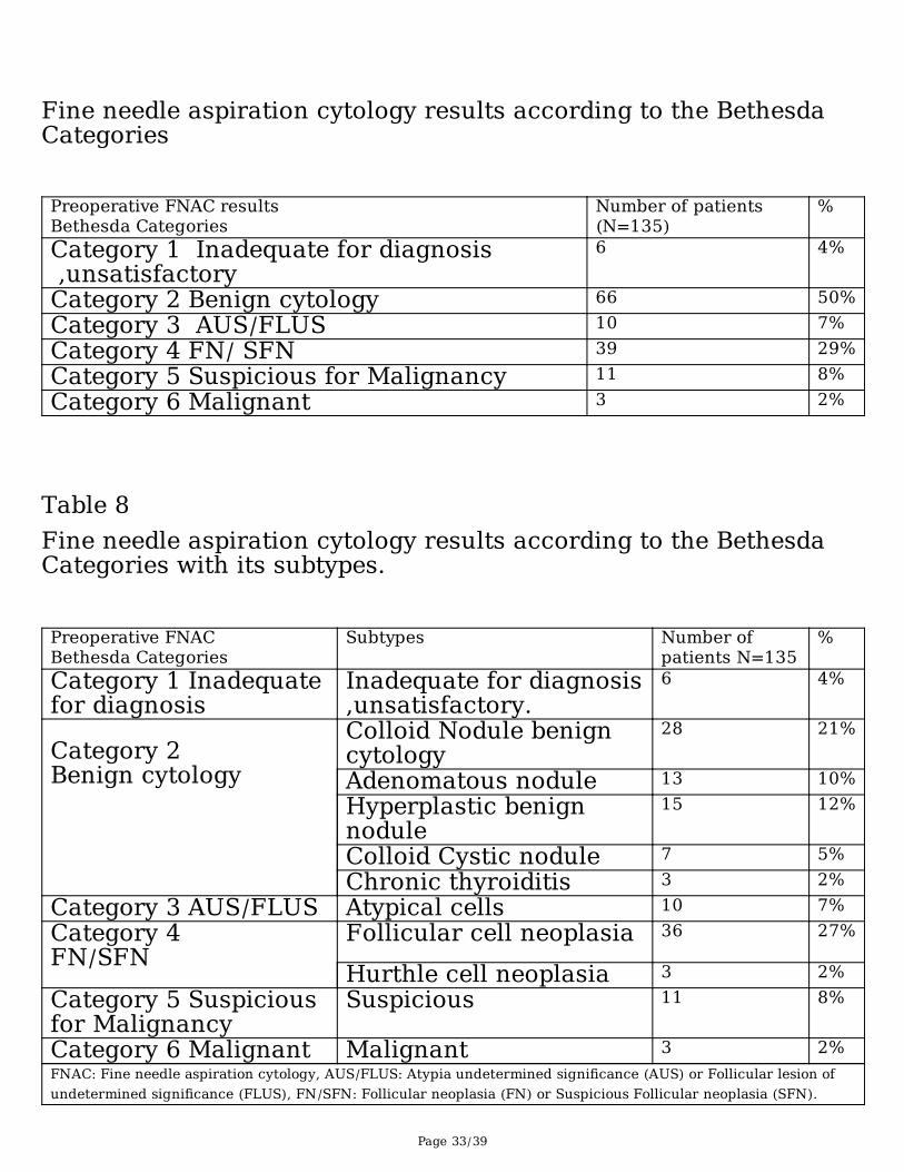
Page 33/39
Fine needle aspiration cytology results according to the BethesdaCategories Preoperative FNAC resultsBethesda Categories
Number of patients(N=135)
%
Category 1 Inadequate for diagnosis ,unsatisfactory
6 4%
Category 2 Benign cytology 66 50%Category 3 AUS/FLUS 10 7%Category 4 FN/ SFN 39 29%Category 5 Suspicious for Malignancy 11 8%Category 6 Malignant 3 2%
Table 8Fine needle aspiration cytology results according to the BethesdaCategories with its subtypes. Preoperative FNACBethesda Categories
Subtypes Number ofpatients N=135
%
Category 1 Inadequatefor diagnosis
Inadequate for diagnosis,unsatisfactory.
6 4%
Category 2 Benign cytology
Colloid Nodule benigncytology
28 21%
Adenomatous nodule 13 10%Hyperplastic benignnodule
15 12%
Colloid Cystic nodule 7 5%Chronic thyroiditis 3 2%
Category 3 AUS/FLUS Atypical cells 10 7%Category 4 FN/SFN
Follicular cell neoplasia 36
27%
Hurthle cell neoplasia 3 2%Category 5 Suspiciousfor Malignancy
Suspicious 11 8%
Category 6 Malignant Malignant 3 2%FNAC: Fine needle aspiration cytology, AUS/FLUS: Atypia undetermined significance (AUS) or Follicular lesion ofundetermined significance (FLUS), FN/SFN: Follicular neoplasia (FN) or Suspicious Follicular neoplasia (SFN).
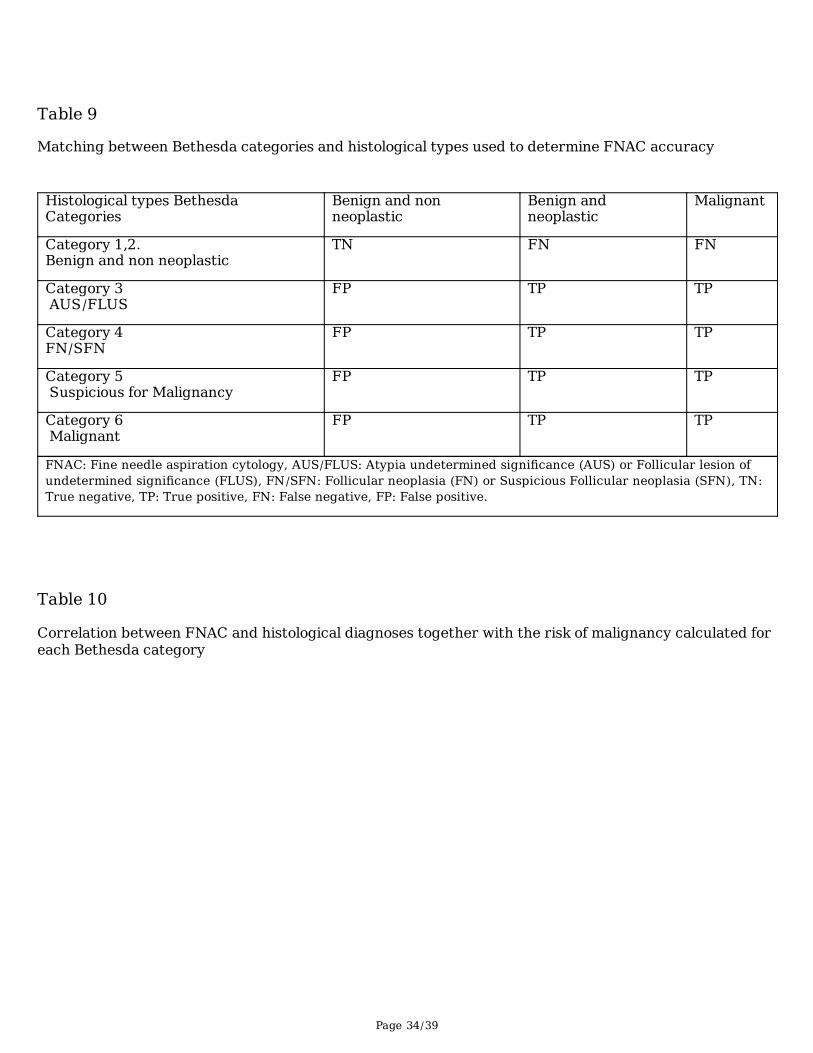
Page 34/39
Table 9
Matching between Bethesda categories and histological types used to determine FNAC accuracy
Histological types BethesdaCategories
Benign and nonneoplastic
Benign andneoplastic
Malignant
Category 1,2.Benign and non neoplastic
TN FN FN
Category 3 AUS/FLUS
FP TP TP
Category 4FN/SFN
FP TP TP
Category 5 Suspicious for Malignancy
FP TP TP
Category 6 Malignant
FP TP TP
FNAC: Fine needle aspiration cytology, AUS/FLUS: Atypia undetermined significance (AUS) or Follicular lesion ofundetermined significance (FLUS), FN/SFN: Follicular neoplasia (FN) or Suspicious Follicular neoplasia (SFN), TN:True negative, TP: True positive, FN: False negative, FP: False positive.
Table 10
Correlation between FNAC and histological diagnoses together with the risk of malignancy calculated foreach Bethesda category
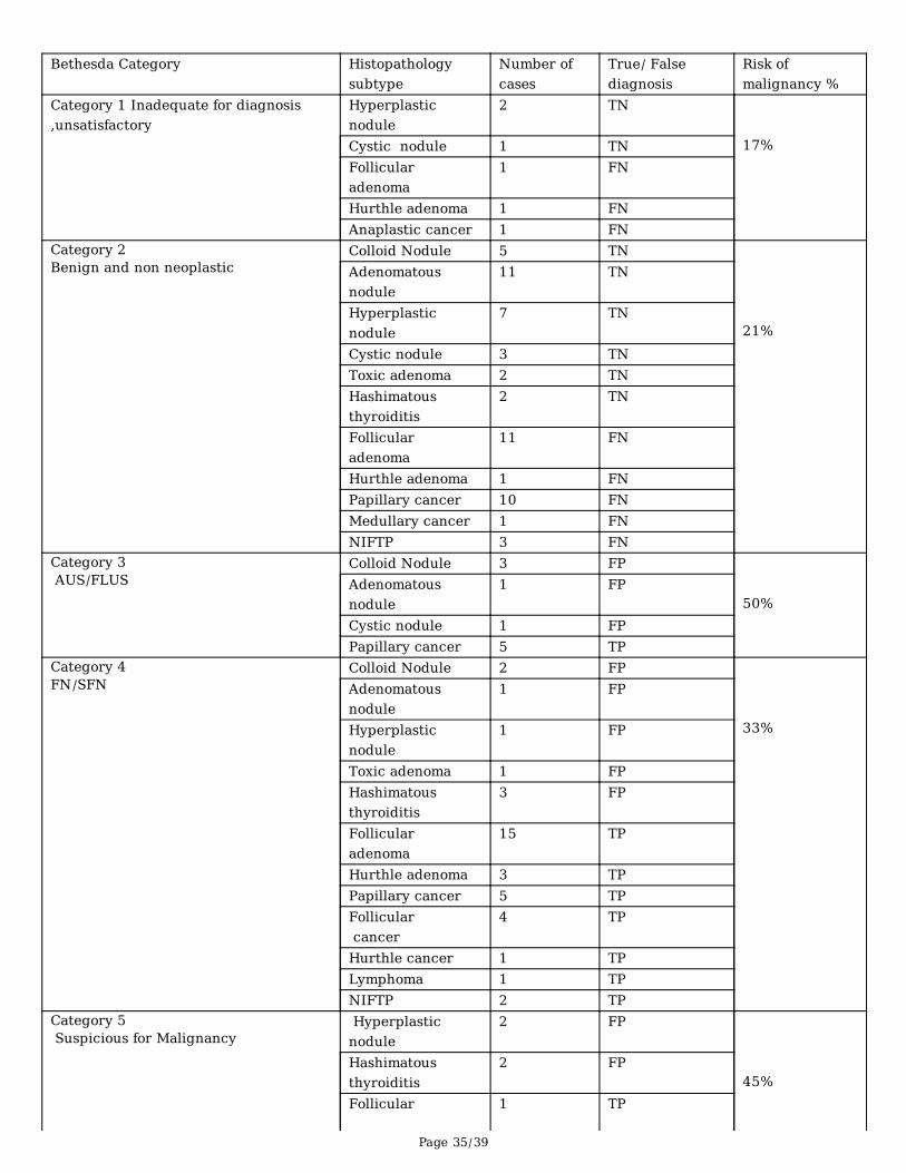
Page 35/39
Bethesda Category Histopathologysubtype
Number ofcases
True/ Falsediagnosis
Risk ofmalignancy %
Category 1 Inadequate for diagnosis,unsatisfactory
Hyperplasticnodule
2 TN 17%Cystic nodule 1 TN
Follicularadenoma
1 FN
Hurthle adenoma 1 FN Anaplastic cancer 1 FN
Category 2Benign and non neoplastic
Colloid Nodule 5 TN 21%
Adenomatousnodule
11 TN
Hyperplasticnodule
7 TN
Cystic nodule 3 TNToxic adenoma 2 TNHashimatousthyroiditis
2 TN
Follicularadenoma
11 FN
Hurthle adenoma 1 FN Papillary cancer 10 FN Medullary cancer 1 FN NIFTP 3 FN
Category 3 AUS/FLUS
Colloid Nodule 3 FP 50%
Adenomatousnodule
1 FP
Cystic nodule 1 FP Papillary cancer 5 TP
Category 4FN/SFN
Colloid Nodule 2 FP 33%
Adenomatousnodule
1 FP
Hyperplasticnodule
1 FP
Toxic adenoma 1 FP Hashimatousthyroiditis
3 FP
Follicularadenoma
15 TP
Hurthle adenoma 3 TP Papillary cancer 5 TP Follicular cancer
4 TP
Hurthle cancer 1 TP Lymphoma 1 TP NIFTP 2 TP
Category 5 Suspicious for Malignancy
Hyperplasticnodule
2 FP 45%
Hashimatousthyroiditis
2 FP
Follicular 1 TP
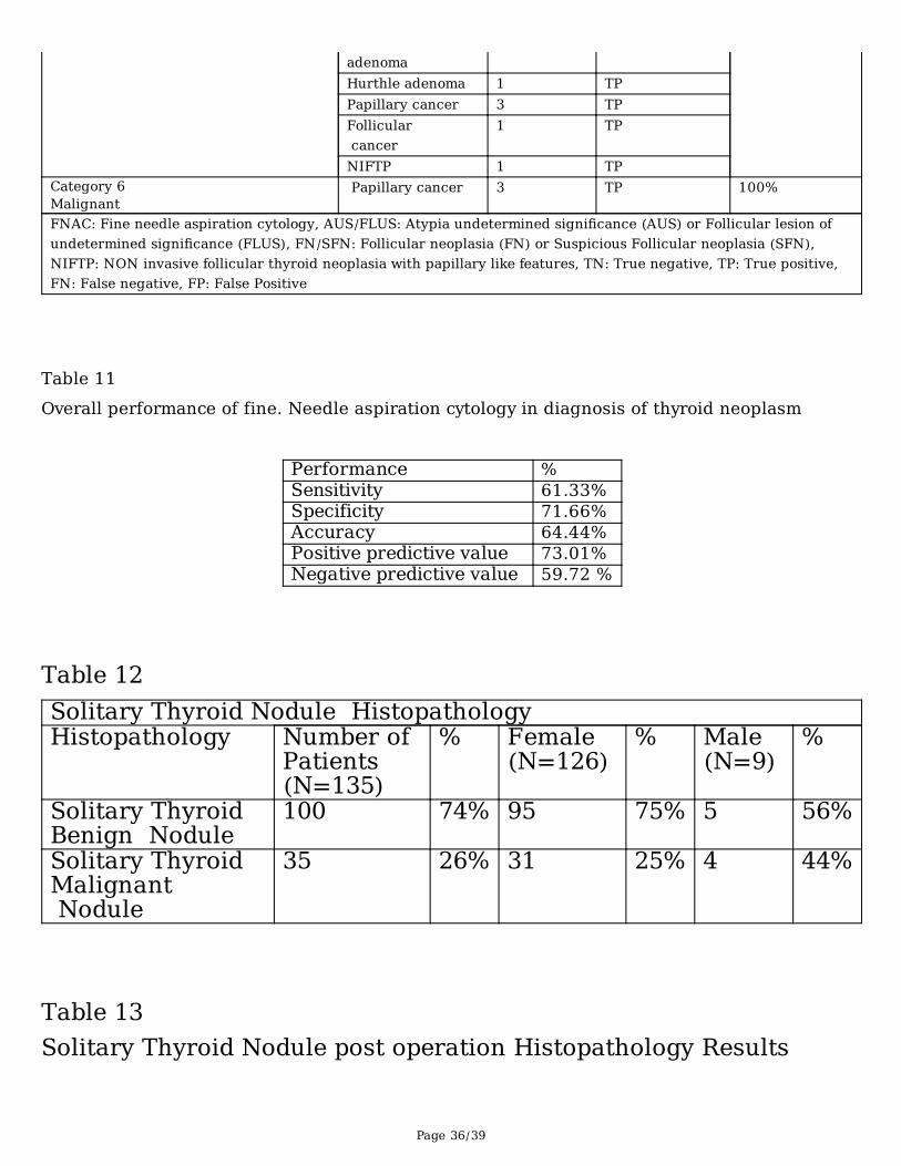
Page 36/39
adenoma Hurthle adenoma 1 TP Papillary cancer 3 TP Follicular cancer
1 TP
NIFTP 1 TP Category 6Malignant
Papillary cancer 3 TP 100%
FNAC: Fine needle aspiration cytology, AUS/FLUS: Atypia undetermined significance (AUS) or Follicular lesion ofundetermined significance (FLUS), FN/SFN: Follicular neoplasia (FN) or Suspicious Follicular neoplasia (SFN),NIFTP: NON invasive follicular thyroid neoplasia with papillary like features, TN: True negative, TP: True positive,FN: False negative, FP: False Positive
Table 11 Overall performance of fine. Needle aspiration cytology in diagnosis of thyroid neoplasm
Performance %Sensitivity 61.33%Specificity 71.66%Accuracy 64.44%Positive predictive value 73.01%Negative predictive value 59.72 %
Table 12Solitary Thyroid Nodule Histopathology Histopathology Number of
Patients(N=135)
% Female(N=126)
% Male(N=9)
%
Solitary ThyroidBenign Nodule
100 74% 95 75% 5 56%
Solitary ThyroidMalignant Nodule
35 26% 31 25% 4 44%
Table 13Solitary Thyroid Nodule post operation Histopathology Results
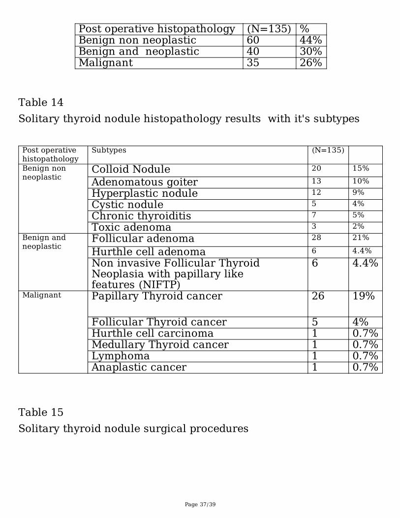
Page 37/39
Post operative histopathology (N=135) %Benign non neoplastic 60 44%Benign and neoplastic 40 30%Malignant 35 26%
Table 14Solitary thyroid nodule histopathology results with it's subtypes Post operativehistopathology
Subtypes (N=135)
Benign nonneoplastic
Colloid Nodule 20 15%
Adenomatous goiter 13 10%Hyperplastic nodule 12 9%Cystic nodule 5 4%Chronic thyroiditis 7 5%Toxic adenoma 3 2%
Benign andneoplastic
Follicular adenoma 28 21%
Hurthle cell adenoma 6 4.4%Non invasive Follicular ThyroidNeoplasia with papillary likefeatures (NIFTP)
6 4.4%
Malignant Papillary Thyroid cancer 26 19%
Follicular Thyroid cancer 5 4%Hurthle cell carcinoma 1 0.7%Medullary Thyroid cancer 1 0.7%Lymphoma 1 0.7%Anaplastic cancer 1 0.7%
Table 15Solitary thyroid nodule surgical procedures
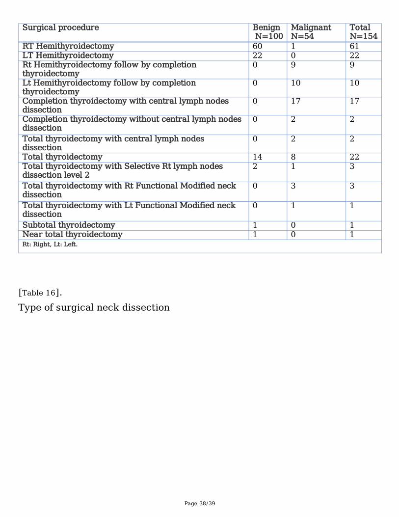
Page 38/39
Surgical procedure Benign N=100
MalignantN=54
Total N=154
RT Hemithyroidectomy 60 1 61LT Hemithyroidectomy 22 0 22Rt Hemithyroidectomy follow by completionthyroidectomy
0 9 9
Lt Hemithyroidectomy follow by completionthyroidectomy
0 10 10
Completion thyroidectomy with central lymph nodesdissection
0 17 17
Completion thyroidectomy without central lymph nodesdissection
0 2 2
Total thyroidectomy with central lymph nodesdissection
0 2 2
Total thyroidectomy 14 8 22Total thyroidectomy with Selective Rt lymph nodesdissection level 2
2 1 3
Total thyroidectomy with Rt Functional Modified neckdissection
0 3 3
Total thyroidectomy with Lt Functional Modified neckdissection
0 1 1
Subtotal thyroidectomy 1 0 1Near total thyroidectomy 1 0 1Rt: Right, Lt: Left.
[Table 16].Type of surgical neck dissection
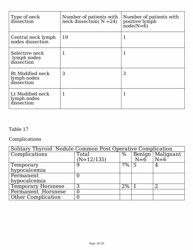
Page 39/39
Type of neckdissection
Number of patients withneck dissection( N =24)
Number of patients withpositive lymphnode(N=6)
Central neck lymphnodes dissection
19 1
Selective neck lymph nodesdissection
1 1
Rt Modified necklymph nodesdissection
3 3
Lt Modified necklymph nodesdissection
1 1
Table 17
Complications
Solitary Thyroid Nodule Common Post Operative Complication Complications Total
(N=12/135)% Benign
N=6Malignant N=6
Temporaryhypocalcemia
9 7% 5 4
Permanenthypocalcemia
0
Temporary Horsnese 3 2% 1 2Permanent Horsnese 0 Other Complication 0