Embed Size (px)
Citation preview
For Peer Review
ATA Guidelines for Management of Thyroid Nodules and Differentiated Thyroid Cancer
Journal: Thyroid
Manuscript ID: THY-2009-0110.R2
Manuscript Type: Invited Paper
Keyword: Thyroid Cancer- clinical, Thyroid Nodules
Abstract:
Abstract Background: Thyroid nodules are a common clinical problem, and
differentiated thyroid cancer is becoming increasingly prevalent. Since the publication of the American Thyroid Association’s guidelines for the management of these disorders was published in 2006, a large amount of new information on these topics has become available, prompting a revision of the guidelines. Methods: Relevant articles through December 2008 were reviewed by the task force and categorized by topic and level of evidence according to a modified schema used by the United States Preventative Services Task Force. Results: The revised guidelines for the management of thyroid
nodules include recommendations regarding initial evaluation, clinical and ultrasound criteria for fine needle aspiration biopsy, interpretation of fine needle aspiration biopsy results, and management of benign thyroid nodules. Recommendations regarding the initial management of thyroid cancer include those relating to optimal surgical management, radioiodine remnant ablation, and suppression therapy using levothyroxine. Recommendations related to long-term management of differentiated thyroid cancer include those related to surveillance for recurrent disease using ultrasound and serum thyroglobulin as well as those related to management of recurrent and metastatic disease.
Conclusions: These evidence-based recommendations were created by the American Thyroid Association to assist in the clinical management of patients with thyroid nodules and differentiated thyroid cancer. They represent, in our opinion, contemporary optimal care for patients with these disorders.
Thyroid

For Peer Review
Page 1 of 141 Thyroid
For Peer Review
1
REVISED MANAGEMENT GUIDELINES FOR PATIENTS WITH THYROID NODULES
AND DIFFERENTIATED THYROID CANCER
Authors are listed in alphabetical order
David S. Cooper, M.D.1 (Chair), Gerard M. Doherty, M.D.
2, Bryan R. Haugen, M.D.
3,Richard T.
Kloos, M.D.4, Stephanie L. Lee, M.D., PhD.
5, Susan J. Mandel, M.D., M.P.H.
6, Ernest L.
Mazzaferri, M.D.7, Bryan McIver, M.D., PhD.
8,Furio Pacini, M.D.
9, Martin Schlumberger,
M.D.10
, Steven I. Sherman, M.D.11
, David L. Steward, M.D.12
, R. Michael Tuttle, M.D.13
.
1 The Johns Hopkins University School of Medicine, Baltimore, MD;
2University of Michigan
Medical Center, Ann Arbor, MI; 3University of Colorado Health Sciences Center, Denver, CO;
4
The Ohio State University, Columbus, OH; 5Boston University Medical Center, Boston, MA;
6University of Pennsylvania School of Medicine, Philadelphia, PA;
7University of Florida
College of Medicine, Gainesville, Fl; 8
The Mayo Clinic, Rochester, MN; 9The University of
Siena, Siena Italy, 10
Institut Gustave Roussy, Paris, France, 11
University of Texas M.D.
Anderson Cancer Center, Houston, TX; 12
University of Cincinnati Medical Center, Cincinnati,
OH; 13
Memorial Sloan-Kettering Cancer Center, New York, NY.
Correspondence
Division of Endocrinology
The Johns Hopkins University School of Medicine
Baltimore, MD 21287
410-955-3663 phone
410-955-8172 fax
Support: These guidelines were funded by the American Thyroid Association without support
from any commercial sources.
Note: The authors are the members of the American Thyroid Association (ATA) Guidelines
Taskforce on Differentiatied Thyroid Cancer. They were appointed by ATA to independently
formulate the content of this manuscript. None of the scientific or medical content of the
manuscript was dictated by the ATA.
Page 2 of 141Thyroid
For Peer Review
2
Abstract
Background: Thyroid nodules are a common clinical problem, and differentiated thyroid cancer
is becoming increasingly prevalent. Since the publication of the American Thyroid Association’s
guidelines for the management of these disorders was published in 2006, a large amount of new
information on these topics has become available, prompting a revision of the guidelines.
Methods: Relevant articles through December 2008 were reviewed by the task force and
categorized by topic and level of evidence according to a modified schema used by the United
States Preventative Services Task Force.
Results: The revised guidelines for the management of thyroid nodules include
recommendations regarding initial evaluation, clinical and ultrasound criteria for fine needle
aspiration biopsy, interpretation of fine needle aspiration biopsy results, and management of
benign thyroid nodules. Recommendations regarding the initial management of thyroid cancer
include those relating to optimal surgical management, radioiodine remnant ablation, and
suppression therapy using levothyroxine. Recommendations related to long-term management
of differentiated thyroid cancer include those related to surveillance for recurrent disease using
ultrasound and serum thyroglobulin as well as those related to management of recurrent and
metastatic disease.
Conclusions: We created evidence-based recommendations in response to our appointment as a
an independent task force by the American Thyroid Association to assist in the clinical
management of patients with thyroid nodules and differentiated thyroid cancer. They represent,
in our opinion, contemporary optimal care for patients with these disorders.
Page 3 of 141 Thyroid
For Peer Review
3
Thyroid nodules are a common clinical problem. Epidemiologic studies have shown the
prevalence of palpable thyroid nodules to be approximately 5% in women and 1% in men living
in iodine sufficient parts of the world (1,2). In contrast, high-resolution ultrasound can detect
thyroid nodules in 19-67% of randomly selected individuals with higher frequencies in women
and the elderly (3). The clinical importance of thyroid nodules rests with the need to exclude
thyroid cancer which occurs in 5-15% depending on age, sex, radiation exposure history, family
history and other factors (4, 5). Differentiated thyroid cancer, which includes papillary and
follicular cancer, comprises the vast majority (90%) of all thyroid cancers (6). In the United
States, approximately 37,200 new cases of thyroid cancer will be diagnosed in 2009 (7). The
yearly incidence has increased from 3.6 per 100,000 in 1973 to 8.7 per 100,000 in 2002, a 2.4-
fold increase (P<.001 for trend) and this trend appears to be continuing (8). Almost the entire
change has been attributed to an increase in the incidence of papillary thyroid cancer, which
increased 2.9-fold between 1988 and 2002. Moreover 49% of the rising incidence consisted of
cancers measuring 1 cm or smaller and 87% consisted of cancers measuring 2 cm or smaller (8).
This tumor shift may be due to the increasing use of neck ultrasonography and early diagnosis
and treatment (9), trends that are changing the initial treatment and follow-up for many patients
with thyroid cancer.
In 1996, the American Thyroid Association (ATA) published treatment guidelines for
patients with thyroid nodules and differentiated thyroid cancer (10). Over the last decade, there
have been many advances in the diagnosis and therapy of both thyroid nodules and differentiated
thyroid cancer. Controversy exists in many areas, including the most cost effective approach in
the diagnostic evaluation of a thyroid nodule, the extent of surgery for small thyroid cancers, the
use of radioactive iodine to ablate remnant tissue following thyroidectomy, the appropriate use of
thyroxine suppression therapy, and the role of human recombinant thyrotropin. In recognition of
Page 4 of 141Thyroid
For Peer Review
4
the changes that have taken place in the overall management of these clinically important
problems, the ATA appointed a task force to re-examine the current strategies that are used to
diagnose and treat thyroid nodules and differentiated thyroid cancer, and to develop clinical
guidelines using principles of evidence based medicine. Members of the taskforce included
experts in thyroid nodule and thyroid cancer management with representation by endocrinology,
surgery, and nuclear medicine. The medical and opinions expressed here are those of the
authors; none were dictated by the ATA. Other groups have previously developed guidelines,
including the American Association of Clinical Endocrinologists and the American Association
of Endocrine Surgeons (11), the British Thyroid Association and The Royal College of
Physicians (12), and the National Comprehensive Cancer Network (13) that have provided
somewhat conflicting recommendations due to the lack of high quality evidence from
randomized controlled trials. The European Thyroid Association has published consensus
guidelines for the management of differentiated thyroid cancer (14). The European Association
of Nuclear Medicine has also recently published consensus guidelines for radioiodine therapy of
differentiated thyroid cancer (15). The organization of management guideline recommendations,
is shown in Table 1.
METHODS
The ATA guidelines taskforce used a strategy similar to that employed by the National
Institutes of Health for its Consensus Development Conferences
(http://consensus.nih.gov/aboutcdp.htm), and developed a series of clinically relevant questions
pertaining to thyroid nodule and thyroid cancer diagnosis and treatment. These questions were as
follows:
Page 5 of 141 Thyroid
For Peer Review
5
Questions regarding thyroid nodules
• What is the appropriate evaluation of clinically or incidentally discovered thyroid nodule(s)?
o What laboratory tests and imaging modalities are indicated?
o What is the role of fine needle aspiration (FNA)?
• What is the best method of long-term follow up of patients with thyroid nodules?
• What is the role of medical therapy of patients with benign thyroid nodules?
• How should thyroid nodules in children and pregnant women be managed?
Questions regarding the initial management of Differentiated Thyroid Cancer (DTC)
• What is the role of preoperative staging with diagnostic imaging and laboratory tests?
• What is the appropriate operation for indeterminate thyroid nodules and differentiated
thyroid cancer?
• What is the role of postoperative staging systems and which should be used?
• What is the role of postoperative radioiodine remnant ablation?
• What is the role of thyrotropin suppression therapy?
• Is there a role for adjunctive external beam irradiation or chemotherapy?
Questions regarding the long term management of DTC
• What are the appropriate features of long-term management?
• What is the role of serum thyroglobulin (Tg) assays?
• What is the role of ultrasound and other imaging techniques during follow-up?
• What is the role of thyrotropin suppression in long-term follow-up?
• What is the most appropriate management of patients with metastatic disease?
• How should Tg positive, scan negative patients be managed?
Page 6 of 141Thyroid
For Peer Review
6
• What is the role of external radiation therapy?
• What is the role of chemotherapy?
• What are directions for future research?
The initial ATA guidelines were published in 2006 (16). Because of the rapid growth of
the literature on this topic, plans for revising the guidelines within 24-36 months of publication
were made at the inception of the project. Relevant articles on thyroid cancer were identified
using the same search criteria employed for the original guidelines (16), and it was agreed to
continue to categorize the published data and strength of recommendations using a modified
schema proposed by the U.S. Preventive Services Task Force (17) (Table 2). Individual task
force members submitted suggestions for clarification of prior recommendations, as well as new
information derived from studies published since 2004. Relevant literature has continued to be
reviewed through December 2008. To begin the revision process, a ½ day meeting was held on
June 2, 2007. The Task Force was broadened to include European experts and a head and neck
surgeon. Three subsequent ½ day meetings were held on October 5, 2007, July 13, 2008, and
October 5, 2008 to review these suggestions and for additional comments to be considered. The
meeting in July 2008 also included a meeting with 6 additional surgeons in an effort to produce
guidelines related to central neck dissection that would be as authoritative as possible.
{A1} THYROID NODULE GUIDELINES
A thyroid nodule is a discrete lesion within the thyroid gland that is radiologically distinct
from the surrounding thyroid parenchyma. Some palpable lesions may not correspond to distinct
radiologic abnormalities (18). Such abnormalities do not meet the strict definition for thyroid
nodules. Nonpalpable nodules detected on ultrasound or other anatomic imaging studies are
Page 7 of 141 Thyroid
For Peer Review
7
termed incidentally discovered nodules or "incidentalomas." Nonpalpable nodules have the
same risk of malignancy as palpable nodules with the same size (19). Generally, only nodules >
1 cm should be evaluated, since they have a greater potential to be clinically significant cancers.
Occasionally, there may be nodules < 1 cm that require evaluation, because of suspicious
ultrasound findings, associated lymphadenopathy, a history of head and neck irradiation, or a
history of thyroid cancer in one or more first degree relatives. However, some nodules < 1 cm
lack these warning signs yet eventually cause morbidity and mortality. These are rare and, given
unfavorable cost/benefit considerations, attempts to diagnose and treat all small thyroid cancers
in an effort to prevent these rare outcomes would likely cause more harm than good.
Approximately 1-2% of people undergoing 18
FDG-PET imaging for other reasons have thyroid
nodules incidentally. Since the risk of malignancy in these 18
FDG positive nodules is about
33%, and the cancers may be more aggressive (20), such lesions require prompt evaluation
(21,22,23). When seen, diffuse 18
FDG uptake is likely related to underlying autoimmune
thyroiditis.
{A2} Appropriate Evaluation of Clinically or
Incidentally Discovered Thyroid Nodule(s)? (See Figure 1 for Algorithm)
With the discovery of a thyroid nodule, a complete history and physical examination
focusing on the thyroid gland and adjacent cervical lymph nodes should be performed. Pertinent
historical factors predicting malignancy include a history of childhood head and neck irradiation,
total body irradiation for bone marrow transplantation (24), family history of thyroid carcinoma
or thyroid cancer syndrome (e.g., Cowden’s syndrome, familial polyposis, Carney Complex,
Page 8 of 141Thyroid
For Peer Review
8
MEN 2, Werner syndrome) in a first-degree relative, exposure to ionizing radiation from fallout
in childhood or adolescence (25), and rapid growth and hoarseness. Pertinent physical findings
suggesting possible malignancy include vocal cord paralysis, lateral cervical lymphadenopathy
and fixation of the nodule to surrounding tissues.
{A3} What laboratory tests and imaging modalities are indicated?
{A4} Serum TSH with ultrasound and with or without scan. With the discovery of a
thyroid nodule >1 cm in any diameter or diffuse or focal thyroidal uptake on 18
FDG-PET scan, a
serum thyrotropin (TSH) level should be obtained. If the serum TSH is subnormal, a
radionuclide thyroid scan should be obtained to document whether the nodule is
hyperfunctioning (i.e., tracer uptake is greater than the surrounding normal thyroid),
isofunctioning or “warm” (i.e., tracer uptake is equal to the surrounding thyroid), or
nonfunctioning (i.e., has uptake less than the surrounding thyroid tissue). Since
hyperfunctioning nodules rarely harbor malignancy, if one is found that corresponds to the
nodule in question, no cytologic evaluation is necessary. If overt or subclinical hyperthyroidism
is present, additional evaluation is required. Higher serum TSH, even within the upper part of
the reference range, is associated with increased risk of malignancy in a thyroid nodule (26).
R1 Measure serum TSH in the initial evaluation of a patient with a thyroid nodule.
If the serum TSH is subnormal, a radionuclide thyroid scan should be performed
using either Tc 99m
pertechnetate or 123
Iodine. Recommendation Rating: A
Diagnostic thyroid ultrasound should be performed in all patients with a suspected
thyroid nodule a nodular goiter, or radiographic abnormality e.g., a nodule found incidentally on
CT or MRI or thyroidal uptake on 18
FDG-PET scan. Thyroid ultrasound can answer the
Page 9 of 141 Thyroid
For Peer Review
9
following questions: Is there truly a nodule that corresponds to the palpable abnormality? How
large is the nodule? Does the nodule have benign or suspicious features? Is suspicious cervical
lymphadenopathy present? Is the nodule greater than 50% cystic? Is the nodule located
posteriorly in the thyroid gland? These last two features might decrease the accuracy of fine
needle aspiration biopsy performed with palpation (27,28). Also, there may be other thyroid
nodules present that require biopsy based on their size and appearance (18,29,30). As noted
above, FNA is recommended especially when the serum TSH is elevated, since, compared with
normal thyroid glands, the rate of malignancy in nodules in thyroid glands involved with
Hashimoto's thyroiditis is as least as high or possibly higher (31,32).
R2 Thyroid sonography should be performed in all patients with known or
suspected thyroid nodules. Recommendation Rating: A
{A5} Serum thyroglobulin (Tg) measurement. Serum Tg levels can be elevated in most
thyroid diseases and are an insensitive and non-specific test for thyroid cancer (33).
R3 Routine measurement of serum thyroglobulin for initial evaluation of thyroid
nodules is not recommended. Recommendation Rating: F
{A6} Serum calcitonin measurement. The utility of serum calcitonin has been evaluated
in a series of prospective, nonrandomized studies (34,35,36,37). The data suggest that the use of
routine serum calcitonin for screening may detect C-cell hyperplasia and medullary thyroid
cancer at an earlier stage and overall survival may be improved. However, most studies rely on
pentagastrin stimulation testing to increase specificity and this drug is no longer available in the
Formatted: Endnote Reference
Formatted: Endnote Reference
Deleted: 18
Deleted: 18
Page 10 of 141Thyroid
For Peer Review
10
United States, and there remain unresolved issues of sensitivity, specificity, assay performance
and cost-effectiveness. A recent cost-effectiveness analysis suggested that calcitonin screening
would be cost-effective in the United States (38). However, the prevalence estimates of
medullary thyroid cancer in this analysis included patients with C cell hyperplasia and
micromedullary carcinoma, which have an uncertain clinical significance. If the unstimulated
serum calcitonin determination has been obtained and the level is greater than 100 pg/ml,
medullary cancer is likely present (39).
R4 The panel cannot recommend either for or against the routine measurement of
serum calcitonin. Recommendation Rating: I
{A7} What is the role of fine needle aspiration biopsy?
FNA is the most accurate and cost effective method for evaluating thyroid nodules.
Retrospective studies have reported lower rates of both nondiagnostic and false-negative
cytology specimens from FNA procedures performed via ultrasound-guidance compared to
palpation (40,41). Therefore, for nodules with a higher likelihood of either a nondiagnostic
cytology (>25-50% cystic component) (28) or sampling error (difficult to palpate or posteriorly-
located nodules), ultrasound-guided FNA is preferred (See Table 3). If the diagnostic ultrasound
confirms the presence of a predominantly solid nodule corresponding to what is palpated, the
FNA may be performed via palpation or ultrasound guidance. Traditionally FNA biopsy results
are divided into four categories: nondiagnostic, malignant (risk of malignancy at surgery >95%),
indeterminate or suspicious for neoplasm, and benign. The recent National Cancer Institute
Thyroid Fine-Needle Aspiration State of the Science Conference proposed a more expanded
classification for FNA cytology that adds two additional categories: suspicious for malignancy
Page 11 of 141 Thyroid
For Peer Review
11
(risk of malignancy 50-75%) and follicular lesion of undetermined significance (risk of
malignancy 5-10%). The conference further recommended that “neoplasm, either follicular or
Hurthle cell neoplasm” be substituted for “indeterminate” (risk of malignancy 15-25%) (42).
{A8} Ultrasound (US) for FNA Decision Making. Various sonographic characteristics of
a thyroid nodule have been associated with a higher likelihood of malignancy
(43,44,45,46,47,48). These include nodule hypoechogenicity compared to the normal thyroid
parenchyma, increased intranodular vascularity, irregular infiltrative margins, the presence of
microcalcifications, an absent halo, and a taller than wide shape measured in the transverse
dimension. With the exception of suspicious cervical lymphadenopathy, which is a specific but
insensitive finding, no single sonographic feature or combinations of features is adequately
sensitive or specific to identify all malignant nodules. However, certain features and
combination of features have high predictive value for malignancy. Furthermore, the most
common sonographic appearances of papillary and follicular thyroid cancer differ. A papillary
thyroid cancer is generally solid or predominantly solid and hypoechoic, often with infiltrative
irregular margins and increased nodular vascularity. Microcalcifications, if present, are highly
specific for papillary thyroid cancer, but may be difficult to distinguish from colloid.
Conversely, follicular cancer is more often iso- to hyperechoic and has a thick and irregular halo,
but does not have microcalcifications (49). Follicular cancers < 2 cm in diameter have not been
shown to be associated with metastatic disease (50).
Certain sonographic appearances may also be highly predictive of a benign nodule. A
pure cystic nodule, although rare (<2% of all nodules), is highly unlikely to be malignant (47).
In addition, a spongiform appearance, defined as an aggregation of multiple microcystic
components in more than 50% of the nodule volume, is 99.7% specific for identification of a
benign thyroid nodule (48,51,52). In a recent study, only 1 of 360 malignant nodules
Page 12 of 141Thyroid
For Peer Review
12
demonstrated this appearance (48) and in another report, a spongiform appearance had a negative
predictive value for malignancy of 98.5% (52). Elastography is an emerging and promising
sonographic technique that requires additional validation with prospective studies (53).
Routine FNA is not recommended for subcentimeter nodules. However, the presence of
a solid hypoechoic nodule with microcalcifications is highly suggestive of papillary thyroid
cancer. Although most micropapillary carcinomas may be incidental findings, a subset may be
more clinically relevant, especially those larger than 5 mm in diameter (54). These include
nodules that have abnormal lymph nodes detected clinically or with imaging at presentation
(55,56). Therefore, after imaging a subcentimeter nodule with a suspicious appearance,
sonographic assessment of lateral neck and central neck lymph nodes (more limited due to the
presence of the thyroid) must be performed. Detection of abnormal lymph nodes should lead to
FNA of the lymph node. Other groups of patients for whom consideration of FNA of a
subcentimeter nodule may be warranted include those with a higher likelihood of malignancy
(high risk history): (1) family history of papillary thyroid cancer (57); (2) history of external
beam radiation exposure as a child (58); (3) exposure to ionizing radiation in childhood or
adolescence (59); (4) history of prior hemithyroidectomy with discovery of thyroid cancer; and
(5) 18
FDG PET positive thyroid nodules.
Mixed cystic/solid nodules and predominantly cystic with >50% cystic component are
generally evaluated by FNA with directed biopsy of the solid component (especially the vascular
component.) Cyst drainage may also be performed, especially in symptomatic patients.
R5a Fine needle aspiration is the procedure of choice in the evaluation of thyroid
nodules. Recommendation Rating: A
Page 13 of 141 Thyroid
For Peer Review
13
R5b FNA should be considered for nodules (See Table 3):
R5c Ultrasound guidance for FNA is recommended for those nodules that are non-
palpable, predominantly cystic, or located posteriorly in the thyroid lobe.
Recommendation Rating: B
R5d Recurrent cystic thyroid nodules with benign cytology should be considered
for surgical removal or PEI based on compressive symptoms and cosmetic concerns.
Recommendation Rating: B
{A9} What are the principals of the cytopathological interpretation of FNA samples?
{A10} Nondiagnostic Cytology. Nondiagnostic biopsies are those which fail to meet
specified criteria for cytologic adequacy that have been previously established (the presence of at
least 6 follicular cell groups, each containing 10 to 15 cells derived from at least 2 aspirates of a
nodule) (5). After an initial nondiagnostic cytology results, repeat FNA with ultrasound
guidance will yield a diagnostic cytology specimen in 75% of solid nodules and 50% of cystic
nodules (28). Therefore, such biopsies need to be repeated using ultrasound guidance (60) and,
if available, on-site evaluation of cytology specimens, which may substantially increase cytology
specimen adequacy (61,62). However, up to 7% of nodules continue to yield non-diagnostic
cytology results despite repeated biopsies, and may be malignant at the time of surgery (63,64).
R6a Ultrasound guidance should be used when repeating the FNA procedure for a
nodule with an initial non-diagnostic cytology result. Recommendation Rating: A
Page 14 of 141Thyroid
For Peer Review
14
R6b Partially cystic nodules that repeatedly yield non-diagnostic aspirates need
close observation or surgical excision. Surgery should be more strongly considered
if the cytologically nondiagnostic nodule is solid. Recommendation Rating: B
{A11} Cytology suggesting papillary thyroid cancer.
R7 If a cytology result is diagnostic of or suspicious for papillary thyroid cancer,
surgery is recommended (65). Recommendation Rating: A
{A12} Indeterminate Cytology (Follicular or Hurthle cell neoplasm Follicular lesion of
undetermined significance, atypia). Indeterminate cytology, reported as “follicular neoplasm” or
“Hurthle cell neoplasm” can be found in 15-30% of FNA specimens (4) and carries a risk of 20-
30% malignancy (42), while lesions reported as atypia or follicular lesion of undetermined
significance are variably reported and have 5-10% risk of malignancy (42). While certain
clinical features such as male sex and nodule size (> 4 cm) (66), older patient age (67), or
cytologic features such as presence of atypia (68) can improve the diagnostic accuracy for
malignancy in patients with indeterminate cytology, overall predictive values are still low. Many
molecular markers (e.g. Galectin-3 (69), cytokeratin, BRAF) have been evaluated to improve
diagnostic accuracy for indeterminate nodules (70,71,72). Recent large prospective studies have
confirmed the ability of genetic markers (BRAF, Ras, RET/PTC) and protein markers (galectin-
3) to improve preoperative diagnostic accuary for patients with indeterminate thyroid nodules
(69,73,74). Many of these markers are available for commercial use in reference laboratories,
but have not yet been widely applied in clinical practice. It is likely that some combination of
Page 15 of 141 Thyroid
For Peer Review
15
molecular markers will be used in the future to optimize management of patients with
indeterminate cytology on FNA specimens.
Recently, 18
FDG-PET scanning has been utilized in an effort to distinguish those
indeterminate nodules that are benign from those that are malignant (75, 76,77,78). 18
FDG-PET
scans appear to have relatively high sensitivity for malignancy but low specificity, but results
vary among studies (79).
R8a The use of molecular markers (e.g., BRAF, Ras, RET/PTC, Pax8-PPARγ or
galectin-3) may be considered for patients with indeterminate cytology on FNA to
help guide management. Recommendation Rating: C
R8b The panel cannot recommend for or against routine clinical use of 18
FDG-PET
scan to improve diagmostic accuracy of indeterminate thyroid nodules.
Recommendation Rating: I
R9 If the cytology reading reports a follicular neoplasm, a radioiodine thyroid scan
may be considered, if not already done, especially if the serum TSH is in the low-
normal range. If a concordant autonomously functioning nodule is not seen,
lobectomy or total thyroidectomy should be considered. Recommendation Rating:
C
R10 If the reading is "suspicious for papillary carcinoma” or “Hurthle cell
neoplasm," a radionuclide scan is not needed, and either lobectomy or total
Page 16 of 141Thyroid
For Peer Review
16
thyroidectomy is recommended, depending on the lesion’s size and other risk
factors. Recommendation Rating: A
{A13} Benign cytology.
R11 If the nodule is benign on cytology, further immediate diagnostic studies or
treatment are not routinely required. Recommendation Rating: A
{A14} How should multinodular thyroid glands or multinodular goiters be evaluated for
malignancy?
Patients with multiple thyroid nodules have the same risk of malignancy as those with
solitary nodules (18,44). However, one large study found that a solitary nodule had a higher
likelihood of malignancy than did a non-solitary nodule (P < 0.01), although the risk of
malignancy per patient was the same and independent of the number of nodules (47). A
diagnostic US should be performed to delineate the nodules, but if only the “dominant” or largest
nodule is aspirated, the thyroid cancer may be missed (44). Radionuclide scanning should also
be considered in patients with multiple thyroid nodules, if the serum TSH is in the low or low-
normal range, with FNA being reserved for those nodules which are shown to be
hypofunctioning.
R12a In the presence of two or more thyroid nodules >1 cm, those with a suspicious
sonographic appearance (see text and Table 3) should be aspirated preferentially.
Recommendation Rating: B
Formatted: Endnote Reference
Formatted: Endnote Reference
Deleted: 18
Deleted: 18
Page 17 of 141 Thyroid
For Peer Review
17
R12b If none of the nodules has a suspicious sonographic appearance, and multiple
sonographically similar coalescent nodules with no intervening normal parenchyma
are present, the likelihood of malignancy is low and it is reasonable to aspirate the
largest nodules only and observe the others with serial ultrasound examinations.
Recommendation Rating: C
R13 A low or low normal serum TSH concentration may suggest the presence of
autonomous nodule(s). A Tc 99m
pertechnetate or 123
iodine scan should be
performed and directly compared to the US images to determine functionality of
each nodule >1-1.5 cm. FNA should then be considered only for those isofunctioning
or nonfunctioning nodules, among which those with suspicious sonographic features
should be aspirated preferentially. Recommendation Rating: B
{A15} Best Methods for Long-Term
Follow-Up of Patients with Thyroid Nodules
Thyroid nodules diagnosed as benign require follow-up because of a low, but not
negligible, false-negative rate of up to 5% with FNA (41,80), which may be even higher with
nodules larger than 4 cm (81). While benign nodules may decrease in size, they often increase in
size, albeit slowly (82). One study of cytologically benign thyroid nodules < 2 cm followed by
ultrasonography for about 38 months found that the rate of thyroid nodule growth did not
distinguish between benign and malignant nodules (83).
Page 18 of 141Thyroid
For Peer Review
18
Nodule growth is not in and of itself pathognomonic of malignancy, but growth is an
indication for repeat biopsy. For mixed cystic/solid nodules, the indication for repeat biopsy
should based upon growth of the solid component. For nodules with benign cytologic results,
recent series report a higher false negative rate with palpation FNA (1-3%) (40,84,85) than with
US FNA (0.6%) (40). Since the accuracy of physical examination for nodule size is likely
inferior to that of ultrasound (30), it is recommended that serial ultrasound be used in follow-up
of thyroid nodules to detect clinically significant changes in size. There is no consensus on the
definition of nodule growth, however, or the threshold that would require rebiopsy. Some
groups suggest a 15% increase in nodule volume, while others recommend measuring a change
in the mean nodule diameter (82,86). One reasonable definition of growth is a 20% increase in
nodule diameter with a minimum increase in two or more dimensions of at least 2 mm. This
approximates the 50% increase in nodule volume that was found by Brauer et al. (87) to be the
minimally significant reproducibly recorded change in nodule size. These authors suggested
that only volume changes of at least 49% or more can be interpreted as nodule shrinkage or
growth and consequently suggest that future investigations should not describe changes in nodule
volume < 50% as significant. A 50% cutoff for nodule volume reduction or growth, which is
used in many studies, appears to appropriate and safe, since the false negative rate for malignant
thyroid nodules on repeat FNA is low (88,89).
R14 It is recommended that all benign thyroid nodules be followed with serial
ultrasound examinations 6-18 months after initial FNA. If nodule size is stable
(i.e., no more than a 50% change in volume or <20% increase in at least 2 nodule
dimensions in solid nodules or in the solid portion of mixed cystic solid nodules), the
Page 19 of 141 Thyroid
For Peer Review
19
interval before the next follow-up clinical examination or ultrasound may be longer,
e.g. every 3 – 5 years. Recommendation Rating: C
R15 If there is evidence for nodule growth either by palpation or sonographically
(more than a 50% change in volume or a 20% increase in at least two nodule
dimensions with a minimal increase of 2 mm in solid nodules or in the solid portion
of mixed cystic solid nodules), the FNA should be repeated, preferably with
ultrasound guidance. Recommendation Rating: B
Cystic nodules that are cytologically benign can be monitored for recurrence (fluid
reaccumulation) which can be seen in 60 – 90% of patients (90,91). For those patients with
subsequent recurrent symptomatic cystic fluid accumulation, surgical removal, generally by
hemithyroidectomy, or percutaneous ethanol injection (PEI) are both reasonable strategies. Four
controlled studies demonstrated a 75-85% success rate after PEI compared with a 7-38% success
rate in controls treated by simple cyst evacuation or saline injection. Success was achieved after
an average of two PEI treatments. Complications included mild to moderate local pain, flushing,
dizziness, and dysphonia (90,91,92,93).
{A16} What is the role of medical therapy for benign thyroid nodules?
Evidence from multiple randomized control trials and three metaanalyses suggest that
thyroid hormone in doses that suppress the serum TSH to subnormal levels may result in a
decrease in nodule size and may prevent the appearance of new nodules in regions of the world
with borderline low iodine intake. Data in iodine sufficient populations are less compelling
Formatted: Endnote Reference
Formatted: Endnote Reference
Formatted: Endnote Reference
Formatted: Endnote Reference
Deleted: 90
Deleted: 90
Deleted: 91
Deleted: 91
Page 20 of 141Thyroid
For Peer Review
20
(94,95,96), with large studies suggesting that only about 17% to 25% of thyroid nodules shrink
more than 50% with levothyroxine suppression of TSH (94,95,96).
R16 Routine suppression therapy of benign thyroid nodules in iodine sufficient
populations is not recommended. Recommendation Rating: F
R17 Patients with growing nodules that are benign after repeat biopsy should be
considered for continued monitoring or intervention with surgery based on
symptoms and clinical concern. There are no data on the use of levothyroxine in
this subpopulation of patients. Recommendation Rating: I
{A17} How should thyroid nodules in children be managed?
Thyroid nodules occur less frequently in children than in adults. In one study in which
approximately 5,000 children aged 11 to 18 years were assessed annually in the southwestern
United States, palpable thyroid nodules occurred in approximately 20 per thousand children, with
an annual incidence of 7 new cases per thousand children (97). Some studies have shown the
frequency of malignancy to be higher in children than adults, in the 15-20% range (98,99,100),
whereas other data have suggested that the frequency of thyroid cancer in childhood thyroid
nodules is similar to that of adults (101,102). Fine needle aspiration biopsy is sensitive and
specific in the diagnosis of childhood thyroid nodules (99,100,101).
R18 The diagnostic and therapeutic approach to one or more thyroid nodules in a
child should be the same as it would be in an adult (clinical evaluation, serum TSH,
ultrasound, FNA). Recommendation Rating: A
Page 21 of 141 Thyroid
For Peer Review
21
{A18} How should thyroid nodules in pregnant women be managed?
It is uncertain if thyroid nodules discovered in pregnant women are more likely to be
malignant than those found in nonpregnant women (103), since there are no population-based
studies on this question. The evaluation is the same as for a nonpregnant patient, with the
exception that a radionuclide scan is contraindicated. In addition, for patients with nodules
diagnosed as differentiated thyroid cancer by FNA during pregnancy, delaying surgery until after
delivery does not affect outcome (104).
R19 For eu- and hypothyroid pregnant women with thyroid nodules, FNA should be
performed. For women with suppressed serum TSH levels that persist after the 1st
trimester, FNA may be deferred until after pregnancy and cessation of lactation,
when a radionuclide scan can be performed to evaluate nodule function.
Recommendation Rating: A
If the FNA cytology is consistent with papillary thyroid cancer, surgery is recommended.
However, there is no consensus about whether surgery should be performed during pregnancy or
after delivery. To minimize the risk of miscarriage, surgery during pregnancy should be done
before 24 weeks gestation (105). However, papillary thyroid cancer discovered during
pregnancy does not behave more aggressively than that diagnosed in a similar aged group of
nonpregnant women (104,106). A retrospective study of pregnant women with differentiated
thyroid cancer found there to be no difference in either recurrence, or survival rates, between
women operated on during or after their pregnancy (104). Further, retrospective data suggest
that treatment delays of less than one year from the time of thyroid cancer discovery do not
Page 22 of 141Thyroid
For Peer Review
22
adversely effect patient outcome (107). Finally, a recent study reported a higher rate of
complications in pregnant women undergoing thyroid surgery compared with nonpregnant
women (108). Some experts recommend thyroid hormone suppression therapy for pregnant
women with FNA suspicious for or diagnostic of papillary thyroid cancer, if surgery is deferred
until the postpartum period (109).
R20a A nodule with cytology indicating papillary thyroid cancer discovered early in
pregnancy should be monitored sonographically and if it grows substantially (as
defined above) by 24 weeks gestation, surgery should be performed at that point.
However, if it remains stable by midgestation or if it is diagnosed in the second half
of pregnancy, surgery may be performed after delivery. In patients with more
advanced disease, surgery in the second trimester is reasonable. Recommendation
Rating: C
R20b In pregnant women with FNA that is suspicious for or diagnostic of papillary
thyroid cancer, consideration could be given to administration of levothyroxine
therapy to keep the TSH in the range of 0.1 – 1 mU/L. Recommendation Rating: C
{B1} DIFFERENTIATED THYROID CANCER (DTC): INITIAL MANAGEMENT
GUIDELINES
Differentiated thyroid cancer, arising from thyroid follicular epithelial cells, accounts for
the vast majority of thyroid cancers. Of the differentiated cancers, papillary cancer comprises
Page 23 of 141 Thyroid
For Peer Review
23
about 85% of cases compared to about 10% that have follicular histology, and 3% that are
Hurthle cell or oxyphil tumors (110). In general, stage for stage, the prognoses of papillary
thyroid cancer and follicular cancer are similar (107,110). Certain histologic subtypes of
papillary thyroid cancer have a worse prognosis (tall cell variant, columnar cell variant, diffuse
sclerosing variant), as do more highly invasive variants of follicular cancer. These are
characterized by extensive vascular invasion and invasion into extrathyroidal tissues or extensive
tumor necrosis and/or mitoses. Other poorly differentiated aggressive tumor histologies include
trabecular, insular, and solid subtypes (111). In contrast, minimally invasive follicular thyroid
cancer, is characterized histologically by microscopic penetration of the tumor capsule without
vascular invasion, and carries no excess mortality (112,113,114,115).
{B2} Goals of Initial Therapy of DTC
The goals of initial therapy of DTC are are follows:
1. To remove the primary tumor, disease that has extended beyond the thyroid capsule, and
involved cervical lymph nodes. Completeness of surgical resection is an important
determinant of outcome, while residual metastatic lymph nodes represent the most common
site of disease persistence/recurrence (116,117,118).
2. To minimize treatment-related morbidity. The extent of surgery and the experience of the
surgeon both play important roles in determining the risk of surgical complications (119,120).
Page 24 of 141Thyroid
For Peer Review
24
3. To permit accurate staging of the disease. Because disease staging can assist with initial
prognostication, disease management, and follow-up strategies, accurate post-operative
staging is a crucial element in the management of patients with differentiated thyroid cancer
(121,122).
4. To facilitate post-operative treatment with radioactive iodine, where appropriate. For
patients undergoing radioiodine remnant ablation, or radioiodine treatment of residual or
metastatic disease, removal of all normal thyroid tissue is an important element of initial
surgery (123). Near total or total thyroidectomy also may reduce the risk for recurrence within
the contralateral lobe (124).
5. To permit accurate long-term surveillance for disease recurrence. Both radioiodine whole
body scanning (WBS) and measurement of serum Tg are affected by residual normal thyroid
tissue. Where these approaches are utilized for long-term monitoring, near-total or total-
thyroidectomy is required (125).
6. To minimize the risk of disease recurrence and metastatic spread. Adequate surgery is the
most important treatment variable influencing prognosis, while radioactive iodine treatment,
thyrotropin suppression, and external beam irradiation each play adjunctive roles in at least
some patients (125,126,127,128).
{B3} What is the role of preoperative staging with diagnostic imaging and laboratory tests?
{B4} Neck Imaging. Differentiated thyroid carcinoma (particularly papillary carcinoma)
involves cervical lymph nodes in 20-50% of patients in most series using standard pathologic
Page 25 of 141 Thyroid
For Peer Review
25
techniques (45,129,130,131,132), and may be present even when the primary tumor is small and
intra-thyroidal (133). The frequency of micrometastases may approach 90%, depending on the
sensitivity of the detection method (134,135). However, the clinical implications of
micrometastases are likely less significant compared to macrometastases. Pre-operative
ultrasound identifies suspicious cervical adenopathy in 20-31% of cases, potentially altering the
surgical approach (136,137) in as many as 20% of patients (138,139). However, preoperative
ultrasound identifies only half of the lymph nodes found at surgery, due to the presence of the
overlying thyroid gland (140).
Sonographic features suggestive of abnormal metastatic lymph nodes include: loss of the
fatty hilus, a rounded rather than oval shape, hyperechogenicity, cystic change, calcifications,
and peripheral vascularity. No single sonographic feature is adequately sensitive for detection of
lymph nodes with metastatic thyroid cancer. A recent study correlated the sonographic features
acquired 4 days pre-operatively directly with the histology of 56 cervical lymph nodes. Some of
the most specific criteria were: short axis >5mm (96%), presence of cystic areas (100%),
presence of hyperechogenic punctuations representing either colloid or microcalcifications
(100%) and peripheral vascularity (82%). Of these, the only one with sufficient sensitivity was
peripheral vascularity (86%). All of the others had sensitivities less than 60% and would not be
adequate to use as single criterion for identification of malignant involvement (140). As shown
by earlier studies (141,142), the feature with the highest sensitivity was absence of a hilus
(100%), but this had a low specificity of only 29%. The location of the lymph nodes may also be
useful for decision-making. Malignant lymph nodes are much more likely to occur in levels III,
IV and VI than in level II (140,142). Figure 2 illustrates the delineation of cervical lymph node
Levels I through VI.
Page 26 of 141Thyroid
For Peer Review
26
Confirmation of malignancy in lymph nodes with a suspicious sonographic appearance is
achieved by US guided FNA aspiration for cytology and/or measurement of Tg in the needle
washout. This FNA measurement of Tg is valid even in patients with circulating Tg
autoantibodies (143,144).
Accurate staging is important in determining the prognosis and tailoring treatment for
patients with differentiated thyroid cancer. However, unlike many tumor types, the presence of
metastatic disease does not obviate the need for surgical excision of the primary tumor in
differentiated thyroid cancer (145). Because metastatic disease may respond to radioiodine
therapy, removal of the thyroid as well as the primary tumor and accessible loco-regional disease
remains an important component of initial treatment even in metastatic disease.
As ultrasound evaluation is uniquely operator dependent, alternative imaging procedures
may be preferable in some clinical settings, though the sensitivities of CT, MRI and PET scan for
the detection of cervical lymph node metastases are all relatively low (30 – 40%) (146). These
alternative imaging modalities, as well as laryngoscopy and endoscopy, may also be useful in the
assessment of large, rapidly growing, or retrosternal or invasive tumors, to assess the
involvement of extrathyroidal tissues (147,148).
R21 Preoperative neck ultrasound for the contralateral lobe and cervical (central
and especially lateral neck compartments) lymph nodes is recommended for all
patients undergoing thyroidectomy for malignant cytologic findings on biopsy.
Ultrasound guided FNA of sonographically suspicious lymph nodes should be
performed to confirm malignancy if this would change management.
Recommendation Rating: B
Page 27 of 141 Thyroid
For Peer Review
27
R22 Routine preoperative use of other imaging studies (CT, MRI, PET) is not
recommended. Recommendation Rating: E
{B5} Measurement of serum Tg. There is limited evidence that high pre-operative
concentrations of serum Tg may predict a higher sensitivity for post-operative surveillance with
serum Tg (149). Evidence that this impacts patient management or outcomes is not yet available.
R23 Routine preoperative measurement of serum thyroglobulin is not
recommended. Recommendation Rating: E
{B6} What is the appropriate operation for indeterminate thyroid nodules and
differentiated thyroid cancer?
The goals of thyroid surgery can include provision of a diagnosis after a nondiagnostic or
indeterminate biopsy, removal of the thyroid cancer, staging, and preparation for radioactive
ablation and serum Tg monitoring. Surgical options to address the primary tumor should be
limited to hemi-thyroidectomy with or without isthmusectomy, near-total thyroidectomy
(removal of all grossly visible thyroid tissue, leaving only a small amount [<1 gm] of tissue
adjacent to the recurrent laryngeal nerve near the ligament of Berry), and total thyroidectomy
(removal of all grossly visible thyroid tissue). Subtotal thyroidectomy, leaving > 1 gm of tissue
with the posterior capsule on the uninvolved side, is an inappropriate operation for thyroid
cancer (150).
{B7} Surgery for a non-diagnostic biopsy, a biopsy suspicious for papillary cancer or
suggestive of “follicular neoplasm”(including special consideration for patients with other risk
factors). Amongst solitary thyroid nodules with an indeterminate (“follicular neoplasm” or
Page 28 of 141Thyroid
For Peer Review
28
Hurthle cell neoplasm) biopsy, the risk of malignancy is approximately 20% (151,152,153). The
risk is higher with large tumors (>4 cm), when atypical features (e.g., cellular pleomorphism) are
seen on biopsy, when the biopsy reading is “suspicious for papillary carcinoma”, in patients with
a family history of thyroid carcinoma, and in patients with a history of radiation exposure
(66,154,155). For solitary nodules that are repeatedly non-diagnostic on biopsy, the risk of
malignancy is unknown but is probably closer to 5-10% (63).
R24 For patients with an isolated indeterminate solitary nodule who prefer a more
limited surgical procedure, thyroid lobectomy is the recommended initial surgical
approach. Recommendation Rating: C
R25a Because of an increased risk for malignancy, total thyroidectomy is indicated
in patients with indeterminate nodules who have large tumors (>4 cm), when
marked atypia is seen on biopsy, when the biopsy reading is “suspicious for
papillary carcinoma”, in patients with a family history of thyroid carcinoma, and in
patients with a history of radiation exposure. Recommendation Rating: A
R25b Patients with indeterminate nodules who have bilateral nodular disease, or
those who prefer to undergo bilateral thyroidectomy to avoid the possibility of
requiring a future surgery on the contralateral lobe, should also undergo total or
near-total thyroidectomy. Recommendation Rating: C
{B8} Surgery for a biopsy diagnostic for malignancy. Near-total or total thyroidectomy
is recommended if the primary thyroid carcinoma is > 1 cm (156), there are contralateral thyroid
Page 29 of 141 Thyroid
For Peer Review
29
nodules present or regional or distant metastases are present, the patient has a personal history of
radiation therapy to the head and neck, or the patient has first-degree family history of
differentiated thyroid cancer. Older age (> 45 years) may also be a criterion for recommending
near-total or total thyroidectomy even with tumors <1-1.5 cm, because of higher recurrence rates
in this age group (112,116,122,123,157,). Increased extent of primary surgery may improve
survival for high-risk patients (158,159,160) and low risk patients (156). A study of over 50,000
patients with papillary thyroid cancer found on multivariate analysis that total thyroidectomy
significantly improved recurrence and survival rates for tumors >1.0 cm. When examined
separately, even patients with 1.0 to 2.0 cm tumors who underwent lobectomy, had a 24%
higher risk of recurrence and a 49% higher risk of thyroid cancer mortality (P=0.04 and P<0.04,
respectively). Other studies have also shown that rates of recurrence are reduced by total or near
total thyroidectomy among low-risk patients (122,161,162).
R26 For patients with thyroid cancer larger than 1 cm, the initial surgical procedure
should be a near-total or total thyroidectomy unless there are contraindications to
this surgery. Thyroid lobectomy alone may be sufficient treatment for small
(<1cm), low risk, unifocal, intrathyroidal papillary carcinomas in the absence of
prior head and neck irradiation or radiologically or clinically involved cervical
nodal metastases. Recommendation Rating: A
{B9} Lymph Node Dissection. Regional lymph node metastases are present at the time of
diagnosis in 20-90% of patients with papillary carcinoma and a lesser proportion of patients with
other histotypes (129,139). Although papillary thyroid carcinoma lymph node metastases are
reported by some to have no clinically important effect on outcome in low risk patients, a study
Page 30 of 141Thyroid
For Peer Review
30
of the Surveillance, Epidemiology, and End Results (SEER) database found, among 9,904
patients with papillary thyroid cancer, that lymph node metastases, age> 45 years, distant
metastasis, and large tumor size significantly predicted poor outcome on multivariate analysis
(163). All-cause survival at 14 years was 82% for papillary thyroid cancer without lymph node
and 79% with lymph node metastases (P< 0.05). Another recent SEER registry study concluded
that cervical lymph node metastases conferred an independent risk of decreased survival, but
only in patients with follicular cancer and patients with papillary cancer over age 45 years (164).
Also, the risk of regional recurrence is higher in patients with lymph node metastases, especially
in those patients with multiple metastases and/or extracapsular nodal extension (165).
In many patients, lymph node metastases in the central compartment (166) do not appear
abnormal preoperatively with imaging (138) or by inspection at the time of surgery. Central
compartment dissection (therapeutic or prophylactic) can be achieved with low morbidity in
experienced hands (167,168,169,170,171), and may convert some patients from clinical N0 to
pathologic N1a, upstaging patients over age 45 from American Joint Committee on Cancer (AJCC)
stage I to III (172). A recent consensus conference statement discusses the relevant anatomy of
the central neck compartment, delineate the nodal subgroups within the central compartment
commonly involved with thyroid cancer, and defines the terminology relevant to central
compartment neck dissection (173).
Comprehensive bilateral central compartment node dissection may improve survival
compared to historic controls and reduce risk for nodal recurrence (174). In addition, selective
unilateral paratracheal central compartment node dissection increases the proportion of patients
who appear disease free with unmeasureable Tg levels six months after surgery (175). Other
studies of central compartment dissection have demonstrated higher morbidity, primarily
recurrent laryngeal nerve injury and transient hypoparathyroidism, with no reduction in
Page 31 of 141 Thyroid
For Peer Review
31
recurrence (176,177). In another study, comprehensive (bilateral) central compartment
dissection demonstrated higher rates of transient hypoparathyroidism compared to selective
(unilateral) dissection with no reduction in rates of undetectable or low Tg levels (178).
Although some lymph node metastases may be treated with radioactive iodine, several
treatments may be necessary, depending upon the histology, size and number of metastases
(179).
R27a1 Therapeutic central-compartment (level VI) neck dissection for patients with
clinically involved central or lateral neck lymph nodes should accompany total
thyroidectomy to provide clearance of disease from the central neck.
Recommendation Rating: B
R27b1 Prophylactic central-compartment neck dissection (ipsilateral or bilateral)
may be performed in patients with papillary thyroid carcinoma with clinically
uninvolved central neck lymph nodes, especially for advanced primary tumors (T3
or T4). Recommendation Rating: C
R27c1 Near-total or total thyroidectomy without prophylactic central neck
dissection may be appropriate for small (T1 or T2), non-invasive clinically node-
negative papillary thyroid cancers, and most follicular cancer. Recommendation
Rating: C
These recommendations (R27a –c ) should be interpreted in light of available surgical
expertise. For patients with small, non-invasive apparently node-negative tumors, the balance of
Page 32 of 141Thyroid
For Peer Review
32
risk and benefit may favor simple near-total thyroidectomy with close intraoperative inspection
of the central compartment with compartmental dissection only in the presence of obviously
involved lymph nodes. This approach may increase the chance of future loco-regional
recurrence, but overall this approach may be safer in less experienced surgical hands.
Lymph nodes in the lateral neck (compartments II – V), level VII (anterior mediastinum),
and rarely in Level I may also be involved by thyroid cancer (129,180). For those patients in
whom nodal disease is evident clinically, on preoperative ultrasound and nodal FNA or Tg
measurement, or at the time of surgery, surgical resection may reduce the risk of recurrence and
possibly mortality (56,139,181). Functional compartmental en-bloc dissection is favored over
selective dissection (berry picking) with limited data suggesting improved mortality
(118,182,183,184).
R281 Therapeutic lateral neck compartmental lymph node dissection should be
performed for patients with biopsy-proven metastatic lateral cervical
lymphadenopathy. RecommendationRating: B
{B10} Completion thyroidectomy. Completion thyroidectomy may be necessary when
the diagnosis of malignancy is made following lobectomy for an indeterminate or non-diagnostic
biopsy. Some patients with malignancy may require completion thyroidectomy to provide
complete resection of multicentric disease (185), and to allow radioiodine therapy. Most
(186,187) but not all (185) studies of papillary cancer have observed a higher rate of cancer in
the opposite lobe when multifocal (≥2 foci), as opposed to unifocal, disease is present in the
1 R27a, 27b, 27c and 28 were developed in collaboration with an ad hoc committee of endocrinologists (David S. Cooper M.D., Richard T.
Kloos, M.D., Susan J. Mandel, M.D., M.P.H., and R. Michael Tuttle, M.D.), otolaryngology-head and neck surgeons (Gregory Randolph, M.D.,
David Steward, M.D., David Terris, M.D. and Ralph Tufano, M.D.) and endocrine surgeons (Sally Carty, M.D., Gerard M. Doherty, M.D., Quan-
Yang Duh, M.D., and Robert Udelsman, M.D., MBA)
Page 33 of 141 Thyroid
For Peer Review
33
ipsilateral lobe. The surgical risks of two-stage thyroidectomy (lobectomy followed by
completion thyroidectomy) are similar to those of a near-total or total thyroidectomy (188).
R29 Completion thyroidectomy should be offered to those patients for whom a near-
total or total thyroidectomy would have been recommended had the diagnosis been
available before the initial surgery. This includes all patients with thyroid cancer
except those with small (<1 cm), unifocal, intrathyroidal, node-negative, low risk
tumors. Therapeutic central neck lymph node dissection should be included if the
lymph nodes are clinically involved. Recommendation Rating: B
R30 Ablation of the remaining lobe with radioactive iodine has been used as an
alternative to completion thyroidectomy (189). It is unknown whether this approach
results in similar long term outcomes. Consequently, routine radioactive iodine
ablation in lieu of completion thyroidectomy is not recommended.
Recommendation Rating: D
{B11} What is the role of postoperative staging systems and which should be used?
{B12} The role of postoperative staging. Postoperative staging for thyroid cancer, as for
other cancer types, is used: (1) to permit prognostication for an individual patient with DTC; (2)
to tailor decisions regarding post-operative adjunctive therapy, including radioiodine therapy and
thyrotropin suppression, to the patient’s risk for disease recurrence and mortality; (3) to make
decisions regarding the frequency and intensity of follow-up, directing more intensive follow-up
towards patients at highest risk; and (4) to enable accurate communication regarding a patient
Page 34 of 141Thyroid
For Peer Review
34
among health care professionals. Staging systems also allow evaluation of differing therapeutic
strategies applied to comparable groups of patients in clinical studies.
{B13} AJCC / UICC TNM staging. Application of the AJCC/International Union against
Cancer (AJCC/UICC) classification system based on pTNM parameters and age is recommended
for tumors of all types, including thyroid cancer (121,190), because it provides a useful
shorthand method to describe the extent of the tumor (191) (Table 4). This classification is also
used for hospital cancer registries and epidemiologic studies. In thyroid cancer, the AJCC/UICC
Stage does not take account of several additional independent prognostic variables and may risk
misclassification of some patients. Numerous other schemes have been developed in an effort to
achieve more accurate risk factor stratification, including CAEORTC, AGES, AMES, U of C,
MACIS, OSU, MSKCC, and NTCTCS systems (107,116,122,159,192,193,194,195). These
schemes take into account a number of factors identified as prognostic for outcome in
multivariate analysis of retrospective studies, with the most predictive factors generally being
regarded as the presence of distant metastases, the age of the patient, and the extent of the tumor.
These and other risk factors are weighted differently among these systems according to their
importance in predicting outcome but no scheme has demonstrated clear superiority (195). Each
of the schemes allows accurate identification of the majority (70 – 85%) of patients at low-risk of
mortality (T1-3, M0 patients), allowing the follow-up and management of these patients to be
less intensive than the higher-risk minority (T4 and M1 patients), who may benefit from a more
aggressive management strategy (195). Nonetheless, none of the examined staging classifications
is able to account for more than a small proportion of the uncertainty in either short term, disease
specific mortality or the likelihood of remaining disease free (121,195,196). AJCC-IUCC
staging was developed to predict risk for death, not recurrence. For assessment of risk of
recurrence, a three level stratification can be used:
Page 35 of 141 Thyroid
For Peer Review
35
� Low risk patients have the following characteristics : (1) no local or distant metastases;
(2) all macroscopic tumor has been resected, (3) there is no tumor invasion of
locoregional tissues or structures, (4) the tumor does not have aggressive histology (e.g.
tall cell, insular, columnar cell carcinoma) or vascular invasion, (5) and, if 131
I is given,
there is no 131
I uptake outside the thyroid bed on the first post-treatment whole body
radioiodine scan (RxWBS) (197,198,199).
� Intermediate risk patients have any of the following: (1) microscopic invasion of tumor
into the perithyroidal soft tissues at initial surgery, (2) cervical lymph node metastases or
131I uptake outside the thyroid bed on the post-treatment scan done after thyroid remnant
ablation (200,201), or (3) tumor with aggressive histology or vascular invasion
(202,203,204).
� High risk patients have (1) macroscopic tumor invasion, (2) incomplete tumor resection,
(3) distant metastases, and possibly (4) thyroglobulinemia out of proportion to what is
seen on the post-treatment scan (205).
Since initial staging is based on clinico-pathologic factors that are available shortly after
diagnosis and initial therapy, the AJCC stage of the patient does not change over time. However,
depending on the clinical course of the disease and response to therapy, the risk of recurrence
and the risk of death may change over time. Appropriate management requires an ongoing re-
assessment of the risk of recurrence and the risk of disease specific mortality as new data are
obtained during follow up (206).
R31 Because of its utility in predicting disease mortality, and its requirement for
cancer registries, AJCC/UICC staging is recommended for all patients with
differentiated thyroid cancer. The use of post-operative clinico-pathologic staging
Page 36 of 141Thyroid
For Peer Review
36
systems is also recommended to improve prognostication and to plan follow-up for
patients with differentiated thyroid carcinoma. Recommendation Rating: B
{B14} What is the role of postoperative radioiodine remnant ablation?
Post-operative radioiodine remnant ablation is increasingly being used to eliminate the
post-surgical thyroid remnant (122). Ablation of the small amount of residual normal thyroid
remaining after total thyroidectomy may facilitate the early detection of recurrence based on
serum Tg measurement and/or RAI whole body scanning. Additionally, the post therapy scan
obtained at the time of remnant ablation may facilitate initial staging by identifying previously
undiagnosed disease, especially in the lateral neck. Furthermore, from a theoretical point of
view, this first dose of RAI may also be considered adjuvant therapy because of the potential
tumoricidal effect on persistent thyroid cancer cells remaining after appropriate surgery in
patients at risk for recurrence or disease specific mortality. Depending on the risk stratification
of the individual patient, the primary goal of the first dose of RAI after total thyroidectomy may
be (1) remnant ablation (to facilitate detection of recurrent disease and initial staging), (2)
adjuvant therapy (to decrease risk of recurrence and disease specific mortality by destroying
suspected, but unproven metastastic disease), or (3) RAI therapy (to treat known persistent
disease). While these three goals are closely inter-related, a clearer understanding of the specific
indications for treatment will improve our ability to select patients most likely to benefit from
RAI after total thyroidectomy and will also influence our recommendations regarding choice of
administered activity for individual patients. Supporting the use of RAI as adjuvant therapy, a
number of large, retrospective studies show a significant reduction in the rates of disease
recurrence (107,159,160,207) and cause-specific mortality (159,160,207,208,209). However,
other similar studies show no such benefit, at least among the majority of patients with papillary
Page 37 of 141 Thyroid
For Peer Review
37
thyroid carcinoma, who are at the lowest risk for mortality (110,122,162,209,210,211,212). In
those studies that show benefit, the advantage appears to be restricted to patients with tumors
>1.5 cm, or with residual disease following surgery, while lower-risk patients do not show
evidence for benefit (122,159,213). The National Thyroid Cancer Treatment Cooperative Study
Group (NTCTCSG) report (214) of 2,936 patients found after a median follow-up of 3 years,
that near-total thyroidectomy followed by radioactive iodine therapy and aggressive thyroid
hormone suppression therapy predicted improved overall survival of patients with NTCTCSG
stage III and IV disease, and was also beneficial for patients with NTCTCSG stage II disease
except that only moderate thyroid hormone suppression of thyrotropin was necessary. No impact
of therapy was observed in patients with stage I disease. It should be noted that the NTCTCSC
staging criteria are similar but not identical to the AJCC criteria. Thus, older patients with
microscopic extrathyroidal extension are Stage II in the NTCTCSG system, but are Stage III in
the AJCC system. There are recent data suggesting a benefit of radioiodine in patients with
more aggressive histologies (215). There are no prospective randomized trials that have
addressed this question (209). Unfortunately, many clinical circumstances have not been
examined with regard to the efficacy of radioiodine ablative therapy. Table 5 presents a
framework for deciding whether radioiodine is worthwhile, solely based on the AJCC
classification, and provides the rationale for therapy and the strength of existing evidence for or
against treatment.
In addition to the major factors listed in Table 5, several other histological features may
place the patient at higher risk of local recurrence or metastases than would have been predicted
by the AJCC staging system. These include worrisome histologic subtypes (such as tall cell,
columnar, insular and solid variants, as well as poorly differentiated thyroid cancer), the presence
of intrathyroidal vascular invasion, or the finding of gross or microscopic multifocal disease.
Page 38 of 141Thyroid

For Peer Review
38
While many of these features have been associated with increased risk, there are inadequate data
to determine whether RAI ablation has a benefit based on specific histologic findings,
independent of tumor size, lymph node status, and the age of the patient. Therefore, while RAI
ablation is not recommended for all patients with these higher risk histologic features, the
presence of these features in combination with size of the tumor, lymph node status, and patient
age may increase the risk of recurrence or metastatic spread to a degree that is high enough to
warrant RAI ablation in selected patients. However, in the absence of data for most of these
factors, clinical judgment must prevail in the decision-making process. For microscopic
multifocal papillary cancer, when all foci are less than 1 cm, recent data suggest that radioiodine
is of no benefit in preventing recurrence (216,217).
Non-papillary histologies (such as follicular thyroid cancer and Hurthle cell cancer) are
generally regarded as higher risk tumors. Expert opinion supports the use of RAI in almost all of
these cases. However, because of the excellent prognosis associated with surgical resection
alone in small follicular thyroid cancers manifesting only capsular invasion (without vascular
invasion (so-called “minimally invasive follicular cancer”), RAI ablation may not be required for
all patients with this histological diagnosis (112).
R32a Radioiodine ablation is recommended for all patients with known distant
metastases, gross extrathyroidal extension of the tumor regardless of tumor size,
or primary tumor size greater than 4 cm even in the absence of other higher risk
features (See Table 5 for strength of evidence).
R32b Radioiodine ablation is recommended for selected patients with 1-4 cm
thyroid cancers confined to the thyroid, who have documented lymph node
Page 39 of 141 Thyroid
For Peer Review
39
metastases, or other higher risk features (see paragraphs above) when the
combination of age, tumor size, lymph node status and individual histology
predicts an intermediate to high risk of recurrence or death from thyroid cancer
(See Table 5 for strength of evidence for individual features). Recommendation
Rating: C (for selective use in higher risk patients)
R32c Radioiodine ablation is not recommended for patients with unifocal cancer
less than 1 cm without other higher risk features (See paragraphs above).
Recommendation Rating: E
R32d Radioiodine ablation is not recommended for patients with multifocal cancer
when all foci are less than 1 cm in the absence other higher risk features (See
paragraphs above). Recommendation Rating: E
{B15} How should patients be prepared for radioiodine ablation?
Remnant ablation requires TSH stimulation. No controlled studies have been performed
to assess adequate levels of endogenous TSH for optimal ablation therapy or follow-up testing.
Non-controlled studies suggest that a TSH of >30 mU/L is associated with increased radioiodine
uptake in tumors (218), while studies using single dose exogenous TSH suggest maximal
thyrocyte stimulation at TSH levels between 51-82 mU/L (219,220). However, the total area
under the TSH curve, and not simply the peak serum TSH concentration, is also potentially
important for optimal radioiodine uptake by thyroid follicular cells. Endogenous TSH elevation
can be achieved by two basic approaches to thyroid hormone withdrawal, stopping L-T4 and
switching to L-T3 for 2-4 weeks followed by withdrawal of L-T3 for 2 weeks, or discontinuation
Page 40 of 141Thyroid
For Peer Review
40
of L-T4 for 3 weeks without use of T3. Both methods of preparation can achieve serum TSH
levels > 30 mU/L in >90% of patients (220,221,222,223,224,225,226,227,228,229). These two
approaches have not been directly compared for efficiency of patient preparation (efficacy of
ablation, iodine uptake, Tg levels, disease detection), although a recent prospective study showed
no difference in hypothyroid symptoms between these two approaches (230). Other preparative
approaches have been proposed, but have not been validated by other investigators (231,232).
Children with thyroid cancer achieve adequate TSH elevation within 14 days of levothyroxine
withdrawal (233). A low serum Tg level at the time of ablation has excellent negative predictive
value for absence of residual disease, and the risk of persistent disease increases with higher
stimulated Tg levels (198,205,234).
R33 Patients undergoing radioiodine therapy or diagnostic testing can be prepared
by L-T4 withdrawal for at least 2-3 weeks or L-T3 treatment for 2-4 weeks and L-
T3 withdrawal for 2 weeks with measurement of serum TSH to determine timing of
testing or therapy (TSH > 30 mU/L). Thyroxine therapy (with or without L-T3 for
7-10 days) may be resumed on the second or third day after radioiodine
administration. Recommendation Rating: B
{B16} Can recombinant human thyrotropin (Thyrogen™) be used in lieu of thyroxine
withdrawal for remnant ablation? For most patients, including those unable to tolerate
hypothyroidism or unable to generate an elevated TSH, remnant ablation can be achieved with
rhTSH (235,236). A prospective randomized study found that thyroid hormone withdrawal and
rhTSH stimulation were equally effective in preparing patients for 131
I remnant ablation with 100
mCi with significantly improved quality of life (237). Another randomized study using rhTSH
Page 41 of 141 Thyroid
For Peer Review
41
showed that ablation rates were comparable with 50 mCi compared to 100 mCi with a significant
decrease (33%) in whole-body irradiation (238). Finally, a recent study has shown that ablation
rates were similar with either withdrawal or preparation with rhTSH using 50 mCi of 131
I (239).
In addition, short term recurrence rates have been found to be similar in patients prepared with
thyroid hormone withdrawal or rhTSH (240). rhTSH is approved for remnant ablation in the
United States, Europe and many other countries around the world.
R34 Remnant ablation can be performed following thyroxine withdrawal or rhTSH
stimulation. Recommendation Rating: A
{B17} Should radioiodine scanning be performed before radioiodine
ablation?Radioiodine whole body scanning provides information on the presence of iodine-avid
thyroid tissue, which may represent the normal thyroid remnant or the presence of residual
disease in the post-operative setting. In the presence of a large thyroid remnant, the scan is
dominated by uptake within the remnant, potentially masking the presence of extrathyroidal
disease within loco-regional lymph nodes, the upper mediastinum or even at distant sites,
reducing the sensitivity of disease detection (241). Furthermore, there is an increasing trend to
avoid pretherapy radioiodine scans altogether because of its low impact on the decision to ablate,
and because of concerns over 131
I induced stunning of normal thyroid remnants (242) and distant
metastases from thyroid cancer (243). Stunning is defined as a reduction in uptake of the 131
I
therapy dose induced by a pretreatment diagnostic activity. Stunning occurs most prominently
with higher activities (5 - 10 mCi) of 131
I (244), with increasing time between the diagnostic dose
and therapy (245), and does not occur if the treatment dose is given within 72 hours of the
scanning dose (246). However, the accuracy of low-activity 131
I scans has been questioned, and
Page 42 of 141Thyroid
For Peer Review
42
some have reported quantitatively the presence of stunning below the threshold of visual
detection (247). Although comparison studies show excellent concordance between 123
I and 131
I
for tumor detection, optimal 123
I activity and time to scan after 123
I administration are not known
(248). Furthermore, 123
I is expensive, is not universally available, its short half life (t½ = 13
hours) makes handling this isotope logistically more difficult (249), and stunning may also occur
though to a lesser degree than with 131
I (245). Furthermore, a recent study showed no difference
in ablation rates between patients that had pre-therapy scans with 123
I (81%) compared to those
who had received diagnostic scans using 2 mCi of 131
I (74%, P>0.05) (250). Alternatively,
determination of the thyroid bed uptake, without scanning, can be achieved using 10-100 µCi
131I.
R35 Pretherapy scans and/or measurement of thyroid bed uptake may be useful
when the extent of the thyroid remnant cannot be accurately ascertained from the
surgical report or neck ultrasonography, or when the results would alter either the
decision to treat or the activity of radioiodine that is administered. If performed,
pre-therapy scans should utilize 123
I (1.5 – 3 mCi) or low-activity 131
I (1 – 3 mCi),
with the therapeutic activity optimally administered within 72 hours of the
diagnostic activity. Recommendation Rating: C
{B18} What activity of 131
I should be used for remnant ablation? Successful remnant
ablation is usually defined as an absence of visible radioiodine uptake on a subsequent diagnostic
radioiodine scan or an undetectable stimulated serum Tg. Activities between 30 and 100 mCi of
131I generally show similar rates of successful remnant ablation (251,252,253,254), and
recurrence rates (213). Although there is a trend toward higher ablation rates with higher
Page 43 of 141 Thyroid
For Peer Review
43
activities (255,256), a recent prospective randomized study found no significant difference in the
remnant ablation rate using 30 or 100 mCi of 131
I (257). Furthermore, there are data showing
that 30 mCi is effective in ablating the remnant with rhTSH preparation (258). In pediatric
patients, it is preferable to adjust the ablation activity according to the patient’s body weight
(259) or surface area (260).
R36 The minimum activity (30 – 100 mCi) necessary to achieve successful remnant
ablation should be utilized, particularly for low risk patients. Recommendation
Rating: B
R37 If residual microscopic disease is suspected or documented, or if there is a more
aggressive tumor histology (e.g. tall cell, insular, columnar cell carcinoma), then
higher activities (100- 200 mCi) may be appropriate. Recommendation Rating: C
{B19} Is a low-iodine diet necessary before remnant ablation? The efficacy of
radioactive iodine depends on the radiation dose delivered to the thyroid tissue (261). Low-
iodine diets (< 50 mcg/day of dietary iodine) and simple recommendations to avoid iodine
contamination have been recommended prior to radioiodine therapy (261,262,263), to increase
the effective radiation dose. A history of possible iodine exposure (e.g., intravenous contrast,
amiodarone use) should be sought. Measurement of iodine excretion with a spot urinary iodine
determination may be a useful way to identify patients whose iodine intake could interfere with
radioiodine remnant ablation (263). Information about low-iodine diets can be obtained at the
Thyroid Cancer Survivors Association website (www.thyca.org.)
Page 44 of 141Thyroid
For Peer Review
44
R38 A low iodine diet for 1-2 weeks is recommended for patients undergoing
radioiodine remnant ablation, particularly for those patients with high iodine
intake. Recommendation Rating: B
{B20} Should a post-therapy scan be performed following remnant ablation?
Post therapy whole body iodine scanning is typically conducted approximately one week
after radioactive iodine therapy to visualize metastases. Additional metastatic foci have been
reported in 10-26% of patients scanned following high dose radioiodine treatment compared with
the diagnostic scan (264,265). The new abnormal uptake was found most often in the neck, lungs
and mediastinum, and the newly discovered disease altered the disease stage in approximately
10% of the patients, affecting clinical management in 9-15% (264,265,266). 131
I SPECT/CT
fusion imaging may provide superior lesion localization after remnant ablation, but it is still a
relatively new imaging modality (267).
R39 A post-therapy scan is recommended following radioiodine remnant ablation.
This is typically done 2-10 days after the therapeutic dose is administered, although
published data supporting this time interval are lacking. Recommendation Rating:
B
{B21} Post Surgery and Radioiodine Therapy
Early Management of DTC
{B22} What is the role of thyrotropin suppression therapy?
Page 45 of 141 Thyroid
For Peer Review
45
Differentiated thyroid cancer expresses the thyrotropin receptor on the cell membrane,
and responds to TSH stimulation by increasing the expression of several thyroid specific proteins
(Tg, sodium-iodide symporter) and by increasing the rates of cell growth (268). Suppression of
TSH, using supra-physiologic doses of levothyroxine, is used commonly to treat patients with
thyroid cancer in an effort to decrease the risk of recurrence (127,214,269). A meta-analysis
supported the efficacy of TSH suppression therapy in preventing major adverse clinical events
(RR = 0.73; CI = 0.60-0.88; P < 0.05) (269).
{B23} What is the appropriate degree of initial thyrotropin suppression?
Retrospective and prospective studies have demonstrated that TSH suppression to below
0.1mU/L may improve outcomes in high-risk thyroid cancer patients (127,270), though no such
evidence of benefit has been documented in low-risk patients. A prospective cohort study (214)
of 2,936 patients found that overall survival improved significantly when the TSH was
suppressed to undetectable levels in patients with NTCTCSG stage III or IV disease and
suppressed to the subnormal to undetectable range in patients with NTCTCSG stage II disease;
however, in the latter group there was no incremental benefit from suppressing TSH to
undetectable levels. Suppression of TSH was not beneficial in patients with stage I disease. In
another study, there was a positive association between serum TSH levels and the risk for
recurrent disease and cancer related mortality (271). Adverse effects of thyrotropin suppression
may include the known consequences of subclinical thyrotoxicosis, including exacerbation of
angina in patients with ischemic heart disease, increased risk for atrial fibrillation in older
patients (272) and increased risk of osteoporosis in post-menopausal women (273).
Page 46 of 141Thyroid
For Peer Review
46
R40 Initial thyrotropin suppression to below 0.1 mU/L is recommended for high-
risk thyroid cancer patients, while maintenance of the TSH at or slightly below the
lower limit of normal (0.1 - 0.5 mU/L) is appropriate for low-risk patients. Similar
recommendations apply to low risk patients who have not undergone remnant
ablation, i.e., serum TSH 0.1-0.5 mU/l. Recommendation Rating: B
{B24} Is there a role for adjunctive external beam irradiation or chemotherapy?
{B25} External beam irradiation. External beam irradiation is used infrequently in the
management of thyroid cancer except as a palliative treatment for locally advanced, otherwise
unresectable disease (274). There are reports of responses among patients with locally advanced
disease (275,276), and improved relapse-free and cause-specific survival in patients over age 60
with extrathyroidal extension but no gross residual disease (277). It remains unknown whether
external beam radiation might reduce the risk for recurrence in the neck following adequate
primary surgery and/or radioiodine treatment in patients with aggressive histologic sub-types
(278).
R41 The use of external beam irradiation to treat the primary tumor should be
considered in patients over age 45 with grossly visible extrathyroidal extension at
the time of surgery and a high likelihood of microscopic residual disease, and for
those patients with gross residual tumor in whom further surgery or radioactive
iodine would likely be ineffective. The sequence of external beam irradiation and
radioiodine therapy depends on the volume of gross residual disease and the
likelihood of the tumor being radioiodine responsive. Recommendation Rating: B
Page 47 of 141 Thyroid
For Peer Review
47
{B26} Chemotherapy. There are no data to support the use of adjunctive chemotherapy
in the management of differentiated thyroid cancer. Doxorubicin (Adriamycin) may act as a
radiation sensitizer in some tumors of thyroid origin (279), and could be considered for patients
with locally advanced disease undergoing external beam radiation.
R42 There is no role for the routine adjunctive use of chemotherapy in patients with
differentiated thyroid cancer. Recommendation Rating: F
{C1} DTC: LONG-TERM MANAGEMENT GUIDELINES
{C2} Appropriate Features of Long-Term Management
Accurate surveillance for possible recurrence in patients thought to be free of disease is a
major goal of long-term follow up. Tests with high negative predictive value allow identification
of patients unlikely to experience disease recurrence, so that less aggressive management
strategies can be used that may be more cost-effective and safe. Similarly, patients with a higher
risk of recurrence are monitored more aggressively, as it is believed that early detection of
recurrent disease offers the best opportunity for effective treatment. A large study (280), found
that the residual life span in disease-free patients treated with total or near-total thyroidectomy
and 131
I for remnant ablation and, in some cases, high dose 131
I for residual disease, was similar
to that in the general Dutch population. In contrast, the life expectancy for patients with
persistent disease was reduced to 60% of that in the general population but varied widely
depending upon tumor features. Age was not a factor in disease-specific mortality when patients
Page 48 of 141Thyroid
For Peer Review
48
were compared with aged matched individuals in the Dutch population. Treatment thus appears
safe and does not shorten life expectancy. Although an increased incidence of second tumors in
thyroid cancer patients has been recognized (157,281) this elevated risk was not found to be
associated with the use of 131I in another study (282), and radioiodine therapy in low risk
patients did not affect median overall survival in another (210). Patients with persistent or
recurrent disease are offered treatment to cure or to delay future morbidity or mortality. In the
absence of such options, therapies to palliate by substantially reducing tumor burden or
preventing tumor growth are utilized, with special attention paid to tumor threatening critical
structures.
A second goal of long term follow up is to monitor thyroxine suppression/replacement
therapy, to avoid underreplacement or overly aggressive therapy (283).
{C3} Appropriate Method for
Following Patients after Surgery with or without Remnant Ablation
(See Figures 3 & 4 for algorithms for first three months and 6-12 months management )
{C4} What are the criteria for absence of persistent tumor?
In patients who have undergone total or near total thyroidectomy and thyroid remnant
ablation, disease free status comprises all of the following:
• (1) no clinical evidence of tumor
• (2) no imaging evidence of tumor (no uptake outside the thyroid bed on the initial post-
treatment whole body scan, or, if uptake outside the thyroid bed had been present, no
imaging evidence of tumor on a recent diagnostic scan and neck ultrasound), and
Page 49 of 141 Thyroid
For Peer Review
49
• (3) undetectable serum Tg levels during TSH suppression and stimulation in the absence
of interfering antibodies.
{C5} What is the role of serum thyroglobulin assays in the follow up of differentiated
thyroid cancer?
Measurement of serum Tg levels is an important modality to monitor patients for residual
or recurrent disease. Most laboratories currently use immunometric (IMA) assays to measure
serum Tg and it is important that these assay are calibrated against the CRM-457 international
standard. Despite improvements in standardization of thyroglobuin assays, there is still a 2-fold
difference between some assays (149), leading to the recommendation that measurements in
individual patients over time be performed in the same assay. Immunometric assays are prone to
interference from Tg autoantibodies, which commonly cause falsely low serum Tg
measurements. Radioimmunoassays (RIA) may be less prone to antibody interference, but are
not as widely available and their role in the clinical care of patients is uncertain. In the absence
of antibody interference, serum Tg has a high degree of sensitivity and specificity to detect
thyroid cancer, especially after total thyroidectomy and remnant ablation, with the highest
degrees of sensitivity noted following thyroid hormone withdrawal or stimulation using
recombinant human thyrotropin (rhTSH) (284). Serum Tg measurements obtained during
thyroid hormone suppression of TSH, and, less commonly during TSH stimulation, may fail to
identify patients with relatively small amounts of residual tumor (197,285, 286.) Conversely,
even TSH-stimulated Tg measurement may fail to identify patients with clinically significant
tumor, due to anti-Tg antibodies, or less commonly, defective or absent production and secretion
of immunoreactive Tg by tumor cells (286) Tg levels should be interpreted in light of the pre-
test probability of clinically significant residual tumor. An aggressive or poorly differentiated
Page 50 of 141Thyroid
For Peer Review
50
tumor may be present despite low basal or stimulated Tg; in contrast, a minimally elevated
stimulated Tg may occur in patients at low risk for clinically significant morbidity (287).
Nevertheless, a single rhTSH-Tg less than 0.5 ng/ml in the absence of anti Tg antibody has an
approximately 98-99.5% likelihood of identifying patients completely free of tumor on follow up
(288,289).
Follow up of low risk patients who have undergone total or near-total thyroidectomy
alone without 131
I remnant ablation or hemithyroidectomy alone may represent a challenge. A
cohort of 80 consecutive patients with very low risk papillary thyroid microcarcinoma who had
undergone near-total thyroidectomy without postoperative radioiodine treatment was studied
over 5 years (290). The rhTSH-stimulated serum Tg levels were ≤1 ng/ml in 45 patients (56%)
and >1 ng/mL in 35 (44%) patients in whom rhTSH-stimulated Tg levels were as high as 25
ng/mL. The diagnostic WBS (DxWBS) revealed uptake in the thyroid bed but showed no
pathological uptake in any patient, and thyroid bed uptake correlated with the rhTSH-stimulated
serum Tg levels (P<0.0001). Neck ultrasonography identified lymph node metastases in both Tg-
positive and Tg-negative patients. The authors concluded that for follow-up of this group of
patients: 1) WBS was ineffective in detecting metastases; 2) neck ultrasonography as the main
surveillance tool was highly sensitive in detecting node metastases; and 3) detectable rhTSH-Tg
levels mainly depended upon the size of thyroid remnants.
Initial follow-up for low risk patients (about 85% of postoperative patients) who have
undergone total or near-total thyroidectomy and 131
I remnant ablation should be based mainly on
TSH-suppressed Tg and cervical ultrasound, followed by TSH-stimulated serum Tg
measurements if the TSH-suppressed Tg testing is undetectable (197,285). However, a Tg assay
with a functional sensitivity of 0.1 ng/mL may reduce the need to perform TSH-stimulated Tg
measurements during the initial follow-up of some patients. In one study of this assay, a T4-
Page 51 of 141 Thyroid
For Peer Review
51
suppressed serum Tg <0.1 ng/ml was only rarely (2.5%) associated with an rhTSH-stimulated Tg
>2 ng/ml; however, 61.5% of the patients had baseline Tg elevation >0.1 ng/mL, but only one
patient was found to have residual tumor (291). In another study of the same assay (292), a
TSH-suppressed serum Tg level was >0.1 ng/mL in 14% of patients, but the false-positive rate
was 35% using an rhTSH stimulated Tg cut off of >2ng/mL, raising the possibility of
unnecessary testing and treatment. The only prospective study also documented increased
sensitivity of detection of disease at the expense of reduced specificity (293).
Approximately 20% of patients who are clinically free of disease with serum Tg levels <
1 ng/mL during thyroid hormone suppression of TSH (285) will have a serum Tg level >2 ng/mL
after rhTSH or thyroid hormone withdrawal at 12 months after initial therapy with surgery and
radioiodine. In this patient population, one third will have identification of persistent or recurrent
disease, and will have increasing Tg levels, and the other two thirds will remain free of clinical
disease and will have stable or decreasing stimulated serum Tg levels over time (294). In about
one-third of this group, persistent tumor can be identified on imaging studies. There is good
evidence that a Tg cutoff level above 2 ng/mL following rhTSH stimulation is highly sensitive in
identifying patients with persistent tumor (285,295,296,297,298,299,300). However, the results
of serum Tg measurements made on the same serum specimen differ among assay methods
(149). Therefore, the Tg cutoff may differ significantly among medical centers and laboratories.
Further, the clinical significance of minimally detectable Tg levels is unclear, especially if only
detected following TSH stimulation. In these patients, the trend in serum Tg over time will
typically identify patients with clinically significant residual disease. A rising unstimulated or
stimulated serum Tg indicates disease that is likely to become clinically apparent (294,301).
The presence of anti-Tg antibodies, which occur in approximately 25% of thyroid cancer
(302) patients and 10% of the general population (303), will falsely lower serum Tg
Page 52 of 141Thyroid
For Peer Review
52
determinations in immunometric assays (304). The use of recovery assays in this setting to detect
significant interference is controversial (201,304). Serial serum anti-Tg antibody quantification
using the same methodology may serve as an imprecise surrogate marker of residual normal
thyroid tissue or tumor (305, 306).
R43 Serum thyroglobulin should be measured every 6-12 months by an
immunometric assay that is calibrated against the CRM-457 standard. Ideally,
serum thyroglobulin should be assessed in the same laboratory and using the same
assay, during follow-up of patients with differentiated thyroid carcinoma who have
undergone total or near total thyroidectomy with or without thyroid remnant
ablation. Thyroglobulin antibodies should be quantitatively assessed with every
measurement of serum thyroglobulin. Recommendation Rating: A
R44 Periodic serum thyroglobulin measurements and neck ultrasonography should
be considered during follow-up of patients with differentiated thyroid carcinoma
who have undergone less than total thyroidectomy, and in patients who have had a
total thyroidectomy but not radioiodine ablation. While specific cutoff levels during
TSH suppression or stimulation that optimally distinguish normal residual thyroid
tissue from persistent thyroid cancer are unknown, rising thyroglobulin values over
time are suspicious for growing thyroid tissue or cancer. Recommendation Rating:
B
R45a In low risk patients who have had remnant ablation and negative cervical
ultrasound and undetectable TSH-suppressed thyroglobulin within the first year
Page 53 of 141 Thyroid
For Peer Review
53
after treatment, serum thyroglobulin should be measured after thyroxine
withdrawal or rhTSH stimulation approximately 12 months after the ablation to
verify absence of disease. Recommendation Rating: A
The timing or necessity of subsequent stimulated testing is uncertain for those found to be
free of disease, because there is infrequent benefit in this patient cohort from repeated TSH-
stimulated Tg testing (289).
R45b Low risk patients who have had remnant ablation, negative cervical
ultrasound and undetectable TSH stimulated thyroglobulin can be followed
primarily with yearly clinical examination and thyroglobulinmeasurements on
thyroid hormone replacement. Recommendation Rating: B
{C6} What are the roles of diagnostic whole body radioiodine scans, ultrasound, and other
imaging techniques during follow-up of differentiated thyroid cancer?
{C7} Diagnostic Whole Body Radioiodine Scans. There are two main issues that affect
the use of diagnostic whole body radioiodine scans (DxWBS) during follow-up: stunning
(described above) and accuracy. A DxWBS is most useful during follow-up when there is little
or no remaining normal thyroid tissue. Disease not visualized on the DxWBS, regardless of the
activity of 131
I employed, may occasionally be visualized on the RxWBS images done after
larger, therapeutic amounts of 131
I (285,307,308, 309,310). Following radioiodine ablation,
when the post therapy scan does not reveal uptake outside the thyroid bed, subsequent DxWBS
have low sensitivity and are usually not necessary in low-risk patients who are clinically free of
Page 54 of 141Thyroid
For Peer Review
54
residual tumor and have an undetectable serum thyroglobulin level on thyroid hormone and
negative cervical ultrasound (197,285,309,311).
R46 After the first RxWBS performed following radioiodine remnant ablation, low
risk patients with an undetectable thyroglobulin on thyroid hormone with negative
antithyrogolublin antibodies and a negative ultrasound do not require routine
DxWBS during follow-up. Recommendation Rating: F
R47 DxWBS, either following thyroid hormone withdrawal or rhTSH, 6-12 months
after remnant ablation may be of value in the follow-up of patients with high or
intermediate risk of persistent disease (see risk stratification system under AJCC
/UICC TNM staging), but should be done with 123
I or low activity 131
I .
Recommendation Rating: C
{C8} Cervical Ultrasonography. Cervical ultrasonography is highly sensitive in the
detection of cervical metastases in patients with differentiated thyroid cancer (139,290,312).
Recent data suggest that measurement of Tg in the needle washout fluid enhances the sensitivity
of FNA of cervical nodes that are suspicious on ultrasound (313,314). Cervical metastases
occasionally may be detected by neck ultrasonography even when TSH-stimulated serum Tg
levels remain undetectable (201,296).
R48a Following surgery, cervical ultrasound to evaluate the thyroid bed and central
and lateral cervical nodal compartments should be performed at 6 to 12 months and
Page 55 of 141 Thyroid
For Peer Review
55
then periodically, depending on the patients’ risk for recurrent disease and
thyroglobulin status. Recommendation Rating: B
R48b If a positive result would change management, ultrasonographically
suspicious lymph nodes greater than 5 – 8 mm in the smallest diameter should be
biopsied for cytology with thyroglobulin measurement in the needle washout fluid.
Recommendation Rating: A
R48c Suspicious lymph nodes less than 5-8 mm in largest diameter may be followed
without biopsy with consideration for intervention if there is growth or if the node
threatens vital structures. Recommendation Rating: C
{C9} 18
FDG PET Scanning. For many years, the primary clinical application of 18
FDG
PET scanning in thyroid cancer was to localize disease in Tg positive (> 10 ng/mL), RAI scan
negative patients (315). When used for this indication, insurance providers have usually required
documentation that the patient had a follicular derived thyroid cancer with suppressed or
stimulated Tg greater than 10 ng/mL in the setting of a negative diagnostic whole body RAI
scan. Still, the impact of 18
FDG PET imaging on biochemical cure, survival, or progression free
survival in this setting are not well defined.
More recently, publications provide data that support the use of 18
FDG PET scanning for
indications beyond simple disease localization in Tg positive, RAI scan negative patients
(315,316).
Current additional clinical uses of 18
FDG PET scanning may include:
Page 56 of 141Thyroid
For Peer Review
56
• Initial staging and follow up of high risk patients with poorly differentiated thyroid
cancers unlikely to concentrate RAI in order to identify sites of disease that may be
missed with RAI scanning and conventional imaging.
• Initial staging and follow up of invasive or metastatic Hurthle cell carcinoma
• As a powerful prognostic tool for identifying which patients with known distant
metastases are at highest risk for disease specific mortality.
• As a selection tool to identify those patients unlikely to respond to additional RAI therapy
• As a measurement of post treatment response following external beam irradiation,
surgical resection, embolization, or systemic therapy.
As can be seen from the list of indications above, low risk patients are very unlikely to
require 18
FDG PET scanning as part of initial staging or follow up. Additionally, inflammatory
lymph nodes, suture granulomas, and increased muscle activity are common causes of false
positive 18
FDG PET findings. Therefore, cytologic or histologic confirmation is required before
one can be certain that an 18
FDG positive lesion represents metastatic disease.
The sensitivity of 18
FDG PET scanning may be marginally improved with TSH
stimulation (especially in patients with low Tg values), but the clinical benefit of identifying
these additional small foci is yet to be proven (316).
R48d In addition to its proven role in the localization of disease in thyroglobulin
positive, RAI scan negative patients, 18
FDG PET scanning may be employed (1) as
part of initial staging in poorly differentiated thyroid cancers and invasive Hurthle
cell carcinomas, especially those with other evidence of disease on imaging or
because of elevated serum thyroglobulin levels, and (2) as a prognostic tool in
patients with metastatic disease to identify those patients at highest risk for rapid
Page 57 of 141 Thyroid
For Peer Review
57
disease progression and disease specific mortality, (3) and as an evaluation of post-
treatment response following systemic or local therapy of metastatic or locally
invasive disease. Recommendation Rating: C
{C10} What is the role of thyroxine TSH suppression during thyroid hormone therapy in
the long-term follow-up of differentiated thyroid cancer?
A meta-analysis has suggested an association (269) between thyroid hormone
suppression therapy and reduction of major adverse clinical events. The appropriate degree of
TSH suppression by levothyroxine (L-T4) is still unknown, especially in high risk patients
rendered free of disease. One study found that a constantly suppressed TSH (<0.05 mU/L) was
associated with a longer relapse-free survival than when serum TSH levels were always 1 mU/L
or greater, and that the degree of TSH suppression was an independent predictor of recurrence in
multivariate analysis (270). Conversely, another large study found that disease stage, patient age
and 131
I therapy independently predicted disease progression, but that the degree of TSH
suppression did not (127). A third study showed that during L-T4 therapy the mean Tg levels
were significantly higher when TSH levels were normal than when TSH levels were suppressed
(<0.5 mU/L) but only in patients with local or distant relapse (317). A fourth study of 2,936
patients found that overall survival improved significantly when the TSH was suppressed to <0.1
mU/L in patients with NTCTCSG stage III or IV disease and to 0.1 to about 0.5 range in patients
with NTCTCSG stage II disease; however, there was no incremental benefit from suppressing
TSH to undetectable levels in stage II patients and suppression of TSH was of no benefit in
patients with stage I disease (214). Another recent study found that a serum TSH threshold of 2
mU/L differentiated best between patients free of disease and those with relapse or cancer-
related mortality (271). No prospective studies have been performed examining the risk of
Page 58 of 141Thyroid
For Peer Review
58
recurrence and death from thyroid cancer associated with varying serum TSH levels, based on
the criteria for the absence of tumor at 6-12 months post surgery and radioiodine ablation
outlined above.
R49a In patients with persistent disease, the serum TSH should be maintained
below 0.1 mU/L indefinitely in the absence of specific contraindications.
Recommendation Rating B
R49b In patients who are clinically and biochemically free of disease but who
presented with high risk disease, consideration should be given to maintaining TSH
suppressive therapy to achieve serum TSH levels of 0.1 to 0.5 mU/L for 5-10 years.
Recommendation Rating: C
R49c In patients free of disease, especially those at low risk for recurrence, the
serum TSH may be kept within the low normal range (0.3 to 2 mU/L).
Recommendation Rating: B
R49d In patients who have not undergone remnant ablation who are clinically free
of disease and have undetectable suppressed serum thyroglobulin and normal neck
ultrasound, the serum TSH may be allowed to rise to the low normal range (0.3 to 2
mU/L). Recommendation Rating: C
{C11} Management of DTC Patients with Metastatic Disease
Page 59 of 141 Thyroid
For Peer Review
59
Metastases discovered during follow-up are likely manifestations of persistent disease
that has survived initial treatment. Some patients will have a reduction in tumor burden with
additional treatments that may offer a survival or palliative benefit (318,319,320,321,322). The
preferred hierarchy of treatment for metastatic disease (in order) is surgical excision of
locoregional disease in potentially curable patients, 131
I therapy for radioiodine avid disease,
external beam radiation, watchful waiting with patients with stable or slowly progressive
asymptomatic disease, and experimental trials, especially for patients with significantly
progressive macroscopic refractory disease. Experimental trials may be tried before external
beam radiation in special circumstances, in part because of the morbidity of external beam
radiation and its relative lack of efficacy. A small fraction of patients may benefit from
radiofrequency ablation (323), ethanol ablation (324), or chemo-embolization (325).
Additionally, surgical therapy in selected incurable patients is important to prevent complications
in targeted areas, such as the CNS and central neck compartment. Conversely, watchful waiting
may be appropriate for selected patients with stable asymptomatic local metastatic disease, and
most patients with stable asymptomatic non-CNS distant metastatic disease.
{C12} What is the surgical management of locoregional metastases?
Surgery is favored for locoregional (i.e. cervical lymph nodes and/or soft tissue tumor in
the neck) recurrences, when distant metastases are not present. Approximately one third to one
half of patients may become free of disease in short-term follow-up (288). It is not clear that
treatment of locoregional disease is beneficial in the setting of untreatable distant metastases,
except for possible palliation of symptoms or prevention of airway or aero-digestive obstruction.
Impalpable metastatic lymph nodes, visualized on ultrasound or other anatomic imaging
modality, have survived initial 131
I therapy and should be considered for resection. Conversely,
Page 60 of 141Thyroid
For Peer Review
60
the benefit to removing asymptomatic small (<5-8 mm) metastatic lymph nodes towards
improving gross clinical disease recurrences or disease-specific survival is unproven. When
surgery is elected, most surgeons endorse comprehensive or selective ipsilateral compartmental
dissection of previously unexplored compartments with clinically significant persistent/recurrent
disease (i.e., lymph nodes > 0.8 cm in diameter,) while sparing vital structures (e.g. ipsilateral
central neck dissection [level VI], selective neck dissection levels II-IV, or modified neck
dissection [levels II-V sparing the spinal accessory nerve, the internal jugular vein, and
sternocleidomastoid muscle] (326) as opposed to “berry picking”, limited lymph node resection
procedures, or ethanol ablation (324), because microscopic lymph node metastases are
commonly more extensive than would appear from imaging studies alone (183,327,328).
Conversely, compartmental surgical dissections may not be feasible in the setting of
compartments that have been previously explored due to extensive scarring, and only a more
limited or targeted lymph node resection may be possible.
R50a Therapeutic comprehensive compartmental lateral and/or central neck
dissection, sparing uninvolved vital structures, should be performed for patients
with persistent/recurrent disease confined to the neck. Recommendation Rating: B
R50b Limited compartmental lateral and/or central compartmental neck dissection
may be a reasonable alternative to more extensive comprehensive dissection for
patients with recurrent disease within compartments having undergone prior
comprehensive dissection and/or external beam radiotherapy. Recommendation
Rating: C
Page 61 of 141 Thyroid
For Peer Review
61
{C13} What is the surgical management of aero-digestive invasion?
For tumors that invade the upper aero-digestive tract, surgery combined with additional
therapy such as 131
I and/or external beam radiation is generally advised (329,330). Patient
outcome is related to complete resection of all gross disease with the preservation of function,
with techniques ranging from shaving tumor off the trachea or esophagus for superficial
invasion, to more aggressive techniques when the trachea is more deeply invaded (e.g. direct
intraluminal invasion) including tracheal resection and anastomosis (331,332,333) or
laryngopharyngoesophagectomy. Patients who are not curable may undergo less aggressive
local treatment in cases of asphyxia or significant hemoptysis, and as a preliminary step prior to
subsequent radical or palliative treatments (330).
R51 When technically feasible, surgery for aerodigestive invasive disease is
recommended in combination with radioiodine and/or external beam radiotherapy.
Recommendation Rating: B
{C14} What is the nature of radioiodine therapy for locoregional or distant metastatic
disease?
For regional nodal metastases discovered on DxWBS, radioiodine may be employed,
although surgery is typically used in the presence of bulky disease or disease amenable to
surgery found on anatomic imaging such as ultrasound, CT scanning or MRI. Radioiodine is
also used adjunctively following surgery for regional nodal disease or aero-digestive invasion if
residual radioiodine avid disease is present or suspected.
Page 62 of 141Thyroid
For Peer Review
62
{C15} Dose and Methods of administering 131
I for locoregional or metastatic disease.
Despite the apparent effectiveness of 131
I therapy in many patients, the optimal therapeutic
activity remains uncertain and controversial (334). There are three approaches to 131
I therapy:
empiric fixed amounts, therapy determined by the upper bound limit of blood and body
dosimetry, and quantitative tumor dosimetry (335). Dosimetric methods are often reserved for
patients with distant metastases or unusual situations such as renal insufficiency (336,337) or
when therapy with rhTSH stimulation is deemed necessary. Comparison of outcome among
these methods from published series is difficult (334). No prospective randomized trial to
address the optimal therapeutic approach has been published. Arguments in favor of higher
activities cite a positive relationship between the total 131
I uptake per tumor mass and outcome
(225), while others have not confirmed this relationship (338). In the future, the use of 123
I or
131I with modern SPECT/CT, or
124I PET based dosimetry may facilitate whole body and lesional
dosimetry (339,340).
The maximum tolerated radiation absorbed dose (MTRD), commonly defined as 200 rads
(cGy) to the blood, is potentially exceeded in a significant number of patients undergoing
empiric treatment with various amounts of 131
I. In one study (341) 1% to 22% of patients treated
with 131
I according to dosimetry calculations would have theoretically exceeded the MTRD had
they been empirically treated with 100 to 300 mCi of 131
I. Another study (342) found that an
empirically administered 131
I activity of 200 mCi would exceed the MTRD in 8%-15% of
patients younger than age 70 and 22%-38% of patients aged 70 years and older. Administering
250 mCi empirically would have exceeded the MTRD in 22% of patients younger than 70 and
50% of patients 70 and older.
R52a In the treatment of locoregional or metastatic disease, no recommendation can
be made about the superiority of one method of radioiodine administration over
Page 63 of 141 Thyroid
For Peer Review
63
another (empiric high dose vs. blood and/or body dosimetry vs. lesional dosimetry.)
Recommendation Rating: I
R52b Empirically administered amounts of 131
I exceeding 200 mCi that often
potentially exceed the maximum tolerable tissue dose should be avoided in patients
over age 70 years. Recommendation Rating: A
No randomized trial comparing thyroid hormone withdrawal therapy to rhTSH mediated
therapy for treatment of metastatic disease has been reported but there is, despite a growing body
of nonrandomized studies regarding this use (343,344,345,346,347,348,349,350,351,352.), one
small comparative study showed that the radiation dose to metastatic foci is lower with rhTSH
than that following withdrawal (353). The use of rhTSH does not eliminate and may even
increase the possibility of rapid swelling of metastatic lesions (348,354,355,356). Many of these
case reports and series report disease stabilization or improvement in some patients following
rhTSH mediated 131
I therapy.
R53 There are currently insufficient outcome data to recommend rhTSH mediated
therapy for all patients with metastatic disease being treated with 131
I.
Recommendation Rating: D
R54 rhTSH mediated therapy may be indicated in selected patients with underlying
comorbidities making iatrogenic hypothyroidism potentially risky, in patients with
pituitary disease who are unable to raise their serum TSH, or in patients in whom a
delay in therapy might be deleterious. Such patients should be given the same or
Page 64 of 141Thyroid
For Peer Review
64
higher activity that would have been given had they been prepared with
hypothyroidism or a dosimetrically determined activity. Recommendation Rating:
C
{C16} Use of lithium in 131
I therapy. Lithium inhibits iodine release from the thyroid
without impairing iodine uptake, thus enhancing 131
I retention in normal thyroid and tumor cells
(357). One study (358) found that lithium increased the estimated 131
I radiation dose in
metastatic tumors an average of more than two-fold, but primarily in those tumors that rapidly
cleared iodine. On the other hand, another more recent study was unable to document any
clinical advantage of lithium therapy on outcome in patients with metastatic disease, despite an
increase in radioiodine uptake in tumor deposits (359.)
R55 Since there are no outcome data that demonstrate a better outcome of patients
treated with lithium as an adjunct to 131
I therapy, the data are insufficient to
recommend lithium therapy. Recommendation Rating: I
{C17} How should distant metastatic disease to various organs be treated?
The overall approach to treatment of distant metastatic thyroid cancer is based upon the
following observations and oncologic principles:
1. Morbidity and mortality are increased in patients with distant metastases, but individual
prognosis depends upon factors including histology of the primary tumor, distribution
and number of sites of metastasis (e.g., brain, bone, lung), tumor burden, age at diagnosis
of metastases, 18
FDG and radioiodine avidity (320,351,360,361,362,363,364,365,366).
Page 65 of 141 Thyroid
For Peer Review
65
2. Improved survival is associated with responsiveness to surgery and/or radioiodine
(320,351,360,361,362,363,364,365,366).
3. In the absence of demonstrated survival benefit, certain interventions can provide
significant palliation or reduce morbidity (325,367,368,369).
4. In the absence of improved survival, palliative benefit or reduced potential morbidity, the
value of empiric therapeutic intervention is significantly limited by the potential for
toxicity.
5. Treatment of a specific metastatic area must be considered in light of the patient’s
performance status and other sites of disease, e.g., 5-20% of patients with distant
metastases die from progressive cervical disease (366,370).
6. Longitudinal re-evaluation of patient status and continuing re-assessment of potential
benefit and risk of intervention is required.
7. The overall poor outcome of patients with radiographically-evident or symptomatic
metastases that do not respond to radioiodine, the complexity of multidisciplinary
treatment considerations and the availability of prospective clinical trials should
encourage the clinician to refer such patients to tertiary centers with particular expertise.
{C18} Treatment of pulmonary metastases. In the management of the patient with
pulmonary metastases, key criteria for therapeutic decisions include 1) size of metastatic lesions
(macronodular typically detected by chest radiography; micronodular typically detected by CT;
lesions beneath the resolution of CT); 2) avidity for radioiodine and, if applicable, response to
prior radioiodine therapy; and 3) stability (or lack thereof) of metastatic lesions. Pulmonary
pneumonitis and fibrosis are rare complications of high dose radioactive iodine treatment.
Dosimetry studies with a limit of 80 mCi whole body retention at 48 hours and 200 cGy to the
Page 66 of 141Thyroid
For Peer Review
66
red bone marrow should be considered in patients with diffuse 131
I pulmonary uptake (371). If
pulmonary fibrosis is suspected, then appropriate periodic pulmonary function testing and
consultation should be obtained. The presence of pulmonary fibrosis may limit the ability to
further treat metastatic disease with radioiodine.
R56 Pulmonary micrometastases should be treated with radioiodine therapy,
repeated every 6-12 months as long as disease continues to concentrate radioiodine
and respond clinically, as the highest rates of complete remission are reported in
these subgroups (360,365,372,373). Recommendation Rating: A
R57 The selection of radioiodine activity to administer for pulmonary
micrometastases can be empiric (100-200 mCi), or estimated by dosimetry to limit
whole body retention to 80 mCi at 48 hours and 200 cGy to the red bone marrow.
Recommendation Rating: B
Macronodular pulmonary metastases may also be treated with radioiodine if
demonstrated to be iodine avid. How many doses of radioiodine to give and how often to give it
is a decision that must be individualized based on the disease response to treatment, the rate of
disease progression in between treatments, age of the patient, and presence/absence of other
metastatic lesions and the availability of other treatment options including clinical trials
(360,365)
R58 Radioiodine-avid macronodular metastases should be treated with radioiodine,
and treatment repeated when objective benefit is demonstrated (decrease in the size
Page 67 of 141 Thyroid
For Peer Review
67
of the lesions, decreasing thyroglobulin), but complete remission is not common and
survival remains poor. The selection of radioiodine activity to administer can be
made empirically (100-200 mCi) or estimated by lesional dosimetry or dosimetry to
limit whole body retention to 80 mCi at 48 hours and 200 cGy to the red bone
marrow. Recommendation Rating: B
{C19} Non-radioiodine avid pulmonary disease. Radioiodine is of no benefit in patients
with nonradioiodine avid disease. In the setting of a negative diagnostic radioiodine scan,
micronodular pulmonary metastases may demonstrate a positive post-treatment scan and
measurable benefit to radioiodine therapy whereas this is unlikely in the setting of macronodular
metastases. In one study, administration of 200-300 mCi of radioiodine to 10 patients with
pulmonary macrometastases who had negative 3 mCi diagnostic scans was associated with a
five-fold increase in the median TSH-suppressed Tg, and death was reported in several patients
within 4 years of treatment (374). Although not specifically limited to pulmonary lesions,
patients with increasing volumes of 18
FDG-avid disease seen on PET scans were less likely to
respond to radioiodine and more likely to die during a 3 year follow-up compared with 18
FDG-
negative patients (375). Another study found that radioiodine therapy of metastatic lesions that
were positive on 18
FDG-PET scanning was of no benefit (376). In other studies of 18
FDG-PET
imaging, however, insufficient details exist in patients known to have pulmonary metastases to
assess the utility of this modality to predict treatment response or prognosis (377). A study
(378) that retrospectively examined the clinical course of 400 thyroid cancer patients with distant
metastases who had undergone 18
FDG-PET scanning found that although age, initial tumor stage,
histology, Tg level, radioiodine uptake, and PET outcomes all correlated with survival by
univariate analysis, only age and PET results were strong predictors of survival. There were
Page 68 of 141Thyroid
For Peer Review
68
significant inverse relationships between survival and both the glycolytic rate of the most active
lesion and the number of 18
FDG-avid lesions. The study found tumors that did not concentrate
18FDG had a significantly better prognosis after a median follow-up of about 8 years than did
tumors that avidly concentrated 18
FDG.
Traditional cytotoxic chemotherapeutic agents such as doxorubicin and cisplatin, are
generally associated with no more than 25% partial response rates, and complete remission has
been rare (379).
Most studies evaluating systemic therapy for metastatic disease have focused on patients
with pulmonary metastases. Traditional cytotoxic chemotherapeutic agents such as doxorubicin
and cisplatin, are generally associated with no more than 25% partial response rates, complete
remission has been rare, and toxicities from these treatments are considerable (379).
Doxorubicin monotherapy, which remains the only FDA-approved treatment for metastatic
thyroid carcinoma, is occasionally effective when dosed appropriately (60-75 mg/m2 every three
weeks) (380,381,382,383) but durable responses are uncommon. Most studies of combination
chemotherapy show no increased response over single agent doxorubicin and increased toxicity
(384). Some specialists recommend consideration of single agent doxorubicin or paclitaxel, or a
combination of these agents based on limited data in anaplastic thyroid carcinoma (385). One
recent study evaluated the effect of combination chemotherapy (carboplatinum and epirubicin)
under TSH stimulation (endogenous or rhTSH) (386), demonstrating an overall rate of complete
and partial response of 37%. These data need to be confirmed prior to consideration for general
use. Recently published phase II trials suggest that anti-angiogenic therapies may produce
partial response rates of up to 31% and stabilize another 40-50% of patients with progressive
metastatic disease (387,388,389,390,391). Clinical benefit lasting at least 24 weeks was
observed in about half of patients. The orally available anti-angiogenic tyrosine kinase inhibitors
Page 69 of 141 Thyroid
For Peer Review
69
(axitinib, motesanib, and sorafenib) have numerous common side effects, including
hypertension, diarrhea, fatigue, skin rashes and erythema, and weight loss, and various drug-
specific toxicities have been reported as well. These side effects, although often mild and
responsive to supportive care measures, justify suggesting that treatment with these agents
should be limited to specialists experienced in their use. Similar results are also being reported
with use of sunitinib, but phase II studies are still ongoing. Serum TSH levels may increase with
the use of these agents. Serum TSH should be monitored, and the thyroxine dose increased as
needed. Multiple other agents are in clinical trials, targeting pathways involved in angiogenesis,
cell cycle regulation, and tumor differentiation.
If the patient qualifies for a clinical trial, they should consider bypassing traditional
chemotherapy and moving directly to clinical trials. However, often patients cannot participate
in clinical trials because of the time and expense required, or failure to meet strict eligibility
criteria. Most available trials can be found listed at www.clinicaltrials.gov, www.nci.nih.gov,
www.centerwatch.com or www.thyroid.org.
R59a Evidence of benefit of routine treatment of non-radioiodine avid pulmonary
metastases is insufficient to recommend any specific systemic therapy. For many
patients, metastatic disease is slowly progressive and patients can often be followed
conservatively on TSH-suppressive therapy with minimal evidence of radiographic
or symptomatic progression. For selected patients, however, other treatment
options need to be considered, such as metastasectomy, endobronchial laser
ablation, or external beam radiation for palliation of symptomatic intrathoracic
lesions (e.g., obstructing or bleeding endobronchial masses), and pleural or
Page 70 of 141Thyroid
For Peer Review
70
pericardial drainage for symptomatic effusions. Referral for participation in
clinical trials should be considered. Recommendation Rating: C
R59b Referral for participation in clinical trials should be considered for patients
with progressive or symptomatic metastatic disease. For those patients who do not
participate in clinical trials, treatment with tyrosine kinase inhibitors should be
considered. Recommendation Rating: B
{C20} Treatment of bone metastases. In the management of the patient with bone
metastases, key criteria for therapeutic decisions include 1) the presence of or the risk for
pathologic fracture, particularly in a weight-bearing structure; 2) risk for neurologic compromise
from vertebral lesions; 3) presence of pain; 4) avidity of radioiodine uptake; and 5) potential
significant marrow exposure from radiation arising from radioiodine-avid pelvic metastases.
R60 Complete surgical resection of isolated symptomatic metastases has been
associated with improved survival and should be considered, especially in patients
<45 years old with slowly progressive disease (320,363). Recommendation Rating:
B
R61 Radioiodine therapy of iodine-avid bone metastases has been associated with
improved survival and should be employed (320,365), although radioiodine is rarely
curative. The radioiodine activity administered can be given empirically (100-200
mCi) or determined by dosimetry (225). Recommendation Rating: B
Page 71 of 141 Thyroid
For Peer Review
71
R62 When skeletal metastatic lesions arise in locations where acute swelling may
produce severe pain, fracture, or neurologic complications, external radiation and
the concomitant use of glucocorticoids to minimize potential TSH-induced and/or
radiation related tumor expansion should be strongly considered (392).
Recommendation Rating: C
R63 Painful lesions that cannot be resected can also be treated by several options
individually or in combination, including: radioiodine, external beam radiotherapy;
intra-arterial embolization (325,393), radiofrequency ablation (394), periodic
pamidronate or zoledronate infusions (with monitoring for development of possible
mandibular osteonecrosis) (369), or verteboplasty/kyphoplasty (395.) While many
of these modalities have been shown to relieve bone pain in cancer, they have not
necessarily been reported to have been used in thyroid cancer patients.
Recommendation Rating: C
R64 Evidence is insufficient to recommend treatment of asymptomatic, non-
radioiodine responsive, stable lesions that do not threaten nearby critical structures.
Recommendation Rating: I
{C21} Treatment of Brain Metastases. Brain metastases typically occur in older patients
with more advanced disease , and are associated with a poor prognosis (351). Surgical resection
and external beam radiotherapy traditionally have been the mainstays of therapy (351,396).
There are few data showing efficacy of radioiodine.
Page 72 of 141Thyroid
For Peer Review
72
R65 Complete surgical resection of CNS metastases should be considered regardless
of radioiodine avidity, as it is associated with significantly longer survival.
Recommendation Rating: B
R66 CNS lesions that are not amenable to surgery should be considered for external
beam irradiation. Optimally, very targeted approaches (such as radiosurgery) are
employed to limit the radiation exposure of the surrounding brain tissue. Whole
brain and spine irradiation could be considered if multiple metastases are present.
Recommendation Rating: C
R67 If CNS metastases do concentrate radioiodine, then radioiodine could be
considered. If radioiodine is being considered, prior external beam radiotherapy
and concomitant glucocorticoid therapy are strongly recommended to minimize the
effects of a potential TSH-induced increase in tumor size and the subsequent
inflammatory effects of the radioiodine (392). Recommendation Rating: C
{C22} Management of Complications of Radioiodine Therapy
While radioiodine appears to be a reasonably safe therapy, it is associated with a
cumulative dose-related low risk of early and late onset complications such as salivary gland
damage, dental caries (397), nasolacrimal duct obstruction (398), and secondary malignancies
(157,281,399,400,). Therefore, it is important to ensure that the benefits of radioiodine therapy,
especially repeated courses, outweigh the potential risks. There is probably no dose of
Page 73 of 141 Thyroid
For Peer Review
73
radioactive iodine that is completely safe nor is there any maximum cumulative dose that could
not be used in selected situations. However, with higher individual and cumulative doses there
are increased risks of side effects as discussed previously.
For acute transient loss of taste or change in taste and sialadentitis, some have
recommended measures to prevent damage to the salivary glands including amifostine,
hydration, sour candies and cholinergic agents (401), but evidence is insufficient to recommend
for or against these modalities. One recent study suggested sour candy may actually increase
salivary gland damage when given within 1 hour of radioiodine therapy, as compared to its use
until 24 hours post-therapy (402). For chronic salivary gland complications, such as dry mouth
and dental caries, cholinergic agents may increase salivary flow (401).
R68 The evidence is insufficient to recommend for or against the routine use of
preventive measures to prevent salivary gland damage after radioiodine therapy.
Recommendation Rating: I
R69 Patients with xerostomia are at increased risk of dental caries and should
discuss preventive strategies with their dentists. Recommendation Rating: C
R70 Surgical correction should be considered for nasolacrimal outflow obstruction,
which often presents as excessive tearing (epiphora) but also predisposes to
infection. Recommendation Rating: B
{C23} What is the risk of second malignancies and leukemia from radioiodine therapy?
Page 74 of 141Thyroid
For Peer Review
74
Most long term follow up studies variably report a very low risk of secondary
malignancies (bone and soft tissue malignancies, breast, colorectal, kidney, and salivary cancers,
and myeloma and leukemia) in long term survivors (157,281). A metaanalysis of these two large
multicenter studies showed that the risk of second malignancies was significantly increased at
1.19 (95% confidence interval [CI] 1.04, 1.36, p<0.010), relative to thyroid cancer survivors not
treated with RAI (403). The risk of leukemia was also significantly increased in thyroid cancer
survivors treated with RAI, with a relative risk of 2.5 (95% CI 1.13, 5.53, p<0.024) (403). The
risk of secondary malignancies is dose related (157), with an excess absolute risk of 14.4 solid
cancers and of 0.8 leukemias per GBq of 131
I at 10,000 person-years of follow-up. Cumulative
131I activities above 500-600 mCi are associated with a significant increase in risk. There
appears to be an increased risk of breast cancer in women with thyroid cancer (281,399,404). It
is unclear whether this is due to screening bias, to radioiodine therapy, or other factors. An
elevated risk of breast cancer with 131
I was not observed in another study (282). The use of
laxatives may decrease radiation exposure of the bowel, and vigorous oral hydration will reduce
exposure of the bladder and gonads (15).
R71 As there is no evidence demonstrating a benefit of more intensive screening, all
thyroid cancer patients should be encouraged to seek age-appropriate screenings for
cancer according to routine health maintenance recommendations. Patients who
receive a cumulative 131
I activity in excess of 500 to 600 mCi should be advised that
they may have a small excess risk of developing leukemia and solid tumors in the
future. Recommendation Rating: C
{C24} What are other risks to the bone marrow from radioiodine therapy?
Page 75 of 141 Thyroid
For Peer Review
75
Published data indicate that when administered activities are selected to remain below
200 cGy to the bone marrow, minimal transient effects are noted in WBC and platelet counts
(371). However, persistent mild decrements in white blood count and/or platelets are not
uncommon in patients who have received multiple radioiodine therapies. Further, radiation to
the bone marrow is impacted by several factors, including renal function.
R72 Patients receiving therapeutic doses of radioiodine should have baseline CBC
and assessment of renal function. Recommendation Rating: C
{C25} What are the effects of radioiodine on gonadal function and in nursing women?
Women about to receive radioactive iodine therapy should first undergo pregnancy
testing. Gonadal tissue is exposed to radiation from radioiodine in the blood, urine and feces.
Temporary amenorrhea/oligomenorrhea lasting 4-10 months occurs in 20-27% of menstruating
women after 131
I therapy for thyroid cancer. Although the numbers of patients studied are small,
long-term rates of infertility, miscarriage, and fetal malformation do not appear to be elevated in
women after radioiodine therapy (405,406,407). One large retrospective study suggested that
pregnancy should be postponed for one year after therapy because of an increase in miscarriage
rate (408), although this was not confirmed in another similarly designed study (409). Ovarian
damage from radioiodine therapy may result in menopause occurring approximately 1 year
earlier than the general population, but this result was not associated with cumulative dose
administered or the age at which the therapy was given (410). In men, radioiodine therapy may
be associated with a temporary reduction in sperm counts and elevated serum FSH levels
(411,412). Higher cumulative activities (500-800 mCi) in men are associated with an increased
risk of persistent elevation of serum FSH levels, but fertility and risks of miscarriage or
Page 76 of 141Thyroid
For Peer Review
76
congenital abnormalities in subsequent pregnancies are not changed with moderate radioiodine
activities (~200mCi) (413,414). Permanent male infertility is unlikely with a single ablative
activity of radioiodine, but theoretically there could be cumulative damage with multiple
treatments. It has been suggested that sperm banking be considered in men who may receive
cumulative radioiodine activities ≥ 400 mCi (412). Gonadal radiation exposure is reduced with
good hydration, frequent micturition to empty the bladder and avoidance of constipation (415).
R73 Women receiving radioactive iodine therapy should avoid pregnancy for 6 – 12
months. Recommendation Rating: C
R74a Radioactive iodine should not be given to nursing women. Depending on the
clinical situation, radioiodine therapy could be deferred until a time when lactating
women have stopped breast-feeding for at least 6-8 weeks. Recommendation
Rating: B
R74b Dopaminergic agents might be useful in decreasing breast exposure in
recently lactating women, although caution should be exercised given the risk of
serious side-effects associated with their routine use to suppress postpartum
lactation. Recommendation Rating: C
{C26} Management of Thyroglobulin
Positive, Radioiodine Scan Negative Patients
Page 77 of 141 Thyroid
For Peer Review
77
If the unstimulated Tg is or becomes detectable, or increases over time, or if stimulated
Tg levels rise to greater than 2 ng/mL, imaging of the neck and chest should be performed to
search for metastatic disease, typically with neck ultrasound and with thin cut (5-7 mm) helical
chest CT. Iodinated contrast should be avoided if radioiodine therapy is planned within the
subsequent few months, although intravenous contrast may aid in identification of cervical and
mediastinal disease. In addition, for patients with a prior history of metastatic cervical lymph
nodes in the anterior compartments, cross-sectional imaging with either neck CT or MRI should
be considered to evaluate the retropharyngeal lymph nodes that cannot be imaged by
sonography. If imaging is negative for disease that is potentially curable by surgery, or the
serum Tg appears out of proportion to the identified surgically resectable disease, then whole
body 18
FDG PET imaging may be obtained if the stimulated serum Tg is > 10 ng/mL. If the
18FDG PET scan is negative, then empiric therapy with radioiodine (100-200 mCi) should be
considered to aid localization or for therapy of surgically incurable disease (Figure 5). This
approach may identify the location of persistent disease in approximately 50% of patients
(307,416) with a wide range of reported success. Some investigators have reported a fall in
serum Tg after empiric radioiodine therapy in patients with negative DxWBS (417,418), but
there is no evidence for improved survival with empiric therapy in this setting (374,418). On the
other hand, Tg levels may decline without specific therapy during the first years of follow up
(418).
When the RxWBS after empiric 131
I therapy is negative, 18
FDG-PET scanning is
indicated if not already obtained. Integrated 18
FDG-PET/CT is able to improve diagnostic
accuracy of 18
FDG-PET in patients with iodine-negative tumors. In a study of 40 such patients,
in whom PET and CT images were scored blindly, the diagnostic accuracy was 93% for
integrated 18
FDG-PET/CT and 78% for PET alone (P <0.5) (419). In 74% of the patients with
Page 78 of 141Thyroid
For Peer Review
78
suspicious 18
FDG foci, integrated 18
FDG-PET/CT added relevant information to the side-by-side
interpretation of PET and CT images by precisely localizing the lesions. 18
FDG-PET/CT fusion
studies led to a change of therapy in 48% of the patients. In another study, 18
FDG-PET/CT
changed the clinical management of 44% of 61 patients, including surgery, radiation therapy, or
chemotherapy (420). The rate of PET scan positivity is low (11-13%) in patients with
stimulated Tg levels < 10 ng/ml (421,422). Some have argued that 18
FDG PET scanning should
be performed prior to empiric radioiodine therapy (423), since tumors that are 18
FDG PET
positive do not generally concentrate radioiodine (376), and radioiodine therapy is unlikely to
alter the poorer outcome in such patients (378).
A cutoff value of Tg above which a patient should be treated with an empiric dose of
radioiodine is difficult to determine, due in part to the wide variation in available Tg assays
(including those used in reports suggesting benefit of such therapy) and the differences in Tg
levels based on method and degree of TSH stimulation or suppression. Recent studies have
reported primarily on patients with Tg levels after T4 withdrawal of 10 ng/mL or higher, and it
has been suggested that a corresponding level after rhTSH stimulation would be 5 ng/mL
(308,374,416,418,424). A Tg level that is rising may warrant greater concern for the need for
empiric therapy, although data regarding the appropriate rate of change are minimal (301).
However a detectable but low Tg level at 9-12 months following remnant ablation may not
warrant further therapy.
R75 Empiric radioactive iodine therapy (100-200 mCi) might be considered in
patients with elevated (thyroglobulin levels after T4 withdrawal of 10 ng/mL or
higher, or a level of 5 ng/mL or higher after rTSH stimulation) or rising serum
thyroglobulin levels in whom imaging has failed to reveal a potential tumor source.
Page 79 of 141 Thyroid
For Peer Review
79
If the post-therapy scan is negative, no further radioactive iodine therapy should be
administered. Recommendation Rating: C
R76 If persistent non-resectable disease is localized after an empiric dose of
radioiodine, and there is objective evidence of significant tumor reduction, then
radioiodine therapy should be repeated until the tumor has been eradicated or the
tumor no longer responds to treatment. The risk of repeated therapeutic doses of
radioiodine must be balanced against uncertain long-term benefits.
Recommendation Rating: C
R77 In the absence of structurally evident disease, stimulated serum Thyroglobulin
less than 10 ng/mL with thyroid hormone withdrawal, or less than 5 ng/mL with
rhTSH can be followed with continued levothyroxine therapy alone reserving
additional therapies for those patients with rising serum Thyroglobulinlevels over
time or other evidence of structural disease progression. Recommendation Rating:
C
{C27} What is the management of patients with a negative posttreatment whole body scans
(RxWBS)?
R78a If an empiric dose (100-200 mCi) of radioiodine fails to localize the persistent
disease, 18
FDG PET/CT scanning should be considered, especially in patients with
unstimulated serum thyroglobulin levels >10-20 ng/ml or in those with aggressive
Page 80 of 141Thyroid
For Peer Review
80
histologies, in order to localize metastatic lesions that may require treatment or
continued close observation (425,426). Recommendation Rating: B
Stimulation with endogenous TSH following thyroxine withdrawal or rhTSH (316) and
CT fusion (427) may minimally enhance the sensitivity and specificity of 18
FDG-PET scanning.
R78b Thyroglobulin positive, RxWBS negative patients with disease that is
incurable with surgery and is structurally evident or visualized on 18
FDG-PET/CT
scan can be managed with thyroid hormone suppression therapy, external beam
radiotherapy, chemotherapy, radiofrequency ablation, chemo-embolization, or
monitoring without additional therapy if stable. Clinical trials should also be
considered. Recommendation Rating: C
R79 Thyroglobulin positive, RxWBS negative patients with no structural evidence
of disease can be followed with serial structural imaging studies and serial
thyroglobulin measurements, with both performed more frequently if the
Thyroglobulin level is rising. When and how often to repeat 18
FDG-PET/CT
imaging in this setting is less certain. Recommendation Rating: C
{C28} What is the role of external beam radiotherapy in treatment of metastatic disease?
R80 External beam radiation should be used in the management of unresectable
gross residual/recurrent cervical disease, painful bone metastases, metastatic lesions
in critical locations likely to result in fracture, neurological, or compressive
Page 81 of 141 Thyroid
For Peer Review
81
symptoms that are not amenable to surgery (e.g., vertebral metastases, CNS
metastases, selected mediastinal or subcarinal lymph nodes, pelvic metastases) (277).
Recommendation Rating: B
{D1} DIRECTIONS FOR FUTURE RESEARCH
{D2} Novel Therapies and Clinical Trials
While surgery and the judicious use of radioactive iodine, as described in these
guidelines, is sufficient treatment for the majority of patients with differentiated thyroid cancer, a
minority of these patients experiences progressive, life-threatening growth and metastatic spread
of the disease. The recent explosion of knowledge regarding the molecular and cellular
pathogenesis of cancer has led to the development of a range of targeted therapies, now
undergoing clinical evaluation. Efficacy has already been demonstrated for several agents in
phase II studies, including axitinib, motesanib, sorafenib, pazopanib, and thalidomide, whereas
many others are in ongoing trials. Randomized phase III trials to demonstrate improved survival,
improved progression free survival, or superiority of one therapy over another have not been
performed, however, and none of these drugs have been specifically approved for treatment of
metastatic thyroid carcinoma. These therapies can be grouped into a number of categories.
{D3} Inhibitors of oncogenic signaling pathways
Tyrosine kinase inhibitors of interest in thyroid carcinoma usually target transmembrane
tyrosine kinase receptors that initiate signaling through the MAP Kinase pathway. This signaling
pathway is activated in the majority of papillary thyroid cancers. Inhibitors of RET, RAS, RAF
Page 82 of 141Thyroid
For Peer Review
82
and MEK kinases target various members of the same signaling pathway. Several of these agents
are in development with several clinical trials completed or underway. Specific oncogene
targeting for FTC and HCC awaits better understanding of the pathways involved in initiation of
these tumor types, although responses in patients with these subtypes have been reported in
clinical trials.
{D4} Modulators of growth or apoptosis
Key components of growth and apoptotic pathways are targeted by PPARγ activators,
including COX2 inhibitors; rexinoids, which activate RXR ; Bortezomib (Velcade®), which
inactivates the cancer proteasome; and derivatives of geldanomycin, which target the hsp-90
protein. Clinical trials in thyroid cancer of each of these agents are available.
{D5} Angiogenesis inhibitors
Targeting of VEGF receptors and other members of the signaling cascades responsible
for neoangiogenesis may limit the growth of cancers by restricting their blood supply. Many of
the kinase inhibitors that have been studied to date are very potent inhibitors of the tyrosine
kinase of the VEGF receptors. Trials of several of these agents are currently underway in all
subtypes of thyroid cancer.
{D6} Immunomodulators
Stimulation of the immune response to cancer may be achieved by augmenting the
activity of antigen-presenting dendritic cells. This approach has shown possible benefits in Phase
I clinical trials, but has not yet been studied in thyroid cancer. The apparent immunogenicity of
thyroid cells makes this an attractive approach for future clinical trials.
Page 83 of 141 Thyroid
For Peer Review
83
{D7} Gene therapy
Preclinical studies have demonstrated some efficacy in thyroid cancer cell lines.
Approaches include introducing toxic genes under the control of thyroid-specific promoters, or
restoration of the p53 tumor suppressor gene in anaplastic thyroid cancer cell lines. Problems
with gene delivery limit the clinical utility of these approaches, which have not yet reached
clinical trials in thyroid cancer.
Each of these targeted approaches holds promise for our future ability to treat patients
with life-threatening disease unresponsive to traditional therapy. In the meantime, for appropriate
patients, entry into one of the available clinical trials may be an attractive option.
{D8} Better Understanding of
the Long Term Risks of Radioiodine
With the more widespread use of radioactive iodine in the management of thyroid cancer,
and the normal life expectancy of most patients with the disease, it is imperative that we have a
better understanding of the long term risks associated with its use. Research that focuses on how
to minimize the impact of radioiodine on the salivary glands in order to prevent sialadenitis and
xerostomia would provide a significant benefit to patients. A better understanding of the long
term effects of radioiodine on reproductive issues in men and women is also an important topic.
Finally, while the risk of second malignancies appears small following the usual activities of
radioiodine used for remnant ablation, we need better understanding of the long term risks for
Page 84 of 141Thyroid
For Peer Review
84
salivary gland tumors, bladder tumors, and colon cancers when repeated doses of radioiodine are
needed in young patients who are potentially long-term survivors of thyroid cancer.
{D9} Clinical Significance
of Persistent Low Levels of Serum Tg
After initial surgery and radioiodine therapy some patients will have persistently
detectable stimulated serum Tg when evaluated 9-12 months later. Most of these patients have
stimulated Tg levels in the range of 1-10 ng/mL, levels typically associated with a small volume
of tissue. Some of these patients demonstrate a subsequent spontaneous fall in Tg over time,
others remain stable, while still others demonstrate rising Tg levels. The optimal management of
these patients is unknown. How often should they undergo neck US or stimulated serum Tg
testing? Will sensitive Tg assays combined with neck ultrasound replace stimulation testing?
Which (if any) of these patients should undergo chest CT, PET, or empiric radioiodine therapy?
Can we improve our abilities to predict and monitor which patients are likely to be harmed by
their disease as opposed to those who will live unaffected by theirs? Does metastatic disease in
small local lymph nodes have the potential to metastasize to distant sites during observation
while on TSH suppression therapy? The current impetus to test and treat all of these patients is
based on the argument that early diagnosis may lead to early treatment of residual disease when
treatment is more likely to be effective, as opposed to less effective treatment when the tumor is
more bulky, more extensive, or spread to inoperable locations. However, there is no current
proof that aggressive treatment of minimal residual disease improves patient outcome. This is
brought into focus by the fact that only about 5% of all PTC patients die of their disease, yet 15-
Page 85 of 141 Thyroid
For Peer Review
85
20% of low risk PTC patients are likely to have persistent disease based on persistent measurable
Tg to stimulation testing.
{D10} The Problem of Tg Antibodies
Anti-Tg antibodies are a common clinical problem in patients with differentiated thyroid
carcinoma (20%) (305). The presence of these antibodies usually interferes with serum Tg
measurement and recovery assays do not appear to accurately predict this interference (305,428).
Decreasing antibody levels are correlated with ‘disease-free’ status while increasing levels
suggest persistent disease (306,429). However, there are clear exceptions to this “rule”. These
patients are therefore a challenge to manage or study because one often can not be certain of
their disease status. This problem limits definitive investigation which, in turn, hampers
development of evidence-based guidelines such as these to assist clinicians. Measurement of Tg
mRNA in the blood may be a sensitive marker for persistent thyroid cells even in the presence of
anti-Tg antibodies (430,431,432), but RNA extraction is not well standardized and some studies
question the specificity of this marker (433,434). Future studies optimizing the measurement of
Tg mRNA and perhaps other thyroid-related substances in blood from DTC patients with anti-Tg
antibodies are needed to better monitor this challenging subgroup of DTC patients. This goal
would also be enhanced by development of Tg assays that have limited interference by anti-Tg
antibodies and by methods to clear anti-Tg antibodies prior to Tg measurement.
{D11} Small Cervical Lymph Node Metastases
Page 86 of 141Thyroid
For Peer Review
86
The rates of cervical lymph node metastases generally range from about 20% to 50% in
most large series of differentiated thyroid carcinoma, with higher rates in children or when
micrometastases are considered. The location and number of lymph node metastases is often
difficult to identify before, during or after surgery, especially micrometastases. Although
postoperative 131I given to ablate the thyroid remnant undoubtedly destroys some
micrometastases, the most common site of recurrence is in cervical lymph nodes, which
comprises the majority of all recurrences. Future research must consider the dilemma of
minimizing iatrogenic patient harm versus preventing cancer morbidity and (perhaps) mortality.
Perhaps techniques will be developed to safely remove or destroy small cervical nodal
metastases, which in some cases would otherwise progress to overt, clinically significant
metastases. Conversely, the clinical significance of very small (< 0.5 cm) nodal metastases needs
to be clarified by long-term follow up studies. Development of a cost-effective method to
determine which metastases can be safely followed without intervention would be of great
benefit.
{D12} Improved Risk Stratification
Current risk stratification schemes rely almost exclusively on clinical, pathological, and
radiological data obtained during the initial evaluation and therapy of the patient. However,
none of the commonly used risk stratification schemes adequately incorporate the prognostic
implications of the very detailed pathological description that are provided (e.g. various
histological subtypes of thyroid cancer, frequent mitoses, areas of tumor necrosis, minor degrees
of extrathyroidal extension or capsular invasion) or the molecular characteristics of the primary
Page 87 of 141 Thyroid
For Peer Review
87
tumor. Furthermore, current staging systems are static representations of the patient at the time
of presentation and are not easily modifiable over time as new data becomes available during
follow up. Therefore, a risk stratification system that incorporates all the important information
available at presentation and also evolves over time as new data becomes available would be
useful in providing ongoing risk assessments that would optimize management throughout the
life of the patient.
Page 88 of 141Thyroid
For Peer Review
88
Acknowledgements
The taskforce wishes to thank Ms. Bobbi Smith, Executive Director, American Thyroid
Association and Ms. Sheri Slaughter, Assistant to the Taskforce for their constant help and
support. We also wish to thank Sally Carty, M.D., Quan-Yang Duh, M.D., Gregory Randolph,
M.D., David Steward, M.D., David Terris, M.D., Ralph Tufano, M.D. and Robert Udelsman,
M.D. for their help in developing recommendations related to central neck dissection.
Author’s and ATA disclaimer
It is our goal in formulating these guidelines, and the ATA’s goal in providing support for the
development of these guidelines, that they assist in the clinical care of patients, and share what
we believe is current, rational, and optimal medical practice. In some circumstances, it may be
apparent that the level of care recommended may be best provided in limited centers with
specific expertise. Finally, it is not the intent of these guidelines to replace individual decision
making, the wishes of the patient or family, or clinical judgment.
Page 89 of 141 Thyroid
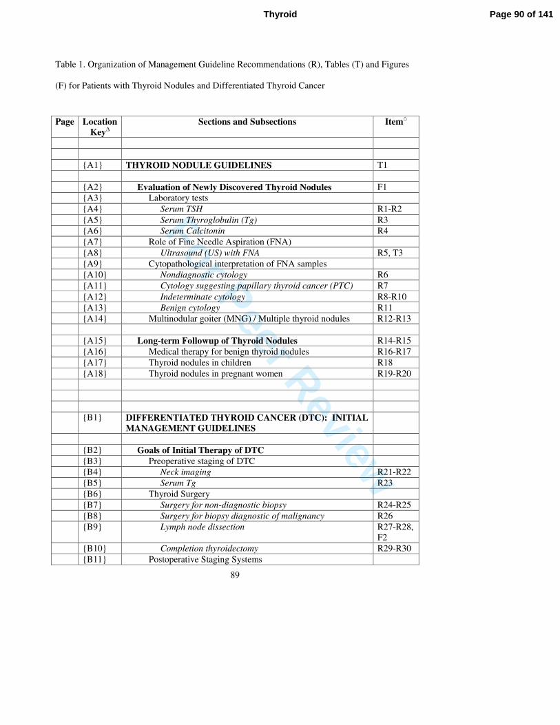
For Peer Review
89
Table 1. Organization of Management Guideline Recommendations (R), Tables (T) and Figures
(F) for Patients with Thyroid Nodules and Differentiated Thyroid Cancer
Page Location
Key∆
Sections and Subsections Item⌂
{A1} THYROID NODULE GUIDELINES T1
{A2} Evaluation of Newly Discovered Thyroid Nodules F1
{A3} Laboratory tests
{A4} Serum TSH R1-R2
{A5} Serum Thyroglobulin (Tg) R3
{A6} Serum Calcitonin R4
{A7} Role of Fine Needle Aspiration (FNA)
{A8} Ultrasound (US) with FNA R5, T3
{A9} Cytopathological interpretation of FNA samples
{A10} Nondiagnostic cytology R6
{A11} Cytology suggesting papillary thyroid cancer (PTC) R7
{A12} Indeterminate cytology R8-R10
{A13} Benign cytology R11
{A14} Multinodular goiter (MNG) / Multiple thyroid nodules R12-R13
{A15} Long-term Followup of Thyroid Nodules R14-R15
{A16} Medical therapy for benign thyroid nodules R16-R17
{A17} Thyroid nodules in children R18
{A18} Thyroid nodules in pregnant women R19-R20
{B1} DIFFERENTIATED THYROID CANCER (DTC): INITIAL
MANAGEMENT GUIDELINES
{B2} Goals of Initial Therapy of DTC
{B3} Preoperative staging of DTC
{B4} Neck imaging R21-R22
{B5} Serum Tg R23
{B6} Thyroid Surgery
{B7} Surgery for non-diagnostic biopsy R24-R25
{B8} Surgery for biopsy diagnostic of malignancy R26
{B9} Lymph node dissection R27-R28,
F2
{B10} Completion thyroidectomy R29-R30
{B11} Postoperative Staging Systems
Page 90 of 141Thyroid
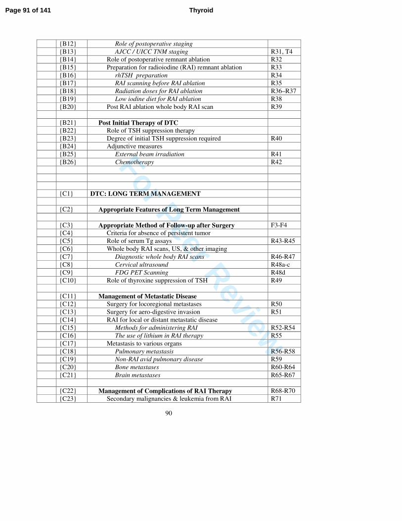
For Peer Review
90
{B12} Role of postoperative staging
{B13} AJCC / UICC TNM staging R31, T4
{B14} Role of postoperative remnant ablation R32
{B15} Preparation for radioiodine (RAI) remnant ablation R33
{B16} rhTSH preparation R34
{B17} RAI scanning before RAI ablation R35
{B18} Radiation doses for RAI ablation R36–R37
{B19} Low iodine diet for RAI ablation R38
{B20} Post RAI ablation whole body RAI scan R39
{B21} Post Initial Therapy of DTC
{B22} Role of TSH suppression therapy
{B23} Degree of initial TSH suppression required R40
{B24} Adjunctive measures
{B25} External beam irradiation R41
{B26} Chemotherapy R42
{C1} DTC: LONG TERM MANAGEMENT
{C2} Appropriate Features of Long Term Management
{C3} Appropriate Method of Follow-up after Surgery F3-F4
{C4} Criteria for absence of persistent tumor
{C5} Role of serum Tg assays R43-R45
{C6} Whole body RAI scans, US, & other imaging
{C7} Diagnostic whole body RAI scans R46-R47
{C8} Cervical ultrasound R48a-c
{C9} FDG PET Scanning R48d
{C10} Role of thyroxine suppression of TSH R49
{C11} Management of Metastatic Disease
{C12} Surgery for locoregional metastases R50
{C13} Surgery for aero-digestive invasion R51
{C14} RAI for local or distant metastatic disease
{C15} Methods for administering RAI R52-R54
{C16} The use of lithium in RAI therapy R55
{C17} Metastasis to various organs
{C18} Pulmonary metastasis R56-R58
{C19} Non-RAI avid pulmonary disease R59
{C20} Bone metastases R60-R64
{C21} Brain metastases R65-R67
{C22} Management of Complications of RAI Therapy R68-R70
{C23} Secondary malignancies & leukemia from RAI R71
Page 91 of 141 Thyroid
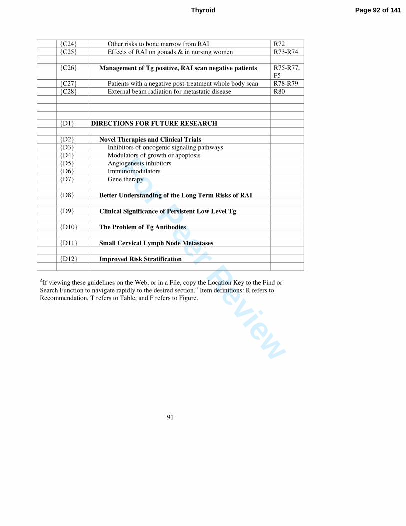
For Peer Review
91
{C24} Other risks to bone marrow from RAI R72
{C25} Effects of RAI on gonads & in nursing women R73-R74
{C26} Management of Tg positive, RAI scan negative patients R75-R77,
F5
{C27} Patients with a negative post-treatment whole body scan R78-R79
{C28} External beam radiation for metastatic disease R80
{D1} DIRECTIONS FOR FUTURE RESEARCH
{D2} Novel Therapies and Clinical Trials
{D3} Inhibitors of oncogenic signaling pathways
{D4} Modulators of growth or apoptosis
{D5} Angiogenesis inhibitors
{D6} Immunomodulators
{D7} Gene therapy
{D8} Better Understanding of the Long Term Risks of RAI
{D9} Clinical Significance of Persistent Low Level Tg
{D10} The Problem of Tg Antibodies
{D11} Small Cervical Lymph Node Metastases
{D12} Improved Risk Stratification
∆If viewing these guidelines on the Web, or in a File, copy the Location Key to the Find or
Search Function to navigate rapidly to the desired section.⌂ Item definitions: R refers to
Recommendation, T refers to Table, and F refers to Figure.
Page 92 of 141Thyroid
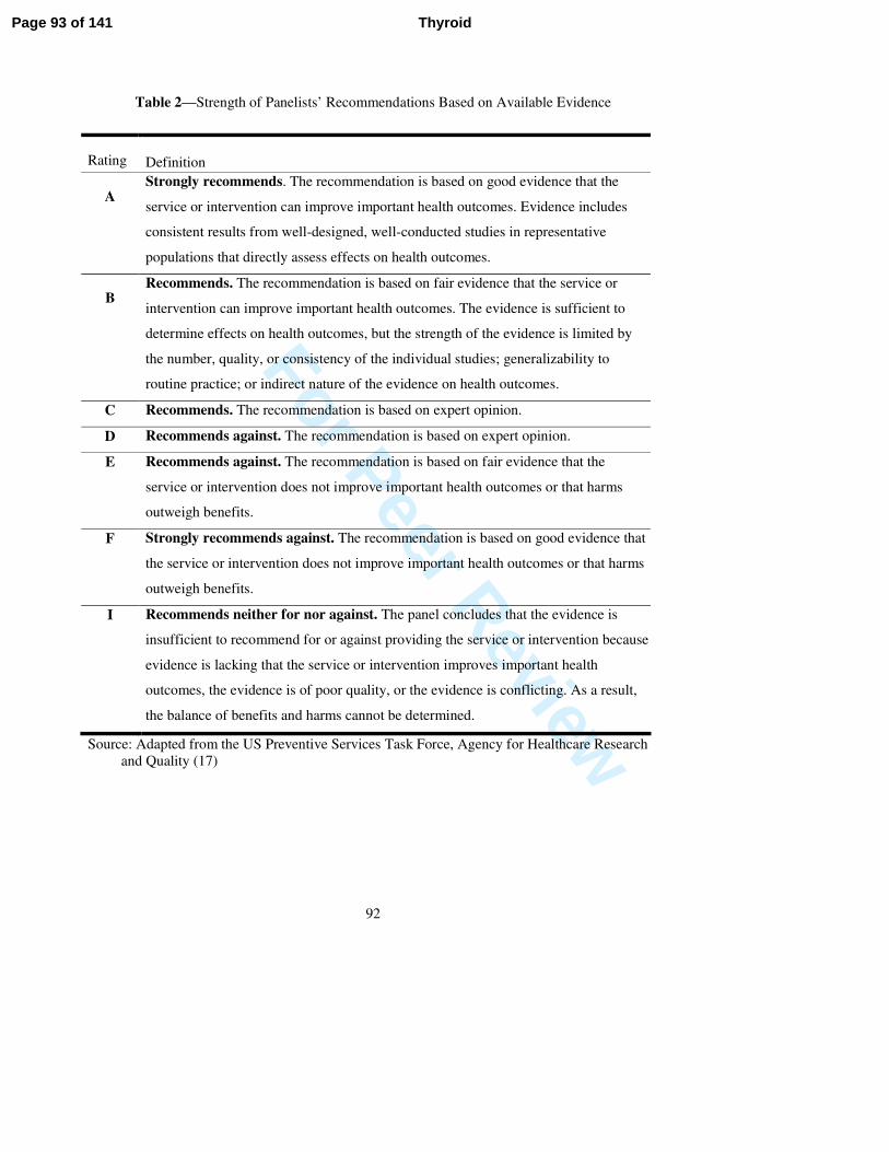
For Peer Review
92
Table 2—Strength of Panelists’ Recommendations Based on Available Evidence
Rating
Definition
A Strongly recommends. The recommendation is based on good evidence that the
service or intervention can improve important health outcomes. Evidence includes
consistent results from well-designed, well-conducted studies in representative
populations that directly assess effects on health outcomes.
B Recommends. The recommendation is based on fair evidence that the service or
intervention can improve important health outcomes. The evidence is sufficient to
determine effects on health outcomes, but the strength of the evidence is limited by
the number, quality, or consistency of the individual studies; generalizability to
routine practice; or indirect nature of the evidence on health outcomes.
C Recommends. The recommendation is based on expert opinion.
D Recommends against. The recommendation is based on expert opinion.
E Recommends against. The recommendation is based on fair evidence that the
service or intervention does not improve important health outcomes or that harms
outweigh benefits.
F Strongly recommends against. The recommendation is based on good evidence that
the service or intervention does not improve important health outcomes or that harms
outweigh benefits.
I Recommends neither for nor against. The panel concludes that the evidence is
insufficient to recommend for or against providing the service or intervention because
evidence is lacking that the service or intervention improves important health
outcomes, the evidence is of poor quality, or the evidence is conflicting. As a result,
the balance of benefits and harms cannot be determined.
Source: Adapted from the US Preventive Services Task Force, Agency for Healthcare Research
and Quality (17)
Page 93 of 141 Thyroid
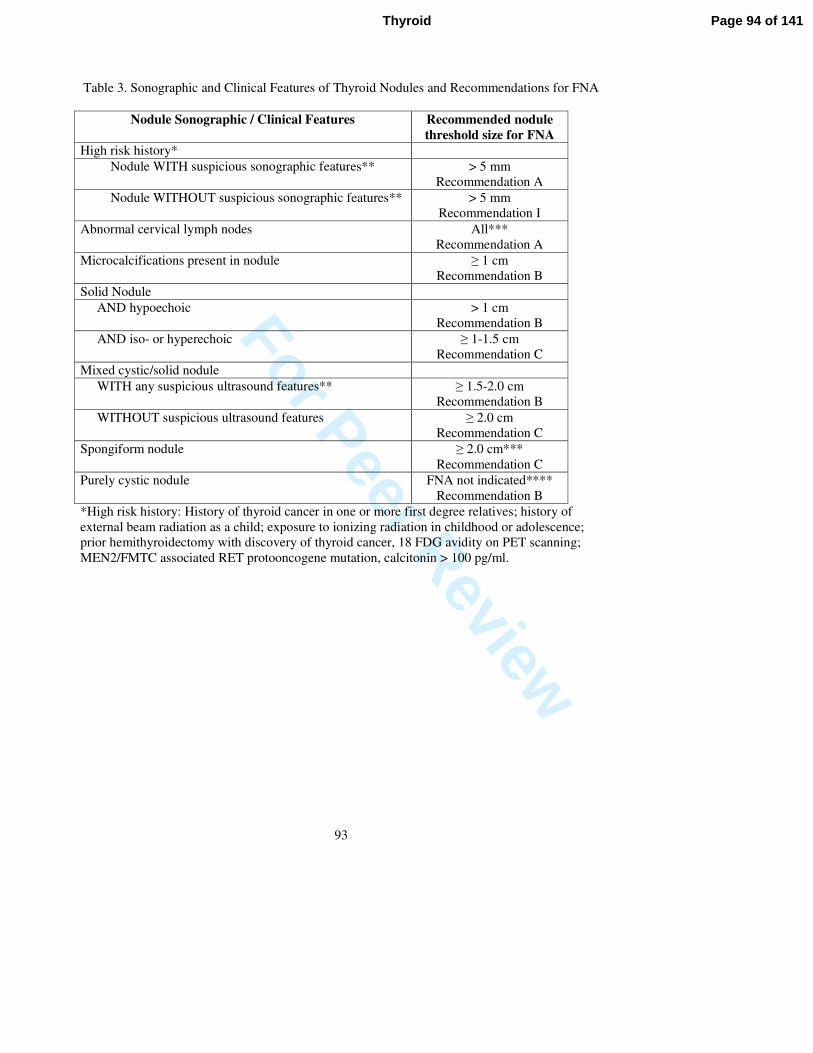
For Peer Review
93
Table 3. Sonographic and Clinical Features of Thyroid Nodules and Recommendations for FNA
Nodule Sonographic / Clinical Features Recommended nodule
threshold size for FNA
High risk history*
Nodule WITH suspicious sonographic features** > 5 mm
Recommendation A
Nodule WITHOUT suspicious sonographic features** > 5 mm
Recommendation I
Abnormal cervical lymph nodes All***
Recommendation A
Microcalcifications present in nodule ≥ 1 cm
Recommendation B
Solid Nodule
AND hypoechoic > 1 cm
Recommendation B
AND iso- or hyperechoic ≥ 1-1.5 cm
Recommendation C
Mixed cystic/solid nodule
WITH any suspicious ultrasound features** ≥ 1.5-2.0 cm
Recommendation B
WITHOUT suspicious ultrasound features ≥ 2.0 cm
Recommendation C
Spongiform nodule ≥ 2.0 cm***
Recommendation C
Purely cystic nodule FNA not indicated****
Recommendation B
*High risk history: History of thyroid cancer in one or more first degree relatives; history of
external beam radiation as a child; exposure to ionizing radiation in childhood or adolescence;
prior hemithyroidectomy with discovery of thyroid cancer, 18 FDG avidity on PET scanning;
MEN2/FMTC associated RET protooncogene mutation, calcitonin > 100 pg/ml.
Page 94 of 141Thyroid
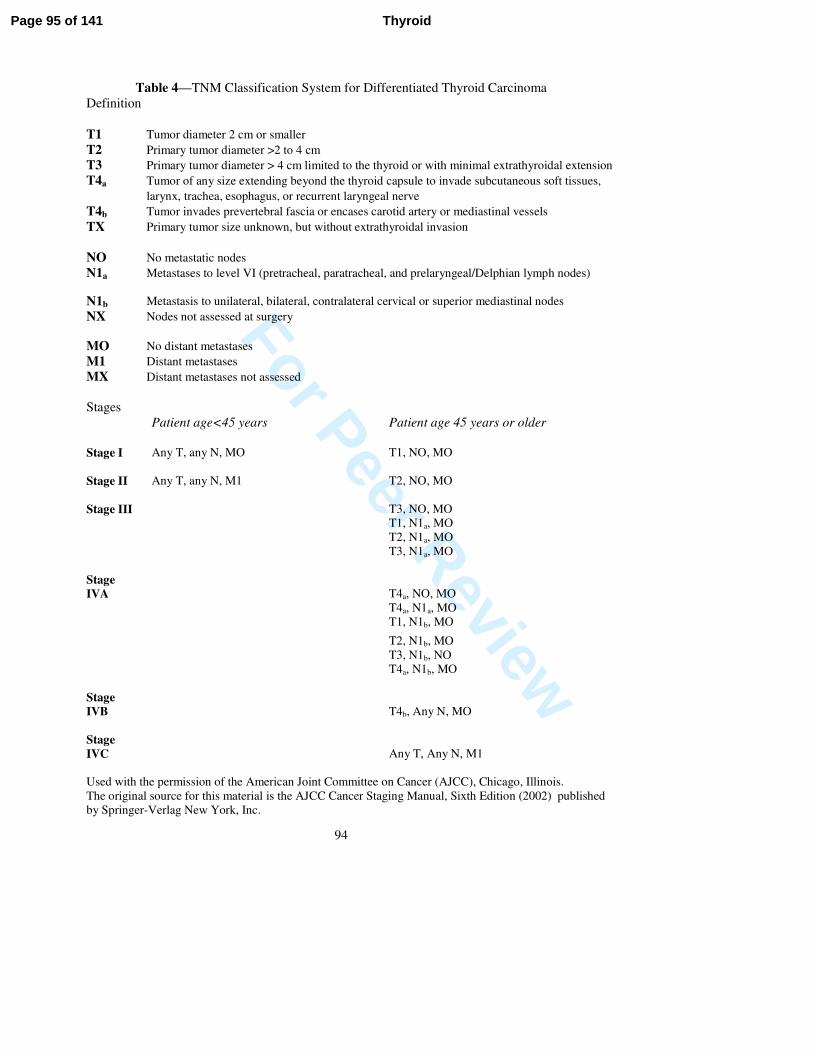
For Peer Review
94
Table 4—TNM Classification System for Differentiated Thyroid Carcinoma
Definition
T1 Tumor diameter 2 cm or smaller
T2 Primary tumor diameter >2 to 4 cm
T3 Primary tumor diameter > 4 cm limited to the thyroid or with minimal extrathyroidal extension
T4a Tumor of any size extending beyond the thyroid capsule to invade subcutaneous soft tissues,
larynx, trachea, esophagus, or recurrent laryngeal nerve
T4b Tumor invades prevertebral fascia or encases carotid artery or mediastinal vessels
TX Primary tumor size unknown, but without extrathyroidal invasion
NO No metastatic nodes
N1a Metastases to level VI (pretracheal, paratracheal, and prelaryngeal/Delphian lymph nodes)
N1b
Metastasis to unilateral, bilateral, contralateral cervical or superior mediastinal nodes
NX Nodes not assessed at surgery
MO No distant metastases
M1 Distant metastases
MX Distant metastases not assessed
Stages
Patient age<45 years Patient age 45 years or older
Stage I Any T, any N, MO T1, NO, MO
Stage II Any T, any N, M1 T2, NO, MO
Stage III T3, NO, MO
T1, N1a, MO
T2, N1a, MO
T3, N1a, MO
Stage
IVA T4a, NO, MO
T4a, N1a, MO
T1, N1b, MO
T2, N1b, MO
T3, N1b, NO
T4a, N1b, MO
Stage
IVB T4b, Any N, MO
Stage
IVC Any T, Any N, M1
Used with the permission of the American Joint Committee on Cancer (AJCC), Chicago, Illinois.
The original source for this material is the AJCC Cancer Staging Manual, Sixth Edition (2002) published
by Springer-Verlag New York, Inc.
Page 95 of 141 Thyroid
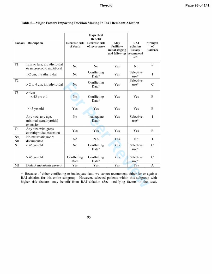
For Peer Review
95
Table 5—Major Factors Impacting Decision Making In RAI Remnant Ablation
Expected
Benefit
Factors Description Decrease risk
of death
Decrease risk
of recurrence
May
facilitate
initial staging
and follow up
RAI
ablation
usually
recommend
-ed
Strength
of
Evidence
T1 1cm or less, intrathyroidal
or microscopic multifocal No No Yes No
E
1-2 cm, intrathyroidal No
Conflicting
Data* Yes
Selective
use* I
T2
> 2 to 4 cm, intrathyroidal No Conflicting
Data* Yes
Selective
use*
C
T3 > 4cm
< 45 yrs old
≥ 45 yrs old
Any size, any age,
minimal extrathyroidal
extension
No
Yes
No
Conflicting
Data*
Yes
Inadequate
Data*
Yes
Yes
Yes
Yes
Yes
Selective
use*
B
B
I
T4 Any size with gross
extrathyroidal extension Yes Yes Yes Yes B
Nx,
N0
No metastatic nodes
documented No N o Yes No I
N1
< 45 yrs old
> 45 yrs old
No
Conflicting
Data
Conflicting
Data*
Conflicting
Data*
Yes
Yes
Selective
use*
Selective
use*
C
C
M1 Distant metastasis present Yes Yes Yes Yes A
* Because of either conflicting or inadequate data, we cannot recommend either for or against
RAI ablation for this entire subgroup. However, selected patients within this subgroup with
higher risk features may benefit from RAI ablation (See modifying factors in the text).
Page 96 of 141Thyroid
For Peer Review
96
Legends to Figures
Figure 1—Algorithm for the evaluation of patients with one or more thyroid nodules.
a If the scan does not show uniform distribution of tracer activity, ultrasound may be considered
to assess for the presence of a cystic component
bHypfnc is hyperfuntioning nodule
Figure 2—Lymph node compartments separated into levels and sublevels. Level VI contains
the thyroid gland, and the adjacent nodes bordered superiorly by the hyoid bone, inferiorly by the
innominate (brachiocephalic) artery, and laterally on each side by the carotid sheaths. The level
II, III and IV nodes are arrayed along the jugular veins on each side, bordered anteromedially by
level VI and laterally by the posterior border of the sternocleidomastoid muscle. The level III
nodes are bounded superiorly by the level of the hyoid bone, and inferiorly by the cricoid
cartilage; levels II and IV are above and below level III, respectively. The level I node
compartment includes the submental and submandibular nodes, above the hyoid bone, and
anterior to the posterior edge of the submandibular gland. Finally, the level V nodes are in the
posterior triangle, lateral to the lateral edge of the sternocleidomastoid muscle. Levels I, II and
V can be further subdivided as noted in the figure. The inferior extent of level VI is defined as
the suprasternal notch. Many authors also include the pretracheal and paratracheal superior
mediastinal lymph nodes above the level of the innominate artery (sometimes referred to as level
VII) in central neck dissection. (166).
Page 97 of 141 Thyroid
For Peer Review
97
Figure 3—Algorithm for initial follow-up of patients with differentiated thyroid carcinoma.
a EBRT is external beam radiotherapy. The usual indication for EBRT is macroscopic
unresectable tumor in a patient older than 45 years; it is not usually recommended for children
and adults less than age 45.
b Neck ultrasonography of operated cervical compartments is often compromised for several
months after surgery.
c Tg is thyroglobulin with anti-thyroglobulin antibody measurement; serum Tg is usually
measured by immunometric assay and may be falsely elevated for several weeks by injury from
surgery or by heterophile antibodies, although a very high serum Tg level after surgery usually
indicates residual disease.
d Some clinicians suspect residual disease when malignant lymph nodes, or tumors with
aggressive histologies (as defined in the text) have been resected, or when there is a
microscopically positive margin of resection.
e rhTSH is recombinant human TSH and is administered as follows: 0.9 mg rhTSH I.M. on two
consecutive days, followed by 131
I therapy on 3rd
day.
f THW is levothyroxine and/or triiodothyronine withdrawal.
g See text for exceptions regarding remnant ablation. The smallest amount of 131I necessary to
ablate normal thyroid remnant tissue should be used. DxWBS (diagnostic whole body
scintigraphy) is not usually necessary at this point, but may be performed if the outcome will
change the decision to treat with radioiodine and/or the amount of administered activity.
h RxWBS is posttreatment whole body scan done 5 to 8 days after therapeutic
131I administration.
iUptake in the thyroid bed may indicate normal remnant tissue or residual central neck nodal
metastases.
Page 98 of 141Thyroid
For Peer Review
98
Figure 4—Longer term follow-up of patients with differentiated thyroid carcinoma
j TgAb is anti-thyroglobulin antibody usually measured by immunometric assay
k Heterophile antibodies may be a cause of falsely elevated serum Tg levels. (Preissner CM,
Dodge LA, O'Kane DJ, Singh RJ, Grebe SK. Prevalence of heterophilic antibody interference in
eight automated tumor marker immunoassays. Clin Chem. 2005;51:208-10. Preissner CM,
O'Kane DJ, Singh RJ, Morris JC, Grebe SK. Phantoms in the assay tube: heterophile antibody
interferences in serum thyroglobulin assays. J Clin Endocrinol Metab. 2003 ;88:3069-74.) The
use of heterophile blocking tubes or heterophile blocking reagents have reduced, but not
completely eliminated this problem. Tg that rises with TSH stimulation and falls with TSH
suppression is unlikely to result from heterophile antibodies.
l See text concerning further information regarding levels of Tg at which therapy should be
considered.
m Tg radioimmunoassay (RIA) may be falsely elevated or suppressed by TgAb. Tg results
following TSH-stimulation with rhTSH or thyroid hormone withdrawal are invalidated by TgAb
in the serum even when Tg is measured by most RIA tests. TgAb levels often decline to
undetectable levels over years following surgery (Chiovato L, Latrofa F, Braverman LE, Pacini
F, Capezzone M, Masserini L, Grasso L, Pinchera A. Disappearance of humoral thyroid
autoimmunity after complete removal of thyroid antigens. Ann Intern Med. 2003;139:346-51).
A rising level of TgAb may be an early indication of recurrent disease (Spencer CA, Takeuchi
M, Kazarosyan M, Wang CC, Guttler RB, Singer PA, Fatemi S, LoPresti JS, Nicoloff JT. Serum
thyroglobulin autoantibodies: prevalence, influence on serum thyroglobulin measurement, and
prognostic significance in patients with differentiated thyroid carcinoma. J Clin Endocrinol
Metab. 1998;83:1121-7).
Page 99 of 141 Thyroid
For Peer Review
99
nSee text for decision regarding surgery vs. medical therapy, and surgical approaches to
locoregional metastases. Fine-needle aspiration confirmation of malignancy is generally advised.
Pre-operative chest CT is recommended as distant metastases may change management.
Figure 5—Considerations for empiric treatment with radioiodine
oEmpiric
131I therapy should be done with meticulous patient preparation, including low iodine
diet and, if iodine contamination is a possibility, urinary iodine measurements. If the RxWBS is
negative or subsequent follow-up studies show no therapeutic benefit, further empiric 131
I should
not be administered.
pTg that rises with TSH stimulation and falls with TSH suppression is unlikely to result from
heterophile antibodies
q National Cancer Institute Common Terminology Criteria for Adverse Events, Version 3.0,
(http://ctep.cancer.gov)
r dosimetry could be considered to allow administration of maximum radioiodine activity if the
tumor is life-threatening
s a dose of 200 mCi could exceed the maximum tolerable dose in older individuals (see R52b)
Page 100 of 141Thyroid
For Peer Review
100
References
1 Tunbridge WMG, Evered DC, Hall R, Appleton D, Brewis M, Clark F, Evans JG, Young E,
Bird T, Smith PA 1977 The spectrum of thyroid disease in a community: the Whickham Survey.
Clin Endocrinol (Oxf).7:481-493.
2 Vander JB, Gaston EA, Dawber TR 1968 The significance of nontoxic thyroid nodules. Ann
Intern Med.69:537-540.
3 Tan GH, Gharib H 1997 Thyroid incidentalomas: management approaches to nonpalpable
nodules discovered incidentally on thyroid imaging. Ann Intern Med.126:226-231.
4 Hegedus L 2004 Clinical practice. The thyroid nodule. N Engl J Med.351:1764-1771.
5 Mandel SJ 2004 A 64-year-old woman with a thyroid nodule. JAMA.292:2632-2642.
6 Sherman SI 2003 Thyroid carcinoma. Lancet.361:501-511.
7 Jemal A, Siegel R, Ward E, Hao Y, Xu J, Thun MJ 2009 Cancer statistics, 2009. CA Cancer J
Clin. Published online before print May 27, 2009
8 Davies L, Welch HG 2006 Increasing incidence of thyroid cancer in the United Stats, 1973-
2002. JAMA.295:2164-2167.
9 Leenhardt L, Bernier MO, Boin-Pineau MH, Conte DB, Marechaud R, Niccoli-Sire P,
Nocaudie M, Orgiazzi J, Schlumberger M, Wémeau JL, Chérie-Challine L, De Vathaire F 2004
Advances in diagnostic practices affect thyroid cancer incidence in France. Eur J
Endocrinol.150:133-139.
10 Singer PA, Cooper DS, Daniels GH, Ladenson PW, Greenspan FS, Levy EG, Braverman LE,
Clark OH, McDougall IR, Ain KV, Dorfman SG 1996 Treatment guidelines for patients with
thyroid nodules and well-differentiated thyroid cancer. American Thyroid Association. Arch
Intern Med.156:2165-2172.
11 AACE/AAES medical/surgical guidelines for clinical practice: management of thyroid
carcinoma. 2001 American Association of Clinical Endocrinologists. Endocr Pract.7:202-220.
12 British Thyroid Association and Royal College of Physicians. 2007 Guidelines for the
management of thyroid cancer, 2nd
Edition. www.british-thyroid-association.org.
13 Thyroid carcinoma. National Comprehensive Cancer Network.
www.nccn.org/professionals/physician_gls/PDF/thyroid.pdf. Accessed 1-28-09
Page 101 of 141 Thyroid
For Peer Review
101
14 Pacini F, Schlumberger M, Dralle H, Elisei R, Smit JW, Wiersinga W 2006 European
consensus for the management of patients with differentiated thyroid carcinoma of the follicular
epithelium. Eur J Endocrinol.154:787-803.
15 Luster M, Clarke SE, Dietlein M, Lassmann M, Lind P, Oyen WJ, Tennvall J, Bombardieri E
2008 Guidelines for radioiodine therapy of differentiated thyroid cancer. Eur J Nucl Med Mol
Imaging.35:1941-1959.
16 Cooper DS, Doherty GM, Haugen BR, Kloos RT, Lee SL, Mandel SJ, Mazzaferri EL, McIver
B, Sherman SI, Tuttle RM 2006 The American Thyroid Association Guidelines Taskforce.
Management guidelines for patients with thyroid nodules and differentiated thyroid cancer.
Thyroid.16:109-142.
17 U.S. Preventive Services Task Force Ratings: Strength of Recommendations and Quality of
Evidence. Guide to Clinical Preventive Services, Third Edition: Periodic Updates, 2000-2003.
Agency for Healthcare Research and Quality, Rockville, MD.
18 Marqusee E, Benson CB, Frates MC, Doubilet PM, Larsen PR, Cibas ES, Mandel SJ 2000
Usefulness of ultrasonography in the management of nodular thyroid disease. Ann Intern
Med.1339:696-700.
19 Hagag P, Strauss S, Weiss M 1998 Role of ultrasound-guided fine-needle aspiration biopsy in
evaluation of nonpalpable thyroid nodules. Thyroid.8:989-995.
20 Are C, Hsu JF, Ghossein RA, Schoder H, Shah JP, Shaha AR 2007 Histological
aggressiveness of fluorodeoxyglucose positron-emission tomogram (FDG-PET)-detected
incidental thyroid carcinomas. Ann Surg Oncol.14:3210-3215.
21 Bogsrud TV, Karantanis D, Nathan MA, Mullan BP, Wiseman GA, Collins DA, Kasperbauer
JL, Strome SE, Reading CC, Hay ID, Lowe VJ 2007 The value of quantifying 18F-FDG uptake
in thyroid nodules found incidentally on whole-body PET-CT. Nucl Med Commun.28:373-381.
22 Kang KW, Kim SK, Kang HS, Lee ES, Sim JS, Lee IG, Jeong SY, Kim SW 2003 Prevalence
and risk of cancer of focal thyroid incidentaloma identified by 18F-fluorodeoxyglucose positron
emission tomography for metastasis evaluation and cancer screening in healthy subjects. J Clin
Endocrinol Metab.88:4100-4104.
23 Choi JY, Lee KS, Kim HJ, Shim YM, Kwon OJ, Park K, Baek CH, Chung JH, Lee KH, Kim
BT 2006 Focal thyroid lesions incidentally identified by integrated 18F-FDG PET/CT: clinical
significance and improved characterization. J Nucl Med.47:609-615.
24 Curtis RE, Rowlings PA, Deeg HJ, Shriner DA, Socié G, Travis LB, Horowitz MM,
Witherspoon RP, Hoover RN, Sobocinski KA, Fraumeni JF, Boice JD, Schoch HG, Sale GE,
Storb R, Travis WD, Kolb HJ, Gale RP, Passweg JR 1997 Solid Cancers after Bone Marrow
Transplantation. N Engl J Med.336:897-904.
Page 102 of 141Thyroid
For Peer Review
102
25 Pacini F, Vorontsova T, Demidchik E, Molinaro E, Agate, L, Romei C, Shavrova E,
Cherstvoy ED, Ivashkevitch Y, Kuchinskaya E, Schlumberger M, Ronga G, Filesi M, Pinchera
A 1997 Post-Chernobyl Thyroid Carcinoma in Belarus Children and Adolescents: Comparison
with Naturally Occurring Thyroid Carcinoma in Italy and France. J Clin Endocrinol
Metab.81:3563-3569.
26 Boelaert K, Horacek J, Holder RL, Watkinson JC, Sheppard MC, Franklyn JA 2006 Serum
thyrotropin concentration as a novel predictor of malignancy in thyroid nodules investigated by
fine-needle aspiration. J Clin Endo Metab.91:4295-4301.
27 Hall TL, Layfield LJ, Philippe A, Rosenthal D 1989 Sources of diagnostic error in fine needle
aspiration of the thyroid. Cancer.63:718-725.
28 Alexander EK, Heering JP, Benson CB, Frates MC, Doubilet PM, Cibas ES, Marqusee E
2002 Assessment of nondiagnostic ultrasound-guided fine needle aspiration of thyroid nodules. J
Clin Endocrinol Metab.87:4924-4927.
29 Brander A, Viikinkoski P, Tuuhea J, Voutilainen L, Kivisaari L 1992 Clinical versus
ultrasound examination of the thyroid gland in common clinical practice. J Clin
Ultrasound.20:37-42.
30 Tan GH, Gharib H, Reading CC 1995 Solitary thyroid nodule. Arch Intern Med.155:2418-
2423.
31 Singh B, Shaha AR, Trivedi H, Carew JF, Poluri A, Shah JP 1999 Coexistent Hashimoto’s
thyroiditis with papillary thyroid carcinoma: impact on presentation, management, and outcome.
Surgery.126:1070–1077.
32 Repplinger D, Bargren A, Zhang YW, Adler JT, Haymart M, Chen H 2008 Is Hashimoto's
thyroiditis a risk factor for papillary thyroid cancer? J Surg Res.150:49-52.
33 Pacini F, Pinchera A, Giani C, Grasso L, Doveri F, Baschieri L 1980. Serum thyroglobulin in
thyroid carcinoma and other thyroid disorders. J Endocrinol Invest.3:283-292.
34 Elisei R, Bottici V, Luchetti F, Di Coscio G, Romei C, Grasso L, Miccoli P, Iacconi P, Basolo
F, Pinchera A, Pacini F 2004 Impact of routine measurement of serum calcitonin on the
diagnosis and outcome of medullary thyroid cancer: experience in 10,864 patients with nodular
thyroid disorders. J Clin Endocrinol Metab.89:163-168.
35 Hahm JR, Lee MS, Min YK, Lee MK, Kim KW, Nam SJ, Yang JH, Chung JH 2001 Routine
measurement of serum calcitonin is useful for early detection of medullary thyroid carcinoma in
patients with nodular thyroid diseases. Thyroid.11:73-80.
Page 103 of 141 Thyroid
For Peer Review
103
36 Niccoli P, Wion-Barbot N, Caron P, Henry JF, de Micco C, Saint Andre JP, Bigorgne JC,
Modigliani E, Conte-Devolx B 1997 Interest of routine measurement of serum calcitonin: study
in a large series of thyroidectomized patients. The French Medullary Study Group. J Clin
Endocrinol Metab.82:338-341.
37 Costante G, Meringolo D, Durante C, Bianchi D, Nocera M, Tumino S Crocetti U, Attard M,
Maranghi M, Torlontano M, Filetti S 2007 Predictive value of serum calcitonin levels for
preoperative diagnosis of medullary thyroid carcinoma in a cohort of 5817 consecutive patients
with thyroid nodules. J Clin Endocrinol Metab.92:450-455.
38 Cheung K, Roman SA, Wang TS, Walker HD, Sosa JA 2008 Calcitonin measurement in the
evaluation of thyroid nodules in the United States: a cost-effectiveness and decision analysis. J
Clin Endocrinol Metab.93:2173-2180.
39 Gagel RF, Hoff AO, Cote GJ 2005 Medullary thyroid carcinoma. In Werner and Ingbar’s
The Thyroid. Lippincott Williams and Wilkins, Philadelphia.967-988.
40 Danese D, Sciacchitano S, Farsetti A, Andreoli M, Pontecorvi A 1998 Diagnostic accuracy of
conventional versus sonography-guided fine-needle aspiration biopsy of thyroid nodules.
Thyroid.8:15-21.
41 Carmeci C, Jeffrey RB, McDougall IR, Nowels KW, Weigel RJ 1998 Ultrasound-guided
fine-needle aspiration biopsy of thyroid masses. Thyroid.8:283-289.
42 Baloch ZW, LiVolsi VA, Asa SL, Rosai J, Merino MJ, Randolph G, Vielh P, DeMay RM,
Sidawy MK, Frable WJ 2008 Diagnostic terminology and morphologic criteria for cytologic
diagnosis of thyroid lesions: a synopsis of the National Cancer Institute Thyroid Fine-Needle
Aspiration State of the Science Conference. Diagn Cytopathol.36:425-437.
43Leenhardt L, Hejblum G, Franc B, Fediaevsky LD, Delbot T, Le Guillouzic D, Ménégaux F,
Guillausseau C, Hoang C, Turpin G, Aurengo A 1999 Indications and limits of ultrasound-
guided cytology in the management of nonpalpable thyroid nodules. J Clin Endocrinol
Metab.84:24-28.
44 Papini E, Guglielmi R, Bianchini A, Crescenzi A, Taccogna S, Nardi F, Panunzi C, Rinaldi R,
Toscano V, Pacella CM 2002 Risk of malignancy in nonpalpable thyroid nodules: predictive
value of ultrasound and color-Doppler features. J Clin Endocrinol Metab.87:1941-1946.
45 Nam-Goong IS, Kim HY, Gong G, Lee HK, Hong SJ, Kim WB, Shong YK 2004
Ultrasonography-guided fine-needle aspiration of thyroid incidentaloma: correlation with
pathological findings. Clin Endocrinol (Oxf).60:21-28.
46 Cappelli C, Castellano M, Pirola I, Cumetti D, Agosti B, Gandossi E, Agabiti Rosei E 2007
The predictive value of ultrasound findings in the management of thyroid nodules. QJM.100:29-
35.
Page 104 of 141Thyroid
For Peer Review
104
47 Frates MC, Benson CB, Doubilet PM, Kunreuther E, Contreras M, Cibas ES, Orcutt J, Moore
FD Jr, Larsen PR, Marqusee E, Alexander EK 2006 Prevalence and distribution of carcinoma in
patients with solitary and multiple thyroid nodules on sonography. J Clin Endocrinol
Metab.91:3411-3417.
48 Moon WJ, Jung SL, Lee JH, Na DG, Baek JH, Lee YH, Kim J, Kim HS, Byun JS, Lee DH;
Thyroid Study Group, Korean Society of Neuro- and Head and Neck Radiology 2008 Benign
and malignant thyroid nodules: US differentiation--multicenter retrospective study.
Radiology.247:762-770.
49 Jeh SK, Jung SL, Kim BS, Lee YS 2007 Evaluating the degree of conformity of papillary
carcinoma and follicular carcinoma to the reported ultrasonographic findings of malignant
thyroid tumor. Korean J Radiol.8:192-197.
50 Machens A, Holzhausen HJ, Dralle H 2005 The prognostic value of primary tumor size in
papillary and follicular thyroid carcinoma. Cancer.103:2269-2273.
51 Moon WJ, Kwag HJ, Na DG 2009 Are There Any Specific Ultrasound Findings of Nodular
Hyperplasia (“Leave Me Alone” Lesion) to Differentiate It from Follicular Adenoma? Acta
Radiologica.50:383-388.
52 Bonavita JA, Mayo J, Babb J, Bennett G, Oweity T, Macari M, Yee J 2009 Pattern
recognition of benign nodules at ultrasound of the thyroid: which nodules can be left alone? AJR
Am J Roentgenol.193:207-213.
53 Rago T, Santini F, Scutari M, Pinchera A, Vitti P 2007 Elastography: new developments in
ultrasound for predicting malignancy in thyroid nodules. J Clin Endocrinol Metab.92:2917-2922.
54 Noguchi S, Yamashita H, Uchino S, Watanabe S 2008 Papillary microcarcinoma.World J
Surg. 32:747-753.
55 Wada N, Duh QY, Sugino K, Iwasaki H, Kameyama K, Mimura T, Ito K, Takami H,
Takanashi Y 2003 Lymph node metastasis from 259 papillary thyroid microcarcinomas:
frequency, pattern of occurrence and recurrence, and optimal strategy for neck dissection. Ann
Surg.237:399-407.
56 Ito Y, Tomoda C, Uruno T, Takamura Y, Miya A, Kobayashi K, Matsuzuka F, Kuma K,
Miyauchi A 2004 Preoperative ultrasonographic examination for lymph node metastasis:
usefulness when designing lymph node dissection for papillary microcarcinoma of the thyroid.
World J Surg.28:498-501.
57 Hemminki K, Eng C, Chen B 2005 Familial risks for nonmedullary thyroid cancer. J Clin
Endocrinol Metab.90:5747-5753.
58 Schneider AB, Ron E, Lubin J, Stovall M, Gierlowski TC 1993 Dose-response relationships
for radiation-induced thyroid cancer and thyroid nodules: evidence for the prolonged effects of
radiation on the thyroid. J Clin Endocrinol Metab.77:362-369.
Page 105 of 141 Thyroid
For Peer Review
105
59 Shibata Y, Yamashita S, Masyakin VB, Panasyuk GD, Nagataki S 2001 15 years after
Chernobyl: new evidence of thyroid cancer. Lancet.358:1965-1966.
60 Braga M, Cavalcanti TC, Collaco LM, Graf H 2001 Efficacy of ultrasound-guided fine-needle
aspiration biopsy in the diagnosis of complex thyroid nodules. J Clin Endocrinol Metab.86:4089-
4091.
61 Redman R, Zalaznick H, Mazzaferri EL, Massoll NA 2006 The impact of assessing specimen
adequacy and number of needle passes for fine-needle aspiration biopsy of thyroid nodules.
Thyroid.16:55-60.
62 Baloch ZW, Tam D, Langer J, Mandel S, LiVolsi VA, Gupta PK 2000 Ultrasound-guided
fine-needle aspiration biopsy of the thyroid: role of on-site assessment and multiple cytologic
preparations. Diagn Cytopathol.23:425-429.
63 de los Santos ET, Keyhani-Rofagha S, Cunningham JJ, Mazzaferri EL 1990 Cystic thyroid
nodules. The dilemma of malignant lesions. Arch Intern Med.150:1422-1427.
64 Yeh MW, Demircan O, Ituarte P, Clark OH 2004 False-negative fine-needle aspiration
cytology results delay treatment and adversely affect outcome in patients with thyroid
carcinoma. Thyroid.14:207-215.
65 Gharib H, Goellner JR, Johnson DA 1993 Fine-needle aspiration cytology of the thyroid. A
12-year experience with 11,000 biopsies. Clin Lab Med.13:699-709.
66 Tuttle RM, Lemar H, Burch HB 1998 Clinical features associated with an increased risk of
thyroid malignancy in patients with follicular neoplasia by fine-needle aspiration. Thyroid.8:377-
383.
67 Tyler DS, Winchester DJ, Caraway NP, Hickey RC, Evans DB 1994 Indeterminate fine-
needle aspiration biopsy of the thyroid: identification of subgroups at high risk for invasive
carcinoma. Surgery.116:1054-1060.
68 Kelman AS, Rathan A, Leibowitz J, Burstein DE, Haber RS 2001 Thyroid cytology and the
risk of malignancy in thyroid nodules: importance of nuclear atypia in indeterminate specimens.
Thyroid.11:271-277.
69 Bartolazzi A, Orlandi F, Saggiorato E, Volante M, Arecco F, Rossetto R, Palestini N, Ghigo
E, Papotti M, Bussolati G, Martegani MP, Pantellini F, Carpi A, Giovagnoli MR, Monti S,
Toscano V, Sciacchitano S, Pennelli GM, Mian C, Pelizzo MR, Rugge M, Troncone G,
Palombini L, Chiappetta G, Botti G, Vecchione A, Bellocco R; Italian Thyroid Cancer Study
Group (ITCSG) 2008 Galectin-3-expression analysis in the surgical selection of follicular
thyroid nodules with indeterminate fine-needle aspiration cytology: a prospective multicentre
study. Lancet Oncol.9:543-549.
Page 106 of 141Thyroid
For Peer Review
106
70 Segev DL, Clark DP, Zeiger MA, Umbricht C 2003 Beyond the suspicious thyroid fine
needle aspirate. A review. Acta Cytol.47:709-722.
71 Haugen BR, Woodmansee WW, McDermott MT 2002 Towards improving the utility of fine-
needle aspiration biopsy for the diagnosis of thyroid tumors. Clin Endo.56:281-290.
72 Sapio MR, Posca D, Raggioli A, Guerra A, Marotta V, Deandrea M, Motta M, Limone PP,
Troncone G, Caleo A, Rossi G, Fenzi G, Vitale M 2007 Detection of RET/PTC, TRK and BRAF
mutations in preoperative diagnosis of thyroid nodules with indeterminate cytological findings.
Clin Endocrinol (Oxf).66:678-683.
73 Nikiforov YE, Nikiforov YE, Steward DL, Robinson-Smith TM, Haugen BR, Klopper JP,
Zhu Z, Fagin JA, Falciglia M, Weber K, Nikiforova MN 2009 Molecular testing for mutations in
improving the fine-needle aspiration diagnosis of thyroid nodules. J Clin Endocrinol
Metab.94:2092-2098.
74 Franco C, Martínez V, Allamand JP, Medina F, Glasinovic A, Osorio M, Schachter D 2009
Molecular markers in thyroid fine-needle aspiration biopsy: a prospective study. Appl
Immunohistochem Mol Morphol.17:211-215.
75 Mitchell JC, Grant F, Evenson AR, Parker JA, Hasselgren PO, Parangi S 2005. Preoperative
evaluation of thyroid nodules with 18FDG-PET/CT. Surgery.138:1166-1174;discussion1174-
1175.
76 de Geus-Oei LF, Pieters GF, Bonenkamp JJ, Mudde AH, Bleeker-Rovers CP, Corstens FH,
Oyen WJ 2006 18F-FDG PET reduces unnecessary hemithyroidectomies for thyroid nodules
with inconclusive cytologic results. J Nucl Med.47:770-775.
77 Kim JM, Ryu JS, Kim TY, Kim WB, Kwon GY, Gong G, Moon DH, Kim SC, Hong SJ,
Shong YK 2007 18F-fluorodeoxyglucose positron emission tomography does not predict
malignancy in thyroid nodules cytologically diagnosed as follicular neoplasm. J Clin Endocrinol
Metab.92:1630-1634.
78 Sebastianes FM, Cerci JJ, Zanoni PH, Soares J Jr, Chibana LK, Tomimori EK, de Camargo
RY, Izaki M, Giorgi MC, Eluf-Neto J, Meneghetti JC, Pereira MA 2007 Role of 18F-
fluorodeoxyglucose positron emission tomography in preoperative assessment of cytologically
indeterminate thyroid nodules. J Clin Endocrinol Metab.92:4485-4488.
79 Hales NW, Krempl GA, Medina JE 2008 Is there a role for fluorodeoxyglucose positron
emission tomography/computed tomography in cytologically indeterminate thyroid nodules?
Am J Otolaryngol.29:113-118.
80 Ylagan LR, Farkas T, Dehner LP 2004 Fine needle aspiration of the thyroid: a cytohistologic
correlation and study of discrepant cases. Thyroid.14:35-41.
Page 107 of 141 Thyroid
For Peer Review
107
81 McCoy KL, Jabbour N, Ogilvie JB, Ohori NP, Carty SE, Yim JH 2007 The incidence of
cancer and rate of false-negative cytology in thyroid nodules greater than or equal to 4 cm in
size. Surgery.142:837-844.
82 Alexander EK, Hurwitz S, Heering JP, Benson CB, Frates MC, Doubilet PM, Cibas ES,
Larsen PR, Marqusee E 2003 Natural history of benign solid and cystic thyroid nodules. Ann Int
Med.138:315-318.
83 Asanuma K, Kobayashi S, Shingu K, Hama Y, Yokoyama S, Fujimori M, Amano J 2001 The
rate of tumour growth does not distinguish between malignant and benign thyroid nodules. Eur J
Surg.167:102-105.
84 Erdogan MF, Kamel N, Aras D, Akdogan A, Baskal N, Erdogan G 1998 Value of re-
aspirations in benign nodular thyroid disease. Thyroid.8:1087-1090.
85 Orlandi A, Puscar A, Capriata E, Fideleff H 2005 Repeated fine-needle aspiration of the
thyroid in benign nodular thyroid disease: critical evaluation of long-term follow-up.
Thyroid.15:274-278.
86 Papini E, Petrucci L, Guglielmi R, Panunzi C, Rinaldi R, Bacci V, Crescenzi A, Nardi F,
Fabbrini R, Pacella CM 1998 Long-term changes in nodular goiter: a 5-year prospective
randomized trial of levothyroxine suppressive therapy for benign cold thyroid nodules. J Clin
Endocrinol Metab.83:780-783.
87 Brauer VF, Eder P, Miehle K, Wiesner TD, Hasenclever H, Paschke R 2005 Interobserver
variation for ultrasound determination of thyroid nodule volumes. Thyroid.15:1169-1175.
88 Brander AE, Viikinkoski VP, Nickels JI, Kivisaari LM 2000 Importance of thyroid
abnormalities detected at US screening: a 5-year follow-up. Radiology.215:801-806.
89 Oertel YC, Miyahara-Felipe L, Mendoza MG, Yu K 2007 Value of repeated fine needle
aspirations of the thyroid: an analysis of over ten thousand FNAs. Thyroid.17:1061-1066.
90 Bennedbaek FN, Hegedüs L 2003 Treatment of recurrent thyroid cysts with ethanol: a
randomized double-blind controlled trial. J Clin Endocrinol Metab.88:5773-5777.
91 Valcavi R, Frasoldati A 2004 Ultrasound-guided percutaneous ethanol injection therapy in
thyroid cystic nodules. Endocr Pract.10:269-275.
92 Antonelli A, Campatelli A, Di Vito A, Alberti B, Baldi V, Salvioni G, Fallahi P, Baschieri L
1994 Comparison between ethanol sclerotherapy and emptying with injection of saline in
treatment of thyroid cysts. Clin Investig.72:971-974.
93 Verde G, Papini E, Pacella CM, Gallotti C, Delpiano S, Strada S, Fabbrini R, Bizzarri G,
Rinaldi R, Panunzi C, Geili D 1994 Ultrasound guided percutaneous ethanol injection in the
treatment of cystic thyroid nodules. Clin Endocrinol (Oxf).41:719-724.
Page 108 of 141Thyroid
For Peer Review
108
94 Zelmanovitz F, Genro S, Gross JL 1998 Suppressive therapy with levothyroxine for solitary
thyroid nodules: a double-blind controlled clinical study and cumulative meta-analyses. J Clin
Endocrinol Metab.83:3881-3885.
95 Wemeau JL, Caron P, Schvartz C, Schlienger JL, Orgiazzi J, Cousty C, Vlaeminck-Guillem
V 2002 Effects of thyroid-stimulating hormone suppression with levothyroxine in reducing the
volume of solitary thyroid nodules and improving extranodular nonpalpable changes: a
randomized, double-blind, placebo-controlled trial by the French Thyroid Research Group. J Clin
Endocrinol Metab.87:4928-4934.
96 Castro MR, Caraballo PJ, Morris JC 2002 Effectiveness of thyroid hormone suppressive
therapy in benign solitary thyroid nodules: a meta-analysis. J Clin Endocrinol Metab.87:4154-
4159.
97 Rallison ML, Dobyns BM, Keating FR Jr, Rall JE, Tyler FH 1975 Thyroid nodularity in
children. JAMA.233:1069-1072.
98 Raab SS, Silverman JF, Elsheikh TM, Thomas PA, Wakely PE 1995 Pediatric thyroid
nodules: disease demographics and clinical management as determined by fine needle aspiration
biopsy. Pediatrics.95:46-49.
99 Corrias A, Einaudi S, Chiorboli E, Weber G, Crino A, Andreo M, Cesaretti G, de Sanctis L,
Messina MF, Segni M, Cicchetti M, Vigone M, Pasquino AM, Spera S, de Luca F, Mussa GC,
Bona G 2001 Accuracy of fine needle aspiration biopsy of thyroid nodules in detecting
malignancy in childhood: comparison with conventional clinical, laboratory, and imaging
approaches. J Clin Endocrinol Metab.86:4644-4648.
100 Hung W 1999 Solitary thyroid nodules in 93 children and adolescents, a 35-years
experience. Horm Res.52:15-18.
101 Gharib H, Zimmerman D, Goellner JR, Bridley SM, LeBlanc SM 1995 Fine-needle
aspiration biopsy: Use in diagnosis and management of pediatric thyroid diseases. Endo
Pract.1:9-13.
102 Arda IS, Yildirim S, Demirhan B, Firat S 2001 Fine needle aspiration biopsy of thyroid
nodules. Arch Dis Child.85:313-317.
103 Tan GH, Gharib H, Goellner JR, van Heerden JA, Bahn RS 1996 Management of thyroid
nodules in pregnancy. Arch Intern Med.156:2317-2320.
104 Moosa M, Mazzaferri EL 1997 Outcome of differentiated thyroid cancer diagnosed in
pregnant women. J Clin Endocrinol Metab.82:2862-2866.
105 Mestman JH, Goodwin TM, Montoro MM 1995 Thyroid disorders of pregnancy. Endocrinol
Metab Clin North Am.24:41-71.
Page 109 of 141 Thyroid
For Peer Review
109
106 Herzon FS, Morris DM, Segal MN, Rauch G, Parnell T 1994 Coexistent thyroid cancer and
pregnancy. Arch Otolaryngol Head Neck Surg.120:1191-1193.
107 Mazzaferri EL, Jhiang SM 1994 Long-term impact of initial surgical and medical therapy on
papillary and follicular thyroid cancer. Am J Med.97:418-428.
108 Kuy S, Roman SA, Desai R, Sosa JA 2009 Outcomes following thyroid and parathyroid
surgery in pregnant women. Arch Surg.144:399-406.
109 Rosen IB, Korman M, Walfish PG 1997 Thyroid nodular disease in pregnancy: current
diagnosis and management. Clin Obstet Gynecol.40:81-89.
110 Hundahl SA, Fleming ID, Fremgen AM, Menck HR 1998 A National Cancer Data Base
report on 53,856 cases of thyroid carcinoma treated in the U.S., 1985-1995. Cancer.83:2638-
2648.
111 Volante M, Landolfi S, Chiusa L, Palestini N, Motta M, Codegone A, Torchio B, Papotti
MG 2004 Poorly differentiated carcinomas of the thyroid with trabecular, insular, and solid
patterns: a clinicopathologic study of 183 patients.Cancer.100:950-957.
112 van Heerden JA, Hay ID, Goellner JR, Salomao D, Ebersold JR, Bergstralh EJ, Grant CS
1992 Follicular thyroid carcinoma with capsular invasion alone: a nonthreatening malignancy.
Surgery. 112:1130-6.
113 Sanders LE, Silverman M 1998 Follicular and Hurthle cell carcinoma: predicting outcome
and directing therapy. Surgery.124:967-974.
114 Lo CY, Chan WF, Lam KY, Wan KY 2005 Follicular thyroid carcinoma: the role of
histology and staging systems in predicting survival. Ann Surg.242:708-715.
115 D'Avanzo A, Treseler P, Ituarte PH, Wong M, Streja L, Greenspan FS, Siperstein AE, Duh
QY, Clark OH 2004. Follicular thyroid carcinoma: histology and prognosis. Cancer.100:1123-
1129.
116 Hay ID, Bergstralh EJ, Goellner JR, Ebersold JR, Grant CS 1993 Predicting outcome in
papillary thyroid carcinoma: development of a reliable prognostic scoring system in a cohort of
1779 patients surgically treated at one institution during 1940 through 1989. Surgery.114:1050-
1057;discussion 1057-1058.
117 Shah MD, Hall FT, Eski SJ, Witterick IJ, Walfish PG, Freeman JL 2003 Clinical course of
thyroid carcinoma after neck dissection. Laryngoscope.113:2102-2107.
118 Wang TS, Dubner S, Sznyter LA, Heller KS 2004 Incidence of metastatic well-differentiated
thyroid cancer in cervical lymph nodes. Arch Otolaryngol Head Neck Surg.130:110-113.
Page 110 of 141Thyroid
For Peer Review
110
119 Sosa JA, Bowman HM, Tielsch JM, Powe NR, Gordon TA, Udelsman R 1998 The
importance of surgeon experience for clinical and economic outcomes from thyroidectomy. Ann
Surg.228:320-330.
120 Friedman M, Pacella BL, Jr 1990 Total versus subtotal thyroidectomy. Arguments,
approaches, and recommendations. Otolaryngol Clin North Am.23:413-427.
121 Brierley JD, Panzarella T, Tsang RW, Gospodarowicz MK, O'Sullivan B 1997 A
comparison of different staging systems predictability of patient outcome. Thyroid carcinoma as
an example. Cancer.79:2414-2423.
122 Hay ID, Thompson GB, Grant CS, Bergstralh EJ, Dvorak CE, Gorman CA, Maurer MS,
McIver B, Mullan BP, Oberg AL, Powell CC, van Heerden JA, Goellner JR 2002 Papillary
thyroid carcinoma managed at the Mayo Clinic during six decades (1940-1999): temporal trends
in initial therapy and long-term outcome in 2444 consecutively treated patients. World J
Surg.26:879-885.
123 Lin JD, Chao TC, Huang MJ, Weng HF, Tzen KY 1998 Use of radioactive iodine for
thyroid remnant ablation in well-differentiated thyroid carcinoma to replace thyroid reoperation.
Am J Clin Oncol.21:77-81.
124 Esnaola NF, Cantor SB, Sherman SI, Lee JE, Evans DB 2001 Optimal treatment strategy in
patients with papillary thyroid cancer: a decision analysis. Surgery.130:921-930.
125 Mazzaferri EL 1999 An overview of the management of papillary and follicular thyroid
carcinoma. Thyroid.9:421-427.
126 Mazzaferri EL 2000 Long-term outcome of patients with differentiated thyroid carcinoma:
effect of therapy. Endocr Pract.6:469-476.
127 Cooper DS, Specker B, Ho M, Sperling M, Ladenson PW, Ross DS, Ain KB, Bigos ST,
Brierley JD, Haugen BR, Klein I, Robbins J, Sherman SI, Taylor T, Maxon HR 3rd 1998
Thyrotropin suppression and disease progression in patients with differentiated thyroid cancer:
results from the National Thyroid Cancer Treatment Cooperative Registry. Thyroid.8:737-744.
128 Kim TH, Yang DS, Jung KY, Kim CY, Choi MS 2003 Value of external irradiation for
locally advanced papillary thyroid cancer. Int J Radiat Oncol Biol Phys.55:1006-1012.
129 Grebe SK, Hay ID 1996 Thyroid cancer nodal metastases: biologic significance and
therapeutic considerations. Surg Oncol Clin N Am.5:43-63.
130 Scheumann GF, Gimm O, Wegener G, Hundeshagen H, Dralle H 1994 Prognostic
significance and surgical management of locoregional lymph node metastases in papillary
thyroid cancer. World J Surg.18:559-568.
131 Ito Y, Uruno T, Nakano K, Takamura Y, Miya A, Kobayashi K, Yokozawa T, Matsuzuka F,
Uchimi T, Kuwano M, Miyoshi E, Matsuura N, Kuma K, Miyauchi A 2003 An observation trial
Page 111 of 141 Thyroid
For Peer Review
111
without surgical treatment in patients with papillary microcarcinoma of the thyroid.
Thyroid.13:381-387.
132 Chow SM, Law SC, Chan JK, Au SK, Yau S, Lau WH 2003 Papillary microcarcinoma of
the thyroid-Prognostic significance of lymph node metastasis and multifocality. Cancer.98:31-
40.
133 Hay ID, Grant CS, van Heerden JA, Goellner JR, Ebersold JR, Bergstralh EJ 1992 Papillary
thyroid microcarcinoma: a study of 535 cases observed in a 50-year period. Surgery.112:1139-
1146;discussion 1146-1147.
134 Qubain SW, Nakano S, Baba M, Takao S, Aikou T 2002 Distribution of lymph node
micrometastasis in pN0 well-differentiated thyroid carcinoma. Surgery.131:249-256.
135 Arturi F, Russo D, Giuffrida D, Ippolito A, Perrotti N, Vigneri R, Filetti S 1997 Early
diagnosis by genetic analysis of differentiated thyroid cancer metastases in small lymph nodes. J
Clin Endocrinol Metab.82:1638-1641.
136 Solorzano CC, Carneiro DM, Ramirez M, Lee TM, Irvin GL3rd 2004 Surgeon-performed
ultrasound in the management of thyroid malignancy. Am Surg.70:576-580;discussion 580-582.
137 Shimamoto K, Satake H, Sawaki A, Ishigaki T, Funahashi H, Imai T 1998 Preoperative
staging of thyroid papillary carcinoma with ultrasonography. Eur J Radiol.29:4-10.
138 Stulak JM, Grant CS, Farley DR, Thompson GB, van Heerden JA, Hay ID, Reading CC,
Charboneau JW 2006 Value of preoperative ultrasonography in the surgical management of
initial and reoperative papillary thyroid cancer. Arch Surg.141:489-494.
139 Kouvaraki MA, Shapiro SE, Fornage BD, Edeiken-Monro BS, Sherman SI, Vassilopoulou-
Sellin R, Lee JE, Evans DB 2003 Role of preoperative ultrasonography in the surgical
management of patients with thyroid cancer. Surgery.134:946-954;discussion 954-955.
140 Leboulleux S, Girard E, Rose M, Travagli JP, Sabbah N, Caillou B, Hartl DM, Lassau N,
Baudin E, Schlumberger M 2007 Ultrasound criteria of malignancy for cervical lymph nodes in
patients followed up for differentiated thyroid cancer. J Clin Endocrinol Metab.92:3590-3594.
141 Frasoldati A, Valcavi R 2004 Challenges in neck ultrasonography: lymphadenopathy and
parathyroid glands. Endocr Pract.10:261-268.
142 Kuna SK, Bracic I, Tesic V, Kuna K, Herceg GH, Dodig D 2006 Ultrasonographic
differentiation of benign from malignant neck lymphadenopathy in thyroid cancer. J Ultrasound
Med.25:1531-1537.
143 Frasoldati A, Pesenti M, Gallo M, Caroggio A, Salvo D, Valcavi R 2003 Diagnosis of neck
recurrences in patients with differentiated thyroid carcinoma. Cancer.97:90-96.
Page 112 of 141Thyroid
For Peer Review
112
144 Boi F, Baghino G, Atzeni F, Lai ML, Faa G, Mariotti S 2006 The diagnostic value for
differentiated thyroid carcinoma metastases of thyroglobulin (Tg) measurement in washout fluid
from fine-needle aspiration biopsy of neck lymph nodes is maintained in the presence of
circulating anti-Tg antibodies. J Clin Endocrinol Metab.91:1364-1369.
145 Stephenson BM, Wheeler MH, Clark OH 1994 The role of total thyroidectomy in the
management of differentiated thyroid cancer. Curr Opin Gen Surg.53-59.
146 Jeong HS, Baek CH, Son YI, Choi JY, Kim HJ, Ko YH, Chung JH, Baek HJ 2006
Integrated 18F-FDG PET/CT for the initial evaluation of cervical node level of patients with
papillary thyroid carcinoma: comparison with ultrasound and contrast-enhanced CT. Clin
Endocrinol (Oxf).65:402-407.
147 Kresnik E, Gallowitsch HJ, Mikosch P, Stettner H, Igerc I, Gomez I, Kumnig G, Lind P
2003 Fluorine-18-fluorodeoxyglucose positron emission tomography in the preoperative
assessment of thyroid nodules in an endemic goiter area. Surgery.133:294-299.
148 Zbaren P, Becker M, Lang H 1997 Pretherapeutic staging of hypopharyngeal carcinoma.
Clinical findings, computed tomography, and magnetic resonance imaging compared with
histopathologic evaluation. Arch Otolaryngol Head Neck Surg.123:908-913.
149 Spencer CA, Bergoglio LM, Kazarosyan M, Fatemi S, LoPresti JS 2005 Clinical Impact of
Thyroglobulin (Tg) and Tg autoantibody Method Differences on the Management of patients
with Differentiated Thyroid Carcinomas. J Clin Endocrinol Metab.90:5566-5575.
150 Duren M, Yavuz N, Bukey Y, Ozyegin MA, Gundogdu S, Açbay O, Hatemi H, Uslu I,
Onsel C, Aksoy F, Oz F, Unal G, Duren E 2000 Impact of initial surgical treatment on survival
of patients with differentiated thyroid cancer: experience of an endocrine surgery center in an
iodine-deficient region. World J Surg.24:1290-1294.
151 Gharib H, Goellner JR, Zinsmeister AR, Grant CS, Van Heerden JA 1984. Fine-needle
aspiration biopsy of the thyroid. The problem of suspicious cytologic findings. Ann Intern
Med.101:25-28.
152 Baloch ZW, Fleisher S, LiVolsi VA, Gupta PK 2002 Diagnosis of "follicular neoplasm": a
gray zone in thyroid fine-needle aspiration cytology. Diagn Cytopathol.26:41-44.
153 Sclabas GM, Staerkel GA, Shapiro SE, Fornage BD, Sherman SI, Vassillopoulou-Sellin R,
Lee JE, Evans DB 2003 Fine-needle aspiration of the thyroid and correlation with histopathology
in a contemporary series of 240 patients. Am J Surg.186:702-709;discussion 709-710.
154 Goldstein RE, Netterville JL, Burkey B, Johnson JE 2002 Implications of follicular
neoplasms, atypia, and lesions suspicious for malignancy diagnosed by fine-needle aspiration of
thyroid nodules. Ann Surg.235:656-662.
Page 113 of 141 Thyroid
For Peer Review
113
155 Schlinkert RT, van Heerden JA, Goellner JR, Gharib H, Smith SL, Rosales RF, Weaver AL
1997 Factors that predict malignant thyroid lesions when fine-needle aspiration is "suspicious for
follicular neoplasm". Mayo Clin Proc.72:913-916.
156 Bilimoria KY, Bentrem DJ, Ko CY, Stewart AK, Winchester DP, Talamonti MS, Sturgeon
C 2007 Extent of surgery affects survival for papillary thyroid cancer. Ann Surg.246:375-381.
157 Rubino C, de Vathaire F, Dottorini ME, Hall P, Schvartz C, Couette JE, Dondon MG,
Abbas MT, Langlois C, Schlumberger M 2003 Second primary malignancies in thyroid cancer
patients. Br J Cancer.89:1638-1644.
158 Mazzaferri EL, Young RL 1981 Papillary thyroid carcinoma: a 10 year follow-up report of
the impact of therapy in 576 patients. Am J Med.70:511-518.
159 DeGroot LJ, Kaplan EL, McCormick M, Straus FH 1990 Natural history, treatment, and
course of papillary thyroid carcinoma. J Clin Endocrinol Metab.71:414-424.
160 Samaan NA, Schultz PN, Hickey RC, Goepfert H, Haynie TP, Johnston DA, Ordonez NG
1992 The results of various modalities of treatment of well differentiated thyroid carcinomas: a
retrospective review of 1599 patients. J Clin Endocrinol Metab.75:714-720.
161 Shaha AR, Shah JP, Loree TR 1997 Differentiated thyroid cancer presenting initially with
distant metastasis. Am J Surg.174:474-476.
162 Sanders LE, Cady B 1998 Differentiated thyroid cancer: reexamination of risk groups and
outcome of treatment. Arch Surg.133:419-425.
163 Podnos YD, Smith D, Wagman LD, Ellenhorn JD 2005 The implication of lymph node
metastasis on survival in patients with well-differentiated thyroid cancer. Am Surg.71:731-734.
164 Zaydfudim V, Feurer ID, Griffin MR, Phay JE 2008 The impact of lymph node involvement
on survival in patients with papillary and follicular thyroid carcinoma. Surgery.144:1070-
1077;discussion 1077-1078.
165 Leboulleux S, Rubino C, Baudin E, Caillou B, Hartl DM, Bidart JM, Travagli JP,
Schlumberger M 2005 Prognostic factors for persistent or recurrent disease of papillary thyroid
carcinoma with neck lymph node metastases and/or tumor extension beyond the thyroid capsule
at initial diagnosis. J Clin Endocrinol Metab.90:5723-5729. 166 Robbins KT, Shaha AR, Medina JE, Califano JA, Wolf GT, Ferlito A, Som PM, Day TA;
Committee for Neck Dissection Classification, American Head and Neck Society 2008
Consensus statement on the classification and terminology of neck dissection. Arch Otolaryngol
Head Neck Surg.134:536-538.
167 Olson JA, Jr., DeBenedetti MK, Baumann DS, Wells SA, Jr 1996 Parathyroid
autotransplantation during thyroidectomy. Results of long-term follow-up. Ann Surg.223:472-
478;discussion 478-480.
Page 114 of 141Thyroid
For Peer Review
114
168 Gimm O, Rath FW, Dralle H 1998 Pattern of lymph node metastases in papillary thyroid
carcinoma. Br J Surg.85:252-254.
169 Henry JF, Gramatica L, Denizot A, Kvachenyuk A, Puccini M, Defechereux T 1998
Morbidity of prophylactic lymph node dissection in the central neck area in patients with
papillary thyroid carcinoma. Langenbecks Arch Surg.383:167-169.
170 Cheah WK, Arici C, Ituarte PH, Siperstein AE, Duh QY, Clark OH 2002 Complications of
neck dissection for thyroid cancer. World J Surg.26:1013-1016.
171 White ML, Gauger PG, Doherty GM 2007 Central lymph node dissection in differentiated
thyroid cancer. World J Surg.31:895-904.
172 Bonnet S, Hartl D, Leboulleux S, Baudin E, Lumbroso JD, Al Ghuzlan A, Chami L,
Schlumberger M, Travagli JP 2009 Prophylactic Lymph Node Dissection for Papillary Thyroid
Cancer Less than 2 cm: Implications for Radioiodine Treatment. J Clin Endocrinol
Metab.94:1162-1167.
173 The ATA Surgery Working Group 2009 Consensus Statement on the Terminology and
Classification of Central Neck Dissection for Thyroid Cancer. Thyroid: Submitted.
174 Tisell LE, Nilsson B, Molne J, Hansson G, Fjälling M, Jansson S, Wingren U 1996
Improved survival of patients with papillary thyroid cancer after surgical microdissection. World
J Surg.20:854-859.
175 Sywak M, Cornford L, Roach P, Stalberg P, Sidhu S, Delbridge L 2006 Routine ipsilateral
level VI lymphadenectomy reduces postoperative thyroglobulin levels in papillary thyroid
cancer. Surgery.140:1000-1007.
176 Roh JL, Park JY, Park CI 2007 Total thyroidectomy plus neck dissection in differentiated
papillary thyroid carcinoma patients: pattern of nodal metastasis, morbidity, recurrence, and
postoperative levels of serum parathyroid hormone. Ann Surg.245:604-610.
177 Cavicchi O, Piccin O, Caliceti U, De Cataldis A, Pasquali R, Ceroni AR 2007 Transient
hypoparathyroidism following thyroidectomy: a prospective study and multivariate analysis of
604 consecutive patients. Otolaryngol Head Neck Surg.137:654-658.
178 Lee YS, Kim SW, Kim SW, Kim SK, Kang HS, Lee ES, Chung KW 2007 Extent of routine
central lymph node dissection with small papillary thyroid carcinoma.World J Surg.31:1954-
1959.
179 Kozak OV, Muzichenko LV, Trembach AM, Voit NU, Turicina VV 2006 First treatment
activity and outcome of radioiodine therapy in thyroid cancer patients with metastases in lymph
nodes: mathematical correlation and clinical implications. Exp Oncol.28:75-79.
Page 115 of 141 Thyroid
For Peer Review
115
180 Machens A, Hinze R, Thomusch O, Dralle H 2002 Pattern of nodal metastasis for primary
and reoperative thyroid cancer.World J Surg.26:22-28.
181 Gemsenjager E, Perren A, Seifert B, Schuler G, Schweizer I, Heitz PU 2003 Lymph node
surgery in papillary thyroid carcinoma. J Am Coll Surg.197:182-190.
182 Kupferman ME, Patterson M, Mandel SJ, LiVolsi V, Weber RS 2004 Patterns of lateral
neck metastasis in papillary thyroid carcinoma. Arch Otolaryngol Head Neck Surg.130:857-860.
183 Kupferman ME, Patterson DM, Mandel SJ, LiVolsi V, Weber RS 2004 Safety of modified
radical neck dissection for differentiated thyroid carcinoma. Laryngoscope.114:403-406.
184 Goropoulos A, Karamoshos K, Christodoulou A, Ntitsias T, Paulou K, Samaras A, Xirou P,
Efstratiou I 2004 Value of the cervical compartments in the surgical treatment of papillary
thyroid carcinoma. World J Surg.28:1275-1281.
185 Pacini F, Elisei R, Capezzone M, Miccoli P, Molinaro E, Basolo F, Agate L, Bottici V,
Raffaelli M, Pinchera A 2001 Contralateral papillary thyroid cancer is frequent at completion
thyroidectomy with no difference in low- and high-risk patients. Thyroid.11:877-881.
186 Pasieka JL, Thompson NW, McLeod MK, Burney RE, Macha M 1992 The incidence of
bilateral well-differentiated thyroid cancer found at completion thyroidectomy. World J
Surg.16:711-716;discussion 716-717.
187 Kim ES, Kim TY, Koh JM, Kim YI, Hong SJ, Kim WB, Shong YK 2004 Completion
thyroidectomy in patients with thyroid cancer who initially underwent unilateral operation. Clin
Endocrinol (Oxf).61:145-148.
188 Erdem E, Gulcelik MA, Kuru B, Alagol H 2003 Comparison of completion thyroidectomy
and primary surgery for differentiated thyroid carcinoma. Eur J Surg Oncol.29:747-749.
189 Randolph GW, Daniels GH 2002 Radioactive iodine lobe ablation as an alternative to
completion thyroidectomy for follicular carcinoma of the thyroid. Thyroid.12:989-996.
190 Loh KC, Greenspan FS, Gee L, Miller TR, Yeo PP 1997 Pathological tumor-node-
metastasis (pTNM) staging for papillary and follicular thyroid carcinomas: a retrospective
analysis of 700 patients. J Clin Endocrinol Metab.82:3553-3562.
191 Wittekind C, Compton CC, Greene FL, Sobin LH 2002 TNM residual tumor classification
revisited. Cancer.94:2511-2516.
192 Byar DP, Green SB, Dor P, Williams ED, Colon J, van Gilse HA, Mayer M, Sylvester RJ,
van Glabbeke M 1979 A prognostic index for thyroid carcinoma. A study of the E.O.R.T.C.
Thyroid Cancer Cooperative Group. Eur J Cancer.15:1033-1041.
193 Cady B, Rossi R 1988 An expanded view of risk-group definition in differentiated thyroid
carcinoma. Surgery.104:947-953.
Page 116 of 141Thyroid
For Peer Review
116
194 Shaha AR, Loree TR, Shah JP 1995 Prognostic factors and risk group analysis in follicular
carcinoma of the thyroid. Surgery.118:1131-1136;discussion 1136-1138.
195 Sherman SI, Brierley JD, Sperling M, Ain KB, Bigos ST, Cooper DS, Haugen BR, Ho M,
Klein I, Ladenson PW, Robbins J, Ross DS, Specker B, Taylor T, Maxon HR 3rd
1998
Prospective multicenter study of thyroid carcinoma treatment: initial analysis of staging and
outcome. National Thyroid Cancer Treatment Cooperative Study Registry Group.
Cancer.83:1012-1021.
196 Eustatia-Rutten CF, Corssmit EP, Biermasz NR, Pereira AM, Romijn JA, Smit JW 2006
Survival and Death Causes in Differentiated Thyroid Carcinoma. J Clin Endocrinol Metab.
91:313-319.
197 Schlumberger M, Berg G, Cohen O, Duntas L, Jamar F, Jarzab B, Limbert E, Lind P, Pacini
F, Reiners C, Sánchez Franco F, Toft A, Wiersinga WM 2004 Follow-up of low-risk patients
with differentiated thyroid carcinoma: a European perspective. Eur J Endocrinol.150:105-112.
198 Toubeau M, Touzery C, Arveux P, Chaplain G, Vaillant G, Berriolo A, Riedinger JM,
Boichot C, Cochet A, Brunotte F 2004 Predictive Value for Disease Progression of Serum
thyroglobulin Levels Measured in the Postoperative Period and After (131)I Ablation Therapy in
Patients with Differentiated Thyroid Cancer. J Nucl Med.45:988-994.
199 Rouxel A, Hejblum G, Bernier MO, Boelle PY, Menegaux F, Mansour G, Hoang C,
Aurengo A, Leenhardt L 2004 Prognostic Factors Associated with the Survival of Patients
Developing Loco-Regional Recurrences of Differentiated Thyroid Carcinomas. J Clin
Endocrinol Metab.89:5362-5368.
200 Cailleux AF, Baudin E, Travagli JP, Ricard M, Schlumberger M 2000 Is diagnostic iodine-
131 scanning useful after total thyroid ablation for differentiated thyroid cancer? J Clin
Endocrinol Metab.85:175-178.
201 Bachelot A, Cailleux AF, Klain M, Baudin E, Ricard M, Bellon N, Caillou B, Travagli JP,
Schlumberger M 2002 Relationship between tumor burden and serum thyroglobulin level in
patients with papillary and follicular thyroid carcinoma. Thyroid.12:707-711.
202 Wenig BM, Thompson LD, Adair CF, Shmookler B, Heffess CS 1998 Thyroid papillary
carcinoma of columnar cell type: a clinicopathologic study of 16 cases. Cancer.82:740-753.
203 Prendiville S, Burman KD, Ringel MD, Shmookler BM, Deeb ZE, Wolfe K, Azumi N,
Wartofsky L, Sessions RB 2000 Tall cell variant: an aggressive form of papillary thyroid
carcinoma. Otolaryngol Head Neck Surg.122:352-357.
204 Akslen LA, Livolsi VA 2000 Prognostic significance of histologic grading compared with
subclassification of papillary thyroid carcinoma. Cancer.88:1902-1908.
Page 117 of 141 Thyroid
For Peer Review
117
205 Kim TY, Kim WB, Kim ES, Ryu JS, Yeo JS, Kim SC, Hong SJ, Shong YK 2005 Serum
thyroglobulin levels at the time of 131I remnant ablation just after thyroidectomy are useful for
early prediction of clinical recurrence in low-risk patients with differentiated thyroid carcinoma.
J Clin Endocrinol Metab.90:1440-1445.
206 Tuttle RM, Leboeuf R 2008 Follow up approaches in thyroid cancer: a risk adapted
paradigm. Endocrinol Metab Clin North Am.37:419-435.
207 Mazzaferri EL, Jhiang SM 1994 Differentiated thyroid cancer long-term impact of initial
therapy. Trans Am Clin Climatol Assoc.106:151-168;discussion 168-170.
208 Taylor T, Specker B, Robbins J, Sperling M, Ho M, Ain K, Bigos ST, Brierley J, Cooper D,
Haugen B, Hay I, Hertzberg V, Klein I, Klein H, Ladenson P, Nishiyama R, Ross D, Sherman S,
Maxon HR 1998 Outcome after treatment of high-risk papillary and non-Hurthle-cell follicular
thyroid carcinoma. Ann Intern Med.129:622-627.
209 Sawka AM, Brierley JD, Tsang RW, Thabane L, Rotstein L, Gafni A, Straus S, Goldstein
DP 2008 An updated systematic review and commentary examining the effectiveness of
radioactive iodine remnant ablation in well-differentiated thyroid cancer. Endocrinol Metab Clin
North Am. 37:457-480.
210 Kim S, Wei JP, Braveman JM, Brams DM 2004 Predicting outcome and directing therapy
for papillary thyroid carcinoma. Arch Surg.139:390-394;discussion 393-394.
211 Sugitani I, Fujimoto Y 1999 Symptomatic versus asymptomatic papillary thyroid
microcarcinoma: a retrospective analysis of surgical outcome and prognostic factors. Endocr
J.46:209-216.
212 Lundgren CI, Hall P, Dickman PW, Zedenius J 2007 Influence of surgical and postoperative
treatment on survival in differentiated thyroid cancer. Br J Surg.94:571-577.
213 Mazzaferri EL1997 Thyroid remnant 131I ablation for papillary and follicular thyroid
carcinoma. Thyroid.7:265-271.
214 Jonklaas J, Sarlis NJ, Litofsky D, Ain KB, Bigos ST, Brierley JD, Cooper DS, Haugen BR,
Ladenson PW, Magner J, Robbins J, Ross DS, Skarulis M, Maxon HR, Sherman SI 2006
Outcomes of patients with differentiated thyroid carcinoma following initial therapy.
Thyroid.16:1229-1242.
215 Jung TS, Kim TY, Kim KW, Oh YL, Park do J, Cho BY, Shong YK, Kim WB, Park YJ,
Jung JH, Chung JH 2007 Clinical features and prognostic factors for survival in patients with
poorly differentiated thyroid carcinoma and comparison to the patients with the aggressive
variants of papillary thyroid carcinoma. Endocr J.54:265-274.
216 Hay ID, Hutchinson ME, Gonzalez-Losada T, McIver B, Reinalda ME, Grant CS,
Thompson GB, Sebo TJ, Goellner JR 2009 Papillary thyroid microcarcinoma: a study of 900
cases observed in a 60-year period. Surgery.144:980-987.
Page 118 of 141Thyroid
For Peer Review
118
217 Ross DS, Litofsky D, Ain KB, Brierley JD, Cooper DS, Haugen BR, Jonklaas J, Ladenson
PW, Magner J, Robbins J, Skarulis MC, Steward DL, Maxon HR, Sherman SI 2009 Recurrence
after treatment of micropapillary thyroid cancer. Thyroid.(in press).
218 Edmonds CJ, Hayes S, Kermode JC., Thompson BD 1977 Measurement of serum TSH and
thyroid hormones in the management of treatment of thyroid carcinoma with radioiodine. Br J
Radiol.50:799-807.
219 Torres MS, Ramirez L, Simkin PH, Braverman LE, Emerson CH 2001 Effect of various
doses of recombinant human thyrotropin on the thyroid radioactive iodine uptake and serum
levels of thyroid hormones and thyroglobulin in normal subjects. J Clin Endocrinol
Metab.86:1660-1664.
220 Hershman JM, Edwards CL 1972 Serum thyrotropin (TSH) levels after thyroid ablation
compared with TSH levels after exogenous bovine TSH: implications for 131-I treatment of
thyroid carcinoma. J Clin Endocrinol Metab.34:814-818.
221 Martin ND 1978 Endogenous serum TSH levels and metastatic survey scans in thyroid
cancer patients using triiodothyronine withdrawal. Clin Nucl Med.3:401-403.
222 Hilts SV, Hellman D, Anderson J, Woolfenden J, Van Antwerp J, Patton D 1979 Serial TSH
determination after T3 withdrawal or thyroidectomy in the therapy of thyroid carcinoma. J Nucl
Med.20:928-932.
223 Goldman JM, Line BR, Aamodt RL, Robbins J 1980 Influence of triiodothyronine
withdrawal time on 131I uptake postthyroidectomy for thyroid cancer. J Clin Endocrinol
Metab.50:734-739.
224 Schneider AB, Line B, Goldman JM, Robbins J 1981 Sequential serum thyroglobulin
determinations, 131I scans, and 131I uptakes after triiodothyronine withdrawal in patients with
thyroid cancer. J Clin Endocrinol Metab.53:1199-1206.
225 Maxon HR, Thomas SR, Hertzberg VS, Kereiakes JG, Chen IW, Sperling MI, Saenger EL
1983 Relation between effective radiation dose and outcome of radioiodine therapy for thyroid
cancer. N Engl J Med.309:937-941.
226 Liel,Y 2002 Preparation for radioactive iodine administration in differentiated thyroid cancer
patients. Clin Endocrinol(Oxf).57:523-527.
227 Sanchez R, Espinosa-de-los-Monteros AL, Mendoza V, Brea E, Hernandez I, Sosa E,
Mercado M 2002 Adequate thyroid-stimulating hormone levels after levothyroxine
discontinuation in the follow-up of patients with well-differentiated thyroid carcinoma. Arch
Med Res.33:478-481.
228 Grigsby PW, Siegel BA, Bekker S, Clutter WE, Moley JF 2004 Preparation of patients with
thyroid cancer for 131I scintigraphy or therapy by 1-3 weeks of thyroxine discontinuation. J
Nucl Med.45:567-570.
Page 119 of 141 Thyroid
For Peer Review
119
229 Serhal DI, Nasrallah MP, Arafah BM 2004 Rapid rise in serum thyrotropin concentrations
after thyroidectomy or withdrawal of suppressive thyroxine therapy in preparation for
radioactive iodine administration to patients with differentiated thyroid cancer.J Clin Endocrinol
Metab.89:3285-3289.
230 Leboeuf R, Perron P, Carpentier AC, Verreault J, Langlois MF 2007 L-T3 preparation for
whole-body scintigraphy: a randomized-controlled trial. Clin Endocrinol (Oxf).67:839-844.
231 Guimaraes V, DeGroot LJ 1996 Moderate hypothyroidism in preparation for whole body
131I scintiscans and thyroglobulin testing. Thyroid.6:69-73.
232 Maxon HR 1999 Detection of residual and recurrent thyroid cancer by radionuclide imaging.
Thyroid.9:443-446.
233 Kuijt WJ, Huang SA 2005 Children with differentiated thyroid cancer achieve adequate
hyperthyrotropinemia within 14 days of levothyroxine withdrawal. J Clin Endocrinol
Metab.90:6123-6125.
234 Heemstra KA, Liu YY, Stokkel M, Kievit J, Corssmit E, Pereira AM, Romijn JA, Smit JW
2007 Serum thyroglobulin concentrations predict disease-free remission and death in
differentiated thyroid carcinoma. Clin Endocrinol (Oxf).66:58-64.
235 Robbins RJ, Larson SM, Sinha N, Shaha A, Divgi C, Pentlow KS, Ghossein R, Tuttle RM
2002 A retrospective review of the effectiveness of recombinant human TSH as a preparation for
radioiodine thyroid remnant ablation. J Nucl Med.43:1482-1488.
236 Pacini F, Molinaro E, Castagna MG, Lippi F, Ceccarelli C, Agate L, Elisei R, Pinchera A
2002 Ablation of thyroid residues with 30 mCi (131)I: a comparison in thyroid cancer patients
prepared with recombinant human TSH or thyroid hormone withdrawal. J Clin Endocrinol
Metab.87:4063-4068.
237 Pacini F, Ladenson PW, Schlumberger M, Driedger A, Luster M, Kloos RT, Sherman S,
Haugen B, Corone C, Molinaro E, Elisei R, Ceccarelli C, Pinchera A, Wahl RL, Leboulleux S,
Ricard M, Yoo J, Busaidy NL, Delpassand E, Hanscheid H, Felbinger R, Lassmann M, Reiners
C 2006 Radioiodine ablation of thyroid remnants after preparation with recombinant human
thyrotropin in differentiated thyroid carcinoma: results of an international, randomized,
controlled study. J Clin Endocrinol Metab.91:926-932.
238 Pilli T, Brianzoni E, Capoccetti F, Castagna MG, Fattori S, Poggiu A, Rossi G, Ferretti F,
Guarino E, Burroni L, Vattimo A, Cipri C, Pacini F 2007 A comparison of 1850 (50 mCi) and
3700 MBq (100 mCi) 131-iodine administered doses for recombinant thyrotropin-stimulated
postoperative thyroid remnant ablation in differentiated thyroid cancer. J Clin Endocrinol
Metab.92:3542-3546.
239 Chianelli M, Todino V, Graziano F, Panunzi C, Pace D, Guglielmi R, Signore A, Papini E
2009 Low dose (2.0 GBq; 54mCi) radioiodine postsurgical remnant ablation in thyroid cancer:
Page 120 of 141Thyroid
For Peer Review
120
comparison between hormone withdrawal and use of rhTSH in low risk patients. Eur J
Endocrinol.160:431-436.
240 Tuttle RM, Brokhin M, Omry G, Martorella AJ, Larson SM, Grewal RK, Fleisher M,
Robbins RJ 2008. Recombinant human TSH-assisted radioactive iodine remnant ablation
achieves short-term clinical recurrence rates similar to those of traditional thyroid hormone
withdrawal. J Nucl Med.49:764-770.
241 Carril JM, Quirce R, Serrano J, Banzo I, Jiménez-Bonilla JF, Tabuenca O, Barquín RG 1997
Total-body scintigraphy with thallium-201 and iodine-131 in the follow-up of differentiated
thyroid cancer. J Nucl Med.38:686-692.
242 Muratet JP, Giraud P, Daver A, Minier JF, Gamelin E, Larra F 1997 Predicting the efficacy
of first iodine-131 treatment in differentiated thyroid carcinoma. J Nucl Med.38:1362-1368.
243 Leger AF, Pellan M, Dagousset F, Chevalier A, Keller I, Clerc J 2005 A case of stunning of
lung and bone metastases of papillary thyroid cancer after a therapeutic dose (3.7 GBq) of 131I
and review of the literature: implications for sequential treatments. Br J Radiol.78:428-432.
244 Park HM, Park YH, Zhou XH 1997 Detection of thyroid remnant/metastasis without
stunning: an ongoing dilemma. Thyroid.7:277-280.
245 Hilditch TE, Dempsey MF, Bolster AA, McMenemin RM, Reed NS 2002 Self-stunning in
thyroid ablation: evidence from comparative studies of diagnostic 131I and 123I. Eur J Nucl
Med Mol Imaging.29:783-788.
246 Morris LF, Waxman AD, Braunstein GD 2001 The nonimpact of thyroid stunning: remnant
ablation rates in 131I-scanned and nonscanned individuals. J Clin Endocrinol Metab.86:3507-
3511
247 Lassmann M, Luster M, Hanscheid H, Reiners C 2004 Impact of (131)I diagnostic activities
on the biokinetics of thyroid remnants. J Nucl Med.45:619-625.
248 Anderson GS, Fish S, Nakhoda K, Zhuang H, Alava A, Mandel SJ 2003 Comparsion of I-
123 and I-131 for whole-body imaging after stimulation by recombinant human thyrotropin: a
preliminary report. Clin Nucl Med.28:93-96.
249 Gerard SK, Cavalieri RR 2002 I-123 diagnostic thyroid tumor whole-body scanning with
imaging at 6, 24, and 48 hours. Clin Nucl Med.27:1-8.
250 Silberstein EB 2007 Comparison of outcomes after (123)I versus (131)I pre-ablation
imaging before radioiodine ablation in differentiated thyroid carcinoma. J Nucl Med.48:1043-
1046.
251 Rosario PW, Reis JS, Barroso AL, Rezende LL, Padrao EL, Fagundes TA 2004 Efficacy of
low and high 131I doses for thyroid remnant ablation in patients with differentiated thyroid
carcinoma based on post-operative cervical uptake. Nucl Med Commun.25:1077-1081.
Page 121 of 141 Thyroid
For Peer Review
121
252 Bal C, Padhy AK, Jana S, Pant GS, Basu AK 1996 Prospective randomized clinical trial to
evaluate the optimal dose of 131 I for remnant ablation in patients with differentiated thyroid
carcinoma. Cancer.77:2574-2580.
253 Creutzig H 1987 High or low dose radioiodine ablation of thyroid remnants? Eur J Nucl
Med.12:500-502.
254 Johansen K, Woodhouse NJ, Odugbesan O1991 Comparison of 1073 MBq and 3700 MBq
iodine-131 in postoperative ablation of residual thyroid tissue in patients with differentiated
thyroid cancer. J Nucl Med.32:252-254.
255 Doi SA, Woodhouse NJ 2000 Ablation of the thyroid remnant and 131I dose in
differentiated thyroid cancer. Clin Endocrinol (Oxf).52:765-773.
256 Hackshaw A, Harmer C, Mallick U, Haq M, Franklyn JA 2007 131I activity for remnant
ablation in patients with differentiated thyroid cancer: a systematic review. J Clin Endocrinol
Metab.92:28-38.
257 Maenpaa HO, Heikkonen J, Vaalavirta L, Tenhunen M, Joensuu H 2008 Low vs. high
radioiodine activity to ablate the thyroid after thyroidectomy for cancer: a randomized study.
PLoS ONE.3:e1885.
258 Barbaro D, Boni G, Meucci G, Simi U, Lapi P, Orsini P, Pasquini C, Piazza F, Caciagli M,
Mariani G 2003 Radioiodine treatment with 30 mCi after recombinant human thyrotropin
stimulation in thyroid cancer: effectiveness for postsurgical remnants ablation and possible role
of iodine content in L-thyroxine in the outcome of ablation. J Clin Endocrinol Metab.88:4110-
4115.
259 Franzius C, Dietlein M, Biermann M, Frühwald M, Linden T, Bucsky P, Reiners C, Schober
O 2007 Procedure guideline for radioiodine therapy and 131iodine whole-body scintigraphy in
paediatric patients with differentiated thyroid cancer. Nuklearmedizin.46:224-231.
260 Jarzab B, Handkiewicz-Junak D, Wloch J 2005 Juvenile differentiated thyroid carcinoma
and the role of radioiodine in its treatment: a qualitative review. Endocr Relat Cancer.12:773-
803.
261 Maxon HR, Thomas SR, Boehringer A, Drilling J, Sperling MI, Sparks JC, Chen IW 1983
Low iodine diet in I-131 ablation of thyroid remnants. Clin Nucl Med.8:123-126.
262 Goslings BM 1975 Proceedings: Effect of a low iodine diet on 131-I therapy in follicular
thyroid carcinomata. J Endocrinol.64:30P.
263 Pluijmen MJ, Eustatia-Rutten C, Goslings BM, Stokkel MP, Arias AM, Diamant M, Romijn
JA, Smit JW 2003 Effects of low-iodide diet on postsurgical radioiodide ablation therapy in
patients with differentiated thyroid carcinoma. Clin Endocrinol (Oxf).58:428-435.
Page 122 of 141Thyroid
For Peer Review
122
264 Fatourechi V, Hay ID, Mullan BP, Wiseman GA, Eghbali-Fatourechi GZ, Thorson LM,
Gorman CA 2000 Are posttherapy radioiodine scans informative and do they influence
subsequent therapy of patients with differentiated thyroid cancer? Thyroid.10:573-577.
265 Sherman SI, Tielens ET, Sostre S, Wharam MD Jr, Ladenson PW 1994 Clinical utility of
posttreatment radioiodine scans in the management of patients with thyroid carcinoma. J Clin
Endocrinol Metab.78:629-634.
266 Souza Rosario PW, Barroso AL, Rezende LL, Padrao EL, Fagundes TA, Penna GC, Purisch
S 2004 Post I-131 therapy scanning in patients with thyroid carcinoma metastases: an
unnecessary cost or a relevant contribution? Clin Nucl Med.29:795-798.
267 Wong KK, Zarzhevsky N, Cahill JM, Frey KA, Avram AM 2008 Incremental value of
diagnostic 131I SPECT/CT fusion imaging in the evaluation of differentiated thyroid carcinoma.
AJR Am J Roentgenol.191:1785-1794.
268 Brabant G 2008 Thyrotropin suppressive therapy in thyroid carcinoma: what are the targets?
J Clin Endocrinol Metab.93:1167-1169.
269 McGriff NJ, Csako G, Gourgiotis L, Lori CG, Pucino F, Sarlis NJ 2002 Effects of thyroid
hormone suppression therapy on adverse clinical outcomes in thyroid cancer. Ann Med.34:554-
564.
270 Pujol P, Daures JP, Nsakala N, Baldet L, Bringer J, Jaffiol C 1996 Degree of thyrotropin
suppression as a prognostic determinant in differentiated thyroid cancer. J Clin Endocrinol
Metab.81:4318-4323.
271 Hovens GC, Stokkel MP, Kievit J, Corssmit EP, Pereira AM, Romijn JA, Smit JW 2007
Associations of serum thyrotropin concentrations with recurrence and death in differentiated
thyroid cancer. J Clin Endocrinol Metab.92:2610-2615.
272 Sawin CT, Geller A, Wolf PA, Belanger AJ, Baker E, Bacharach P, Wilson PW, Benjamin
EJ, D'Agostino RB 1994 Low serum thyrotropin concentrations as a risk factor for atrial
fibrillation in older persons. N Engl J Med.33:1249-1252.
273 Toft AD 2001 Clinical practice. Subclinical hyperthyroidism. N Engl J Med.345:512-516.
274 Wilson PC, Millar BM, Brierley JD 2004 The management of advanced thyroid cancer. Clin
Oncol (R Coll Radiol).16:561-568.
275 Ford D, Giridharan S, McConkey C, Hartley A, Brammer C, Watkinson JC, Glaholm J 2003
External beam radiotherapy in the management of differentiated thyroid cancer. Clin Oncol (R
Coll Radiol).15:337-341.
276 Terezakis SA, Lee KS, Ghossein RA, Rivera M, Tuttle RM, Wolden SL, Zelefsky MJ,
Wong RJ, Patel SG, Pfister DG, Shaha AR, Lee NY 2008 Role of External Beam Radiotherapy
Page 123 of 141 Thyroid
For Peer Review
123
in Patients with Advanced or Recurrent Nonanaplastic Thyroid Cancer: Memorial Sloan-
Kettering Cancer Center Experience. Int J Radiat Oncol Biol Phys.73:795-801.
277 Brierley J, Tsang R, Panzarella T, Bana N 2005 Prognostic factors and the effect of
treatment with radioactive iodine and external beam radiation on patients with differentiated
thyroid cancer seen at a single institution over 40 years. Clin Endocrinol (Oxf).63:418-427.
278 Sanders EM Jr, LiVolsi VA, Brierley J, Shin J, Randolph GW 2007 An evidence-based
review of poorly differentiated thyroid cancer. World J Surg.31:934-945.
279 Kim JH, Leeper RD 1987 Treatment of locally advanced thyroid carcinoma with
combination doxorubicin and radiation therapy. Cancer.60:2372-2375.
280 Links TP, Van Tol KM, Jager PL, Plukker JT, Piers DA, Boezen HM, Dullaart RP, de Vries
EG, Sluiter WJ 2005 Life expectancy in differentiated thyroid cancer: a novel approach to
survival analysis. Endocr Relat Cancer.12:273-280.
281 Brown AP, Chen J, Hitchcock YJ, Szabo A, Schrieve DC, Tward JD 2008 The risk of
second primary malignancies up to three decades after the treatment of differentiated thyroid
cancer. J Clin Endocrinol Metab.93:504-515.
282 Berthe E, Henry-Amar M, Michels JJ, Rame JP, Berthet P, Babin E, Icard P, Samama G,
Galateau-Sallé F, Mahoudeau J, Bardet S 2004 Risk of second primary cancer following
differentiated thyroid cancer. Eur J Nucl Med Mol Imaging.31:685-691.
283 Biondi B, Filetti S, Schlumberger M 2005 Thyroid-hormone therapy and thyroid cancer: a
reassessment. Nat Clin Pract Endocrinol Metab.1:32-40.
284 Eustatia-Rutten CF, Smit JW, Romijn JA, van der Kleij-Corssmit EP, Pereira AM, Stokkel
MP, Kievit J 2004 Diagnostic value of serum thyroglobulin measurements in the follow-up of
differentiated thyroid carcinoma, a structured meta-analysis. Clin Endocrinol (Oxf).61:61-74.
285 Mazzaferri EL, Robbins RJ, Braverman LE, Pacini F, Haugen B, Wartofsky L, Haugen BR,
Sherman SI, Cooper DS, Braunstein GD, Lee S, Davies TF, Arafah BM, Ladenson PW, Pinchera
A 2003 Authors' response: a consensus report of the role of serum thyroglobulin as a monitoring
method for low-risk patients with papillary thyroid carcinoma. J Clin Endocrinol Metab;88:4508-
4509.
286 Bachelot A, Leboulleux S, Baudin E, Hartl DM, Caillou B, Travagli JP, Schlumberger M
2005 Neck recurrence from thyroid carcinoma: serum thyroglobulin and high-dose total body
scan are not reliable criteria for cure after radioiodine treatment. Clin Endocrinol (Oxf).62:376-
379.
287 Robbins RJ, Srivastava S, Shaha A, Ghossein R, Larson SM, Fleisher M, Tuttle RM 2004
Factors influencing the Basal and recombinant human thyrotropin-stimulated serum
thyroglobulin in patients with metastatic thyroid carcinoma. J Clin Endocrinol Metab.89:6010-
6016.
Page 124 of 141Thyroid
For Peer Review
124
288 Kloos RT, Mazzaferri EL 2005 A Single Recombinant Human Thyrotrophin-stimulated
Serum Thyroglobulin Measurement Predicts Differentiated Thyroid Carcinoma Metastases Three
to Five Years Later. J Clin Endocrinol Metab.90:5047-5057.
289 Castagna MG, Brilli L, Pilli T, Montanaro A, Cipri C, Fioravanti C, Sestini F, Capezzone M,
Pacini F 2008 Limited Value of Repeat Recombinant Thyrotropin (rhTSH)-Stimulated
Thyroglobulin Testing in Differentiated Thyroid Carcinoma Patients With Previous Negative
rhTSH-Stimulated Thyroglobulin and Undetectable Basal Serum Thyroglobulin Levels. J Clin
Endocrinol Metab.93:76-81.
290 Torlontano M, Crocetti U, Augello G, D'Aloiso L, Bonfitto N, Varraso A, Dicembrino F,
Modoni S, Frusciante V, Di Giorgio A, Bruno R, Filetti S, Trischitta V 2006 Comparative
evaluation of recombinant human thyrotropin-stimulated thyroglobulin levels, 131I whole-body
scintigraphy, and neck ultrasonography in the follow-up of patients with papillary thyroid
microcarcinoma who have not undergone radioiodine therapy. J Clin Endocrinol Metab.91:60-
63.
291 Smallridge RC, Meek SE, Morgan MA, Gates GS, Fox TP, Grebe S, Fatourechi V 2007
Monitoring thyroglobulin in a sensitive immunoassay has comparable sensitivity to recombinant
human TSH-stimulated thyroglobulin in follow-up of thyroid cancer patients. J Clin Endocrinol
Metab.92:82-87.
292 Iervasi A, Iervasi G, Ferdeghini M, Solimeo C, Bottoni A, Rossi L, Colato C, Zucchelli GC
2007 Clinical relevance of highly sensitive Tg assay in monitoring patients treated for
differentiated thyroid cancer. Clin Endocrinol (Oxf).67:434-441.
293 Schlumberger M, Hitzel A, Toubert ME, Corone C, Troalen F, Schlageter MH, Claustrat F,
Koscielny S, Taieb D, Toubeau M, Bonichon F, Borson-Chazot F, Leenhardt L, Schvartz C,
Dejax C, Brenot-Rossi I, Torlontano M, Tenenbaum F, Bardet S, Bussière F, Girard JJ, Morel O,
Schneegans O, Schlienger JL, Prost A, So D, Archambeaud F, Ricard M, Benhamou E 2007
Comparison of seven serum thyroglobulin assays in the follow-up of papillary and follicular
thyroid cancer patients. J Clin Endocrinol Metab.92:2487-2495.
294 Baudin E, Do Cao C, Cailleux AF, Leboulleux S, Travagli JP, Schlumberger M 2003
Positive predictive value of serum thyroglobulin levels, measured during the first year of follow-
up after thyroid hormone withdrawal, in thyroid cancer patients. J Clin Endocrinol
Metab.88:1107-1111.
295 Haugen BR, Pacini F, Reiners C, Schlumberger M, Ladenson PW, Sherman SI, Cooper DS,
Graham KE, Braverman LE, Skarulis MC, Davies TF, DeGroot LJ, Mazzaferri EL, Daniels GH,
Ross DS, Luster M, Samuels MH, Becker DV, Maxon HR 3rd, Cavalieri RR, Spencer CA,
McEllin K, Weintraub BD, Ridgway EC 1999 A comparison of recombinant human thyrotropin
and thyroid hormone withdrawal for the detection of thyroid remnant or cancer. J Clin
Endocrinol Metab.84:3877-3885.
Page 125 of 141 Thyroid
For Peer Review
125
296 David A, Blotta A, Bondanelli M, Rossi R, Roti E, Braverman LE, Busutti L, degli Uberti
EC 2001 Serum thyroglobulin concentrations and (131)I whole-body scan results in patients with
differentiated thyroid carcinoma after administration of recombinant human thyroid-stimulating
hormone. J Nucl Med.42:1470-1475.
297 Mazzaferri EL, Kloos RT 2002 Is Diagnostic Iodine-131 Scanning With Recombinant
Human TSH (rhTSH) Useful in the Follow-up of Differentiated Thyroid Cancer after Thyroid
Ablation? J Clin Endocrinol Metab.87:1490-1498.
298 Haugen BR, Ridgway EC, McLaughlin BA, McDermott MT 2002 Clinical comparison of
whole-body radioiodine scan and serum thyroglobulin after stimulation with recombinant human
thyrotropin. Thyroid.12:37-43.
299 Lima N, Cavaliere H, Tomimori E, Knobel M, Medeiros-Neto G 2002 Prognostic value of
serial serum thyroglobulin determinations after total thyroidectomy for differentiated thyroid
cancer. J Endocrinol Invest.25:110-115.
300 Wartofsky L 2002 Management of low-risk well-differentiated thyroid cancer based only on
thyroglobulin measurement after recombinant human thyrotropin. Thyroid.12:583-590.
301 Schaap J, Eustatia-Rutten CF, Stokkel M, Links TP, Diamant M, van der Velde EA, Romijn
JA, Smit JW 2002 Does radioiodine therapy have disadvantageous effects in non-iodine
accumulating differentiated thyroid carcinoma? Clin Endocrinol (Oxf).57:117-124.
302 Spencer CA, LoPresti JS, Fatemi S, Nicoloff JT 1999 Detection of residual and recurrent
differentiated thyroid carcinoma by serum thyroglobulin measurement.Thyroid.9:435-441.
Review.
303 Hollowell JG, Staehling NW, Flanders WD, Hannon WH, Gunter EW, Spencer CA,
Braverman LE 2002 Serum TSH, T(4), and thyroid antibodies in the United States population
(1988 to 1994): National Health and Nutrition Examination Survey (NHANES III). J Clin
Endocrinol Metab.87:489-499.
304 Spencer CA 2004 Challenges of serum thyroglobulin (thyroglobulin) measurement in the
presence of thyroglobulin autoantibodies. J Clin Endocrinol Metab.89:3702-3704.
305 Spencer CA, Takeuchi M, Kazarosyan M, Wang CC, Guttler RB, Singer PA, Fatemi S,
LoPresti JS, Nicoloff JT 1998 Serum thyroglobulin autoantibodies: Prevalence, influence on
serum thyroglobulin measurement, and prognostic significance in patients with differentiated
thyroid carcinoma. J Clin Endocrinol Metab.83:1121-1127.
306 Chiovato L, Latrofa F, Braverman LE, Pacini F, Capezzone M, Masserini L, Grasso L,
Pinchera A 2003 Disappearance of humoral thyroid autoimmunity after complete removal of
thyroid antigens. Ann Intern Med.139:346-351.
307 Schlumberger M, Mancusi F, Baudin E, Pacini F 1997 131-I Therapy for elevated
thyroglobulin levels. Thyroid.7:273-276.
Page 126 of 141Thyroid
For Peer Review
126
308 Mazzaferri EL, Kloos RT 2001 Current approaches to primary therapy for papillary and
follicular thyroid cancer. J Clin Endocrinol Metab.86:1447-1463.
309 Pacini F, Capezzone M, Elisei R, Ceccarelli C, Taddei D, Pinchera A 2002 Diagnostic 131-
iodine whole-body scan may be avoided in thyroid cancer patients who have undetectable
stimulated serum thyroglobulin levels after initial treatment. J Clin Endocrinol Metab.87:1499-
1501.
310 Koh JM, Kim ES, Ryu JS, Hong SJ, Kim WB, Shong YK 2003 Effects of therapeutic doses
of 131I in thyroid papillary carcinoma patients with elevated thyroglobulin level and negative
131I whole-body scan: comparative study. Clin Endocrinol (Oxf).58:421-427.
311 Torlontano M, Crocetti U, D'Aloiso L, Bonfitto N, Di Giorgio A, Modoni S, Valle G,
Frusciante V, Bisceglia M, Filetti S, Schlumberger M, Trischitta V 2003 Serum thyroglobulin
and 131I whole body scan after recombinant human TSH stimulation in the follow-up of low-
risk patients with differentiated thyroid cancer. Eur J Endocrinol.148:19-24.
312 Pacini F, Molinaro E, Castagna MG, Agate L, Elisei R, Ceccarelli C, Lippi F, Taddei D,
Grasso L, Pinchera A 2003 Recombinant human thyrotropin-stimulated serum thyroglobulin
combined with neck ultrasonography has the highest sensitivity in monitoring differentiated
thyroid carcinoma. J Clin Endocrinol Metab.88:3668-3673.
313 Snozek CL, Chambers EP, Reading CC, Sebo TJ, Sistrunk JW, Singh RJ, Grebe SK 2007
Serum thyroglobulin, high-resolution ultrasound, and lymph node thyroglobulin in diagnosis of
differentiated thyroid carcinoma nodal metastases. J Clin Endocrinol Metab.92:4278-4281.
314 Cunha N, Rodrigues F, Curado F, Ilhéu O, Cruz C, Naidenov P, Rascão MJ, Ganho J,
Gomes I, Pereira H, Real O, Figueiredo P, Campos B, Valido F 2007 Thyroglobulin detection in
fine-needle aspirates of cervical lymph nodes: a technique for the diagnosis of metastatic
differentiated thyroid cancer. Eur J Endocrinol.157:101-107.
315 Larson SM, Robbins R 2002 Positron emission tomography in thyroid cancer management.
Semin Roentgenol.37:169-174.
316 Leboulleux S, Schroeder PR, Busaidy NL, Auperin A, Corone C, Jacene HA, Ewertz ME,
Bournaud C, Wahl RL, Sherman SI, Ladenson PW, Schlumberger M 2009 Assessment of the
Incremental Value of Recombinant TSH Stimulation before FDG PET/CT Imaging to Localize
Residual Differentiated Thyroid Cancer. J Clin Endocrinol Metab.94:1310-1316.
317 Wang PW, Wang ST, Liu RT, Chien WY, Tung SC, Lu YC, Chen HY, Lee CH 1999
Levothyroxine suppression of thyroglobulin in patients with differentiated thyroid carcinoma. J
Clin Endocrinol Metab.84:4549-4553.
318 Leeper RD 1973 The effect of 131 I therapy on survival of patients with metastatic papillary
or follicular thyroid carcinoma. J Clin Endocrinol Metab.36:1143-1152.
Page 127 of 141 Thyroid
For Peer Review
127
319 Beierwaltes WH, Nishiyama RH, Thompson NW, Copp JE, Kubo A 1982 Survival time and
"cure" in papillary and follicular thyroid carcinoma with distant metastases: statistics following
University of Michigan therapy. J Nucl Med.23:561-568.
320 Bernier MO, Leenhardt L, Hoang C, Aurengo A, Mary JY, Menegaux F, Enkaoua E, Turpin
G, Chiras J, Saillant G, Hejblum G 2001 Survival and therapeutic modalities in patients with
bone metastases of differentiated thyroid carcinomas. J Clin Endocrinol Metab.86:1568-1573.
321 Mazzaferri EL, Kloos RT 2001 Current approaches to primary therapy for papillary and
follicular thyroid cancer. J Clin Endocrinol Metab.86:1447-1463.
322 Durante C, Haddy N, Baudin E, Leboulleux S, Hartl D, Travagli JP, Caillou B, Ricard M,
Lumbroso JD, De Vathaire F, Schlumberger M 2006 Long term outcome of 444 patients with
distant metastases from papillary and follicular thyroid carcinoma: benefits and limits of
radioiodine therapy. J Clin Endocrinol Metab.92:450-455.
323 Dupuy DE, Monchik JM, Decrea C, Pisharodi L 2001 Radiofrequency ablation of regional
recurrence from well-differentiated thyroid malignancy. Surgery.130:971-977.
324 Lewis BD, Hay ID, Charboneau JW, McIver B, Reading CC, Goellner JR 2002
Percutaneous ethanol injection for treatment of cervical lymph node metastases in patients with
papillary thyroid carcinoma. Am J Roentgenol.178:699-704.
325 Eustatia-Rutten CF, Romijn JA, Guijt MJ, Vielvoye GJ, van den Berg R, Corssmit EP,
Pereira AM, Smit JW 2003 Outcome of palliative embolization of bone metastases in
differentiated thyroid carcinoma. J Clin Endocrinol Metab.88:3184-3189.
326 Uchino S, Noguchi S, Yamashita H, Watanabe S 2004 Modified radical neck dissection for
differentiated thyroid cancer: operative technique. World J Surg.28:1199-1203.
327 Noguchi S, Yamashita H, Murakami N, Nakayama I, Toda M, Kawamoto H 1996 Small
carcinomas of the thyroid. A long-term follow-up of 867 patients. Arch Surg.131:187-191.
328 Marchesi M, Biffoni M, Biancari F, Berni A, Campana FP 2003 Predictors of outcome for
patients with differentiated and aggressive thyroid carcinoma. Eur J Surg Suppl.588:46-50.
329 Ge JH, Zhao RL, Hu JL, Zhou WA 2004 Surgical treatment of advanced thyroid carcinoma
with aero-digestive invasion. Zhonghua Er Bi Yan Hou Ke Za Zhi.39:237-240.
330 Avenia N, Ragusa M, Monacelli M, Calzolari F, Daddi N, Di Carlo L, Semeraro A, Puma F
2004 Locally advanced thyroid cancer: therapeutic options. Chir Ital.56:501-508.
331 McCaffrey JC 2000 Evaluation and treatment of aerodigestive tract invasion by well-
differentiated thyroid carcinoma. Cancer Control.7:246-252.
Page 128 of 141Thyroid
For Peer Review
128
332 Musholt TJ, Musholt PB, Behrend M, Raab R, Scheumann GF, Klempnauer J 1999 Invasive
differentiated thyroid carcinoma: tracheal resection and reconstruction procedures in the hands of
the endocrine surgeon. Surgery.126:1078-1087;discussion 1087-1088.
333 Czaja JM, McCaffrey TV 1997 The surgical management of laryngotracheal invasion by
well-differentiated papillary thyroid carcinoma. Arch Otolaryngol Head Neck Surg.123:484-490.
334 Van Nostrand D, Atkins F, Yeganeh F, Acio E, Bursaw R, Wartofsky L 2002 Dosimetrically
determined doses of radioiodine for the treatment of metastatic thyroid carcinoma.
Thyroid.12:121-134.
335 Robbins RJ, Schlumberger MJ 2005 The evolving role of (131)I for the treatment of
differentiated thyroid carcinoma. J Nucl Med.46:28S-37S.
336 Holst JP, Burman KD, Atkins F, Umans JG, Jonklaas J 2005 Radioiodine therapy for thyroid
cancer and hyperthyroidism in patients with end-stage renal disease on hemodialysis.
Thyroid.15:1321-1331.
337 Driedger AA, Quirk S, McDonald TJ, Ledger S, Gray D, Wall W, Yoo J 2006 A pragmatic
protocol for I-131 rhTSH-stimulated ablation therapy in patients with renal failure. Clin Nucl
Med.31:454-457.
338 Samuel AM, Rajashekharrao B, Shah DH 1998 Pulmonary metastases in children and
adolescents with well-differentiated thyroid cancer. J Nucl Med.39:1531-1536.
339 Sgouros G, Kolbert KS, Sheikh A, Pentlow KS, Mun EF, Barth A, Robbins RJ, Larson SM
2004 Patient-specific dosimetry for 131I thyroid cancer therapy using 124I PET and 3-
dimensional-internal dosimetry (3D-ID) software. J Nucl Med.45:1366-1372.
340 Jentzen W, Freudenberg L, Eising EG, Sonnenschein W, Knust J, Bockisch A 2008
Optimized 124I PET dosimetry protocol for radioiodine therapy of differentiated thyroid cancer.
J Nucl Med.49:1017-1023.
341 Kulkarni K, Nostrand DV, Atkins F, Aiken M, Burman K, Wartofsky L 2006 The relative
frequency in which empiric dosages of radioiodine would potentially overtreat or undertreat
patients who have metastatic well-differentiated thyroid cancer. Thyroid.16:1019-1023.
342 Tuttle RM, Leboeuf R, Robbins RJ, Qualey R, Pentlow K, Larson SM, Chan CY 2006
Empiric radioactive iodine dosing regimens frequently exceed maximum tolerated activity levels
in elderly patients with thyroid cancer. J Nucl Med.47:1587-1591.
343 Rudavsky AZ, Freeman LM 1997 Treatment of scan-negative, thyroglobulin-positive
metastatic thyroid cancer using radioiodine 131I and recombinant human thyroid stimulating
hormone. J Clin Endocrinol Metab.82:11-14.
Page 129 of 141 Thyroid
For Peer Review
129
344 Ringel MD, Ladenson PW 1996 Diagnostic accuracy of 131I scanning with recombinant
human thyrotropin versus thyroid hormone withdrawal in a patient with metastatic thyroid
carcinoma and hypopituitarism. J Clin Endocrinol Metab.81:1724-1725.
345 Luster M, Lassmann M, Haenscheid H, Michalowski U, Incerti C, Reiners C 2000 Use of
recombinant human thyrotropin before radioiodine therapy in patients with advanced
differentiated thyroid carcinoma. J Clin Endocrinol Metab.85:3640-3645.
346 Mariani G, Ferdeghini M, Augeri C, Villa G, Taddei GZ, Scopinaro G, Boni G, Bodei L,
Rabitti C, Molinari E, Bianchi R 2000 Clinical experience with recombinant human thyrotrophin
(rhTSH) in the management of patients with differentiated thyroid cancer. Cancer Biother
Radiopharm.15:211-217.
347 Perros P 1999 Recombinant human thyroid-stimulating hormone (rhTSH) in the
radioablation of well-differentiated thyroid cancer: preliminary therapeutic experience. J
Endocrinol Invest.22:30-34.
348 Lippi F, Capezzone M, Angelini F, Taddei D, Molinaro E, Pinchera A, Pacini F 2001
Radioiodine treatment of metastatic differentiated thyroid cancer in patients on L-thyroxine,
using recombinant human TSH. Eur J Endocrinol.144:5-11.
349 Pellegriti G, Scollo C, Giuffrida D, Vigneri R, Squatrito S, Pezzino V 2001 Usefulness of
recombinant human thyrotropin in the radiometabolic treatment of selected patients with thyroid
cancer. Thyroid.11:1025-1030.
350 Adler ML, Macapinlac HA, Robbins RJ 1998 Radioiodine treatment of thyroid cancer with
the aid of recombinant human thyrotropin. Endocr Pract.4:282-286.
351 Chiu AC, Delpassand ES, Sherman SI 1997 Prognosis and treatment of brain metastases in
thyroid carcinoma. J Clin Endocrinol Metab.82:3637-3642.
352 Lau WF, Zacharin MR, Waters K, Wheeler G, Johnston V, Hicks RJ 2006 Management of
paediatric thyroid carcinoma: recent experience with recombinant human thyroid stimulating
hormone in preparation for radioiodine therapy. Intern Med J.36:564-570.
353 Pötzi C, Moameni A, Karanikas G, Preitfellner J, Becherer A, Pirich C, Dudczak R 2006
Comparison of iodine uptake in tumour and nontumour tissue under thyroid hormone deprivation
and with recombinant human thyrotropin in thyroid cancer patients. Clin Endocrinol
(Oxf).65:519-523.
354 Vargas GE, Uy H, Bazan C, Guise TA, Bruder JM 1999 Hemiplegia after thyrotropin alfa in
a hypothyroid patient with thyroid carcinoma metastatic to the brain. J Clin Endocrinol
Metab.84:3867-3871.
355 Robbins RJ, Voelker E, Wang W, Macapinlac HA, Larson SM 2000 Compassionate use of
recombinant human thyrotropin to facilitate radioiodine therapy: case report and review of
literature. Endocr Pract.6:460-464.
Page 130 of 141Thyroid
For Peer Review
130
356 Braga M, Ringel MD, Cooper DS 2001 Sudden enlargement of local recurrent thyroid tumor
after recombinant human TSH administration. J Clin Endocrinol Metab.86:5148-5151.
357 Pons F, Carrio I, Estorch M, Ginjaume M, Pons J, Milian R 1987 Lithium as an adjuvant of
iodine-131 uptake when treating patients with well-differentiated thyroid carcinoma. Clin Nucl
Med.12:644-647.
358 Koong SS, Reynolds JC, Movius EG, Keenan AM, Ain KB, Lakshmanan MC, Robbins J
1999 Lithium as a potential adjuvant to 131I therapy of metastatic, well differentiated thyroid
carcinoma. J Clin Endocrinol Metab.84:912-916.
359 Liu YY, van der Pluijm G, Karperien M, Stokkel MP, Pereira AM, Morreau J, Kievit J,
Romijn JA, Smit JW 2006 Lithium as adjuvant to radioiodine therapy in differentiated thyroid
carcinoma: clinical and in vitro studies. Clin Endocrinol (Oxf).64:617-624.
360 Ronga G, Filesi M, Montesano T, Di Nicola AD, Pace C, Travascio L, Ventroni G, Antonaci
A, Vestri AR 2004 Lung metastases from differentiated thyroid carcinoma. A 40 years'
experience. Q J Nucl Med Mol Imaging.48:12-19.
361 Lin JD, Chao TC, Chou SC, Hsueh C 2004 Papillary thyroid carcinomas with lung
metastases. Thyroid.14:1091-1096.
362 Shoup M, Stojadinovic A, Nissan A, Ghossein RA, Freedman S, Brennan MF, Shah JP,
Shaha AR 2003 Prognostic indicators of outcomes in patients with distant metastases from
differentiated thyroid carcinoma. J Am Coll Surg.197:191-197.
363 Zettinig G, Fueger BJ, Passler C, Kaserer K, Pirich C, Dudczak R, Niederle B 2002 Long-
term follow-up of patients with bone metastases from differentiated thyroid carcinoma -- surgery
or conventional therapy? Clin Endocrinol (Oxf).56:377-382.
364 Pittas AG, Adler M, Fazzari M, Tickoo S, Rosai J, Larson SM, Robbins RJ 2000 Bone
metastases from thyroid carcinoma: clinical characteristics and prognostic variables in one
hundred forty-six patients. Thyroid.10:261-268.
365 Schlumberger M, Challeton C, De Vathaire F, Travagli J-P, Gardet P, Lumbroso J-D,
Francese C, Fontaine F, Ricard M, Parmentier C 1996 Radioactive iodine treatment and external
radiotherapy for lung and bone metastases from thyroid carcinoma. J Nucl Med.37:598-605.
366 Dinneen SF, Valimaki MJ, Bergstralh EJ, Goellner JR, Gorman CA, Hay ID 1995 Distant
metastases in papillary thyroid carcinoma: 100 cases observed at one institution during 5
decades. J Clin Endocrinol Metab.80:2041-2045.
367 Foote RL, Brown PD, Garces YI, McIver B, Kasperbauer JL 2003 Is there a role for
radiation therapy in the management of Hurthle cell carcinoma? Int J Radiat Oncol Biol
Phys.56:1067-1072.
Page 131 of 141 Thyroid
For Peer Review
131
368 Pak H, Gourgiotis L, Chang WI, Guthrie LC, Skarulis MC, Reynolds JC, Merino MJ,
Schrump DS, Libutti SK, Alexander HR, Jr, Sarlis NJ 2003 Role of metastasectomy in the
management of thyroid carcinoma: the NIH experience. J Surg Oncol.82:10-18.
369 Vitale G, Fonderico F, Martignetti A, Caraglia M, Ciccarelli A, Nuzzo V, Abbruzzese A,
Lupoli G 2001 Pamidronate improves the quality of life and induces clinical remission of bone
metastases in patients with thyroid cancer. Br J Cancer.84:1586-1590.
370 Kitamura Y, Shimizu K, Nagahama M, Sugino K, Ozaki O, Mimura T, Ito K, Tanaka S
1999 Immediate causes of death in thyroid carcinoma: clinicopathological analysis of 161 fatal
cases. J Clin Endocrinol Metab.84:4043-4049.
371 Benua RS, Cicale NR, Sonenberg M, Rawson RW 1962 The relation of radioiodine
dosimetry to results and complications in the treatment of metastatic thyroid cancer. AJR.87:171.
372 Ilgan S, Karacalioglu AO, Pabuscu Y, Atac GK, Arslan N, Ozturk E, Gunalp B, Ozguven
MA 2004 Iodine-131 treatment and high-resolution CT: results in patients with lung metastases
from differentiated thyroid carcinoma. Eur J Nucl Med Mol Imaging.3:825-830.
373 Hod N, Hagag P, Baumer M, Sandbank J, Horne T 2005 Differentiated thyroid carcinoma in
children and young adults: evaluation of response to treatment. Clin Nucl Med.30:387-390.
374 Fatourechi V, Hay ID, Javedan H, Wiseman GA, Mullan BP, Gorman CA 2002 Lack of
impact of radioiodine therapy in thyroglobulin-positive, diagnostic whole- body scan-negative
patients with follicular cell-derived thyroid cancer. J Clin Endocrinol Metab.87:1521-1526.
375 Wang W, Larson SM, Fazzari M, Tickoo SK, Kolbert K, Sgouros G, Yeung H, Macapinlac
H, Rosai J, Robbins RJ 2000 Prognostic value of [18F]fluorodeoxyglucose positron emission
tomographic scanning in patients with thyroid cancer. J Clin Endocrinol Metab.85:1107-1113.
376 Wang W, Larson SM, Tuttle RM, Kalaigian H, Kolbert K, Sonenberg M, Robbins RJ 2001
Resistance of [18f]-fluorodeoxyglucose-avid metastatic thyroid cancer lesions to treatment with
high-dose radioactive iodine. Thyroid.11:1169-1175.
377 Hooft L, Hoekstra OS, Deville W, Lips P, Teule GJ, Boers M, van Tulder MW 2001
Diagnostic accuracy of 18F-fluorodeoxyglucose positron emission tomography in the follow-up
of papillary or follicular thyroid cancer. J Clin Endocrinol Metab.86:3779-3786.
378 Robbins RJ, Wan Q, Grewal RK, Reibke R, Gonen M, Strauss HW, Tuttle RM, Drucker W,
Larson SM 2006 Real-time prognosis for metastatic thyroid carcinoma based on 2-[18F]fluoro-
2-deoxy-D-glucose-positron emission tomography scanning. J Clin Endocrinol Metab.91:498-
505.
379 Sarlis NJ 2001 Metastatic thyroid cancer unresponsive to conventional therapies: novel
management approaches through translational clinical research. Curr Drug Targets Immune
Endocr Metabol Disord.1:103-115.
Page 132 of 141Thyroid
For Peer Review
132
380 Gottlieb JA, Hill CS, Jr., Ibanez ML, Clark RL 1972 Chemotherapy of thyroid cancer. An
evaluation of experience with 37 patients. Cancer.30:848-853.
381 Gottlieb JA, Hill CS, Jr 1974 Chemotherapy of thyroid cancer with adriamycin. Experience
with 30 patients. N Engl J Med.290:93-197.
382 O'Bryan RM, Baker LH, Gottlieb JE, Rivkin SE, Balcerzak SP, Grumet GN, Salmon SE,
Moon TE, Hoogstraten B 1977 Dose response evaluation of adriamycin in human neoplasia.
Cancer.39:1940-1948.
383 Pacini F, Vitti P, Martino E, Giani C, Bambini G, Pinchera A, Bascheri L 1984 Treatment of
refractory thyroid cancer with adriamycin. Drugs Experimental Clinical Research.10:911-916.
384 Haugen BR 1999 Management of the patient with progressive radioiodine non-responsive
disease. Semin Surg Oncol.16:34-41.
385 Ain KB, Egorin MJ, DeSimone PA 2000 Treatment of anaplastic thyroid carcinoma with
paclitaxel: phase 2 trial using ninety-six-hour infusion. Collaborative Anaplastic Thyroid Cancer
Health Intervention Trials (CATCHIT) Group. Thyroid.10:587-594.
386 Santini F, Bottici V, Elisei R, Montanelli L, Mazzeo S, Basolo F, Pinchera A, Pacini F 2002
Cytotoxic effects of carboplatinum and epirubicin in the setting of an elevated serum thyrotropin
for advanced poorly differentiated thyroid cancer. J Clin Endocrinol Metab.87:4160-4165.
387 Ain KB, Lee C, Williams KD 2007 Phase II trial of thalidomide for therapy of radioiodine-
unresponsive and rapidly progressive thyroid carcinomas. Thyroid.17:663-670.
388 Cohen EE, Rosen LS, Vokes EE, Kies MS, Forastiere AA, Worden FP, Kane MA, Sherman
E, Kim S, Bycott P, Tortorici M, Shalinsky DR, Liau KF, Cohen RB 2008 Axitinib Is an Active
Treatment for All Histologic Subtypes of Advanced Thyroid Cancer: Results From a Phase II
Study. J Clin Oncol.26:4708-4713.
389 Gupta-Abramson V, Troxel AB, Nellore A, Puttaswamy K, Redlinger M, Ransone K,
Mandel SJ, Flaherty KT, Loevner LA, O'Dwyer PJ, Brose MS 2008 Phase II Trial of Sorafenib
in Advanced Thyroid Cancer. J Clin Oncol.26:4714-4719.
390 Sherman SI, Wirth LJ, Droz JP, Hofmann M, Bastholt L, Martins RG, Licitra L, Eschenberg
MJ, Sun YN, Juan T, Stepan DE, Schlumberger MJ 2008 Motesanib diphosphate in progressive
differentiated thyroid cancer. N Engl J Med.359:31-42.
391 Kloos RT, Ringel MD, Knopp MV, Hall NC, King M, Stevens R, Liang J, Wakely PE Jr,
Vasko VV, Saji M, Rittenberry J, Wei L, Arbogast D, Collamore M, Wright JJ, Grever M, Shah
MH 2009 Phase II trial of sorafenib in metastatic thyroid cancer. J Clin Oncol.27:1675-1684.
392 Luster M, Lippi F, Jarzab B, Perros P, Lassmann M, Reiners C, Pacini F 2005 rhTSH-aided
radioiodine ablation and treatment of differentiated thyroid carcinoma: a comprehensive review.
Endocr Relat Cancer.12:49-64.
Page 133 of 141 Thyroid
For Peer Review
133
393 Van Tol KM, Hew JM, Jager PL, Vermey A, Dullaart RP, Links TP 2000 Embolization in
combination with radioiodine therapy for bone metastases from differentiated thyroid carcinoma.
Clin Endocrinol (Oxf).52:653-659.
394 Posteraro AF, Dupuy DE, Mayo-Smith WW 2004 Radiofrequency ablation of bony
metastatic disease. Clin Radiol.59:803-811.
395 Masala S, Fiori R, Massari F, Simonetti G 2003 Vertebroplasty and kyphoplasty: new
equipment for malignant vertebral fractures treatment. J Exp Clin Cancer Res.22:75-79.
396 McWilliams RR, Giannini C, Hay ID, Atkinson JL, Stafford SL, Buckner JC 2003
Management of brain metastases from thyroid carcinoma: a study of 16 pathologically confirmed
cases over 25 years. Cancer.98:356-362.
397 Walter MA, Turtschi CP, Schindler C, Minnig P, Müller-Brand J, Müller B 2007 The dental
safety profile of high-dose radioiodine therapy for thyroid cancer: long-term results of a
longitudinal cohort study. J Nucl Med.48:1620-1625.
398 Kloos RT, Duvuuri V, Jhiang SM, Cahill KV, Foster JA, Burns JA 2002 Nasolacrimal
drainage system obstruction from radioactive iodine therapy for thyroid carcinoma. J Clin
Endocrinol Metab.87:5817-5820.
399 Sandeep TC, Strachan MW, Reynolds RM, Brewster DH, Scélo G, Pukkala E, Hemminki K,
Anderson A, Tracey E, Friis S, McBride ML, Kee-Seng C, Pompe-Kirn V, Kliewer EV, Tonita
JM, Jonasson JG, Martos C, Boffetta P, Brennan P 2006 Second primary cancers in thyroid
cancer patients: a multinational record linkage study. J Clin Endocrinol Metab.91:1819-1825.
400 Subramanian S, Goldstein DP, Parlea L, Thabane L, Ezzat S, Ibrahim-Zada I, Straus S,
Brierley JD, Tsang RW, Gafni A, Rotstein L, Sawka AM 2007 Second primary malignancy risk
in thyroid cancer survivors: a systematic review and meta-analysis. Thyroid.17:1277-1288.
401 Mandel SJ, Mandel L 2003 Radioactive iodine and the salivary glands. Thyroid.13:265-271.
402 Nakada K, Ishibashi T, Takei T, Hirata K, Shinohara K, Katoh S, Zhao S, Tamaki N,
Noguchi Y, Noguchi S 2005 Does lemon candy decrease salivary gland damage after radioiodine
therapy for thyroid cancer? J Nucl Med.46:261-266.
403 Sawka AM, Thabane L, Parlea L, Ibrahim-Zada I, Tsang RW, Brierley JD, Straus S, Ezzat
S, Goldstein DP 2009 Second primary malignancy risk after radioactive iodine treatment for
thyroid cancer: a systematic review and meta-analysis. Thyroid.19:451-457.
404 Chen AY, Levy L, Goepfert H, Brown BW, Spitz MR, Vassilopoulou-Sellin R 2001 The
development of breast carcinoma in women with thyroid carcinoma. Cancer.92:225-231.
405 Vini, L, Hyer S, Al-Saadi A, Pratt B, Harmer C 2002 Prognosis for fertility and ovarian
function after treatment with radioiodine for thyroid cancer. Postgrad Med J.78:92.
Page 134 of 141Thyroid
For Peer Review
134
406 Dottorini ME, Lomuscio G, Mazzucchelli L, Vignati A, Colombo L 1995 Assessment of
female fertility and carcinogenesis after iodine-131 therapy for differentiated thyroid carcinoma.
J Nucl Med.36:21-27.
407 Sawka AM, Lakra DC, Lea J, Alshehri B, Tsang RW, Brierley JD, Straus S, Thabane L,
Gafni A, Ezzat S, George SR, Goldstein DP 2008 A systematic review examining the effects of
therapeutic radioactive iodine on ovarian function and future pregnancy in female thyroid cancer
survivors. Clin Endocrinol (Oxf).69:479-490.
408 Schlumberger M, De Vathaire F, Ceccarelli C, Delisle MJ, Francese C, Couette JE, Pinchera
A, Parmentier C 1996 Exposure to radioactive iodine-131 for scintigraphy or therapy does not
preclude pregnancy in thyroid cancer patients. J Nucl Med.37:606-612.
409 Garsi JP, Schlumberger M, Rubino C, Ricard M, Labbé M, Paoletti C, Ceccarelli C,
Schvartz C, Henri-Amar M, Couette JE, de Vathaire F 2008 Therapeutic administration of 131I
for differentiated thyroid cancer, radiation dose to ovaries and outcome of pregnancies. J Nucl
Med.49:845-852.
410 Ceccarelli C, Benicivelli W, Morciano D, Pinchera A, Pacini F 2001 I-131 therapy for
differentiated thyroid cancer leads to an earlier onset of menopause: Results of a retrospective
study. J Clin Endocrinol Metab.86:3512.
411 Wichers M, Benz E, Palmedo H, Biersack HJ, Grunwald F, Klingmuller D 2000 Testicular
function after radioiodine therapy for thyroid carcinoma. Eur J Nucl Med.27:503-507.
412 Hyer S, Vini L, O’Connell M, Pratt B, Harmer C 2002 Testicular dose and fertility in men
following I(131) therapy for thyroid cancer. Clin Endocrinol (Oxf).56:755-758.
413 Lushbaugh CC, Casarett GW 1976 The effects of gonadal irradiation in clinical radiation
therapy: a review. Cancer.37:1111-1125.
414 Sarkar SD, Beierwaltes WH,Gill SP, Cowley BJ 1976 Subsequent fertility and birth histories
of children and adolescents treated with I-131 for thyroid cancer. J Nucl Med.17:460-464.
415 Mazzaferri E 2002 Gonadal damage from 131I therapy for thyroid cancer. Clin
Endocrinol.57:313-314.
416 van Tol KM, Jager PL, de Vries EG, Piers DA, Boezen HM, Sluiter WJ, Dullaart RP, Links
TP 2003 Outcome in patients with differentiated thyroid cancer with negative diagnostic whole-
body scanning and detectable stimulated thyroglobulin. Eur J Endocrinol.148:589-596.
417 Pineda JD, Lee T, Ain K, Reynolds JC, Robbins J 1995 Iodine-131 therapy for thyroid
cancer patients with elevated thyroglobulin and negative diagnostic scan. J Clin Endocrinol
Metab.80:1488-1492.
Page 135 of 141 Thyroid
For Peer Review
135
418 Pacini F, Agate L, Elisei R, Capezzone M, Ceccarelli C, Lippi F, Molinaro E, Pinchera A
2001 Outcome of differentiated thyroid cancer with detectable serum thyroglobulin and negative
diagnostic (131)I whole body scan: comparison of patients treated with high (131)I activities
versus untreated patients. J Clin Endocrinol Metab.86:4092-4097.
419 Palmedo H, Bucerius J, Joe A, Strunk H, Hortling N, Meyka S, Roedel R, Wolff M,
Wardelmann E, Biersack HJ, Jaeger U 2006 Integrated PET/CT in differentiated thyroid cancer:
diagnostic accuracy and impact on patient management. J Nucl Med.47:616-624.
420 Shammas A, Degirmenci B, Mountz JM, McCook BM, Branstetter B, Bencherif B, Joyce
JM, Carty SE, Kuffner HA, Avril N 2007 18F-FDG PET/CT in patients with suspected recurrent
or metastatic well-differentiated thyroid cancer. J Nucl Med.48:221-226.
421 Dietlein M, Scheidhauer K, Voth E, Theissen P, Schicha H 1997 Fluorine-18
fluorodeoxyglucose positron emission tomography and iodine-131 whole-body scintigraphy in
the follow-up of differentiated thyroid cancer. Eur J Nucl Med.24:1342-1348.
422 Schlüter B, Bohuslavizki KH, Beyer W, Plotkin M, Buchert R, Clausen M 2001 Impact of
FDG PET on patients with differentiated thyroid cancer who present with elevated thyroglobulin
and negative 131I scan. J Nucl Med.42:71-76.
423 Kloos RT 2008 Approach to the patient with a positive serum thyroglobulin and a negative
radioiodine scan after initial therapy for differentiated thyroid cancer. J Clin Endocrinol
Metab.93:1519-1525.
424 Kabasakal L, Selcuk NA, Shafipour H, Ozmen O, Onsel C, Uslu I 2004 Treatment of iodine-
negative thyroglobulin-positive thyroid cancer: differences in outcome in patients with
macrometastases and patients with micrometastases. Eur J Nucl Med Mol Imaging.31:1500-
1504.
425 Wang W, Macapinlac H, Larson SM, Yeh SD, Akhurst T, Finn RD, Rosai J, Robbins RJ
1999 [18F]-2-fluoro-2-deoxy-D-glucose positron emission tomography localizes residual thyroid
cancer in patients with negative diagnostic (131)I whole body scans and elevated serum
thyroglobulin levels. J Clin Endocrinol Metab.84:2291-2302.
426 Helal BO, Merlet P, Toubert ME, Franc B, Schvartz C, Gauthier-Koelesnikov H, Prigent A,
Syrota A 2001 Clinical impact of (18)F-FDG PET in thyroid carcinoma patients with elevated
thyroglobulin levels and negative (131)I scanning results after therapy. J Nucl Med.42:1464-
1469.
427 Nahas Z, Goldenberg D, Fakhry C, Ewertz M, Zeiger M, Ladenson PW, Wahl R, Tufano RP
2005 The role of positron emission tomography/computed tomography in the management of
recurrent papillary thyroid carcinoma. Laryngoscope.115:237-243.
428 Rosario PW, Maia FF, Fagundes TA, Vasconcelos FP, Cardoso LD, Purisch S 2004
Antithyroglobulin antibodies in patients with differentiated thyroid carcinoma: methods of
Page 136 of 141Thyroid
For Peer Review
136
detection, interference with serum thyroglobulin measurement and clinical significance. Arq
Bras Endocrinol Metabol.48:487-492.
429 Chung JK, Park YJ, Kim TY, So Y, Kim SK, Park DJ, Lee DS, Lee MC, Cho BY 2002
Clinical significance of elevated level of serum antithyroglobulin antibody in patients with
differentiated thyroid cancer after thyroid ablation. Clin Endocrinol (Oxf).57:215-221.
430 Chinnappa P, Taguba L, Arciaga R, Faiman C, Siperstein A, Mehta AE, Reddy SK, Nasr C,
Gupta MK 2004 Detection of thyrotropin-receptor messenger ribonucleic acid (mRNA) and
thyroglobulin mRNA transcripts in peripheral blood of patients with thyroid disease: sensitive
and specific markers for thyroid cancer. J Clin Endocrinol Metab.89:3705-3709.
431 Li D, Butt A, Clarke S, Swaminathana R 2004 Real-time quantitative PCR measurement of
thyroglobulin mRNA in peripheral blood of thyroid cancer patients and healthy subjects. Ann N
Y Acad Sci.1022:147-151.
432 Grammatopoulos D, Elliott Y, Smith SC, Brown I, Grieve RJ, Hillhouse EW, Levine MA,
Ringel MD 2003 Measurement of thyroglobulin mRNA in peripheral blood as an adjunctive test
for monitoring thyroid cancer. Mol Pathol.56:162-166.
433 Elisei R, Vivaldi A, Agate L, Molinaro E, Nencetti C, Grasso L, Pinchera A, Pacini F 2004
Low specificity of blood thyroglobulin messenger ribonucleic acid assay prevents its use in the
follow-up of differentiated thyroid cancer patients. J Clin Endocrinol Metab.89:33-39.
434 Bellantone R, Lombardi CP, Bossola M, Ferrante A, Princi P, Boscherini M, Maussier L,
Salvatori M, Rufini V, Reale F, Romano L, Tallini G, Zelano G, Pontecorvi A 2001 Validity of
thyroglobulin mRNA assay in peripheral blood of postoperative thyroid carcinoma patients in
predicting tumor recurrences varies according to the histologic type: results of a prospective
study. Cancer.92:2273-2279.
Page 137 of 141 Thyroid
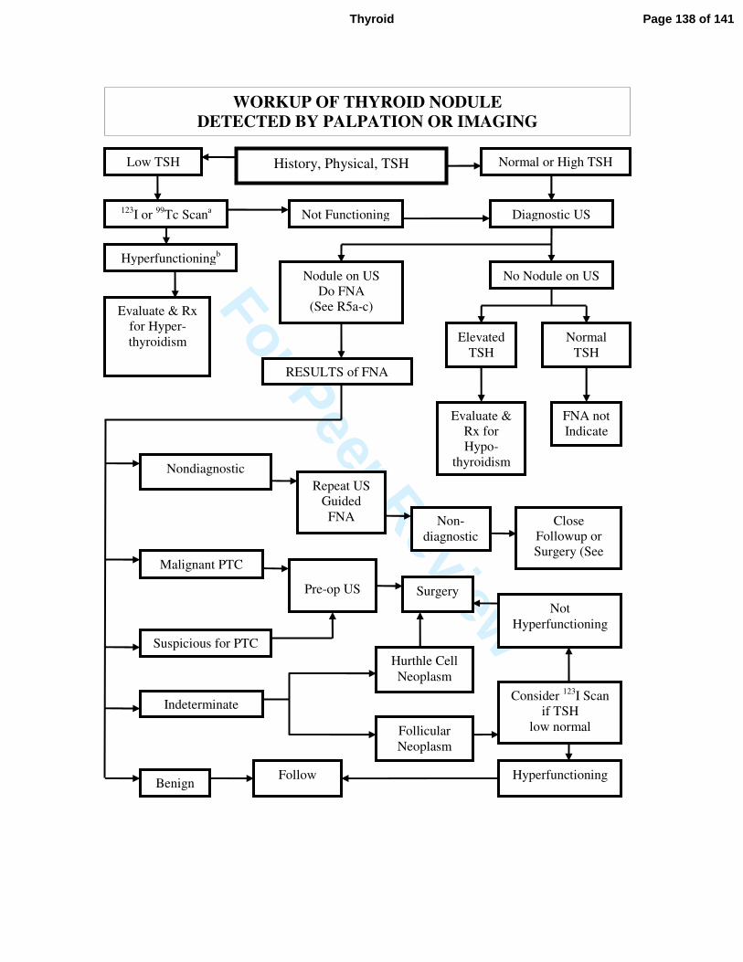
For Peer Review
123I or
99Tc Scan
a
Normal or High TSH History, Physical, TSH Low TSH
Diagnostic US
RESULTS of FNA
Elevated
TSH
Evaluate & Rx
for Hyper-
thyroidism
Not Functioning
Hyperfunctioningb
Nodule on US
Do FNA
(See R5a-c)
No Nodule on US
Normal
TSH
Evaluate &
Rx for
Hypo-
thyroidism
FNA not
Indicate
d
Nondiagnostic
Malignant PTC
Suspicious for PTC
Benign
Indeterminate
Repeat US
Guided
FNA Non-
diagnostic
Close
Followup or
Surgery (See
Text)
Pre-op US Surgery
Follicular
Neoplasm
Hurthle Cell
Neoplasm
Follow
Consider 123
I Scan
if TSH
low normal
Not
Hyperfunctioning
Hyperfunctioning
WORKUP OF THYROID NODULE
DETECTED BY PALPATION OR IMAGING
Page 138 of 141Thyroid
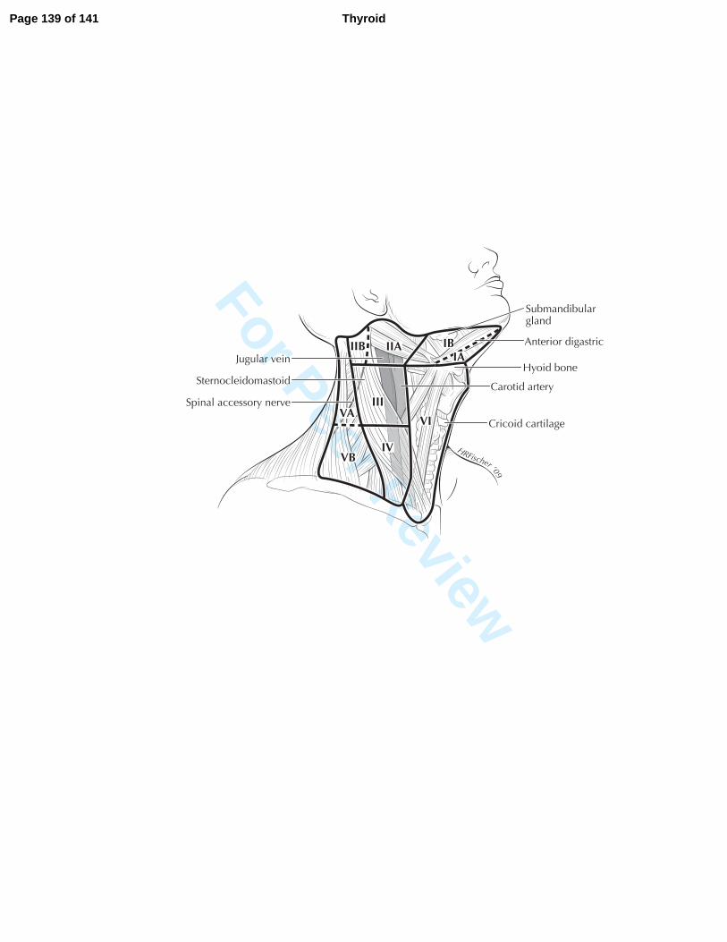
For Peer Review
HRFischer ‘09
Sternocleidomastoid
Jugular vein
Carotid arterySpinal accessory nerve
Submandibulargland
Anterior digastric
Hyoid bone
Cricoid cartilage
IBIIA
III
IVVB
VAVI
IIBIA
Page 139 of 141 Thyroid
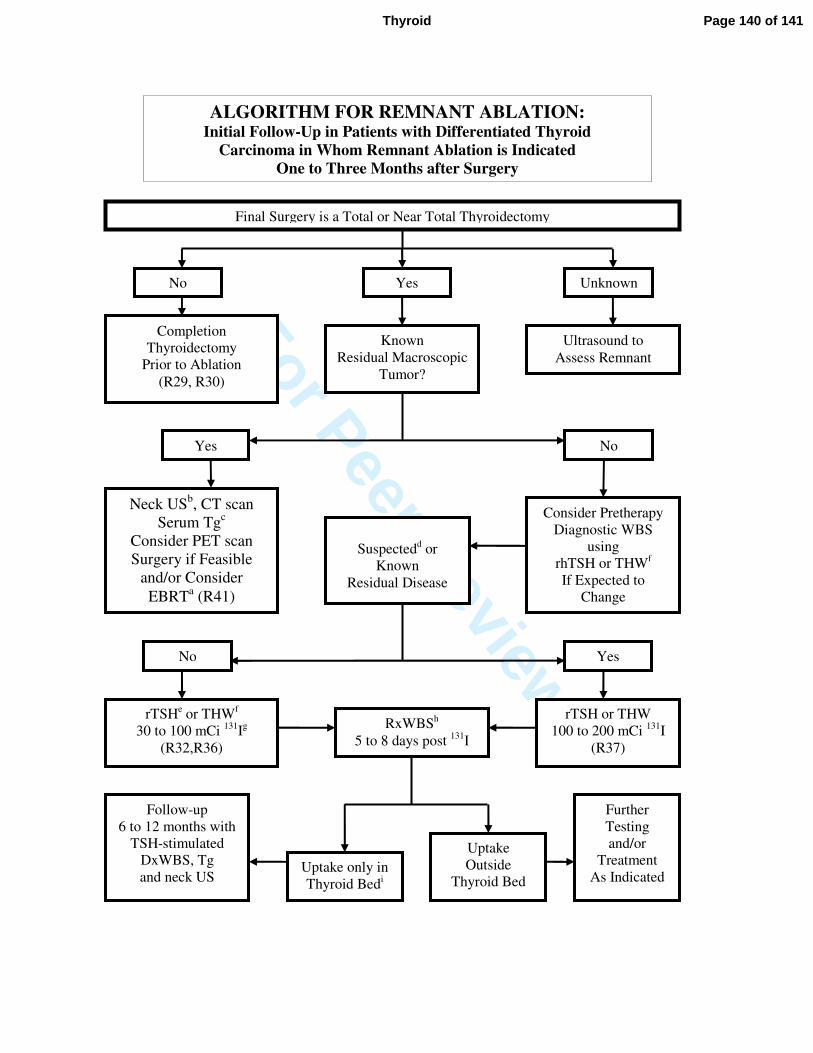
For Peer Review
ALGORITHM FOR REMNANT ABLATION: Initial Follow-Up in Patients with Differentiated Thyroid
Carcinoma in Whom Remnant Ablation is Indicated
One to Three Months after Surgery
Uptake only in
Thyroid Bedi
Final Surgery is a Total or Near Total Thyroidectomy
Completion
Thyroidectomy
Prior to Ablation
(R29, R30)
Unknown Yes No
Known
Residual Macroscopic
Tumor?
Ultrasound to
Assess Remnant
Neck USb, CT scan
Serum Tgc
Consider PET scan
Surgery if Feasible
and/or Consider
EBRTa (R41)
Consider Pretherapy
Diagnostic WBS
using
rhTSH or THWf
If Expected to
Change
Management (R35)
Suspectedd or
Known
Residual Disease
rTSHe or THW
f
30 to 100 mCi 131
Ig
(R32,R36)
RxWBSh
5 to 8 days post 131
I
rTSH or THW
100 to 200 mCi 131
I
(R37)
Uptake
Outside
Thyroid Bed
Follow-up
6 to 12 months with
TSH-stimulated
DxWBS, Tg
and neck US
Further
Testing
and/or
Treatment
As Indicated
Yes No
Yes No
Page 140 of 141Thyroid
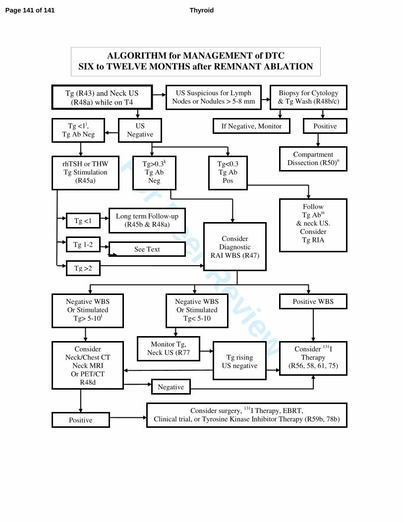
For Peer Review
ALGORITHM for MANAGEMENT of DTC
SIX to TWELVE MONTHS after REMNANT ABLATION
Tg (R43) and Neck US
(R48a) while on T4
US Suspicious for Lymph
Nodes or Nodules > 5-8 mm
US
Negative
Biopsy for Cytology
& Tg Wash (R48b/c)
If Negative, Monitor Positive Tg <1j,
Tg Ab Neg
rhTSH or THW
Tg Stimulation
(R45a)
Tg <1
Tg >2
Tg 1-2
Long term Follow-up
(R45b & R48a)
See Text
Tg>0.3k
Tg Ab
Neg
Tg<0.3
Tg Ab
Pos
Consider
Diagnostic
RAI WBS (R47)
Follow
Tg Abm
& neck US.
Consider
Tg RIA
Compartment
Dissection (R50)n
Negative WBS
Or Stimulated
Tg> 5-10I
Negative WBS
Or Stimulated
Tg< 5-10
Positive WBS
Consider
Neck/Chest CT
Neck MRI
Or PET/CT
R48d
Consider 131
I
Therapy
(R56, 58, 61, 75)
Negative
Positive
Tg rising
US negative
Monitor Tg,
Neck US (R77
Consider surgery, 131
I Therapy, EBRT,
Clinical trial, or Tyrosine Kinase Inhibitor Therapy (R59b, 78b)
Page 141 of 141 Thyroid
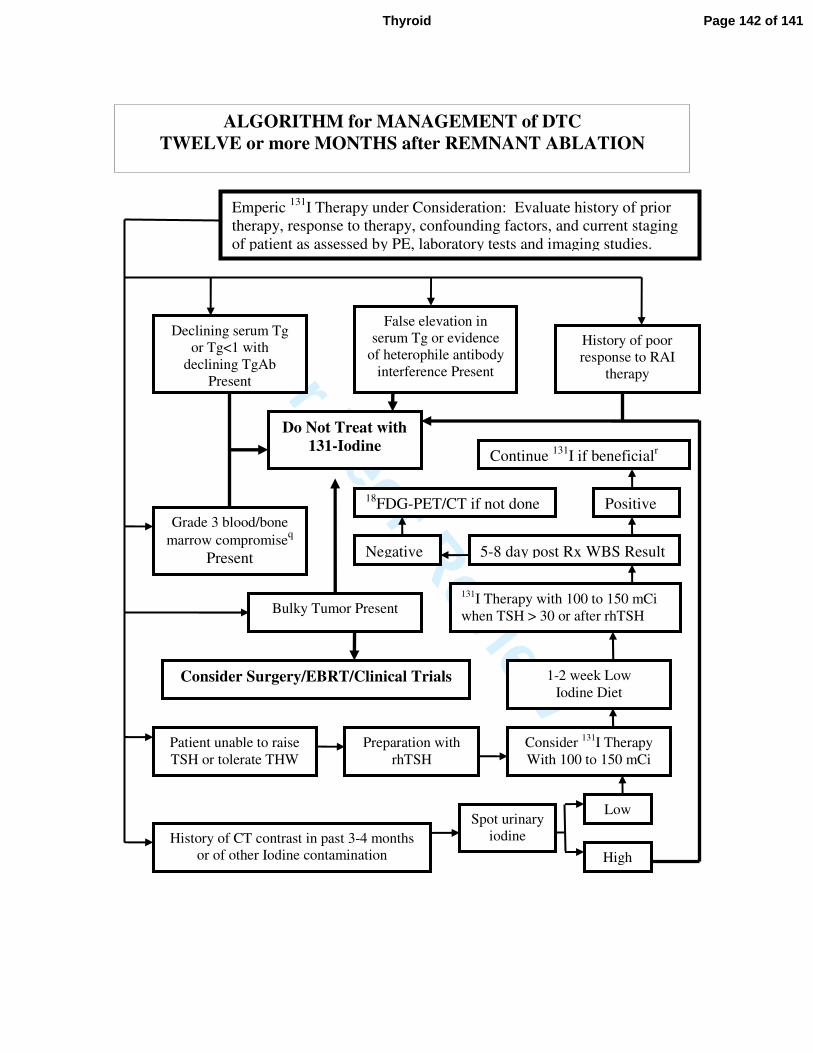
For Peer Review
ALGORITHM for MANAGEMENT of DTC
TWELVE or more MONTHS after REMNANT ABLATION
Emperic 131
I Therapy under Consideration: Evaluate history of prior
therapy, response to therapy, confounding factors, and current staging
of patient as assessed by PE, laboratory tests and imaging studies.
Declining serum Tg
or Tg<1 with
declining TgAb
Present
False elevation in
serum Tg or evidence
of heterophile antibody
interference Present
History of poor
response to RAI
therapy
Bulky Tumor Present
Grade 3 blood/bone
marrow compromiseq
Present
Do Not Treat with
131-Iodine
Consider Surgery/EBRT/Clinical Trials
Patient unable to raise
TSH or tolerate THW
History of CT contrast in past 3-4 months
or of other Iodine contamination
Spot urinary
iodine
Preparation with
rhTSH
Consider 131
I Therapy
With 100 to 150 mCi
131I Therapy with 100 to 150 mCi
when TSH > 30 or after rhTSH
1-2 week Low
Iodine Diet
Low
High
5-8 day post Rx WBS Result Negative
Positive
Continue 131
I if beneficialr
18FDG-PET/CT if not done
Page 142 of 141Thyroid