Embed Size (px)
Citation preview
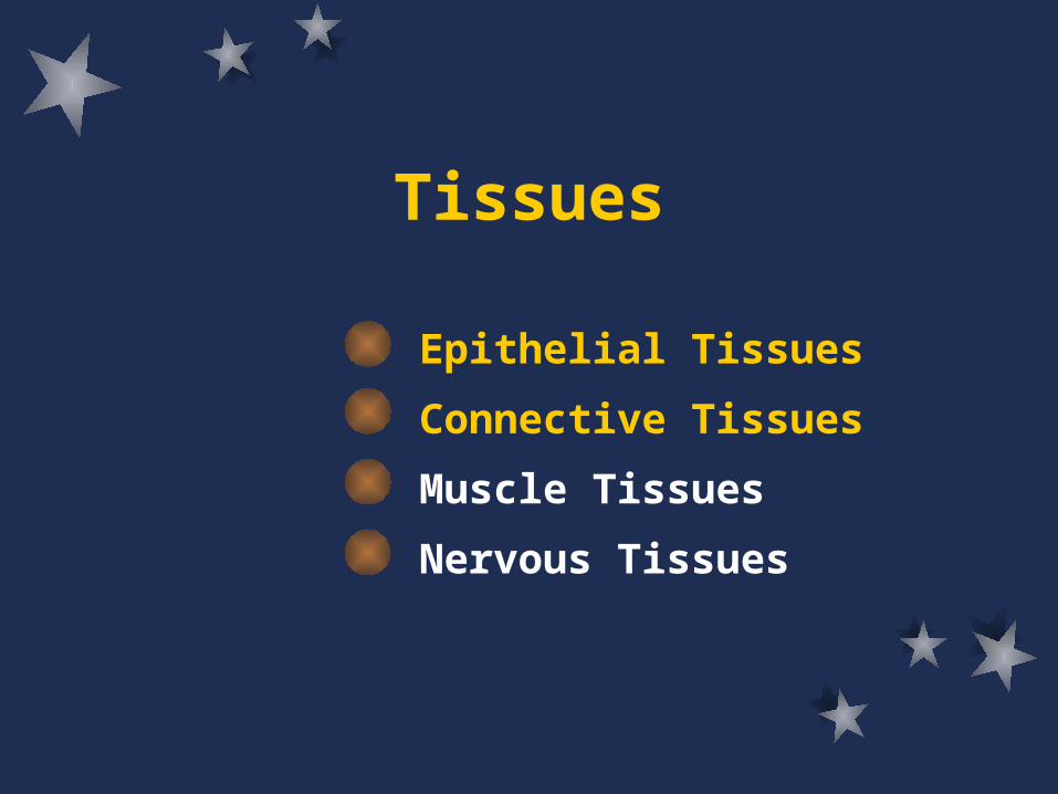
Tissues
Epithelial Tissues
Connective Tissues
Muscle Tissues
Nervous Tissues
Objectives
Upon completion of this chapter, you should be able to:– Describe the general characteristics and functions
of epithelial tissue.– Name the types of epithelium and identify an organ
in which each is found.– Explain how glands are classified.– Describe the general characteristics of connective
tissue.– Describe the major cell types and fibers of
connective tissue.

Objectives
Upon completion of this chapter, you should be able to:– List the types of connective tissue that occur within
the body.– Describe the major functions of each type of
connective tissue.– Distinguish among the three types of muscle tissue.– Describe the general characteristics and functions
of nervous tissue.– Complete the review activities at the end of the
chapter.
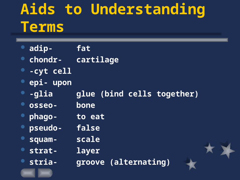
Aids to Understanding Terms adip- fat chondr- cartilage -cyt cell epi- upon -glia glue (bind cells together) osseo- bone phago- to eat pseudo- false squam- scale strat- layer stria- groove (alternating)
Epithelial Tissues
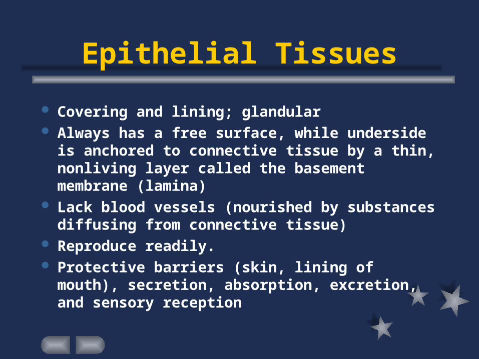
Covering and lining; glandular Always has a free surface, while underside is
anchored to connective tissue by a thin, nonliving layer called the basement membrane (lamina)
Lack blood vessels (nourished by substances diffusing from connective tissue)
Reproduce readily. Protective barriers (skin, lining of mouth),
secretion, absorption, excretion, and sensory reception
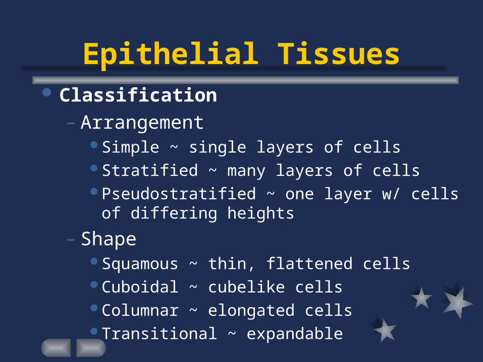
Epithelial Tissues Classification
– ArrangementSimple ~ single layers of cellsStratified ~ many layers of cellsPseudostratified ~ one layer w/ cells of differing
heights
– ShapeSquamous ~ thin, flattened cellsCuboidal ~ cubelike cellsColumnar ~ elongated cellsTransitional ~ expandable
Simple Squamous Epithelium
Single layer of thin, flattened cells Occurs commonly where diffusion, osmosis, and
filtration are taking place– air sacs of lungs– walls of capillaries– insides of blood and lymph vessels– covers membranes that line body cavities
Simple Cuboidal Epithelium
Single layer of cube-shaped cells w/ centrally located nucleus
Secretion, Absorption– Kidney tubules – Glands -- secretion of glandular products
salivary glandspancreasliverovaries
Simple Columnar Epithelium
Single layer of elongated cells w/ nuclei located near basement membrane
Protection, secretion, absorption– Lining of uterus– Lining of various organs of digestive tract, e.g.,
stomach and intestines Microvilli often cover surface, increasing surface
area for more effective absorption Goblet cells scattered throughout, secreting
protective fluid (mucus) onto free surface
Pseudostratified Columnar Epithelium
Nuclei located at two or more levels within cells Cilia extend from free surface Goblet cells scattered throughout tissue Protection, secretion, movement of mucus and
cells– Lines passages of respiratory system– Lines tubes of reproductive system
Stratified Squamous Epithelium
Many layers of cells; thick Flattened near surface, cuboidal or columnar deeper Protection
– Skin (epidermis) As older cells are pushed outward, they accumulate the protein
keratin, harden, and die
– Lines mouth cavity, throat– Lines vagina, anal canal
Transitional Epithelium
Also called uroepithelium Specialized to undergo changes w/ tension Contracted, several layers of cuboidal cells; distended,
appears to contain only a few layers of cells Distensibility, protection, barrier from diffusion
– inner lining of urinary bladder – passageways of urinary system
Glandular Epithelium
Specialized to produce and secrete various substances into ducts or into body fluids
Found within columnar or cuboidal epithelium Exocrine glands
– glands that secrete products into ducts that open onto some internal or external surface
Endocrine glands– glands that secrete products into tissue fluid or
blood
Types of Exocrine Glands
Unicellular glands– Single secretory cell
mucus-secreting goblet cell
Multicellular glands– Simple glands
communicate with surface by means of unbranched ducts
– Compound glands communicate with surface by means of branched ducts
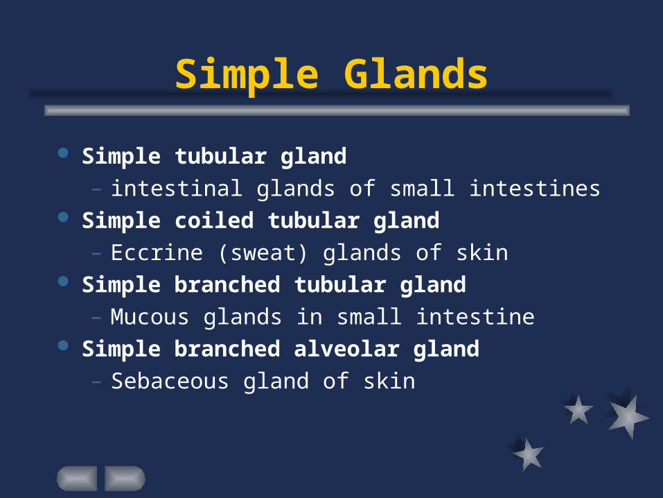
Simple Glands
Simple tubular gland – intestinal glands of small intestines
Simple coiled tubular gland– Eccrine (sweat) glands of skin
Simple branched tubular gland– Mucous glands in small intestine
Simple branched alveolar gland– Sebaceous gland of skin
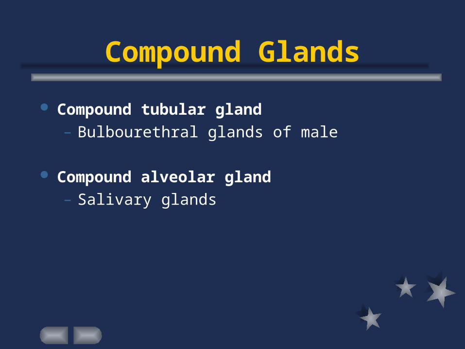
Compound Glands
Compound tubular gland– Bulbourethral glands of male
Compound alveolar gland– Salivary glands
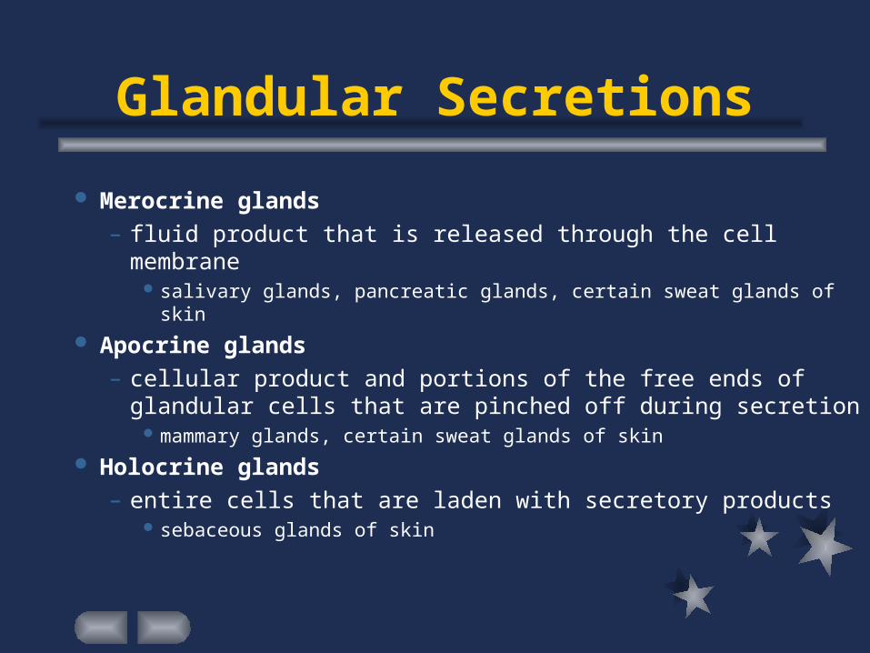
Glandular Secretions
Merocrine glands – fluid product that is released through the cell membrane
salivary glands, pancreatic glands, certain sweat glands of skin
Apocrine glands– cellular product and portions of the free ends of
glandular cells that are pinched off during secretion mammary glands, certain sweat glands of skin
Holocrine glands– entire cells that are laden with secretory products
sebaceous glands of skin
Connective Tissues
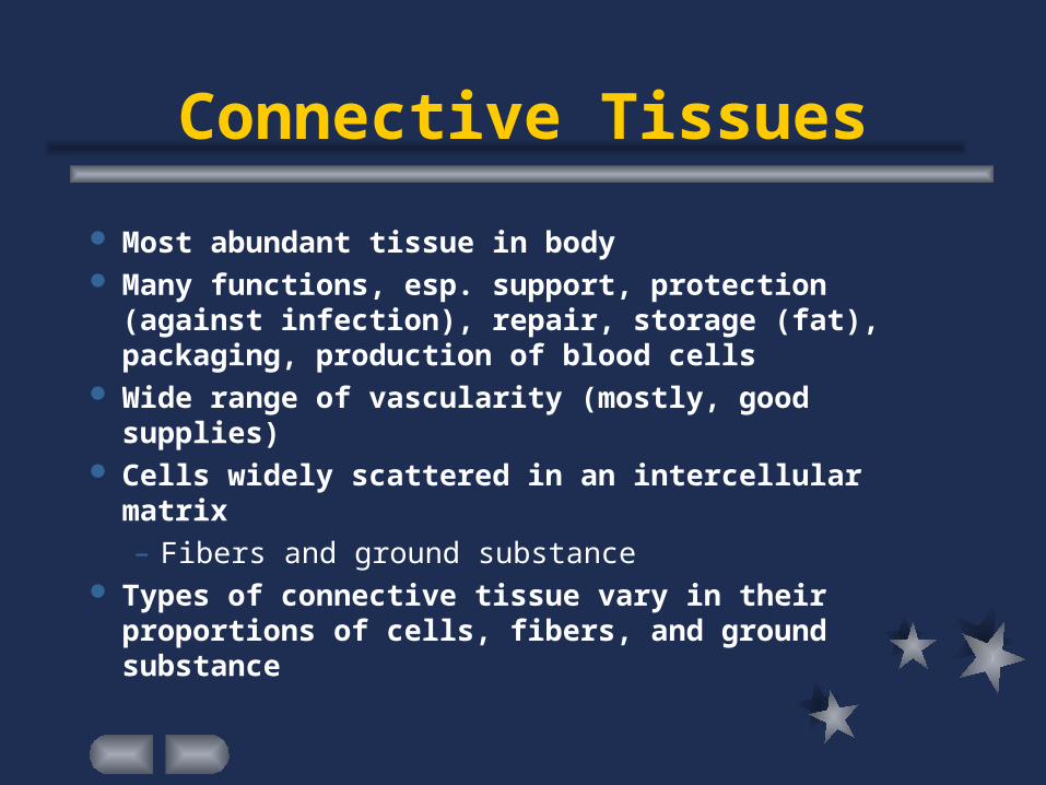
Most abundant tissue in body Many functions, esp. support, protection (against
infection), repair, storage (fat), packaging, production of blood cells
Wide range of vascularity (mostly, good supplies) Cells widely scattered in an intercellular matrix
– Fibers and ground substance Types of connective tissue vary in their
proportions of cells, fibers, and ground substance
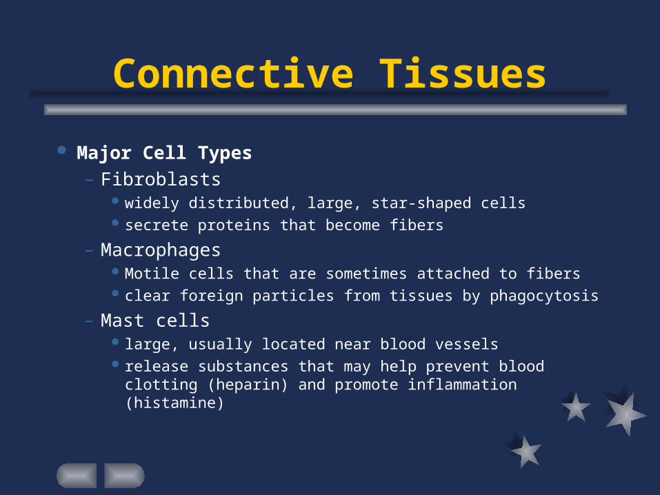
Connective Tissues
Major Cell Types– Fibroblasts
widely distributed, large, star-shaped cells secrete proteins that become fibers
– Macrophages Motile cells that are sometimes attached to fibers clear foreign particles from tissues by phagocytosis
– Mast cells large, usually located near blood vessels release substances that may help prevent blood clotting
(heparin) and promote inflammation (histamine)
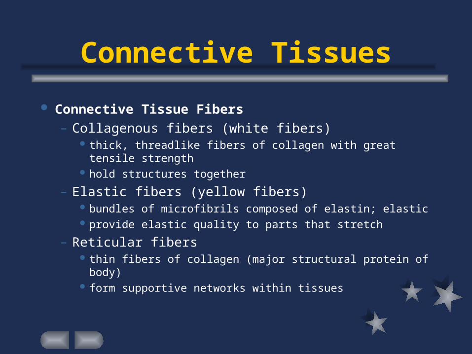
Connective Tissues
Connective Tissue Fibers– Collagenous fibers (white fibers)
thick, threadlike fibers of collagen with great tensile strength
hold structures together
– Elastic fibers (yellow fibers) bundles of microfibrils composed of elastin; elastic provide elastic quality to parts that stretch
– Reticular fibers thin fibers of collagen (major structural protein of body) form supportive networks within tissues
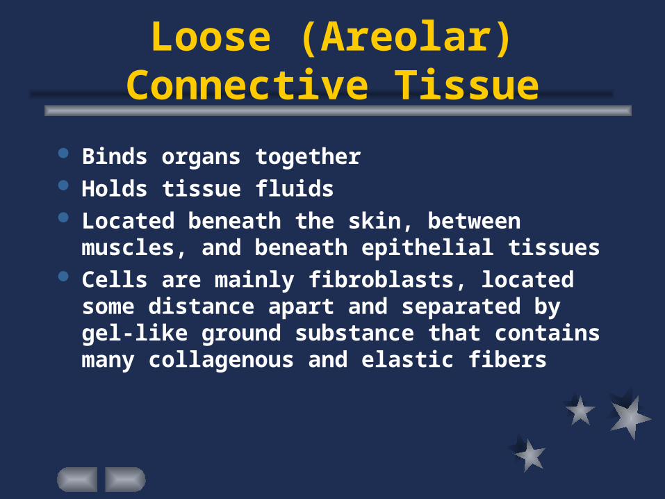
Loose (Areolar) Connective Tissue
Binds organs together Holds tissue fluids Located beneath the skin, between muscles, and
beneath epithelial tissues Cells are mainly fibroblasts, located some
distance apart and separated by gel-like ground substance that contains many collagenous and elastic fibers
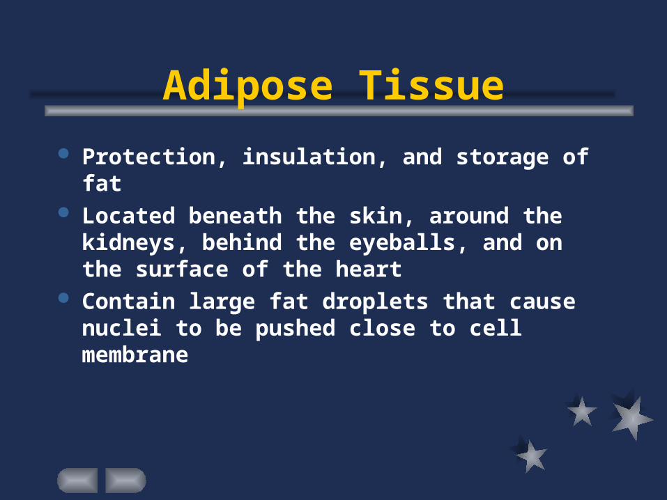
Adipose Tissue
Protection, insulation, and storage of fat Located beneath the skin, around the kidneys,
behind the eyeballs, and on the surface of the heart
Contain large fat droplets that cause nuclei to be pushed close to cell membrane
Fibrous Connective Tissue
Binds organs together Located in tendons (connect muscles to bones),
ligaments (connect bones to bones at joints), protective white layer of eyeball, and deep layer of skin
Contains many closely packed, thick, collagenous fibers and a fine network of elastic fibers; few cells -- fibroblasts
Blood supply relatively poor -- slow repair

Elastic Connective Tissue
Provides elastic quality Located between adjacent vertebrae, in walls of
arteries and airways Consists mainly of yellow, elastic fibers in
parallel strands or branching networks
Reticular Connective Tissue
Support Located in walls of liver, spleen, and lymphatic
organs Composed of thin, collagenous fibers arranged in
a three-dimensional network
Cartilage
Supports parts, provides frameworks and attachments, protects underlying tissues, and forms structural models for many developing bones
Chondrocytes occupy small chambers called lacunae and are completely surrounded by matrix
Enclosed in a covering of fibrous connective tissue called the perichondrium (location of blood supply)
Lacks direct blood supply Three types: hyaline, elastic, and fibrocartilage
Hyaline Cartilage
Support, protection, provides framework Located in ends of bones, nose, and rings in walls of
respiratory passages Most common type of cartilage Role in the growth of most bones and repair of bone
fractures
Elastic Cartilage Support, protection, provides flexible framework Located in framework of external ear and part of larynx Matrix contains many elastic fibers
Fibrocartilage Support, protection, shock absorption Located between bony parts of backbone, pelvic girdle,
and knee Very tough; many collagenous fibers
Bone (Osseous Tissue) Support, protection, provides framework Located in bones of skeleton Most rigid connective tissue, due largely to calcium
phosphate and calcium carbonate in matrix Protects vital parts in the cranial and thoracic cavities,
and serves as attachment for muscles Contains red marrow -- forms blood cells Matrix deposited in thin layers called lamellae Osteocytes clustered in concentric circles around
osteonic (Haversian) canals
Blood
Cells (red, white, platelets) are suspended in a fluid intercellular matrix called plasma
Most blood cells form in hematopoietic tissues in red marrow within the hollow parts of certain bones
Muscle Tissues Contractile; elongated cells (muscle fibers) Three types: skeletal, smooth muscle, cardiac muscle Skeletal muscle tissue
– found in muscles attached to bones– controlled by conscious effort (voluntary muscle tissue)– cells have many nuclei, plus alternating light and dark
cross-markings called striations– Responsible for moving head, trunk, and limbs, as well as
movements involved with facial expressions, writing, talking, singing, chewing, swallowing, and breathing
Muscle Tissues Smooth Muscle Tissue
– Lacks striations; single, centrally located nucleus– Found in walls of hollow internal organs (stomach,
intestines, urinary bladder, uterus, blood vessels)– Involuntary muscle tissue– Responsible for moving food through GI tract, constricting
blood vessels, and emptying bladder Cardiac Muscle Tissue
– Only in the heart; involuntary muscle tissue– striated cells joined end to end by intercalated disk– single nucleus
Nervous Tissue Found in the brain, spinal cord, and peripheral nerves Basic cells -- nerve cells or neurons plus neuroglial cells
(support and bind components of nervous tissue together, carry on phagocytosis, supply nutrients to neurons by connecting them to blood vessels)
Coordinate, regulate, and integrate many body functions