Embed Size (px)
Citation preview
TissuesHonors Anatomy & PhysiologyChapter 4Human Anatomy & Physiology

•4 basic types of tissues in human body contribute to homeostasis by providing diverse functions including
•protection•support•communication among cells• resistance to disease •& many more
Tissues & Homeostasis
•a tissue is a group of similar cells that usually have a common embryonic origin & function together to carry out specialized activities
Definition
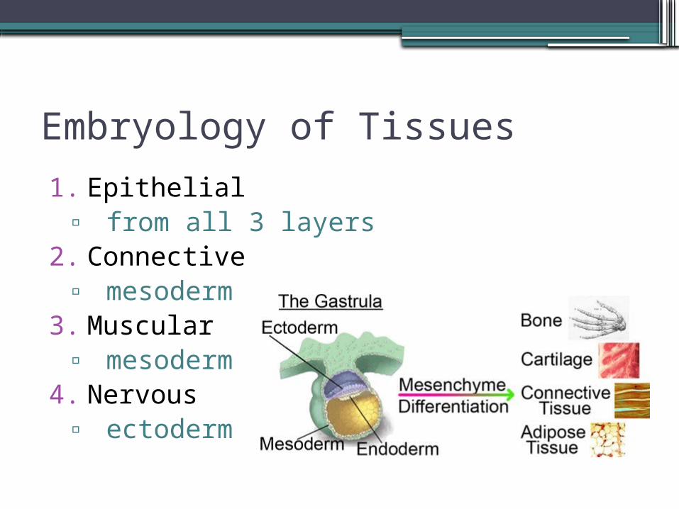
1. Epithelial▫ from all 3 layers
2. Connective▫ mesoderm
3. Muscular▫ mesoderm
4. Nervous ▫ ectoderm
Embryology of Tissues
• their structure & properties are influenced by factors such as: ▫nature of the extracellular material
surrounding tissue cells▫type of connections between cells
Tissues
Classification of Tissues
1. Epithelial▫ covers body surfaces & lines
hollow organs, cavities, & ducts▫ forms glands
2. Connective 3. Muscle4. Nerve
Functions of Epithelial Tissues
•protection•absorption•filtration•excretion•secretion•sensory reception
Epithelial Tissue
•5 distinguishing characteristics1. polarity2. specialized contacts3. supported by CT (connective
tissue)4. avascular but innervated5. ability to regenerate
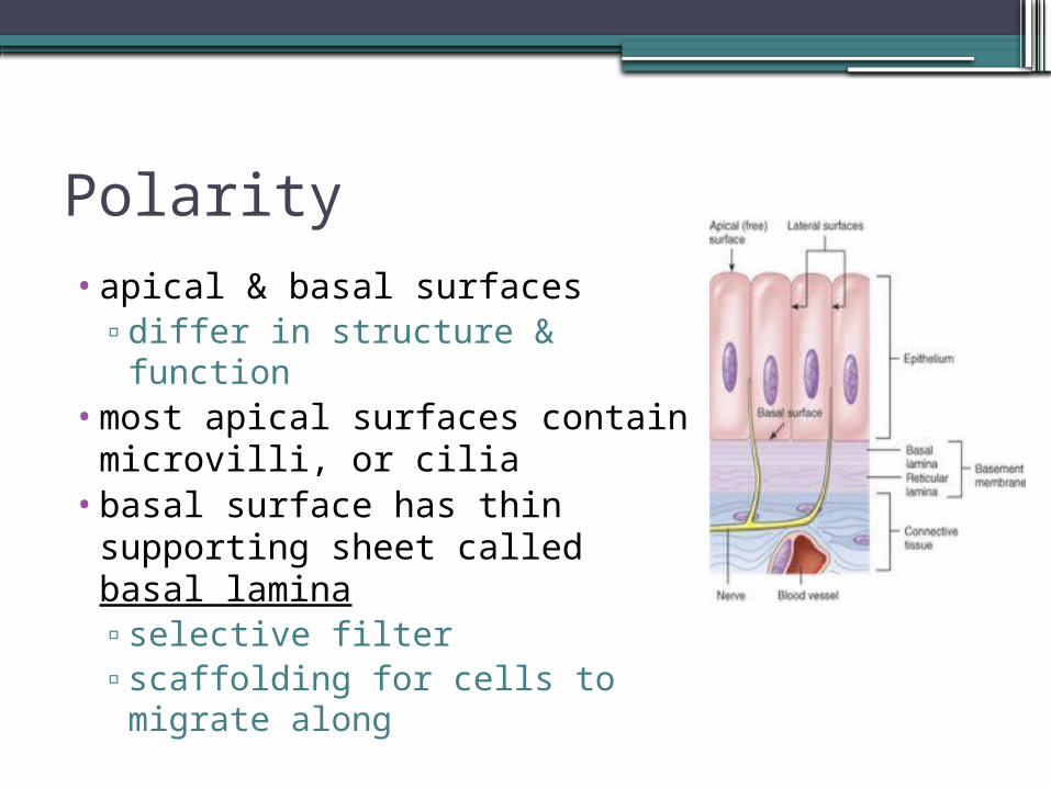
Polarity•apical & basal surfaces
▫differ in structure & function
•most apical surfaces contain microvilli, or cilia
•basal surface has thin supporting sheet called basal lamina▫selective filter▫scaffolding for cells to
migrate along
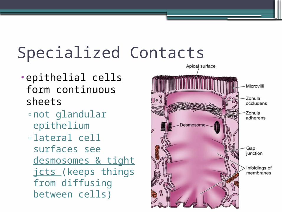
Specialized Contacts•epithelial cells form
continuous sheets ▫not glandular
epithelium▫lateral cell surfaces
see desmosomes & tight jcts (keeps things from diffusing between cells)
Supported by CT
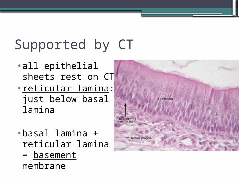
•all epithelial sheets rest on CT
•reticular lamina: just below basal lamina
•basal lamina + reticular lamina = basement membrane
Avascular / Innervated
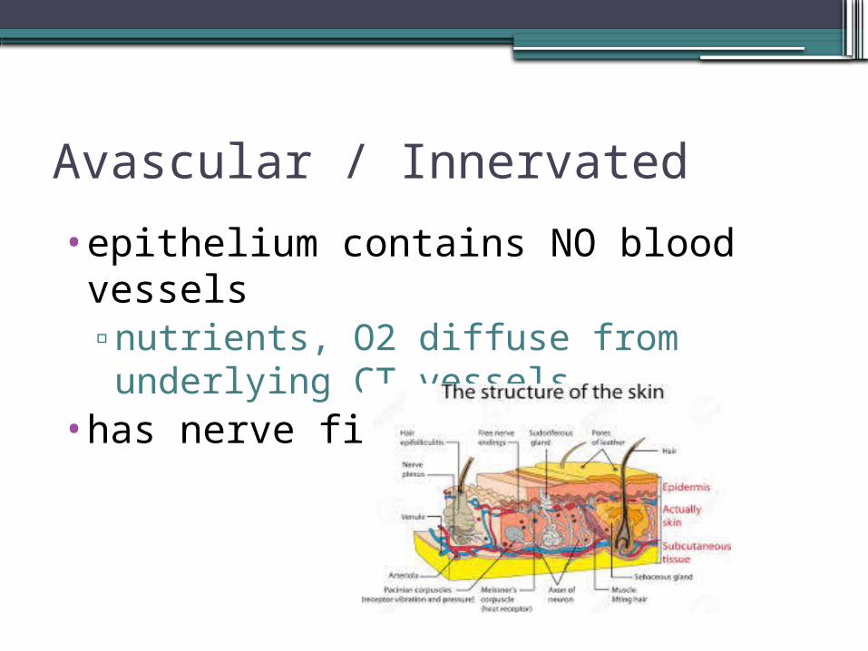
•epithelium contains NO blood vessels▫nutrients, O2 diffuse from underlying
CT vessels•has nerve fibers
Regeneration
•epithelial cells will reproduce rapidly when neighboring cells damaged or destroyed▫as long as necessary nutrients can
get to epithelium
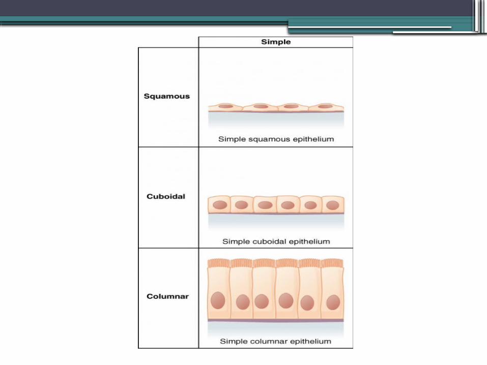
Classification of Epithelia•classified according to 2 characteristics:1. # of layers• single layer = simple• multiple layers = stratified• single layer that looks like multiple =
pseudostratified 2. cell shape• squamous• cuboidal• columnar
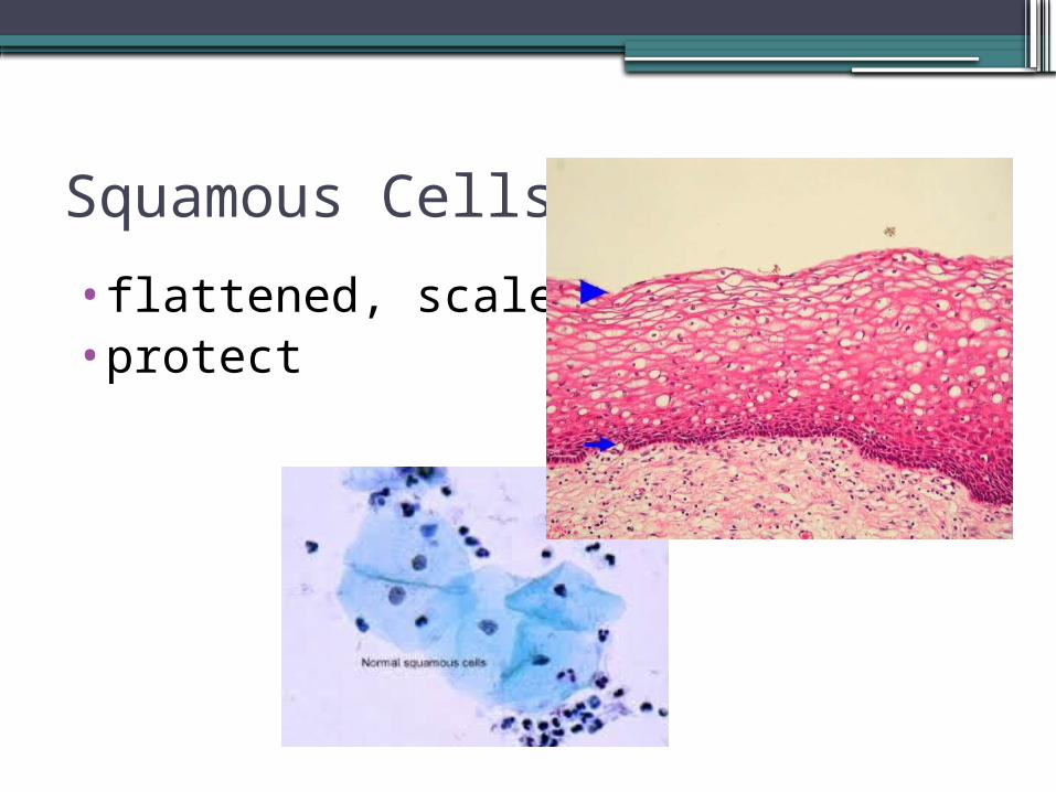
Squamous Cells
•flattened, scalelike•protect
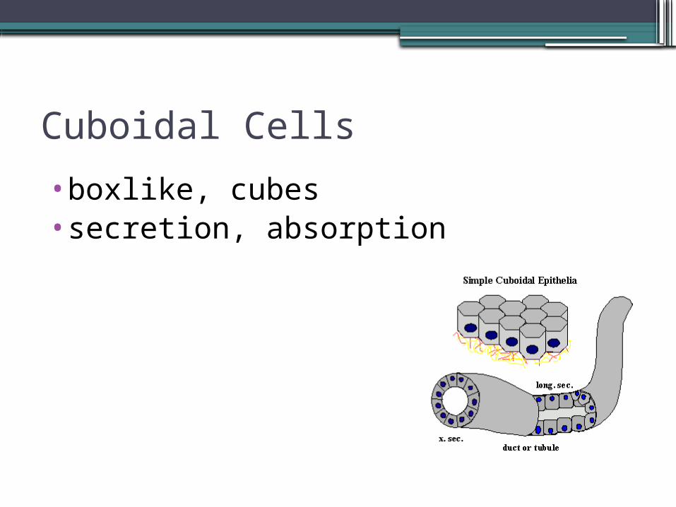
Cuboidal Cells
•boxlike, cubes•secretion, absorption
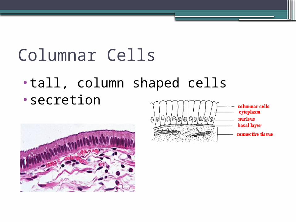
Columnar Cells
•tall, column shaped cells•secretion
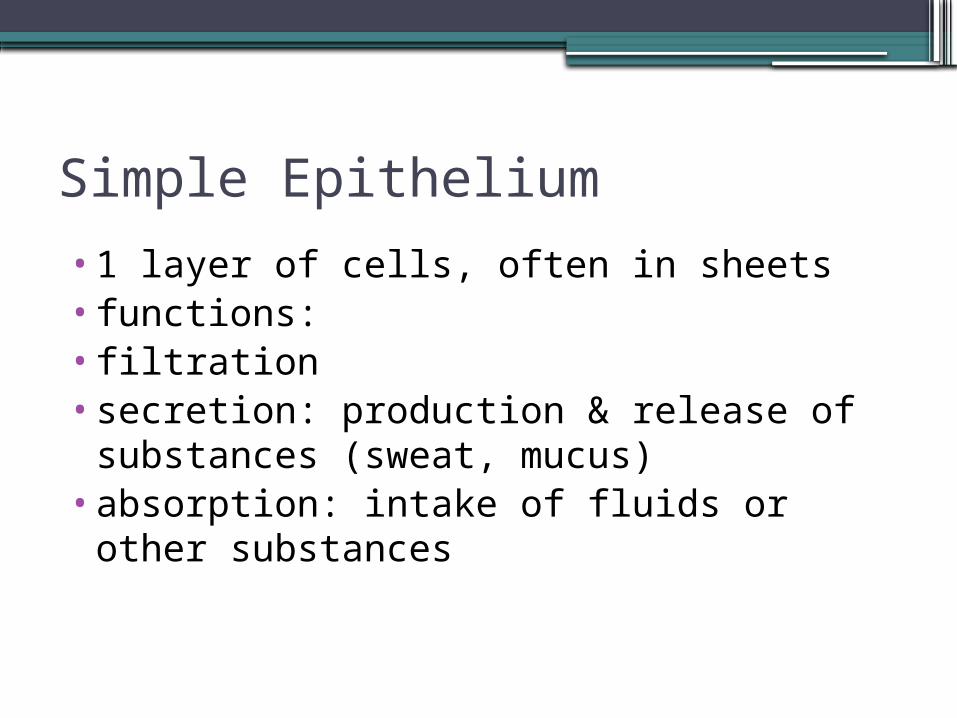
•1 layer of cells, often in sheets• functions:•filtration•secretion: production & release of
substances (sweat, mucus)•absorption: intake of fluids or other
substances
Simple Epithelium
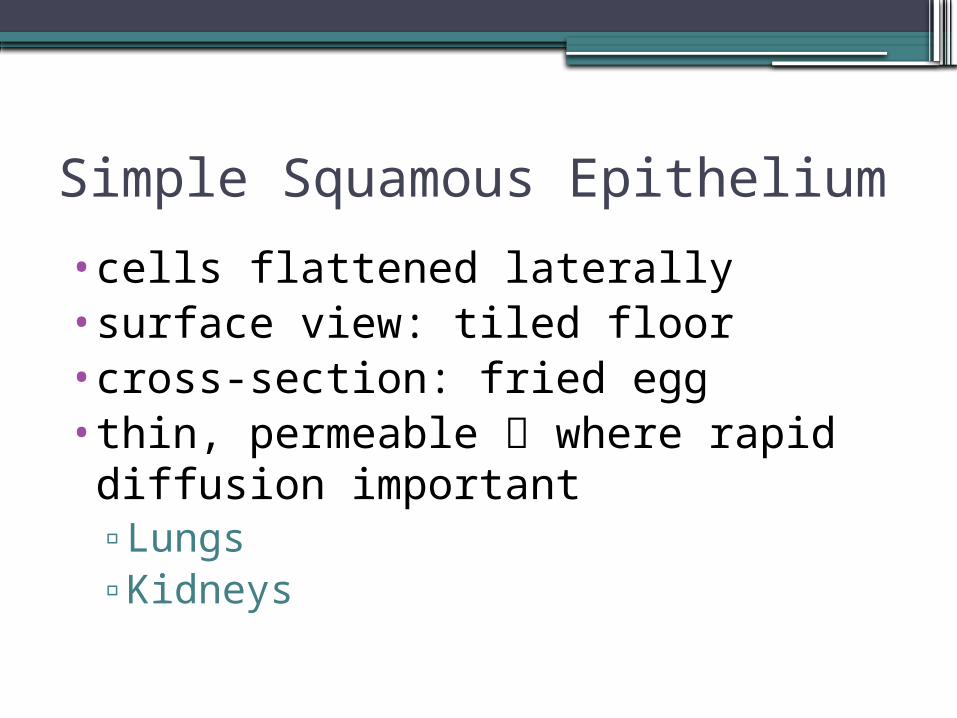
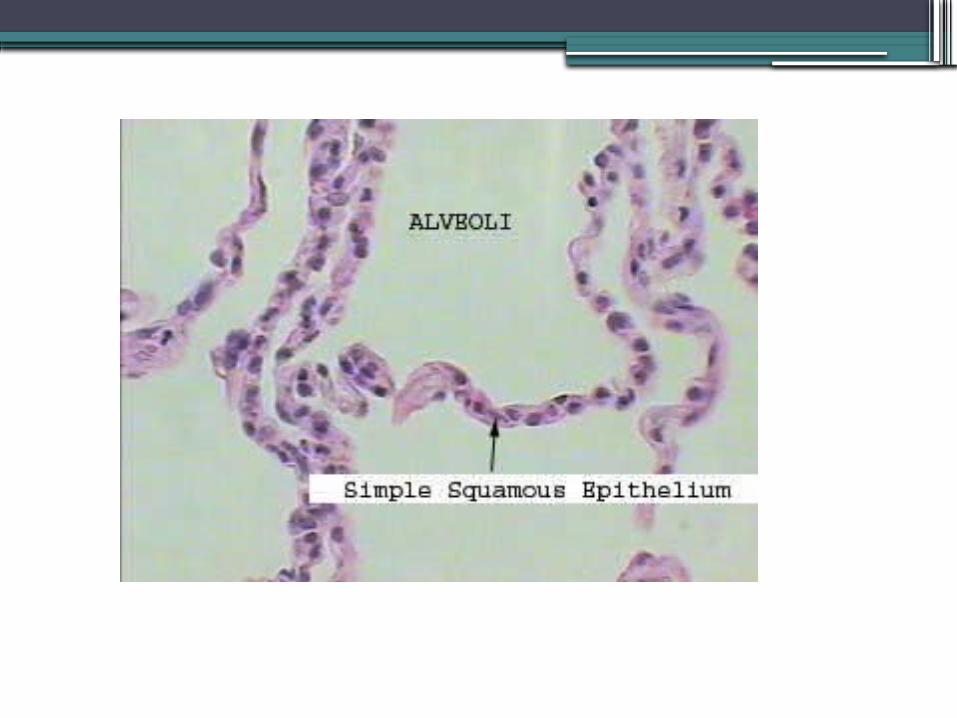
Simple Squamous Epithelium
•cells flattened laterally•surface view: tiled floor•cross-section: fried egg•thin, permeable where rapid diffusion important▫Lungs▫Kidneys
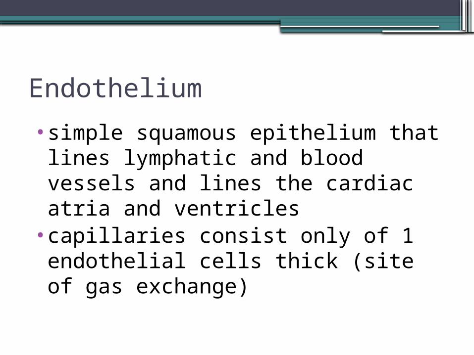
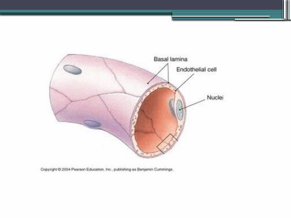
Endothelium
•simple squamous epithelium that lines lymphatic and blood vessels and lines the cardiac atria and ventricles
•capillaries consist only of 1 endothelial cells thick (site of gas exchange)
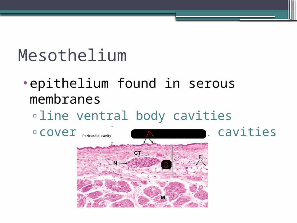
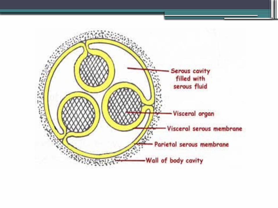
Mesothelium
•epithelium found in serous membranes▫line ventral body cavities▫cover organs in ventral cavities
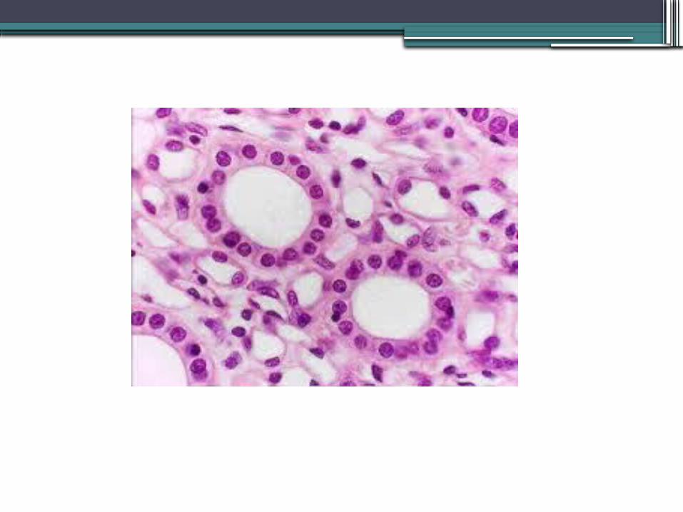
Simple Cuboidal Epithelium
•square cells with round, central nuclei in a sheet
•secretion•absorption•forms walls of small ducts of glands & kidney tubules
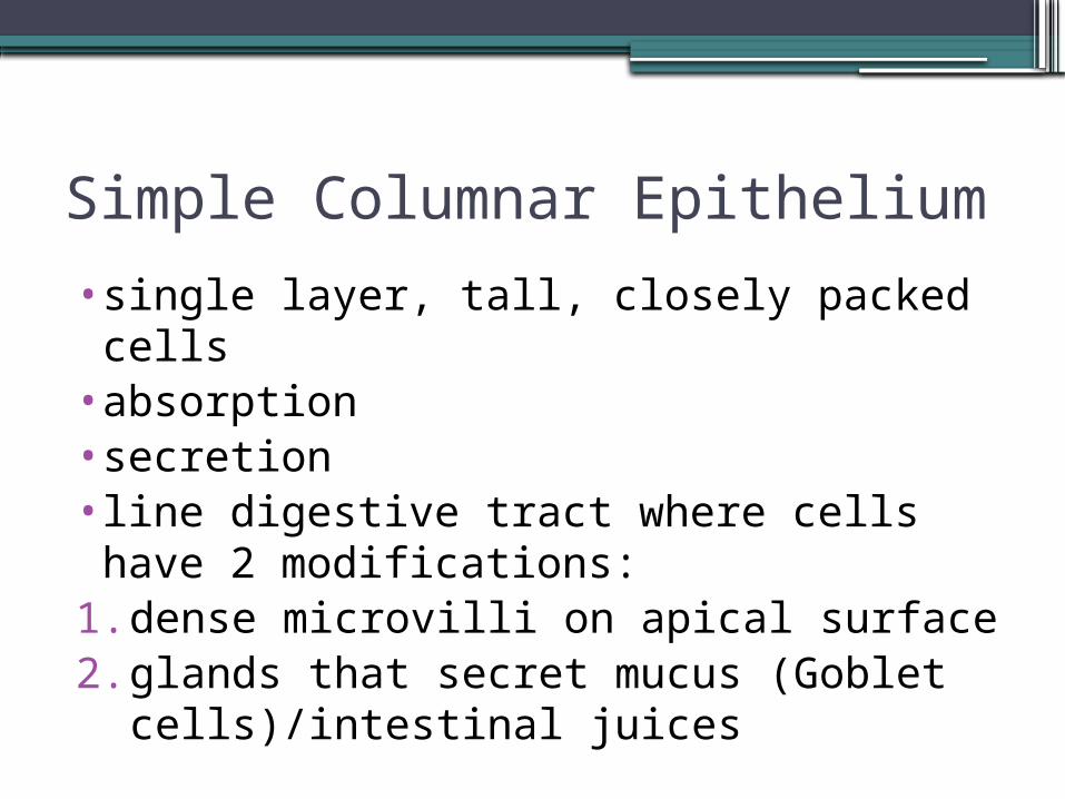
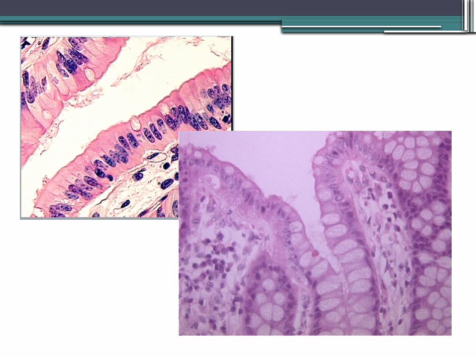
Simple Columnar Epithelium
•single layer, tall, closely packed cells•absorption•secretion•line digestive tract where cells have 2 modifications:
1. dense microvilli on apical surface2. glands that secret mucus (Goblet
cells)/intestinal juices
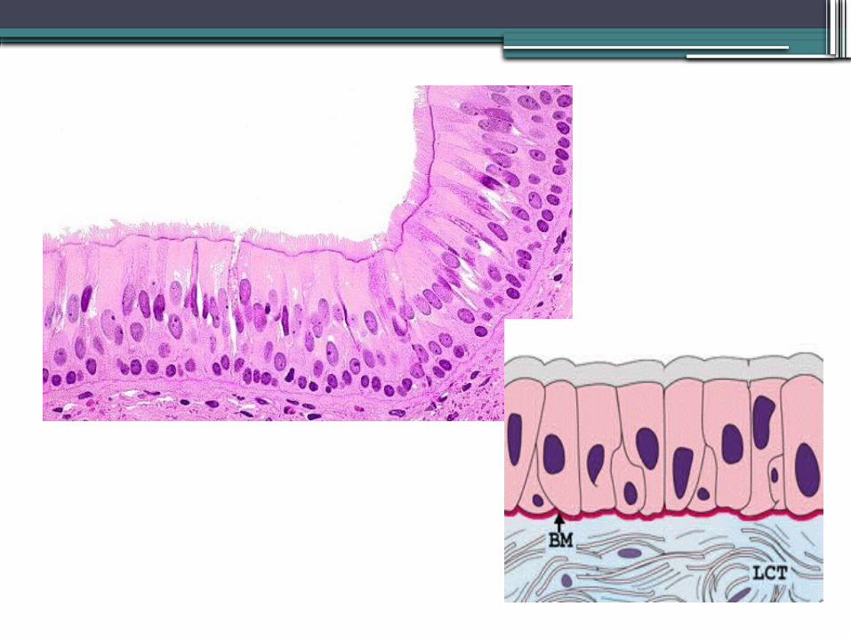
Pseudostratified Columnar Epithelium•cells vary in height•nuclei at variable heights•ciliated or nonciliated•all cells rest on BM (basement membrane) but not all reach free surface▫short cells replace tall ones if
damaged
Stratified Epithelium
•2 or more cell layers•cells are generated from below•more durable than simple epithelium•protection: major function•type of stratified epithelium determined by cell shape of top layer
Stratified Squamous Epithelium•found in areas subjected to wear and tear
▫upper layers rubbed away ▫lower cells replace
•top cells are squamous•as cells pushed up further from BM fewer nutrients reach them die lipids from atrophied cells add some water proofing
•keratinized/not keratinized▫keratin: tough protein
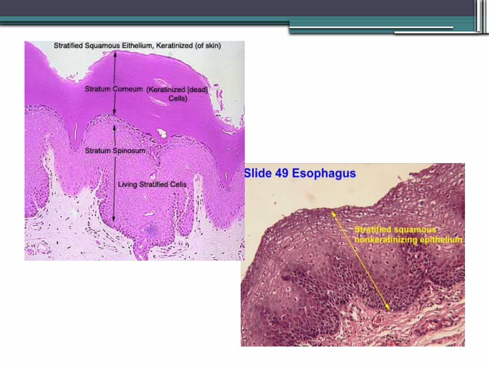
Stratified Squamous Epithelium
•Keratinized
•upper layers contain keratin: tough, fibrous protein that protects underlying tissues from heat, microbes, chemicals
• found: skin•Nonkeratinized
• found: lining mouth (buccal mucosa) & esophagus
•protect underlying tissues from wear & tear and from invasion by microbes
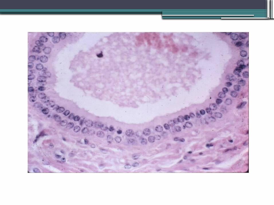
Stratified Cuboidal Epithelium
•rare•found in some ducts of larger glands
▫mammary glands, some sweat glands•2 layers of cuboidal cells
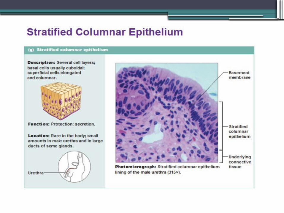
Stratified Columnar Epithelium•limited distribution in body
▫male urethra▫some glandular ducts▫@junction between 2 other types of
epithelial cells
Glandular Epithelium
•gland: 1 or more cells that make & secrete a product called a secretion
•secretions are aqueous solutions▫composition varies by gland
Secretion
•active process of gland releasing its product (also called secretion)
2 Classes of Glands
1. Endocrine glands (ductless glands)▫ internal secretion: cells exocytose
extracellular space capillaries target cells prompts some response in cell
▫ hormones: messenger chemicals2. Exocrine glands (glands with ducts)
▫ external secretion ▫ secrete product onto surfaces or into
body cavities
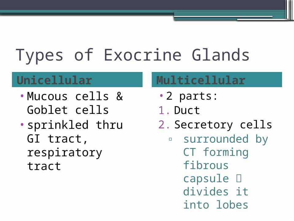
Types of Exocrine GlandsUnicellular Multicellular •Mucous cells &
Goblet cells•sprinkled thru GI
tract, respiratory tract
•2 parts:1. Duct 2. Secretory cells
▫ surrounded by CT forming fibrous capsule divides it into lobes
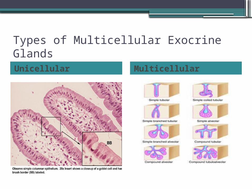
Types of Multicellular Exocrine GlandsUnicellular Multicellular
Connective Tissue
•most abundant & widely distributed of the 4 tissue types
•major functions:1. binding & supporting2. protecting3. insulating4. storing5. transporting
Classification of CT
•CT proper▫fat▫fibrous tissue
•cartilage•bone•blood
Characteristics of CT
•common origin▫mesenchyme
•degrees of vascularity▫cartilage: avascular▫dense CT poorly vascularized▫other types richly vascularized
•extracellular matrix▫few cells/ lots extracellular matrix
3 Main Elements in CT
1. ground substance2. fibers3. cells
• 1 + 2 = extracellular matrix• large variation in composition &
arrangement of the three large diversity of CTs each adapted to specific function
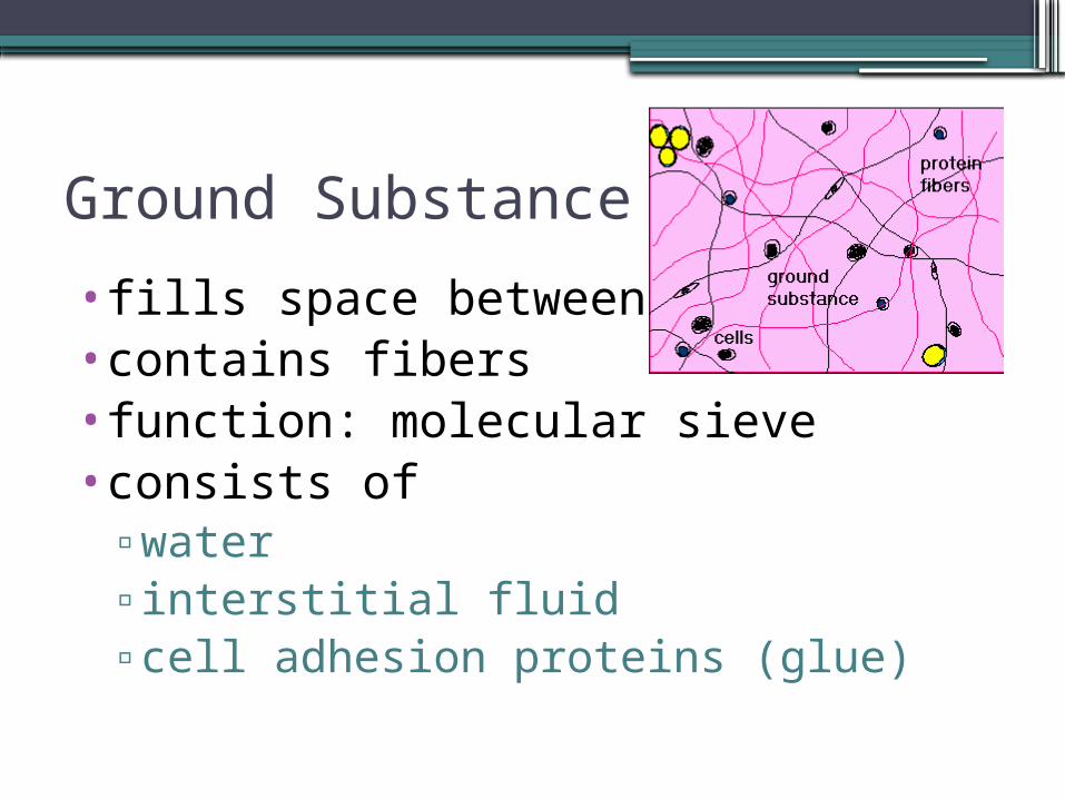
Ground Substance
•fills space between cells•contains fibers•function: molecular sieve•consists of
▫water▫interstitial fluid▫cell adhesion proteins (glue)
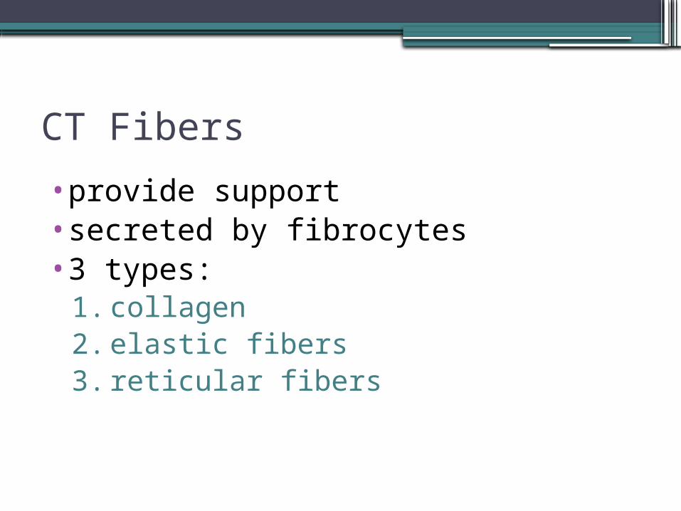
CT Fibers
•provide support•secreted by fibrocytes•3 types:
1. collagen2. elastic fibers3. reticular fibers
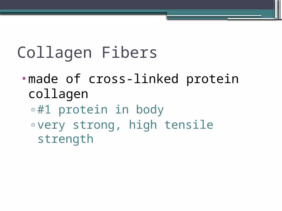
Collagen Fibers
•made of cross-linked protein collagen▫#1 protein in body▫very strong, high tensile strength
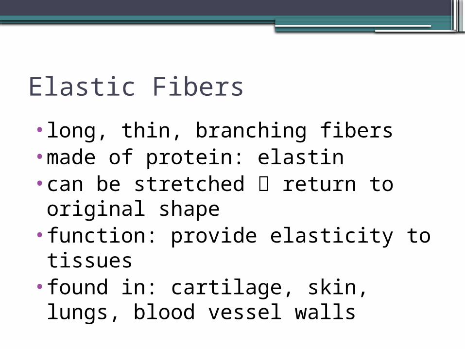
Elastic Fibers
•long, thin, branching fibers•made of protein: elastin•can be stretched return to original shape
•function: provide elasticity to tissues•found in: cartilage, skin, lungs, blood vessel walls
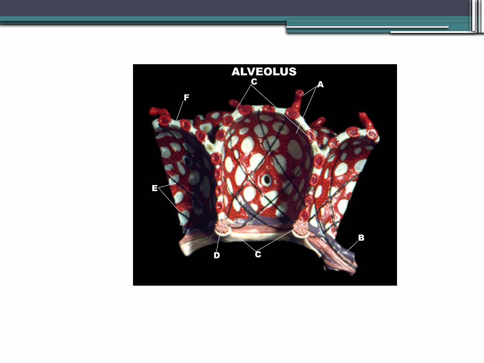
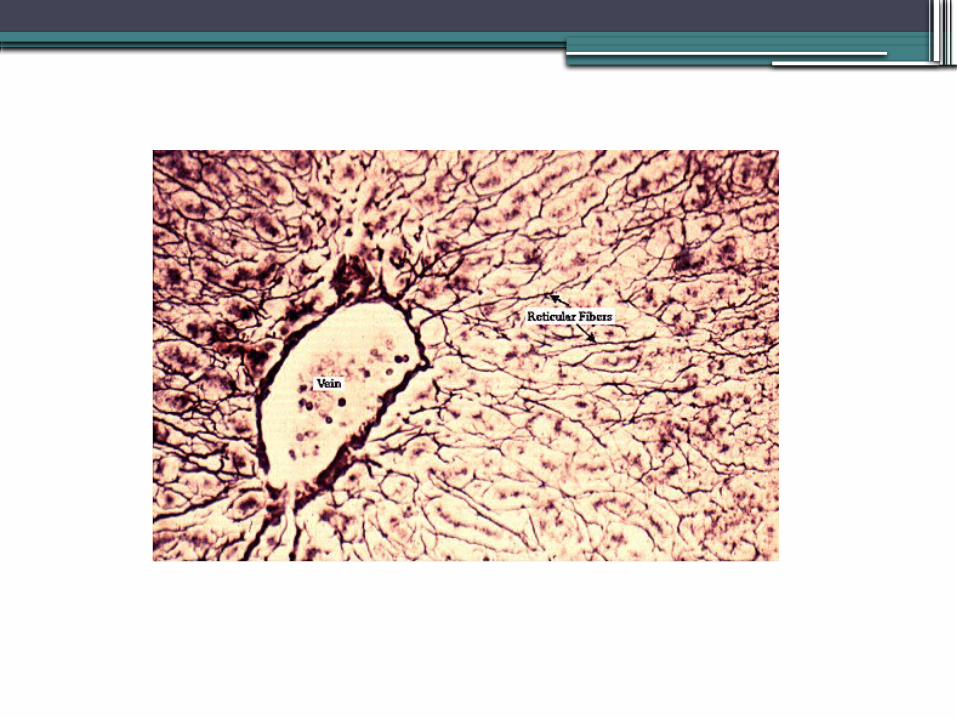
Reticular Fibers
•form delicate networks that surround small blood vessels& support soft tissue organs (liver, spleen)
•made of collagen derived protein•also found in BM of epithelium, around capillaries
CT Cells
•each class of CT has resident cell type:▫immature form –blast▫mature form - cyte
Loose CT
•3 types:1. Areolar2. Adipose3. Reticular
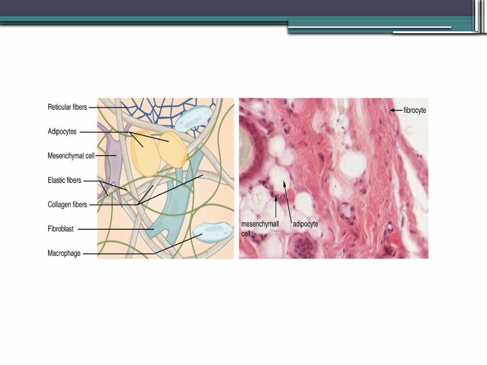
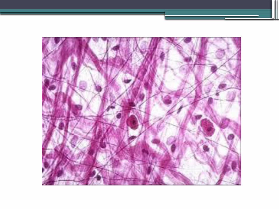
Areolar CT
•Functions:▫support & bind other tissues▫hold body fluids (reservoir of water &
salts)▫store nutrients
•Cells: all CT cells•Features: loose arrangement of fibers•Found: subcutaneous tissue
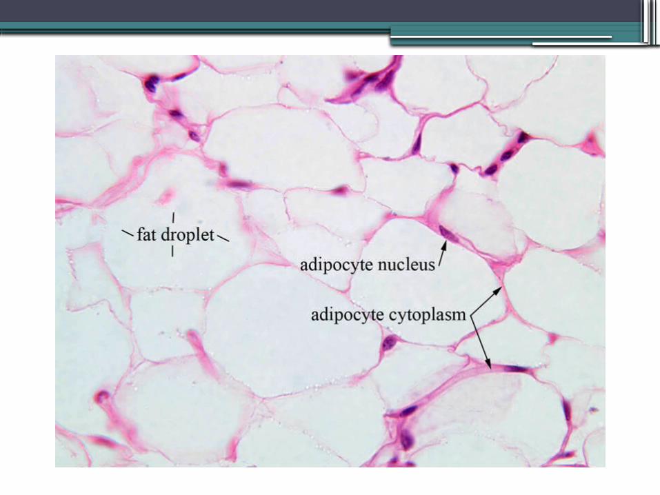
Adipose Tissue
•Functions: store triglycerides, insulation, protection
•Cells: adipocytes•Features: scant matrix, closely packed cells, rich blood supply
•Found: ass’c w/areolar CT, around heart, kidneys, lymph nodes
Adipose Tissue
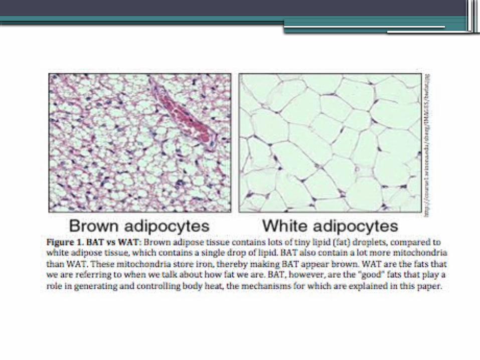
•2 types:1. White: most adipose tissue 2. Brown: abundant mitochondria
▫ use fat to generate heat (less ATP, more heat)
▫ found on back of newborns: unable to shiver if chilled
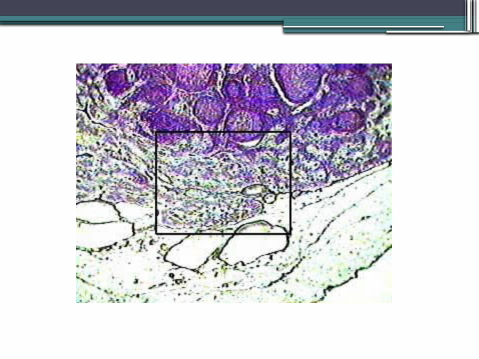
Reticular Connective Tissue
•Function: reticular fibers form internal framework = stroma
•Cells: fibroblasts = reticulocytes•Features: only fibers are reticular•Found: lymph nodes, spleen, bone marrow
Dense CT
•3 types:1. Dense Regular2. Dense Irregular3. Elastic
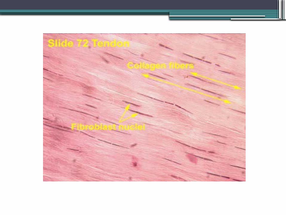
Dense Regular CT
•Functions: resistance to tension•Cells: fibroblasts•Features: closely packed, parallel collagen fibers (parallel to direction of pull); appears white, flexible
•Found: tendons (muscle to bone) & most ligaments (bone to bone)
Cartilage •Function: withstands tension & compression•Cells: chondroblasts (lay down cartilage), chondrocytes found in lacunae
•Features: qualities intermediate between dense CT and bone, no nerve or blood supply, surrounded by perichondrium
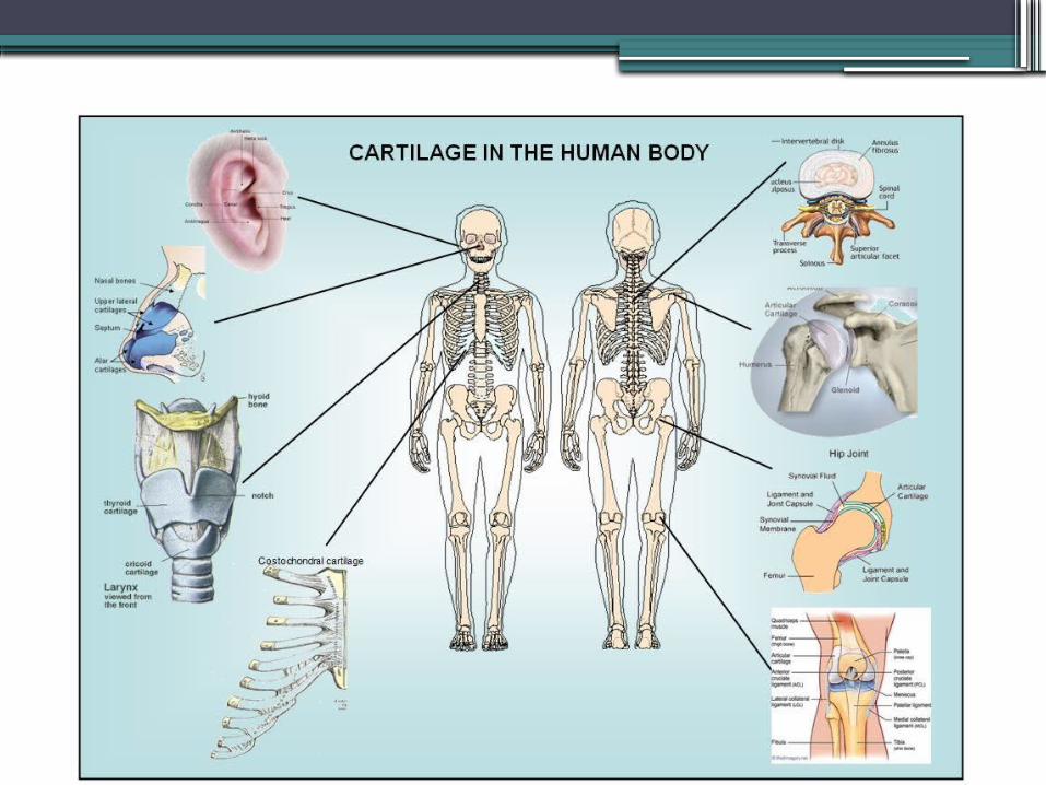
•Found: ends of long bones, see next slide
Types of Cartilage
1. Hyaline2. Elastic3. Fibrocartilage
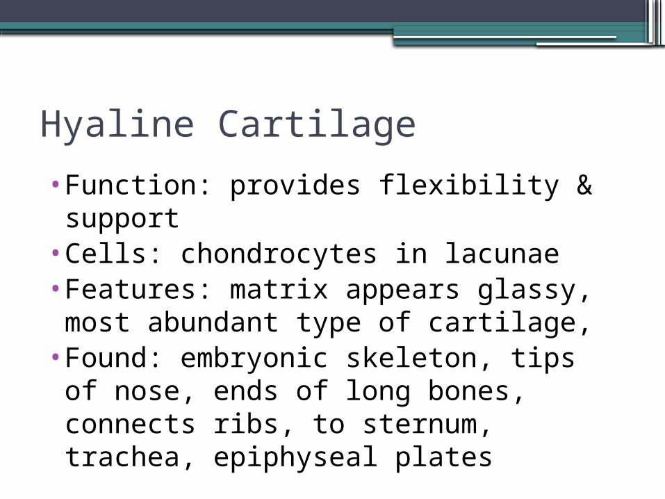
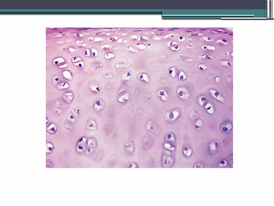
Hyaline Cartilage
•Function: provides flexibility & support
•Cells: chondrocytes in lacunae•Features: matrix appears glassy, most abundant type of cartilage,
•Found: embryonic skeleton, tips of nose, ends of long bones, connects ribs, to sternum, trachea, epiphyseal plates
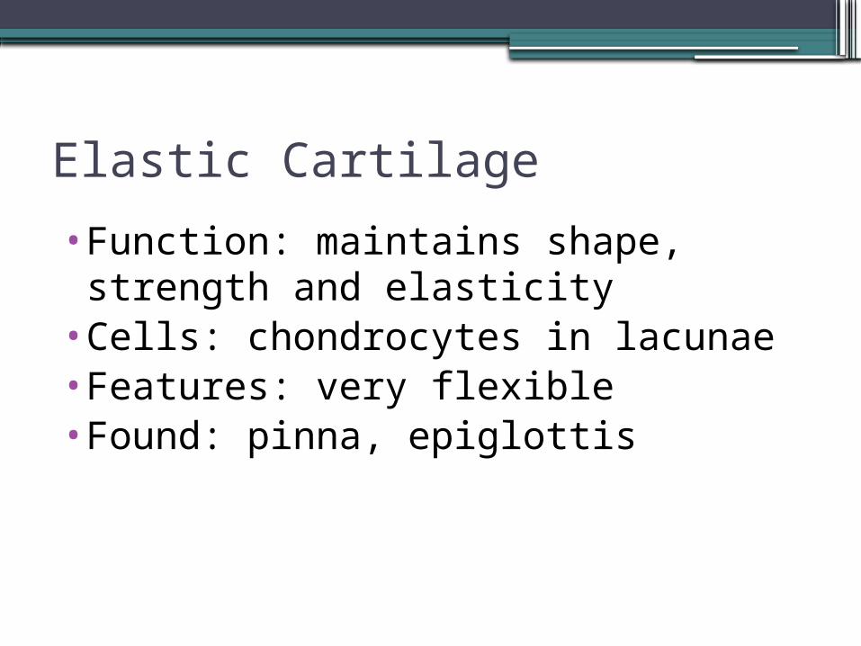
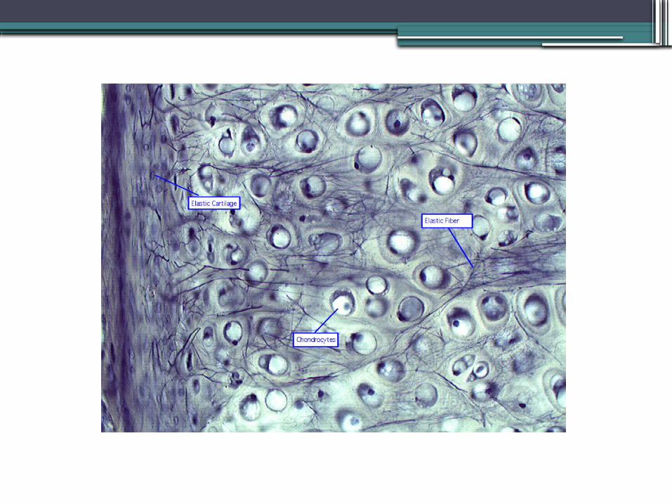
Elastic Cartilage
•Function: maintains shape, strength and elasticity
•Cells: chondrocytes in lacunae•Features: very flexible•Found: pinna, epiglottis
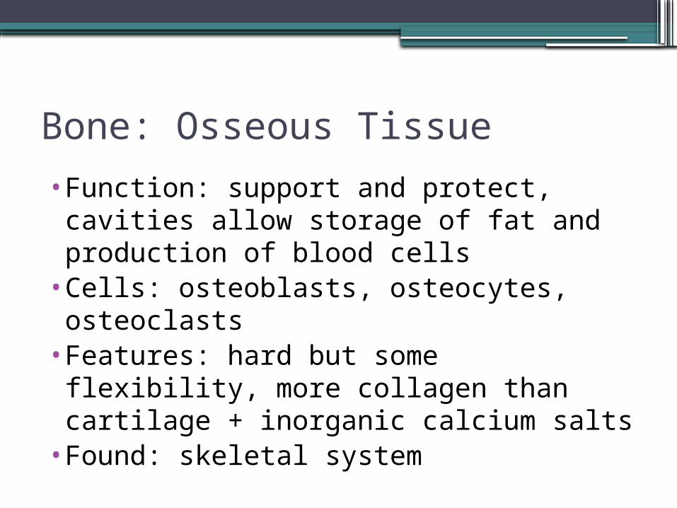
Bone: Osseous Tissue
•Function: support and protect, cavities allow storage of fat and production of blood cells
•Cells: osteoblasts, osteocytes, osteoclasts
•Features: hard but some flexibility, more collagen than cartilage + inorganic calcium salts
•Found: skeletal system
Bone
•2 types:1. Compact Bone2. Spongy Bone
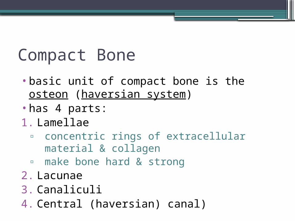
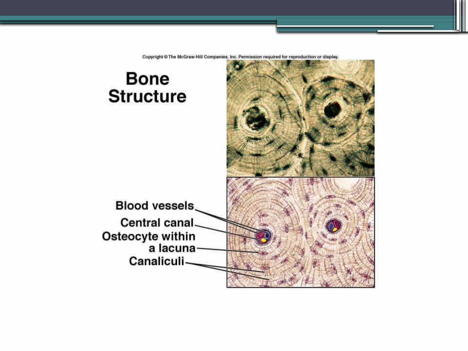
Compact Bone
•basic unit of compact bone is the osteon (haversian system)
•has 4 parts:1. Lamellae
▫ concentric rings of extracellular material & collagen
▫ make bone hard & strong2. Lacunae3. Canaliculi4. Central (haversian) canal)
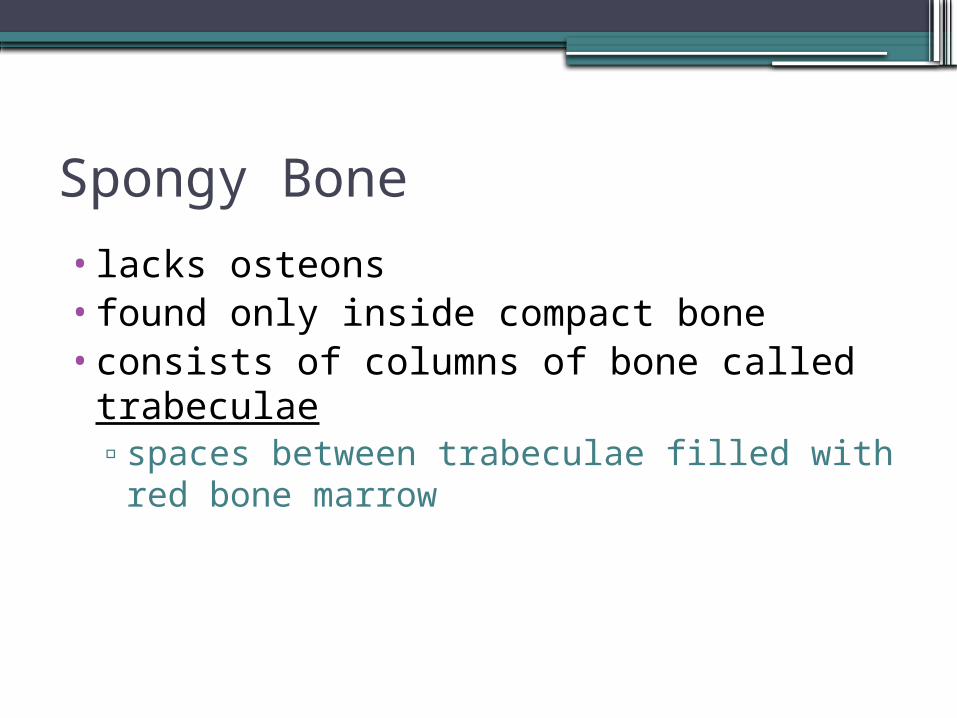
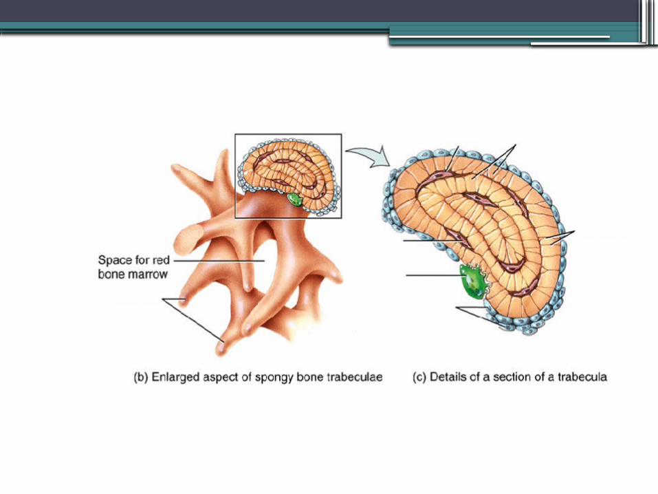
Spongy Bone
• lacks osteons• found only inside compact bone•consists of columns of bone called
trabeculae▫spaces between trabeculae filled with red
bone marrow
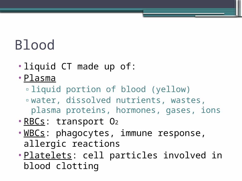
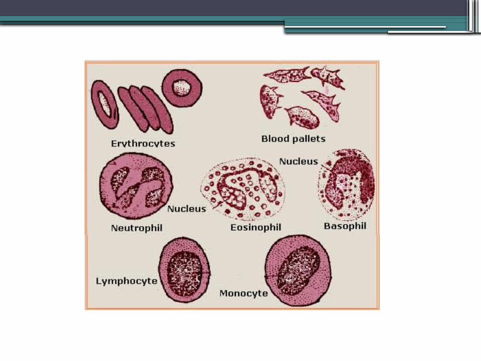
Blood
• liquid CT made up of:•Plasma
▫liquid portion of blood (yellow)▫water, dissolved nutrients, wastes, plasma
proteins, hormones, gases, ions•RBCs: transport O2
•WBCs: phagocytes, immune response, allergic reactions
•Platelets: cell particles involved in blood clotting
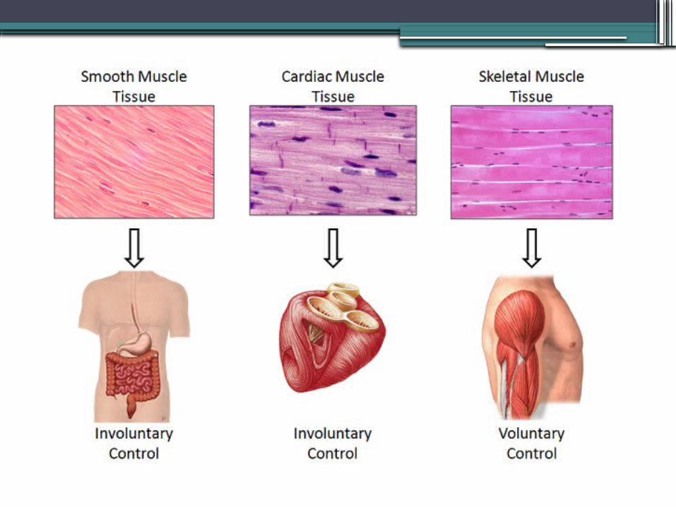
Muscle Tissue
•Function: movement, maintain posture, generate heat
•Cells: muscle fibers containing actin and myosin
•Features: contraction & relaxation•Found: skeletal system, walls of vessels and hollow organs, heart
Smooth Muscle
•Function: movement •Cells: 1 central nucleus, fusiform cells
•Features: involuntary & not striated, innervated by ANS,
•Found: walls of vessels & hollow organs, between hair follicles and dermis
Cardiac Muscle
•Function: propel blood thru body•Cells: branching with gap jcts = intercalated discs
•Features: striated, involuntary (ANS)•Found: only in wall of heart
Skeletal Muscle
•Function: move bones•Cells: multinucleated (peripherally located) muscle fibers
•Features: striated and voluntary•Found: attached to bones or skin
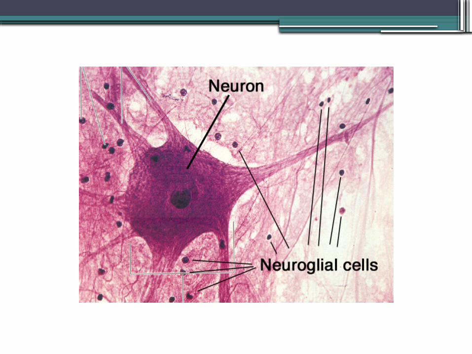
Nervous Tissue
•Function: regulate and control body functions
•Cells: neurons, neuroglial cells•Features: neurons can generate and/or respond to action potentials
•Found: nervous system
Membranes
•are flat sheets of pliable tissue that cover or line a part of the body
•2 types:1. epithelial membrane:
▫ epithelial layer + underlying CT▫ types: mucous membrane, serous
membrane, cutaneous membrane (skin)2. synovial membrane:
▫ + CT but - epithelium▫ line joints
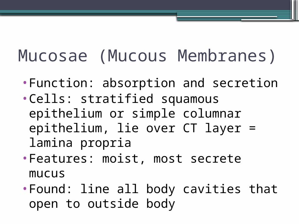
Mucosae (Mucous Membranes)•Function: absorption and secretion•Cells: stratified squamous epithelium or simple columnar epithelium, lie over CT layer = lamina propria
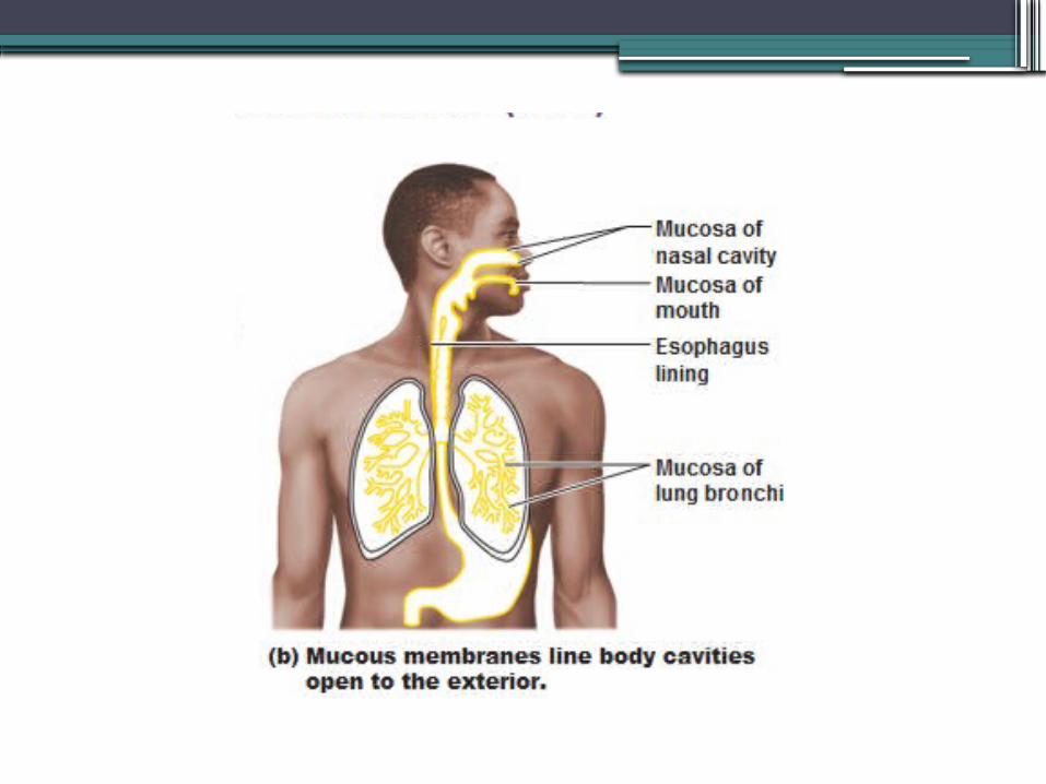
•Features: moist, most secrete mucus•Found: line all body cavities that open to outside body
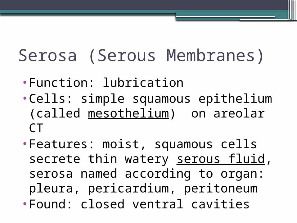
Serosa (Serous Membranes)
•Function: lubrication •Cells: simple squamous epithelium (called mesothelium) on areolar CT
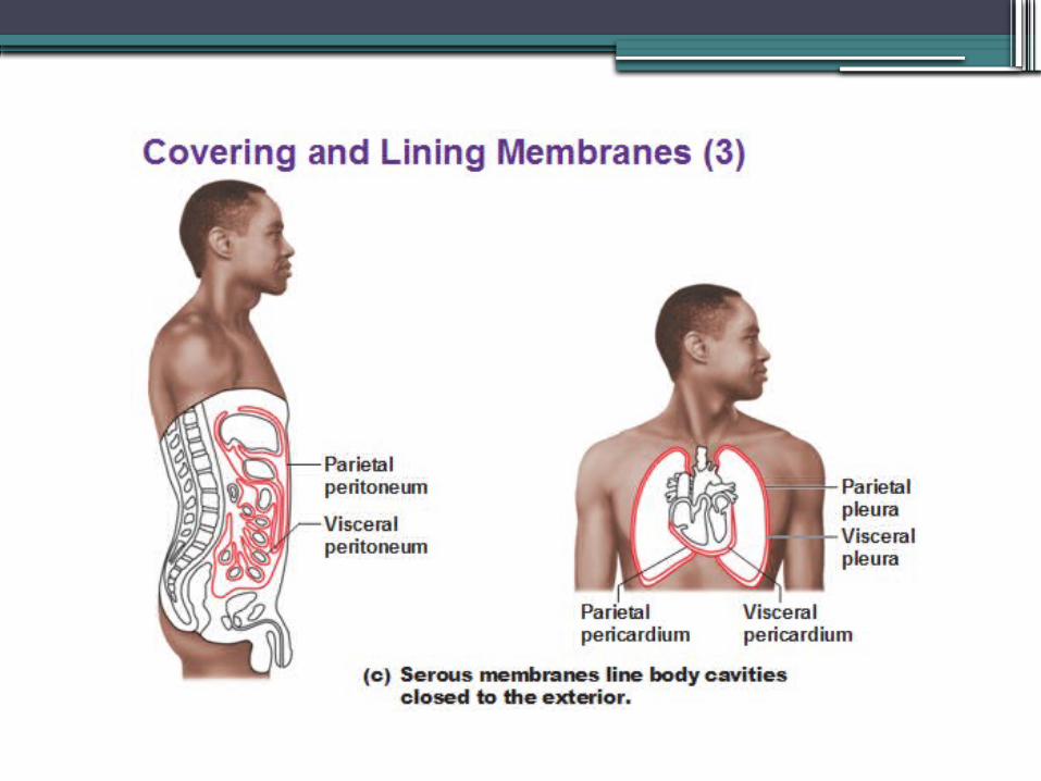
•Features: moist, squamous cells secrete thin watery serous fluid, serosa named according to organ: pleura, pericardium, peritoneum
•Found: closed ventral cavities
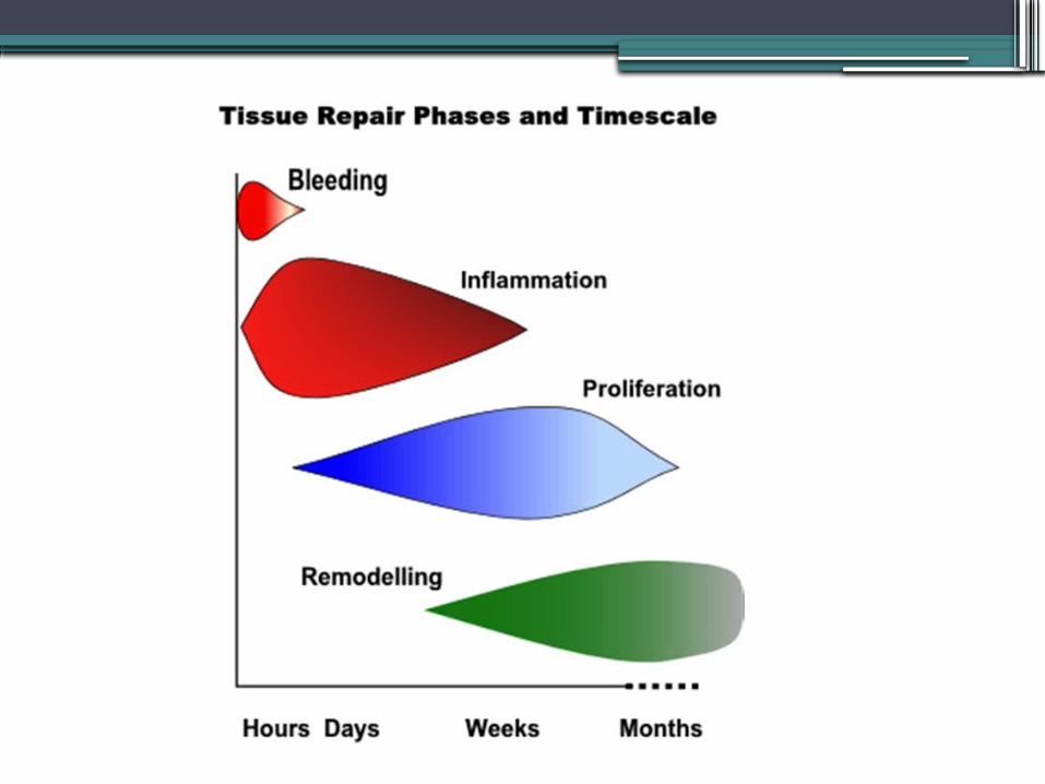
Tissue Repair
•3 phases:1. Inflammation2. Organization3. Regeneration/Fibrosis
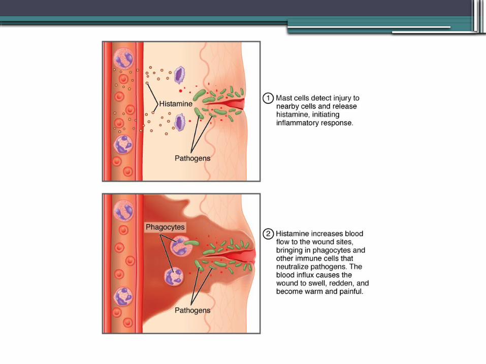
Inflammation
•nonspecific reaction • injury causes tissue cells, macrophages,
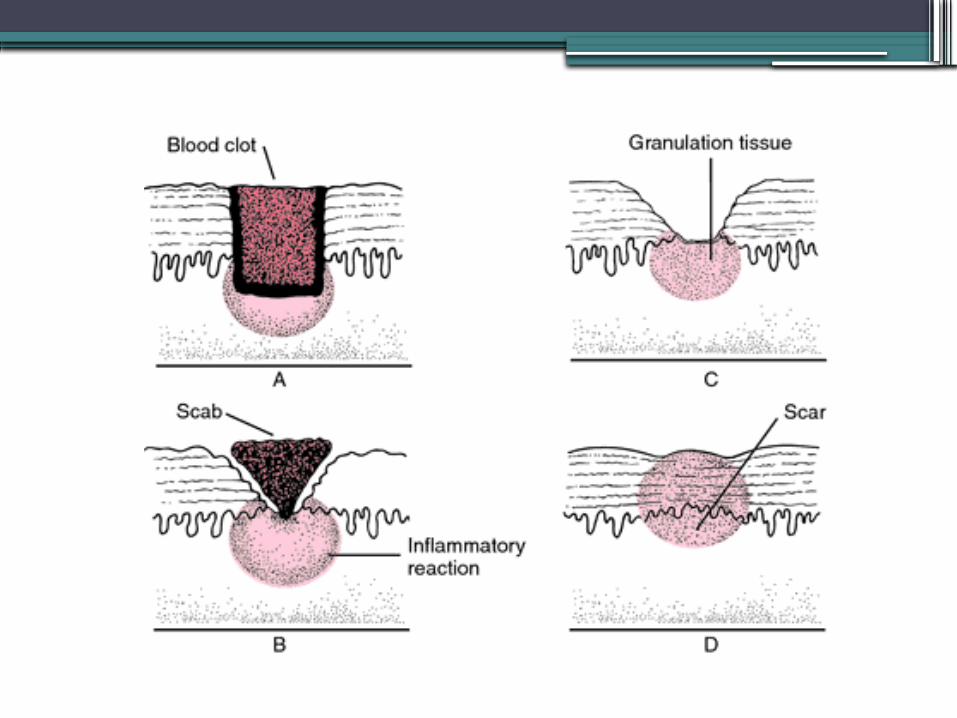
mast cells inflammatory chemicals capillaries dilate & become more permeable neutrophils, monocytes & plasma fluid (clotting factors, abys) enter injured area form clot stops bleeding, holds wound together, isolating injured area giving some protection to infection scab forms
Inflammation
•Characterized by area of:•redness (rubor)•swelling •+/- pain (dolor)•warmth
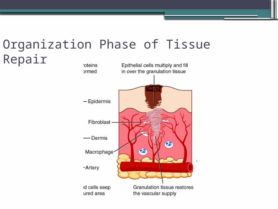
Organization Phase of Tissue Repair
•begins during inflammatory pahse•blood clot replaced by granulation tissue:a friable, delicate tissue▫capillaries ▫fibroblasts collagen, growth factors
when phase complete become inactive or apoptosis
▫macrophages phagocytize injured cells/cell debris, clot
Organization Phase of Tissue Repair
Regeneration & Fibrosis Phase
•fibrous tissue beneath scab matures epithelium regenerate until it resembles surrounding skin
•+/- scar tissue
Injury Below Skin
•skin surface not breached•usually involves infection (pimple, sore throat)▫minor : heals by regeneration only▫more serious clot formation
possibly scarring
Scar Tissue
•permanent fibrous patch•if wound was extensive replaced only
with scar tissue does not replace function of normal tissue
•adhesions: scar tissue that forms around irritated organs in abdominopelvic cavity or w/in hollow organs
Healing in Different Tissues
•heal well: epithelium, areolar CT, dense irregular CT, bone, blood forming tissue
•moderate capacity to heal: smooth muscle, dense regular CT
•poor healing: skeletal muscle, cartilage•NO healing: cardiac muscle, nerve tissue

•https://www.dnalc.org/resources/3d/cellsignals.html