Embed Size (px)
Citation preview
Tumour necrosis factor (TNF) is a key signalling protein in the immune system. As a regulatory cytokine, TNF orchestrates communication between immune cells and controls many of their functions1. TNF is best known for its role in leading immune defences to protect a localized area from invasion or injury but it is also involved in controlling whether target cells live or die.
In autoimmunity, defective immune cells provoke destruction of the body’s own proteins, cells and tissues. This can lead to the development of autoimmune dis-eases including rheumatoid arthritis, Crohn’s disease, multiple sclerosis, lupus, type 1 diabetes and Sjogren’s syndrome. Rheumatoid arthritis and Crohn’s disease, among others, have benefited from regulatory approved anti-TNF therapies when administered with other immunosuppressive therapies2,3. This is because rheuma-toid arthritis, as well as other diseases, may be associ-ated with excessive levels of TNF. However, although anti-TNF therapies can reduce the disease-associated inflammation, they do not reverse the underlying mechanisms of autoimmunity and sometimes lead to adverse effects. These can include the development of additional autoimmune diseases such as lupus, type 1 diabetes, uveitis, multiple sclerosis, psoriasis, as well as lymphomas (see FDA’s MedWatch Safety Alerts — September 2009: Stronger Warnings for TNF Blockers; see Further information)4–20. Moreover, current autoim-mune therapies cannot reverse the course of illness, are associated with adverse effects and cannot successfully control morbidity and mortality associated with the disease21. Increased understanding of the complex roles of TNF, its receptor activity and signalling pathways is
therefore crucial for the identification of new molecular targets and the development of safer and more effective medications.
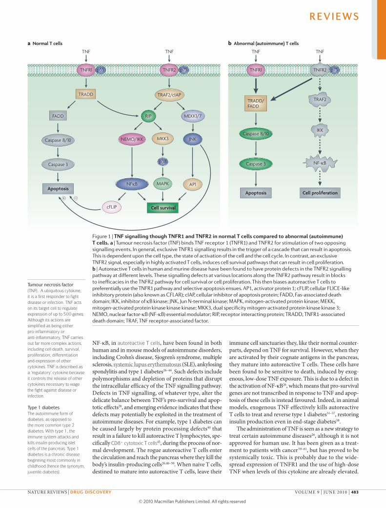
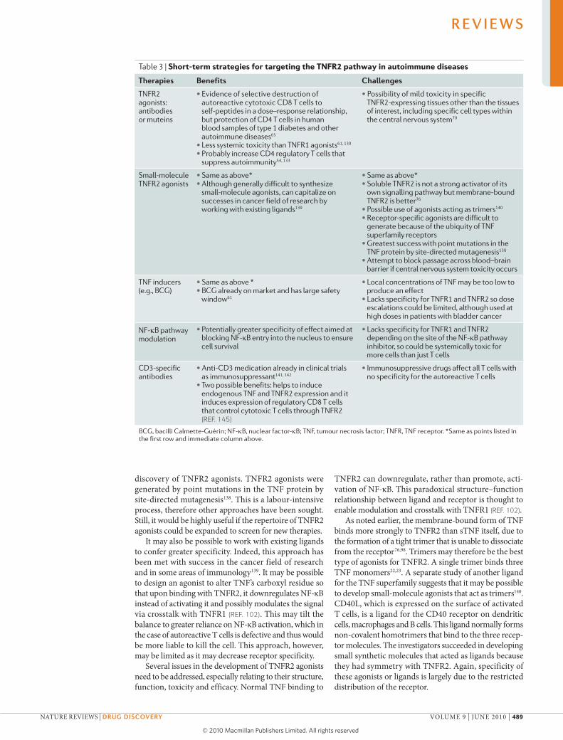
The diversity of TNF’s regulatory functions is becom-ing better understood. TNF can bind to two structurally distinct membrane receptors on target cells — TNFR1 (also known as p55 and TNFRSF1A) or TNFR2 (also known as p75 and TNFRSF1B)22,23 — to activate two separate intracellular signalling pathways to gene tran-scription24. TNFR1 is expressed on nearly all cells of the body, including the entire lymphoid system, whereas TNFR2 exhibits more restricted expression, being found on certain subpopulations of immune cells and a few other cell types. In general, TNF largely relies on TNFR1 for apoptosis and on TNFR2 for any function related to T-cell survival. However, there is some degree of recep-tor crosstalk and overlapping function, depending on the activation state of the cell, among a host of other factors25. TNF binding to TNFR1 activates apoptosis through two pathways, involving the adaptor proteins TNFR1-associated death domain (TRADD) and Fas-associated death domain (FADD). By contrast, TNFR2 signalling involves the mobilization and nuclear entry of the transcription factor nuclear factor-κB (NF-κB) to promote transcription of pro-survival genes (FIG. 1).
The precise aetiologies of autoimmune diseases are heterogeneous, yet overlap to some degree in their genetic, post-translational and environmental origin. The specific causes of some autoimmune diseases are now emerging and are facilitating the identification of potential new therapeutic approaches. Various defects in the TNF signalling pathway that act through TNFR2 and
Immunobiology Laboratory, Room 3602, Building 149, Massachusetts General Hospital and Harvard Medical School, 13th Street, Charlestown, Massachusetts 02129, USA.Correspondence to D.F. e-mail: [email protected]:10.1038/nrd3030 Published online 21 May 2010
CytokineA small protein that is responsible for signalling between immune cells or cells of other types. Once the cytokine is released from an immune cell, it migrates to a target cell with a matching receptor. On activation by the cytokine, the receptor triggers a signal through an internal signal induction pathway that, on reaching the nucleus, causes specific genes to be transcribed.
TNF receptor 2 pathway: drug target for autoimmune diseasesDenise Faustman and Miriam Davis
Abstract | Although drug development has advanced for autoimmune diseases, many current therapies are hampered by adverse effects and the frequent destruction or inactivation of healthy cells in addition to pathological cells. Targeted autoimmune therapies capable of eradicating the rare autoreactive immune cells that are responsible for the attack on the body’s own cells are yet to be identified. This Review presents a new emerging approach aimed at selectively destroying autoreactive immune cells by specific activation of tumour necrosis factor receptor 2 (TNFR2), which is found on autoreactive and normal T lymphocytes, with the potential of avoiding or reducing the toxicity observed with existing therapies.
R E V I E W S
482 | juNe 2010 | VoluMe 9 www.nature.com/reviews/drugdisc
© 20 Macmillan Publishers Limited. All rights reserved10
Nature Reviews | Drug Discovery
TNFR1
Caspase 8/10
Caspase 3
Apoptosis
TRADD/FADD
TNF
TNFR2 p75
NF-κB
IKK
Cell proliferation
TRAF2
TNF
b Abnormal (autoimmune) T cells
TNFR1
Caspase 8/10
Caspase 3
TRADD
FADD
TNF
p75p55
NFκB
NEMO/IKK MKK3
p38
MAPK
cFLIP
JNK
AP1
RIP MEKK1/7
Cell survival
TRAF2/cIAP
TNF
a Normal T cells
Apoptosis
TNFR2
–+
Tumour necrosis factor(TNF). A ubiquitous cytokine, it is a first responder to fight disease or infection. TNF acts on its target cell to regulate expression of up to 500 genes. Although its actions are simplified as being either pro-inflammatory or anti-inflammatory, TNF carries out far more complex actions, including cell death, survival, proliferation, differentiation and expression of other cytokines. TNF is described as a ‘regulatory’ cytokine because it controls the release of other cytokines necessary to wage the fight against disease or infection.
Type 1 diabetesThe autoimmune form of diabetes, as opposed to the more common type 2 diabetes. With type 1, the immune system attacks and kills insulin-producing islet cells of the pancreas. Type 1 diabetes is a chronic disease beginning most commonly in childhood (hence the synonym, juvenile diabetes).
NF-κB, in autoreactive T cells, have been found in both human and in mouse models of autoimmune disorders, including Crohn’s disease, Sjogren’s syndrome, multiple sclerosis, systemic lupus erythematosus (Sle), ankylosing spondylitis and type 1 diabetes26–44. Such defects include polymorphisms and depletion of proteins that disrupt the intracellular efficacy of the TNF signalling pathway. Defects in TNF signalling, of whatever type, alter the delicate balance between TNF’s pro-survival and apop-totic effects26, and emerging evidence indicates that these defects may potentially be exploited in the treatment of autoimmune diseases. For example, type 1 diabetes can be caused largely by protein processing defects40 that result in a failure to kill autoreactive T lymphocytes, spe-cifically CD8+ cytotoxic T cells45, during the process of nor-mal development. The rogue autoreactive T cells enter the circulation and reach the pancreas where they kill the body’s insulin-producing cells26,46–50. When naive T cells, destined to mature into autoreactive T cells, leave their
immune cell sanctuaries they, like their normal counter-parts, depend on TNF for survival. However, when they are activated by their cognate antigens in the pancreas, they mature into autoreactive T cells. These cells have been found to be sensitive to death, induced by exog-enous, low-dose TNF exposure. This is due to a defect in the activation of NF-κB26, which means that pro-survival genes are not transcribed in response to TNF and apop-tosis of these cells is instead favoured. Indeed, in animal models, exogenous TNF effectively kills autoreactive T cells to treat and reverse type 1 diabetes51–57, restoring insulin production even in end-stage diabetes58.
The administration of TNF is seen as a new strategy to treat certain autoimmune diseases26, although it is not approved for human use. It has been given as a treat-ment to patients with cancer59–61, but has proved to be systemically toxic. This is probably due to the wide-spread expression of TNFR1 and the use of high-dose TNF when levels of this cytokine are already elevated.
Figure 1 | TNF signalling though TNFR1 and TNFR2 in normal T cells compared to abnormal (autoimmune) T cells. a | Tumour necrosis factor (TNF) binds TNF receptor 1 (TNFR1) and TNFR2 for stimulation of two opposing signalling events. In general, exclusive TNFR1 signalling results in the trigger of a cascade that can result in apoptosis. This is dependent upon the cell type, the state of activation of the cell and the cell cycle. In contrast, an exclusive TNFR2 signal, especially in highly activated T cells, induces cell survival pathways that can result in cell proliferation. b | Autoreactive T cells in human and murine disease have been found to have protein defects in the TNFR2 signalling pathway at different levels. These signalling defects at various locations along the TNFR2 pathway result in blocks to inefficacies in the TNFR2 pathway for cell survival or cell proliferation. This then biases autoreactive T cells to preferentially use the TNFR1 pathway and selective apoptosis ensues. AP1, activator protein 1; cFLIP, cellular FLICE-like inhibitory protein (also known as CFLAR); cIAP, cellular inhibitor of apoptosis protein; FADD, Fas-associated death domain; IKK, inhibitor of κB kinase; JNK, Jun N-terminal kinase; MAPK, mitogen-activated protein kinase; MEKK, mitogen-activated protein kinase kinase kinase; MKK3, dual specificity mitogen-activated protein kinase kinase 3; NEMO, nuclear factor-κB (NF-κB) essential modulator; RIP, receptor interacting protein; TRADD, TNFR1-associated death domain; TRAF, TNF receptor-associated factor.
R E V I E W S
NATuRe ReVIeWS | DRug DiscoveRy VoluMe 9 | juNe 2010 | 483
© 20 Macmillan Publishers Limited. All rights reserved10

Nuclear factor-κB(NF-κB). One of the most prevalent and important transcription factors. It is an intracellular protein that, when activated, directly stimulates genes to induce expression. T cells rely on NF-κB for their survival.
Autoreactive T cellA defective subset of T lymphocytes, or T cells, that attacks and kills the body’s own regular T-cell target proteins, for example, insulin, instead of attacking the same external targets. Although autoreactive T cells are supposed to be destroyed early in life, this rare type of T cell survives to mount autoimmune diseases.
CD8+ cytotoxic T cellA subset of T cells that directly kills its target proteins on cells. CD8 refers to a particular type of protein on the surface of these killer cells. When CD8 T cells are autoreactive, they kill the T cell’s natural target proteins on cells.
T-cell educationDescribes the removal of newly formed autoreactive T cells and is the process by which antigen presenting cells properly present self peptides in the exterior facing human leukocyte antigen class I grooves. For autoreactive T cells this binding to self peptides results in death of these potentially bad cells prior to their release into the circulation.
CD4+ T cellA subset of T cells not directly responsible for killing target cells. Instead this type recruits other types of immune cells to assist in the battle. These cells bear CD4 proteins on their cellular surface. Some subpopulations of CD4 T cells can assist CD8+ cytotoxic T cells. Other subsets of CD4+ cells, called regulatory T cells function to turn off CD8+ cytotoxic T cells.
However, given the limited distribution of TNFR2 in contrast to TNFR1, the possibility of specifically targeting the TNFR2 protein has emerged as a safer therapeutic strategy. Several studies have now demonstrated the efficacy and safety of TNFR2-specific agonists in pro-moting apoptosis of autoreactive T cells in primates and in mouse models39,54,62–65.
TNF in development and autoimmunityTNF has several pre-eminent roles during normal devel-opment: it shapes the efficacy of the immune system and guards against infectious diseases, cancer and auto-immune diseases24. Released primarily by macrophages in the early stages of normal growth and development, TNF continues throughout life to exert regulatory roles over immune cells by activating genes responsible for inflammation, proliferation, differentiation and apop-tosis. These functions facilitate the proliferation of immune cell clones, especially of T cells, to counter a pathological infection or invasion. They also allow the differentiation and recruitment of naive immune cells to continue waging the battle, as well as the destruc-tion of superfluous immune cell clones to limit internal inflammation and tissue damage once the infection or invasion has resolved. To carry out the complex array of functions, TNF acts on the two receptors TNFR1 and TNFR2; generally relying on TNFR2 for any function related to T-cell survival and on TNFR1 for apoptosis. However, research in the past decade has surprisingly shown some degree of overlap in function of the two receptors (see below).
During the development of autoimmunity, patho-logical progenitors to T cells and other immune cell types proliferate and mature in the thymus, along with their normal counterparts. Most of these immature immune cells die by apoptosis, an orderly demise that is essential to remove defective immune-cell progenitors, but some will subsequently differentiate into autoreac-tive T cells. Determining which cells live or die is the purpose of T-cell education. Failures in T-cell education can generate autoreactive but still naive T cells. They escape from apoptosis during T-cell education and enter the circulation. once there, removed from immunologi-cally privileged sites, they differentiate into autoreactive T cells on encountering specific self-antigens46. Failed T-cell education can lead to various autoimmune dis-eases, including type 1 diabetes, Crohn’s disease, multiple sclerosis and Sjogren’s syndrome. Produced by thymic stromal cells and associated with immune-cell differen-tiation, TNF may even contribute to the pathogenesis of autoimmunity by playing a role in lineage commitment of T cells66,67. Although TNF exposure in the circulation could potentially reach sufficient levels to kill these cells once they are activated and before they reach their targets, why this generally does not happen is unknown. That said, low TNF gene versions are found in some animal models of spontaneous autoimmunity. one hypothesis is that TNF-induced death is concentration dependent and that intrinsic defects exist in the signalling pathway of the naive cells at the time of TNF selection, prior to release into the circulation68.
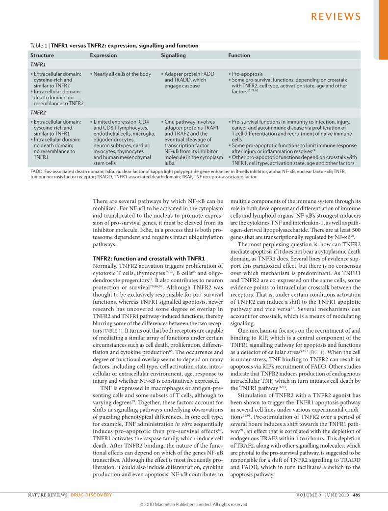
TNFR expression, structure and signallingTNFR2 and TNFR1 have marked differences in expres-sion patterns, structure, signalling and function (TABLE 1). TNFR2 exhibits far more limited expression than TNFR1, with the latter being expressed on almost every cell type in the body. TNFR2 expression is restricted to certain T-cell subpopulations, including lymphocytes (CD4 and CD8 cells)69, endothelial cells, microglia and specific neuron subtypes70,71, oligodendrocytes72,73, cardiac myo-cytes74, thymocytes75,76 and human mesenchymal stem cells77. Human mesenchymal stem cells can differentiate into various cell types that are needed for tissue regener-ation or repair, including the insulin-secreting islets of the pancreas78,79. Neither TNF receptor type is expressed on erythrocytes.
TNFR2’s more restricted expression does not neces-sarily reflect the role, level of expression, circumstance or importance of its signalling function, as will be seen in the following section. For example, each of the cell types bearing TNFR2 also has TNFR1. The ratio of expression of TNFR1 to TNFR2 and the signalling behind these receptors typically fluctuates in relation to the cell type and its functional roles. For immune cells, the state of prior activation of the cell is also a key variable25,68.
Structural differences between TNFR2 and TNFR1 have been reported22,23 and distinctions in TNFR1-specific versus TNFR2-specific binding to TNF or variants of TNF, often referred to as muteins, have been identified. The TNFR1-selective candidates were highly mutated near residue 30, whereas TNFR2-selective candidates were highly mutated near residue 140. As in past studies of designer muteins with receptor specificity to TNFR1 or to TNFR2, it is possible to modify the endogenous TNF structure to obtain receptor-specific binding. Such structure–function analysis makes the development of new and more specific TNFR ligands possible.
TNFR1 and TNFR2 are proteins with extracellular, transmembrane and cytoplasmic components. The two receptors have similar extracellular sequences that are rich in cysteine, the hallmark of the TNF superfamily. However, TNFR1 alone possesses a cytoplasmic death domain, an 80 amino-acid sequence that rapidly engages the apoptotic signalling pathway of the cells. The cyto-plasmic domain of TNFR2 bears no structural or functional resemblance to that of TNFR1 (REFS 80,81).
of TNF’s two receptors, most is known about TNFR1 because of its pervasive expression. In general, TNF binding to TNFR1 mediates apoptosis by two major signalling pathways. It can recruit and cluster the adaptor protein TRADD, which in turn activates one apoptotic pathway. The second pathway relies on TNFR1’s recruit-ment of the intracellular adaptor proteins, receptor inter-acting protein (RIP; also known as RIPK1) and FADD, to coordinate downstream signalling by the caspase cascade82 (FIG. 1a).
TNFR2 also uses distinct signalling pathways (FIG. 1a). Signalling begins with recruitment of the adaptor proteins TNF receptor-associated factor 1 (TRAF1) and TRAF2 (REFS 83,84) and ends with mobilization of the pro-survival transcription factor NF-κB so that it can enter the nucleus to promote gene transcription.
R E V I E W S
484 | juNe 2010 | VoluMe 9 www.nature.com/reviews/drugdisc
© 20 Macmillan Publishers Limited. All rights reserved10
There are several pathways by which NF-κB can be mobilized. For NF-κB to be activated in the cytoplasm and translocated to the nucleus to promote expres-sion of pro-survival genes, it must be cleaved from its inhibitor molecule, IκBα, in a process that is both pro-teasome dependent and requires intact ubiquitylation pathways.
TNFR2: function and crosstalk with TNFR1Normally, TNFR2 activation triggers proliferation of cytotoxic T cells, thymocytes75,76, B cells85 and oligo-dendrocyte progenitors72. It also contributes to neuron protection or survival70,86,87. Although TNFR2 was thought to be exclusively responsible for pro-survival functions, whereas TNFR1 signalled apoptosis, newer research has uncovered some degree of overlap in TNFR2 and TNFR1 pathway-induced functions, thereby blurring some of the differences between the two recep-tors (TABLE 1). It turns out that both receptors are capable of mediating a similar array of functions under certain circumstances such as cell death, proliferation, differen-tiation and cytokine production88. The occurrence and degree of functional overlap seems to depend on many factors, including cell type, cell activation state, intra-cellular or extracellular environment, age, response to injury and whether NF-κB is constitutively expressed.
TNF is expressed in macrophages or antigen-pre-senting cells and some subsets of T cells, although to varying degrees79. Together, these factors account for shifts in signalling pathways underlying observations of puzzling phenotypical differences. In one cell type, for example, TNF administration in vitro sequentially induces pro-apoptotic then pro-survival effects89. TNFR1 activates the caspase family, which induce cell death. After TNFR2 binding, the nature of the func-tional effects can depend on which of the genes NF-κB transcribes. Although the effect is most frequently pro-liferation, it could also include differentiation, cytokine production and even apoptosis. NF-κB contributes to
multiple components of the immune system through its role in both development and differentiation of immune cells and lymphoid organs. NF-κB’s strongest inducers are the cytokines TNF and interleukin-1, as well as path-ogen-derived lipopolysaccharide. There are at least 500 genes that are transcriptionally regulated by NF-κB90.
The most perplexing question is: how can TNFR2 mediate apoptosis if it does not bear a cytoplasmic death domain, as TNFR1 does. Several lines of evidence sup-port this paradoxical effect, but there is no consensus over which mechanism is predominant. As TNFR1 and TNFR2 are co-expressed on the same cells, some evidence points to intracellular crosstalk between the receptors. That is, under certain conditions activation of TNFR2 can induce a shift to the TNFR1 apoptotic pathway and vice versa91. Several mechanisms can account for crosstalk, which is a means of modulating signalling.
one mechanism focuses on the recruitment of and binding to RIP, which is a central component of the TNFR1 signalling pathway for apoptosis and functions as a detector of cellular stress92,93 (FIG. 1). When the cell is under stress, TNF binding to TNFR2 can result in apoptosis via RIP’s recruitment of FADD. other studies indicate that TNFR2 induces production of endogenous intracellular TNF, which in turn initiates cell death by the TNFR1 pathway76,94.
Stimulation of TNFR2 with a TNFR2 agonist has been shown to trigger the TNFR1 apoptosis pathway in several cell lines under various experimental condi-tions91,95. Pre-stimulation of TNFR2 over a period of several hours induces a shift towards the TNFR1 path-way91, an effect that is correlated with the depletion of endogenous TRAF2 within 1 to 6 hours. This depletion of TRAF2, along with other signalling molecules, which are pivotal to the pro-survival pathway, is suggested to be responsible for a shift of TNFR2 signalling to TRADD and FADD, which in turn facilitates a switch to the apoptosis pathway.
Table 1 | TNFR1 versus TNFR2: expression, signalling and function
structure expression signalling Function
TNFR1
• Extracellular domain: cysteine-rich and similar to TNFR2
• Intracellular domain: death domain; no resemblance to TNFR2
• Nearly all cells of the body • Adapter protein FADD and TRADD, which engage caspase
• Pro-apoptosis• Some pro-survival functions, depending on crosstalk
with TNFR2, cell type, activation state, age and other factors25,79,91
TNFR2
• Extracellular domain: cysteine-rich and similar to TNFR1
• Intracellular domain: no death domain; no resemblance to TNFR1
• Limited expression: CD4 and CD8 T lymphocytes, endothelial cells, microglia, oligodendrocytes, neuron subtypes, cardiac myocytes, thymocytes and human mesenchymal stem cells
• One pathway involves adapter proteins TRAF1 and TRAF2 and the eventual cleavage of transcription factor NF-κB from its inhibitor molecule in the cytoplasm IκBα
• Pro-survival functions in immunity to infection, injury, cancer and autoimmune disease via proliferation of T cell differentiation and recruitment of naive immune cells
• Some pro-apoptotic functions to limit immune response after injury or inflammation resolves24
• Other pro-apoptotic functions depend on crosstalk with TNFR1, cell type, activation state, age and other factors
FADD, Fas-associated death domain; IκBα, nuclear factor of kappa light polypeptide gene enhancer in B-cells inhibitor, alpha; NF-κB, nuclear factor-κB; TNFR, tumour necrosis factor receptor; TRADD, TNFR1-associated death domain; TRAF, TNF receptor-associated factor.
R E V I E W S
NATuRe ReVIeWS | DRug DiscoveRy VoluMe 9 | juNe 2010 | 485
© 20 Macmillan Publishers Limited. All rights reserved10
The prior stimulation of TNFR2 with a TNFR2 ago-nist, may also result in depletion of the cellular inhibitor of apoptosis protein (cIAP) in the cytoplasmic domain of TRAF2. under normal conditions cIAP is recruited by TRAF2 and becomes part of the signalling complex that steers TNFR2 activation towards the pro-survival pathway. With prior stimulation of TNFR2, less cIAP is available and TRAF2 is less capable of forming the pro-survival signalling complex. There is then a shift towards reliance upon the TNFR1 pathway leading to apop-tosis, an event some think could even be independent of TNF91,99–101.
The binding kinetics of TNF to TNFR2 is another factor known to govern the frequency or type of TNF signalling. First synthesized by the body as a membrane-bound protein, TNF can remain membrane-bound yet still act as a TNFR2 ligand, or it can be cleaved from the membrane to become soluble. Membrane-bound TNF (mTNF) binds to the T-cell receptor through cell-to-cell contact. In fact, mTNF binds more strongly to TNFR2 than soluble TNF does76,96. It may be that TNFR2 only becomes fully activated with mTNF because it is a tight trimer that cannot dissociate once it binds96–98.
Finally, TNFR2 does not strongly activate its own signalling pathway. TNFR2 carries an unusual carboxy-terminal TRAF2-binding site, T2bs-C, which negatively regulates TNFR2’s binding to TRAF2, the first step in the TNFR2 intracellular signalling pathway102. Consequently, normal TNF binding to TNFR2 can downregulate, rather than promote, activation of NF-κB. This paradoxical structure–function relation-ship between ligand and receptor is thought to allow another type of modulation and crosstalk with TNFR1 (REF. 102).
In summary, research has discovered several mecha-nisms that affect the functional consequences of TNF signalling through TNFR2. They are determined by a host of factors, including conformation of TNFR1 and TNFR2, recruitment of adaptor molecules, cell type, cIAP concentration, activation state and intracellular versus extracellular concentration of TNF103,104.
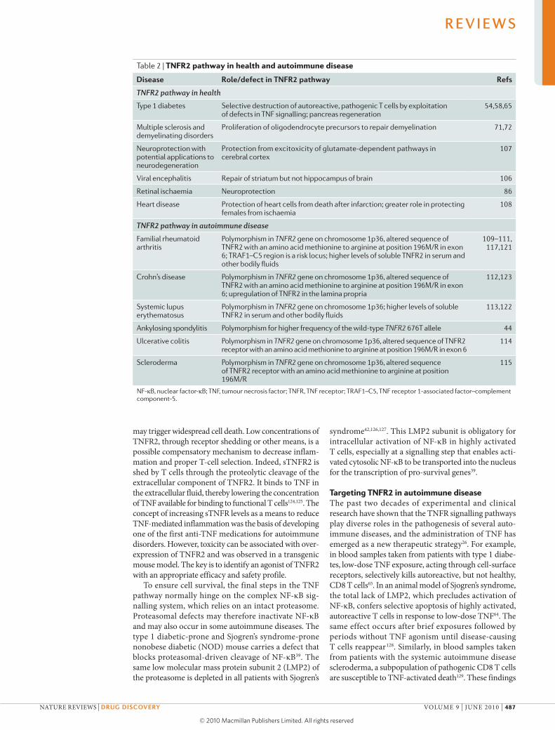
TNFR2 pathways benefit healthSignalling through the TNFR2 pathway may offer pro-tective roles in several disorders, including autoimmune diseases, heart disease, demyelinating and neurodegen-erative disorders and infectious disease (TABLE 2). Several in vitro and in vivo studies suggest that TNFR2 agonism is associated with pancreatic regeneration, cardioprotec-tion, remyelination, survival of some neuron subtypes and stem cell proliferation65,71,72,86,105–108.
In a mouse model, targeted deletion of the Tnfr2 gene is associated with higher rates of heart failure and reduced survival after infarction108. In female mice, the TNFR2 sig-nalling pathway is the one by which TNF protects heart cells after ischaemia in an isolated heart preparation105, which may help explain why females recover better from heart attacks than males do. In addition, in blood speci-mens taken from patients with type 1 diabetes, an agonist for the TNFR2 signalling pathway selectively kills auto-reactive T cells but not healthy T cells65. In animal models
of diabetic autoimmunity, the elimination of autoreac-tive T cells with low-dose TNF was also associated with massive regeneration in the pancreas54,58. Furthermore, the combination of TNF with a TNFR2 agonist neither potentiated nor inhibited the effect, suggesting that it is mediated through the TNFR2 pathway65. Another study found that TNFR2 is essential for TNF-induced regenera-tion of oligodendrocyte precursor cells that form myelin72, which may be of importance in the treatment of multiple sclerosis and other demyelinating disorders, whether or not they are of autoimmune origin. In addition, after viral encephalitis in knockout mice, the TNFR2 pathway is used by TNF to specifically repair the brain’s stria-tum, whereas the TNFR1 pathway is used to repair the hippo campus106. Finally, in a model of retinal ischaemia in knockout mice, TNFR2 promotes neuroprotection, whereas TNFR1 promotes neurodegeneration86.
TNFR2 signalling in autoimmune diseasesDisruption of TNF and the downstream NF-κB signal-ling pathway occurs across several human autoimmune disorders, including Crohn’s disease, Sjogren’s syndrome, multiple sclerosis, Sle, ankylosing spondylitis and type 1 diabetes26–44, and is seen in several murine models of autoimmune diseases. Various defects in the TNFR2 pathway, due to polymorphisms in the TNFR2 gene, upregulated expression of TNFR2 and TNFR2 shedding, have been implicated in the pathology of several auto-immune disorders (FIG. 1b; TABLE 2).
In controlled studies, polymorphisms in TNFR2 have been found in some patients with familial rheumatoid arthritis109–111, Crohn’s disease112, Sle113, ankylosing spondylitis44, ulcerative colitis114 and autoimmune-related conditions such as graft-versus-host disease that is asso-ciated with scleroderma risk115. Common to several autoimmune disorders, but not to type 1 diabetes, is a polymorphism on chromosome 1p36, involving a substi-tution of the amino acid methionine to arginine at posi-tion 196 (196M/R) in exon 6 (REF. 79). The consequence of this polymorphism may be to alter binding kinetics between TNF and TNFR2, which in turn reduces signal activation through NF-κB116. In a large genome-wide study, the TRAF1 and complement component-5 (C5) region, which is essential to TNFR2 signalling, was found to be a risk locus for rheumatoid arthritis, with the highest risk-conferring region outside the human leukocyte antigen region117.
TNFR2 expression may also be upregulated in autoimmune disease79. Administration of TNF to patients produces higher systemic levels of soluble TNFR1 and TNFR2 (sTNFR1 and sTNFR2), presumably through receptor shedding into the extracellular space118,119. The degree of the increase in healthy humans and in primates correlates with greater TNF stimulation120. Patients with familial rheumatoid arthritis121 and Sle who have poly-morphisms in TNFR2 (REF. 122), exhibit higher levels of sTNFR2 but not sTNFR1 in serum or other bodily fluids. unlike TNFR1, TNFR2 is upregulated in the lamina pro-pria of mice with Crohn’s disease and it causes in vivo experimental colitis123. The concentration of TNFR2 in bodily fluids plays a pivotal role. Too high a concentration
R E V I E W S
486 | juNe 2010 | VoluMe 9 www.nature.com/reviews/drugdisc
© 20 Macmillan Publishers Limited. All rights reserved10
may trigger widespread cell death. low concentrations of TNFR2, through receptor shedding or other means, is a possible compensatory mechanism to decrease inflam-mation and proper T-cell selection. Indeed, sTNFR2 is shed by T cells through the proteolytic cleavage of the extracellular component of TNFR2. It binds to TNF in the extracellular fluid, thereby lowering the concentration of TNF available for binding to functional T cells124,125. The concept of increasing sTNFR levels as a means to reduce TNF-mediated inflammation was the basis of developing one of the first anti-TNF medications for autoimmune disorders. However, toxicity can be associated with over-expression of TNFR2 and was observed in a transgenic mouse model. The key is to identify an agonist of TNFR2 with an appropriate efficacy and safety profile.
To ensure cell survival, the final steps in the TNF pathway normally hinge on the complex NF-κB sig-nalling system, which relies on an intact proteasome. Proteasomal defects may therefore inactivate NF-κB and may also occur in some autoimmune diseases. The type 1 diabetic-prone and Sjogren’s syndrome-prone nonobese diabetic (NoD) mouse carries a defect that blocks proteasomal-driven cleavage of NF-κB39. The same low molecular mass protein subunit 2 (lMP2) of the proteasome is depleted in all patients with Sjogren’s
syndrome42,126,127. This lMP2 subunit is obligatory for intracellular activation of NF-κB in highly activated T cells, especially at a signalling step that enables acti-vated cytosolic NF-κB to be transported into the nucleus for the transcription of pro-survival genes39.
Targeting TNFR2 in autoimmune disease The past two decades of experimental and clinical research have shown that the TNFR signalling pathways play diverse roles in the pathogenesis of several auto-immune diseases, and the administration of TNF has emerged as a new therapeutic strategy26. For example, in blood samples taken from patients with type 1 diabe-tes, low-dose TNF exposure, acting through cell-surface receptors, selectively kills autoreactive, but not healthy, CD8 T cells65. In an animal model of Sjogren’s syndrome, the total lack of lMP2, which precludes activation of NF-κB, confers selective apoptosis of highly activated, autoreactive T cells in response to low-dose TNF64. The same effect occurs after brief exposures followed by periods without TNF agonism until disease-causing T cells reappear128. Similarly, in blood samples taken from patients with the systemic autoimmune disease scleroderma, a subpopulation of pathogenic CD8 T cells are susceptible to TNF-activated death129. These findings
Table 2 | TNFR2 pathway in health and autoimmune disease
Disease Role/defect in TNFR2 pathway Refs
TNFR2 pathway in health
Type 1 diabetes Selective destruction of autoreactive, pathogenic T cells by exploitation of defects in TNF signalling; pancreas regeneration
54,58,65
Multiple sclerosis and demyelinating disorders
Proliferation of oligodendrocyte precursors to repair demyelination 71,72
Neuroprotection with potential applications to neurodegeneration
Protection from excitoxicity of glutamate-dependent pathways in cerebral cortex
107
Viral encephalitis Repair of striatum but not hippocampus of brain 106
Retinal ischaemia Neuroprotection 86
Heart disease Protection of heart cells from death after infarction; greater role in protecting females from ischaemia
108
TNFR2 pathway in autoimmune disease
Familial rheumatoid arthritis
Polymorphism in TNFR2 gene on chromosome 1p36, altered sequence of TNFR2 with an amino acid methionine to arginine at position 196M/R in exon 6; TRAF1–C5 region is a risk locus; higher levels of soluble TNFR2 in serum and other bodily fluids
109–111, 117,121
Crohn’s disease Polymorphism in TNFR2 gene on chromosome 1p36, altered sequence of TNFR2 with an amino acid methionine to arginine at position 196M/R in exon 6; upregulation of TNFR2 in the lamina propria
112,123
Systemic lupus erythematosus
Polymorphism in TNFR2 gene on chromosome 1p36; higher levels of soluble TNFR2 in serum and other bodily fluids
113,122
Ankylosing spondylitis Polymorphism for higher frequency of the wild-type TNFR2 676T allele 44
Ulcerative colitis Polymorphism in TNFR2 gene on chromosome 1p36, altered sequence of TNFR2 receptor with an amino acid methionine to arginine at position 196M/R in exon 6
114
Scleroderma Polymorphism in TNFR2 gene on chromosome 1p36, altered sequence of TNFR2 receptor with an amino acid methionine to arginine at position 196M/R
115
NF-κB, nuclear factor-κB; TNF, tumour necrosis factor; TNFR, TNF receptor; TRAF1–C5, TNF receptor 1-associated factor–complement component-5.
R E V I E W S
NATuRe ReVIeWS | DRug DiscoveRy VoluMe 9 | juNe 2010 | 487
© 20 Macmillan Publishers Limited. All rights reserved10
are relevant to other autoimmune diseases that share similar pathogenic defects that impede the function of NF-κB, the key pro-survival transcription factor26–43. The approach of directly killing autoreactive T cells is highly different to the use of anti-cytokine or immunosuppres-sive drug therapies in which continuous drug levels are necessary for a sustained therapeutic effect.
However, the administration of TNF itself is not an immediately feasible approach. It has proved to be sys-temically toxic when given in very high doses in clinical trials of patients with cancer, who already have high TNF levels due to an intrinsic defence system59–61. This toxicity is probably due to the widespread expression of TNFR1. The limited expression of TNFR2 relative to TNFR1, in combination with the functional overlap and crosstalk between these receptors, suggests that TNFR2 agonism may represent a safer therapeutic approach.
Rationale, efficacy and safetyThe rationale for the use of a TNFR2 agonist in the treat-ment of certain autoimmune diseases has been clearly defined and successfully tested in type 1 diabetes. It seems that low-dose TNF exposure or agonism of TNFR2 is a selective means of destroying pathogenic, autoreactive CD8 T cells in animal models, in human cells in vitro39,54,62–64 and in blood samples taken from patients with type 1 diabetes65. examination of blood samples from diabetic and control subjects found a subpopulation of CD8, but not CD4 T cells, that were vulnerable to TNF or TNF agonist-induced death65 in the diabetic subjects. The pattern of CD8 T-cell toxicity exhibited a dose–response relationship and was traced to CD8 T cells that were autoreactive to insulin, a well-established autoantigen in type 1 diabetes. Insulin autoreactive T cells in both mice and humans with type 1 diabetes selectively die in this dose–response manner to TNF. Interestingly, TNFR1 agonists, as opposed to TNFR2 agonists, were ineffective at targeted killing of autoreactive T cells induced in both diabetic and control CD4 T cells. Mild proliferation and, at very high dose, uniform killing of all CD4 T cells65 was seen. Although the safety profile of the TNFR2 ago-nist was not determined, as it was not studied in vivo, it neither destroyed CD4 T cells nor activated cytotoxic T cells to non-autoimmune viral proteins such as epstein–Barr virus or cytomegalovirus65.
owing to the restricted cellular distribution of its receptor, a TNFR2 agonist may demonstrate less systemic toxicity than TNF130. Additionally, because the agonist is given at low doses and intermittently, adverse health effects are minimized. However, in a study conducted in baboons, some adverse effects of a specific TNFR2 ago-nist were observed. These included enhanced thymocyte proliferation in vitro and in vivo, a febrile reaction and a small, transient inflammation produced by infiltration of mononuclear cells upon intradermal administration of the agonist130.
In animal models of non-autoimmune and autoim-mune disorders, TNFR2 stimulation has been shown to result in the generation and expansion of a subpopula-tion of protective CD4 regulatory T (TReg) cells that may suppress autoimmunity54,131–133. TReg cells act in diverse
ways to control CD8-induced autoimmunity. The most common is the destruction of excess CD8 T cells at inflammation sites by inhibition of pro-inflammatory cytokines and release of immunomodulatory cytokines that restore the inflammatory site to normality133.
However, like any new class of therapeutics, there are risks associated with the development of TNFR2 agonists. Indeed, there is evidence of systemic toxicity with over-expression of TNFR2 in a transgenic mouse model134 and higher production of TNFR2 is associated with expe-dited development of inflammatory pathologies, regard-less of TNF levels. upregulated expression of TNFR2 is also associated with several autoimmune disorders, as noted previously109–113,117,121–123. Inflammation should be carefully monitored in drug development, especially if it is sustained or systemic. The finding suggests that it may be necessary to develop tissue-specific or cell-specific therapies or modes of selective administration of TNFR2 agonists. All TNFR2 agonists, especially the antibodies created so far, are not the same in terms of how they mechanistically signal through the receptor. Some TNFR2 antibodies can be agonists, some can be antago-nists and others may bind without eliciting an immune response. For example, a TNFR2 agonist antibody that promotes crosstalk between TNFR1 and TNFR2 and that is independent of defects in the TNFR2 pathway, might carry the risk of widespread toxicity, as it may trig-ger the death of normal as well as abnormal T cells.
It is clear that many factors have profound effects on the nature of TNFR1 versus TNFR2 signalling. Changes in concentration of ligand, receptor, adaptor proteins or other members of the pathway, therefore have the poten-tial to affect drug safety and efficacy. once the receptor is activated, subtle differences in TNF stimulation can alter intracellular signalling, as well as induce post-translational modifications of the signalling proteins, such as the adap-tor protein TRAF2 or others that require ubiquitylation and phosphorylation for a full cycle of activation fol-lowed by degradation135. experimental findings can vary dramatically according to whether the cells are freshly cultured, whether they are of a particular origin and activation state or whether the signalling events through TNF are based on cultured tumour cell lines. Studies of new drugs must examine any changes in members of the TNF pathway and their physiological consequences.
Short-term strategies for targeting TNFR2Short-term strategies for targeting TNFR2 are summa-rized in TABLE 3 and discussed below.
Small-molecule agonists. Medicinal chemists are familiar with the challenges of generating receptor-specific ago-nists for the entire TNF superfamily. Indeed, finding a blocking antagonist is usually an easier feat than finding a receptor-specific agonist. Nevertheless, peptides, anti-bodies and small molecules that engage the TNF super-family have been generated to TNFR2 (REFS 136–138). of the three types of agents, antibody agonists have dem-onstrated the most success at modulating a specific sig-nalling pathway. efforts primarily aimed at finding TNF agonists and antagonists, have included the simultaneous
R E V I E W S
488 | juNe 2010 | VoluMe 9 www.nature.com/reviews/drugdisc
© 20 Macmillan Publishers Limited. All rights reserved10
discovery of TNFR2 agonists. TNFR2 agonists were generated by point mutations in the TNF protein by site-directed mutagenesis138. This is a labour-intensive process, therefore other approaches have been sought. Still, it would be highly useful if the repertoire of TNFR2 agonists could be expanded to screen for new therapies.
It may also be possible to work with existing ligands to confer greater specificity. Indeed, this approach has been met with success in the cancer field of research and in some areas of immunology139. It may be possible to design an agonist to alter TNF’s carboxyl residue so that upon binding with TNFR2, it downregulates NF-κB instead of activating it and possibly modulates the signal via crosstalk with TNFR1 (REF. 102). This may tilt the balance to greater reliance on NF-κB activation, which in the case of autoreactive T cells is defective and thus would be more liable to kill the cell. This approach, however, may be limited as it may decrease receptor specificity.
Several issues in the development of TNFR2 agonists need to be addressed, especially relating to their structure, function, toxicity and efficacy. Normal TNF binding to
TNFR2 can downregulate, rather than promote, acti-vation of NF-κB. This paradoxical structure–function relationship between ligand and receptor is thought to enable modulation and crosstalk with TNFR1 (REF. 102).
As noted earlier, the membrane-bound form of TNF binds more strongly to TNFR2 than sTNF itself, due to the formation of a tight trimer that is unable to dissociate from the receptor76,98. Trimers may therefore be the best type of agonists for TNFR2. A single trimer binds three TNF monomers22,23. A separate study of another ligand for the TNF superfamily suggests that it may be possible to develop small-molecule agonists that act as trimers140. CD40l, which is expressed on the surface of activated T cells, is a ligand for the CD40 receptor on dendritic cells, macrophages and B cells. This ligand normally forms non-covalent homotrimers that bind to the three recep-tor molecules. The investigators succeeded in developing small synthetic molecules that acted as ligands because they had symmetry with TNFR2. Again, specificity of these agonists or ligands is largely due to the restricted distribution of the receptor.
Table 3 | Short-term strategies for targeting the TNFR2 pathway in autoimmune diseases
Therapies Benefits challenges
TNFR2 agonists: antibodies or muteins
• Evidence of selective destruction of autoreactive cytotoxic CD8 T cells to self-peptides in a dose–response relationship, but protection of CD4 T cells in human blood samples of type 1 diabetes and other autoimmune diseases65
• Less systemic toxicity than TNFR1 agonists61, 130
• Probably increase CD4 regulatory T cells that suppress autoimmunity54, 133
• Possibility of mild toxicity in specific TNFR2-expressing tissues other than the tissues of interest, including specific cell types within the central nervous system79
Small-molecule TNFR2 agonists
• Same as above*• Although generally difficult to synthesize
small-molecule agonists, can capitalize on successes in cancer field of research by working with existing ligands139
• Same as above*• Soluble TNFR2 is not a strong activator of its
own signalling pathway but membrane-bound TNFR2 is better76
• Possible use of agonists acting as trimers140
• Receptor-specific agonists are difficult to generate because of the ubiquity of TNF superfamily receptors
• Greatest success with point mutations in the TNF protein by site-directed mutagenesis138
• Attempt to block passage across blood–brain barrier if central nervous system toxicity occurs
TNF inducers (e.g., BCG)
• Same as above *• BCG already on market and has large safety
window61
• Local concentrations of TNF may be too low to produce an effect
• Lacks specificity for TNFR1 and TNFR2 so dose escalations could be limited, although used at high doses in patients with bladder cancer
NF-κB pathway modulation
• Potentially greater specificity of effect aimed at blocking NF-κB entry into the nucleus to ensure cell survival
• Lacks specificity for TNFR1 and TNFR2 depending on the site of the NF-κB pathway inhibitor, so could be systemically toxic for more cells than just T cells
CD3-specific antibodies
• Anti-CD3 medication already in clinical trials as immunosuppressant141, 142
• Two possible benefits: helps to induce endogenous TNF and TNFR2 expression and it induces expression of regulatory CD8 T cells that control cytotoxic T cells through TNFR2 (REF. 145)
• Immunosuppressive drugs affect all T cells with no specificity for the autoreactive T cells
BCG, bacilli Calmette-Guérin; NF-κB, nuclear factor-κB; TNF, tumour necrosis factor; TNFR, TNF receptor. *Same as points listed in the first row and immediate column above.
R E V I E W S
NATuRe ReVIeWS | DRug DiscoveRy VoluMe 9 | juNe 2010 | 489
© 20 Macmillan Publishers Limited. All rights reserved10
TNF inducers. There are some TNF inducers available for study. one is the mycobacterium bovis bacille Calmette-Guérin (BCG) vaccine. Another is the BCG equivalent, a non-formulated version that is commonly called com-plete Freund’s adjuvant (CFA). Although CFA is made with the same bacilli as BCG it does not meet the uS Food and Drug Administration standards of purity and manufacturing for human use. BCG is produced using good manufacturing practices and used as a vaccine for tuberculosis or at very high doses as a treatment for bladder cancer. As a cancer therapeutic, BCG has a wide dose-window of at least 100–1,000 times greater than that of a single vaccination to prevent tuberculosis infections61. Although BCG induction of TNF is not receptor-specific for TNFR1 or TNFR2, it may still be of benefit in treating autoimmune disease as it induces TNF at low levels, thereby possibly avoiding systemic toxicity. Newly synthesized TNF inducers, designed to be more specific to the TNFR2 receptor, may hold promise as another targeted treatment for type 1 diabetes.
NF-κB pathway modulation. In autoimmune diseases, defects in the TNF signalling pathway prevent the pro-survival transcription factor NF-κB from entering the nucleus following TNF therapy39,40. The final stage of typi-cal TNF signalling depends on NF-κB’s cleavage from the inhibitory molecule IκBα to which it is bound in the cyto-plasm, enabling nuclear entry and gene transcription39–41. In the type 1 diabetic-prone and Sjogren’s syndrome-prone NoD mouse, a genetic defect blocks proteasomal-driven cleavage of NF-κB40. This same defect has similarly been identified in all patients suffering from Sjogren’s syndrome127. Therefore, blocking NF-κB’s translocation to the nucleus may provide another potential therapeutic approach to autoimmune disease. However, the challenge with NF-κB pathway modulation is that it would not be specific for TNFR2-expressing cell receptors and thus it is likely to have a less favourable toxicity profile.
CD3-specific antibodies. CD3-specific antibodies offer another approach to modulating TNFR2 activity. T cells of all types possess the distinctive cell-surface marker CD3. CD3-specific antibodies are routinely used as immunosuppressive agents to prevent organ trans-plant rejection. A humanized form of a CD3-specific antibody is being tested in clinical trials for its capacity to slow the onset of complete insulin dependence in new-onset type 1 diabetes141,142. For children with new-onset diabetes, results using two slightly different humanized forms of CD3-specific antibodies141,143 have thus far been promising. A recently published study reported insulin production to be preserved for up to 5 years in patients with type 1 diabetes receiving a single course of the CD3-specific antibody teplizumab144. While a CD3-specific antibody is a general immuno-suppressant, studies of its mechanism of action suggest additional effects to control type 1 diabetes and possibly other autoimmune diseases. Indeed, CD3-specific anti-bodies may help to induce endogenous TNF secretion and TNFR2 expression145, and could therefore be anticipated to exert the same beneficial effects as TNFR2 agonism. The
upregulation of TNF and TNFR1 or TNFR2 is thought to be a consequence of the release of another cytokine, transforming growth factor-β, as a result of the interaction between CD8 T cells and natural killer cells145.
Long-term strategies for targeting TNFR2There are several possible long-term strategies for tar-geting the TNFR2 pathway and selectively destroying autoreactive T cells, although these are at present only speculative. one set of strategies focuses on developing better TNFR2 agonists by improving their specificity, binding duration and affinity to TNFR2. The rationale is that autoreactive T cells are rare and that any means to prolong or to facilitate the actions of TNFR2 ago-nists are more likely to be effective, considering the dose–response relationship between TNFR2 agonists and death of autoreactive target cells65. The likelihood of systemic toxicity with improvements of this kind is still expected to be low, given the large dose-window of tolerability to a TNFR2 agonist130. The cancer field of research has been at the forefront of studying TNFR2 agonists for their utility as adjuncts to anticancer thera-pies. TNF has been found to kill mammalian tumour cells in vitro and in vivo, since its first discovery as an antitumour agent in 1975 (REF. 146). The cancer field of research may offer clues to achieving greater TNFR2 specificity and/or duration of binding.
A related approach would be to inject TNFR2 agonists directly into sites in the body where autoreactive CD8 T cells are generated or sequestered. Sequestered sites could be lymphoid organs such as the thymus or bone marrow. However, injections into the pancreas would be potentially dangerous owing to the possible stimulation of pancreatitis. In other cases, the use of the salivary glands for autoimmune Sjogren’s syndrome would be attractive as direct salivary gland access is possible through the draining of the salivary duct in the oral cavity. The efficacy of direct target delivery into a lymphoid organ would have to be countenanced by the possibility that autoreactive T cells might still be naive at local sites of production or sequestration. They typically mature once they encounter their self-antigen (insulin for example), an encounter that induces them to become activated. It is likely that only activated, autoreactive T cells are vulnerable to TNF expo-sure or to TNFR2 agonists65. TNFR2 agonists can also be of use as topical treatments for skin disease, which may allow the local delivery of high concentrations without causing systemic toxicity. However, the potential toxic-ity of local therapy must be considered when adminis-tering prolonged or continuous TNF. Depending on the binding traits of the TNFR2 agonist, high levels of the agonist may overwhelm TNFR2 signalling, thereby possibly shifting it through crosstalk to the TNFR1 pro-apoptotic pathway. This shift, being independent of underlying defects in TNFR2 signalling, would lead to death of both autoreactive and healthy cells. Although traditionally most autoimmune intervention trials have been centred on newly diagnosed autoimmune patients like type 1 diabetics or even pre-diabetics, accumulating evidence suggests that even in cases in which the auto-immune organ appears to be long destroyed, autoreactive
R E V I E W S
490 | juNe 2010 | VoluMe 9 www.nature.com/reviews/drugdisc
© 20 Macmillan Publishers Limited. All rights reserved10
T cells are present decades after disease onset. This does not mean that the autoreactive T cells are long-lived but instead means the autoreactive T cells are continuously being produced due to attempted but failed regeneration in the organ. This hypothesis is supported by the brisk rejection of transplanted pancreatic islets after years of long-standing disease, and by the continuous production of an autoantibody54,58. Therefore, this treatment approach would not be like immunosuppressive drugs, which must be administered continuously to achieve steady blood levels, but instead involves a bolus therapy to rid the host of autoreactive T cells followed by drug-free intervals, which may eliminate or minimize side effects.
Inactivation or destruction of the pivotal signalling protein TRAF2 offers another approach to enhance the likelihood of apoptosis of autoreactive CD8 T cells. TRAF2 is the adaptor protein that is recruited upon agonist bind-ing to TNFR2. TRAF2 inhibitors have been identified147. As TRAF2 is the locus of crosstalk between the two TNF receptors, its inactivation should shift TNFR2 signalling to the TNFR1 pro-apoptotic pathway. The question of whether TNFR2 inactivation is toxic to normal T cells or even non-immune cells, which would also be shunted to the pro-apoptotic pathway, remains to be answered. As noted earlier, much remains to be understood about crosstalk between the two TNF receptors, the functional overlap between these receptors and the array of condi-tions under which crosstalk occurs, in order to support the rationale for this approach. Targeting any signalling proteins in the TNFR2 pathway that are downstream of TRAF2 also presents limitations as most of them are used for other signalling purposes throughout the body.
An alternative approach is to use antisense therapy as a means to alter TNF signalling, by inhibiting specific mRNAs from being translated into proteins. As poly-morphisms and defects in the NF-κB pathway associated
with various autoimmune diseases33 allow the use of TNF or TNFR2 agonists to selectively kill autoreactive T cells, perhaps NF-κB-targeted antisense therapy, which could block NF-κB function by inhibiting specific mRNAs for NF-κB, could be used to achieve or potentiate this effect. However, there are limitations to the therapeutic use of antisense oligonucleotides, including poor efficacy, off-target effects and achieving targeted delivery to cells and tissues.
Concluding remarksThis Review has identified the untapped potential of the TNFR2 pathway as a therapeutic target for several autoimmune diseases, which share the same or similar defects that render their autoreactive, but not normal, T cells selectively vulnerable to TNF-induced death. Although TNF as a therapy is successful at killing those pathogenic cells, it is likely to be too toxic or to have a narrow range for dosing as one type of TNF receptor (TNFR1) is expressed ubiquitously throughout the body. However, TNFR2, the only other TNF receptor, has a more limited bodily expression and agonists specifically targeting the TNFR2 pathway hold promise as a safer and more effective treatment than TNF or current therapies for several autoimmune diseases.
Research has already demonstrated that TNFR2 ago-nism successfully destroys autoreactive T cells in human blood specimens from hundreds of patients with type 1 diabetes and several other autoimmune diseases. This Review identifies several short-term and long-term therapeutic strategies for developing new treatments aimed at the TNFR2 pathway, with the hope of gal-vanizing the development of safer and more effective treatments for several autoimmune diseases, including type 1 diabetes, Sjogren’s syndrome, multiple sclerosis and scleroderma.
1. Croft, M. The role of TNF superfamily members in T-cell function and diseases. Nature Rev. Immunol. 9, 271–285 (2009).
2. Breedveld, F. C. et al. The PREMIER study: a multicenter, randomized, double-blind clinical trial of combination therapy with adalimumab plus methotrexate versus methotrexate alone or adalimumab alone in patients with early, aggressive rheumatoid arthritis who had not had previous methotrexate treatment. Arthritis Rheum. 54, 26–37 (2006).
3. Klareskog, L. et al. Therapeutic effect of the combination of etanercept and methotrexate compared with each treatment alone in patients with rheumatoid arthritis: double-blind randomised controlled trial. Lancet 363, 675–681 (2004).
4. Chaudhari, U. et al. Efficacy and safety of infliximab monotherapy for plaque-type psoriasis: a randomised trial. Lancet 357, 1842–1847 (2001).
5. Dereure, O. et al. Psoriatic lesions induced by antitumour necrosis factor-alpha treatment: two cases. Br. J. Dermatol. 151, 506–507 (2004).
6. Flendrie, M. et al. Dermatological conditions during TNF-alpha-blocking therapy in patients with rheumatoid arthritis: a prospective study. Arthritis Res. Ther. 7, R666–R676 (2005).
7. Kary, S. et al. New onset or exacerbation of psoriatic skin lesions in patients with definite rheumatoid arthritis receiving tumour necrosis factor alpha antagonists. Ann. Rheum. Dis. 65, 405–407 (2006).
8. Thurber, M., Feasel, A., Stroehlein, J. & Hymes, S. R. Pustular psoriasis induced by infliximab. J. Drugs Dermatol. 3, 439–440 (2004).
9. Verea, M. M., Del Pozo, J., Yebra-Pimentel, M. T., Porta, A. & Fonseca, E. Psoriasiform eruption induced by infliximab. Ann. Pharmacother. 38, 54–57 (2004).
10. Galaria, N. A., Werth, V. P. & Schumacher, H. R. Leukocytoclastic vasculitis due to etanercept. J. Rheumatol. 27, 2041–2044 (2000).
11. Jarrett, S. J. et al. Anti-tumor necrosis factor-alpha therapy-induced vasculitis: case series. J. Rheumatol. 30, 2287–2291 (2003).
12. Charles, P. J., Smeenk, R. J., De Jong, J., Feldmann, M. & Maini, R. N. Assessment of antibodies to double-stranded DNA induced in rheumatoid arthritis patients following treatment with infliximab, a monoclonal antibody to tumor necrosis factor alpha: findings in open-label and randomized placebo-controlled trials. Arthritis Rheum. 43, 2383–2390 (2000).
13. Feldmann, M., Brennan, F. M. & Maini, R. N. Role of cytokines in rheumatoid arthritis. Annu. Rev. Immunol. 14, 397–440 (1996).
14. US Food and Drug Administration. Update on the TNF-alpha blocking agents. US FDA website [online], http://www.fda.gov/ohrms/dockets/ac/03/briefing/3930B1_01_B-TNF.Briefing.htm (2006).
15. Klinkhoff, A. Biological agents for rheumatoid arthritis: targeting both physical function and structural damage. Drugs 64, 1267–1283 (2004).
16. Stokes, M. B. et al. Development of glomerulonephritis during anti-TNF-alpha therapy for rheumatoid arthritis. Nephrol. Dial. Transplant. 20, 1400–1406 (2005).
17. Lipsky, P. E. et al. Infliximab and methotrexate in the treatment of rheumatoid arthritis. Anti-tumor
necrosis factor trial in rheumatoid arthritis with concomitant therapy study group. N. Engl. J. Med. 343, 1594–1602 (2000).
18. Moreland, L. W. et al. Etanercept therapy in rheumatoid arthritis. A randomized, controlled trial. Ann. Intern. Med. 130, 478–486 (1999).
19. Vermeire, S. et al. Autoimmunity associated with anti-tumor necrosis factor alpha treatment in Crohn’s disease: a prospective cohort study. Gastroenterology 125, 32–39 (2003).
20. Tack, C. J., Kleijwegt, F. S., Van Riel, P. L. & Roep, B. O. Development of type 1 diabetes in a patient treated with anti-TNF-alpha therapy for active rheumatoid arthritis. Diabetologia 52, 1442–1444 (2009).
21. Dorman, J. S. et al. The Pittsburgh insulin-dependent diabetes mellitus (IDDM) morbidity and mortality study. Mortality results. Diabetes 33, 271–276 (1984).
22. Mukai, Y. et al. Crystallization and preliminary X-ray analysis of the tumour necrosis factor alpha-tumour necrosis factor receptor type 2 complex. Acta Crystallogr. Sect. F Struct. Biol. Cryst. Commun. 65, 295–298 (2009).This article is the first to use crystallography to find the structure of TNFR1.
23. Mukai, Y. et al. Structure–function relationship of tumor necrosis factor (TNF) and its receptor interaction based on 3D structural analysis of a fully active TNFR1-selective TNF mutant. J. Mol. Biol. 385, 1221–1229 (2009).This article is the first to use phage display techniques to produce receptor-specific TNF mutants to activate only one type of TNFR.
R E V I E W S
NATuRe ReVIeWS | DRug DiscoveRy VoluMe 9 | juNe 2010 | 491
© 20 Macmillan Publishers Limited. All rights reserved10
24. Aggarwal, B. B. Signalling pathways of the TNF superfamily: a double-edged sword. Nature Rev. Immunol. 3, 745–756 (2003).
25. Pimentel-Muinos, F. X. & Seed, B. Regulated commitment of TNF receptor signaling: a molecular switch for death or activation. Immunity 11, 783–793 (1999).
26. Kodama, S., Davis, M. & Faustman, D. L. The therapeutic potential of tumor necrosis factor for autoimmune disease: a mechanistically based hypothesis. Cell. Mol. Life Sci. 62, 1850–1862 (2005).This review article explains the evidence behind the counter-intuitive rationale to investigate TNF for its potential to selectively and effectively kill autoreactive T cells in several autoimmune disorders.
27. Abbott, D. W., Wilkins, A., Asara, J. M. & Cantley, L. C. The Crohn’s disease protein, NOD2, requires RIP2 in order to induce ubiquitinylation of a novel site on NEMO. Curr. Biol. 14, 2217–2227 (2004).
28. Eckmann, L. & Karin, M. NOD2 and Crohn’s disease: loss or gain of function? Immunity 22, 661–667 (2005).
29. Maeda, S. et al. Nod2 mutation in Crohn’s disease potentiates NF-kappaB activity and IL-1beta processing. Science 307, 734–738 (2005).
30. Watanabe, T., Kitani, A., Murray, P. J. & Strober, W. NOD2 is a negative regulator of Toll-like receptor 2-mediated T helper type 1 responses. Nature Immunol. 5, 800–808 (2004).
31. Levine, A. et al. TNF promoter polymorphisms and modulation of growth retardation and disease severity in pediatric Crohn’s disease. Am. J. Gastroenterol. 100, 1598–1604 (2005).
32. van Heel, D. A. et al. Inflammatory bowel disease is associated with a TNF polymorphism that affects an interaction between the OCT1 and NF-κB transcription factors. Hum. Mol. Genet. 11, 1281–1289 (2002).
33. Karban, A. S. et al. Functional annotation of a novel NFKB1 promoter polymorphism that increases risk for ulcerative colitis. Hum. Mol. Genet. 13, 35–45 (2004).
34. Kammer, G. M. & Tsokos, G. C. Abnormal T lymphocyte signal transduction in systemic lupus erythematosus. Curr. Dir. Autoimmun. 5, 131–150 (2002).
35. Hegazy, D. M. et al. NF-κB polymorphisms and susceptibility to type 1 diabetes. Genes Immun. 2, 304–308 (2001).
36. Deng, G. Y., Muir, A., Maclaren, N. K. & She, J. X. Association of LMP2 and LMP7 genes within the major histocompatibility complex with insulin-dependent diabetes mellitus: population and family studies. Am. J. Hum. Genet. 56, 528–534 (1995).
37. Ding, H., Cheng, H., Fu, Z., Yan, L. & Yang, G. Relationship of large multifunctional proteasome 7 gene polymorphism with susceptibility to type 1 diabetes mellitus and DR3 gene. Chin. Med. J. (Engl.) 114, 1263–1266 (2001).
38. Fu, Y., Yan, G., Shi, L. & Faustman, D. Antigen processing and autoimmunity. Evaluation of mRNA abundance and function of HLA-linked genes. Ann. NY Acad. Sci. 842, 138–155 (1998).
39. Hayashi, T. & Faustman, D. Essential role of HLA-encoded proteasome subunits in NF-κB activation and prevention of TNF-α induced apoptosis. J. Biol. Chem. 275, 5238–5247 (2000).
40. Hayashi, T. & Faustman, D. NOD mice are defective in proteasome production and activation of NF-κB. Mol. Cell Biol. 19, 8646–8659 (1999).This article was the first to demonstrate proteasomal defects prevent cleavage of NF-κB from its chaperone protein, IκBα, in a mouse model of type 1 diabetes.
41. Hayashi, T. & Faustman, D. L. Selected contribution: association of gender-related LMP2 inactivation with autoimmune pathogenesis. J. Appl. Physiol. 91, 2804–2815 (2001).
42. Yan, G., Fu, Y. & Faustman, D. L. Reduced expression of Tap1 and Lmp2 antigen processing genes in the nonobese diabetic (NOD) mouse due to a mutation in their shared bidirectional promoter. J. Immunol. 159, 3068–3080 (1997).This article was the first to demonstrate the LMP2 subunit of the proteasome was lacking in subpopulations of the immune system, thus influencing both MHC class I education and interrupting proper NF-κB activation in CD8 T cells, in a mouse model of autoimmunity.
43. Miterski, B. et al. Inhibitors in the NF-κB cascade comprise prime candidate genes predisposing to multiple sclerosis, especially in selected combinations. Genes Immun. 3, 211–219 (2002).
44. Chatzikyriakidou, A., Georgiou, I., Voulgari, P. V. & Drosos, A. A. The role of tumor necrosis factor (TNF)-alpha and TNF receptor polymorphisms in susceptibility to ankylosing spondylitis. Clin. Exp. Rheumatol. 27, 645–648 (2009).
45. Faustman, D. et al. Linkage of faulty major histocompatibility complex class I to autoimmune diabetes. Science 254, 1756–1761 (1991).
46. Faustman, D. L. & Davis, M. The primacy of CD8 T lymphocytes in type 1 diabetes and implications for therapies. J. Mol. Med. 87, 1173–1178 (2009).
47. Wong, F. S. et al. Identification of an MHC class I-restricted autoantigen in type 1 diabetes by screening an organ-specific cDNA library. Nature Med. 5, 1026–1031 (1999).
48. Christianson, S. W., Shultz, L. D. & Leiter, E. H. Adoptive transfer of diabetes into immunodeficient NOD-scid/scid mice. Relative contributions of CD4+ and CD8+ T-cells from diabetic versus prediabetic NOD.NON-Thy-1a donors. Diabetes 42, 44–55 (1993).
49. Yagi, H. et al. Analysis of the roles of CD4+ and CD8+ T cells in autoimmune diabetes of NOD mice using transfer to NOD athymic nude mice. Eur. J. Immunol. 22, 2387–2393 (1992).
50. Pinkse, G. G. et al. Autoreactive CD8 T cells associated with beta cell destruction in type 1 diabetes. Proc. Natl Acad. Sci. USA 102, 18425–18430 (2005).
51. Satoh, J., Seino, H. & Abo, T. Recombinant human tumor necrosis factor α suppresses autoimmune diabetes in nonobese diabetic mice. J. Clin. Invest. 84, 1345–1348 (1989).
52. Grewal, I. S. et al. Local expression of transgene encoded TNF alpha in islets prevents autoimmune diabetes in non-obese diabetic (NOD) mice by preventing the development of autoreactive islet specific T cells. J. Exp. Med. 184, 1963–1974 (1996).
53. Sadelain, M. W. et al. Prevention of diabetes in the BB rat by early immunotherapy using Freund’s adjuvant. J. Autoimmun. 3, 671–680 (1990).
54. Ryu, S., Kodama, S., Ryu, K., Schoenfeld, D. A. & Faustman, D. L. Reversal of established autoimmune diabetes by restoration of endogenous beta cell function. J. Clin. Invest. 108, 63–72 (2001).
55. Nishio, J. et al. Islet recovery and reversal of murine type 1 diabetes in the absence of any infused spleen cell contribution. Science 311, 1775–1778 (2006).
56. Suri, A. et al. Immunological reversal of autoimmune diabetes without hematopoietic replacement of beta cells. Science 311, 1778–1780 (2006).
57. Chong, A. S. et al. Reversal of diabetes in non-obese diabetic mice without spleen cell-derived beta cell regeneration. Science 311, 1774–1775 (2006).
58. Kodama, S., Kuhtreiber, W., Fujimura, S., Dale, E. A. & Faustman, D. L. Islet regeneration during the reversal of autoimmune diabetes in NOD mice. Science 302, 1223–1227 (2003).This article was the first to show that treatment with a TNF inducer was capable of stimulating pancreatic islet cells to be restored and function normally by returning glycaemic levels back to healthy levels in a mouse model of advanced type 1 diabetes.
59. Sidhu, R. S. & Bollon, A. P. Tumor necrosis factor activities and cancer therapy — a perspective. Pharmacol. Ther. 57, 79–128 (1993).
60. Hieber, U. & Heim, M. E. Tumor necrosis factor for the treatment of malignancies. Oncology 51, 142–153 (1994).
61. Balkwill, F. Tumour necrosis factor and cancer. Nature Rev. Cancer 9, 361–371 (2009).
62. Qin, H. Y., Chaturvedi, P. & Singh, B. In vivo apoptosis of diabetogenic T cells in NOD mice by IFN-γ/TNF-α. Int. Immunol. 16, 1723–1732 (2004).
63. Christen, U. et al. A dual role for TNF-alpha in type 1 diabetes: islet-specific expression abrogates the ongoing autoimmune process when induced late but not early during pathogenesis. J. Immunol. 166, 7023–7032 (2001).
64. Tran, S. D. et al. Reversal of Sjogren’s-like syndrome in non-obese diabetic mice. Ann. Rheum. Dis. 66, 812–814 (2007).
65. Ban, L. et al. Selective death of autoreactive T cells in human diabetes by TNF or TNF receptor 2 agonism. Proc. Natl Acad. Sci. USA 105, 13644–13649 (2008).This article was the first to demonstrate that a specific TNFR2 ligand was capable of selectively killing insulin-specific autoreactive T cells in type 1 diabetes.
66. Zuniga-Pflucker, J. C., Di, J. & Lenardo, M. J. Requirement for TNF-alpha and IL-1 alpha in fetal thymocyte commitment and differentiation. Science 268, 1906–1909 (1995).
67. Samira, S. et al. Tumor necrosis factor promotes human T-cell development in nonobese diabetic/severe combined immunodeficient mice. Stem Cells 22, 1085–1100 (2004).
68. Lenardo, M. J. Interleukin-2 programs mouse alpha beta T lymphocytes for apoptosis. Nature 353, 858–861 (1991).
69. Ware, C. F. et al. Tumor necrosis factor (TNF) receptor expression in T lymphocytes. Differential regulation of the type I TNF receptor during activation of resting and effector T cells. J. Immunol. 147, 4229–4238 (1991).
70. Yang, L., Lindholm, K., Konishi, Y., Li, R. & Shen, Y. Target depletion of distinct tumor necrosis factor receptor subtypes reveals hippocampal neuron death and survival through different signal transduction pathways. J. Neurosci. 22, 3025–3032 (2002).
71. McCoy, M. K. & Tansey, M. G. TNF signaling inhibition in the CNS: implications for normal brain function and neurodegenerative disease. J. Neuroinflammation 5, 45 (2008).
72. Arnett, H. A. et al. TNF alpha promotes proliferation of oligodendrocyte progenitors and remyelination. Nature Neurosci. 4, 1116–1122 (2001).
73. Dopp, J. M. et al. Expression of the p75 TNF receptor is linked to TNF-induced NF-κB translocation and oxyradical neutralization in glial cells. Neurochem. Res. 27, 1535–1542 (2002).
74. Irwin, M. W. et al. Tissue expression and immunolocalization of tumor necrosis factor-alpha in postinfarction dysfunctional myocardium. Circulation 99, 1492–1498 (1999).
75. Tartaglia, L. A. et al. The two different receptors for tumor necrosis factor mediate distinct cellular responses. Proc. Natl Acad. Sci. USA 88, 9292–9296 (1991).
76. Grell, M., Becke, F. M., Wajant, H., Mannel, D. N. & Scheurich, P. TNF receptor type 2 mediates thymocyte proliferation independently of TNF receptor type 1. Eur. J. Immunol. 28, 257–263 (1998).
77. Bocker, W. et al. IKK-2 is required for TNF-α-induced invasion and proliferation of human mesenchymal stem cells. J. Mol. Med. 86, 1183–1192 (2008).
78. Chen, L. B., Jiang, X. B. & Yang, L. Differentiation of rat marrow mesenchymal stem cells into pancreatic islet beta-cells. World J. Gastroenterol. 10, 3016–3020 (2004).
79. Carpentier, I., Coornaert, B. & Beyaert, R. Function and regulation of tumor necrosis factor type 2. Curr. Med. Chem. 11, 2205–2212 (2004).This review article provides the most comprehensive portrait and analysis of TNFR2.
80. Tartaglia, L. A., Ayres, T. M., Wong, G. H. & Goeddel, D. V. A novel domain within the 55 kd TNF receptor signals cell death. Cell 74, 845–853 (1993).
81. Hsu, H., Xiong, J. & Goeddel, D. V. The TNF receptor 1-associated protein TRADD signals cell death and NF-κB activation. Cell 81, 495–504 (1995).
82. Wilson, N. S., Dixit, V. & Ashkenazi, A. Death receptor signal transducers: nodes of coordination in immune signaling networks. Nature Immunol. 10, 348–355 (2009).
83. Rothe, M., Sarma, V., Dixit, V. M. & Goeddel, D. V. TRAF2-mediated activation of NF-kappaB by TNF receptor 2 and CD40. Science 269, 1424–1427 (1995).
84. Rothe, M., Wong, S. C., Henzel, W. J. & Goeddel, D. V. A novel family of putative signal transducers associated with the cytoplasmic domain of the 75 kDa tumor necrosis factor receptor. Cell 78, 681–692 (1994).
85. Gehr, G., Gentz, R., Brockhaus, M., Loetscher, H. & Lesslauer, W. Both tumor necrosis factor receptor types mediate proliferative signals in human mononuclear cell activation. J. Immunol. 149, 911–917 (1992).
86. Fontaine, V. et al. Neurodegenerative and neuroprotective effects of tumor necrosis factor (TNF) in retinal ischemia: opposite roles of TNF receptor 1 and TNF receptor 2. J. Neurosci. 22, RC216 (2002).
87. Shen, Y., Li, R. & Shiosaki, K. Inhibition of p75 tumor necrosis factor receptor by antisense oligonucleotides increases hypoxic injury and beta-amyloid toxicity in human neuronal cell line. J. Biol. Chem. 272, 3550–3553 (1997).
88. Shen, H. M. & Pervaiz, S. TNF receptor superfamily-induced cell death: redox-dependent execution. FASEB J. 20, 1589–1598 (2006).
89. Milani, D. et al. Tumour necrosis factor-related apoptosis-inducing ligand sequentially activates pro-survival and pro-apoptotic pathways in SK-N-MC neuronal cells. J. Neurochem. 86, 126–135 (2003).
R E V I E W S
492 | juNe 2010 | VoluMe 9 www.nature.com/reviews/drugdisc
© 20 Macmillan Publishers Limited. All rights reserved10
90. Hoffmann, A. & Baltimore, D. Circuitry of nuclear factor kappaB signaling. Immunol. Rev. 210, 171–186 (2006).
91. Fotin-Mleczek, M. et al. Apoptotic crosstalk of TNF receptors: TNF-R2-induces depletion of TRAF2 and IAP proteins and accelerates TNF-R1-dependent activation of caspase-8. J. Cell Sci. 115, 2757–2770 (2002).This article was one of the first to demonstrate in vitro crosstalk between activation of either TNFR1 and TNFR2, and the mechanisms behind crosstalk.
92. Dembic, Z. et al. Two human TNF receptors have similar extracellular, but distinct intracellular, domain sequences. Cytokine 2, 231–237 (1990).
93. Meylan, E. & Tschopp, J. The RIP kinases: crucial integrators of cellular stress. Trends Biochem. Sci. 30, 151–159 (2005).
94. Vercammen, D. et al. Cytotoxicity in L929 murine fibrosarcoma cells after triggering of transfected human p75 tumour necrosis factor (TNF) receptor is mediated by endogenous murine TNF. Cytokine 7, 463–470 (1995).
95. Zheng, L. et al. Induction of apoptosis in mature T cells by tumour necrosis factor. Nature 377, 348–351 (1995).This article was the first to find that stimulation of TNFR2 induces cell death in mature T cells of mice.
96. Grell, M. et al. The transmembrane form of tumor necrosis factor is the prime activating ligand of the 80 kDa tumor necrosis factor receptor. Cell 83, 793–802 (1995).
97. Linseman, D. A. et al. Suppression of death receptor signaling in cerebellar Purkinje neurons protects neighboring granule neurons from apoptosis via an insulin-like growth factor I-dependent mechanism. J. Biol. Chem. 277, 24546–24553 (2002).
98. Krippner-Heidenreich, A. et al. Control of receptor-induced signaling complex formation by the kinetics of ligand/receptor interaction. J. Biol. Chem. 277, 44155–44163 (2002).
99. Weiss, T. et al. TNFR80-dependent enhancement of TNFR60-induced cell death is mediated by TNFR-associated factor 2 and is specific for TNFR60. J. Immunol. 161, 3136–3142 (1998).
100. Weiss, T. et al. Enhancement of TNF receptor p60-mediated cytotoxicity by TNF receptor p80: requirement of the TNF receptor-associated factor-2 binding site. J. Immunol. 158, 2398–2404 (1997).
101. Li, X., Yang, Y. & Ashwell, J. D. TNF-RII and c-IAP1 mediate ubiquitination and degradation of TRAF2. Nature 416, 345–347 (2002).
102. Grech, A. P. et al. Tumor necrosis factor receptor 2 (TNFR2) signaling is negatively regulated by a novel, carboxyl-terminal TNFR-associated factor 2 (TRAF2)-binding site. J. Biol. Chem. 280, 31572–31581 (2005).
103. Micheau, O. & Tschopp, J. Induction of TNF receptor I-mediated apoptosis via two sequential signaling complexes. Cell 114, 181–190 (2003).
104. Ihnatko, R. & Kubes, M. TNF signaling: early events and phosphorylation. Gen. Physiol. Biophys. 26, 159–167 (2007).
105. Wang, M., Crisostomo, P. R., Markel, T. A., Wang, Y. & Meldrum, D. R. Mechanisms of sex differences in TNFR2-mediated cardioprotection. Circulation 118, S38–S45 (2008).
106. Rodriguez, M. et al. Tumor necrosis factor alpha is reparative via TNFR1 in the hippocampus and via TNFR2 in the striatum after virus-induced encephalitis. Brain Pathol. 19, 12–26 (2009).
107. Marchetti, L., Klein, M., Schlett, K., Pfizenmaier, K. & Eisel, U. L. Tumor necrosis factor (TNF)-mediated neuroprotection against glutamate-induced excitotoxicity is enhanced by N-methyl-d-aspartate receptor activation. Essential role of a TNF receptor 2-mediated phosphatidylinositol 3-kinase-dependent NF-κB pathway. J. Biol. Chem. 279, 32869–32881 (2004).
108. Monden, Y. et al. Tumor necrosis factor-alpha is toxic via receptor 1 and protective via receptor 2 in a murine model of myocardial infarction. Am. J. Physiol. Heart Circ. Physiol. 293, H743–H753 (2007).
109. Barton, A., John, S., Ollier, W. E., Silman, A. & Worthington, J. Association between rheumatoid arthritis and polymorphism of tumor necrosis factor receptor II, but not tumor necrosis factor receptor I, in Caucasians. Arthritis Rheum. 44, 61–65 (2001).
110. Dieude, P. et al. Association between tumor necrosis factor receptor II and familial, but not sporadic, rheumatoid arthritis: evidence for genetic heterogeneity. Arthritis Rheum. 46, 2039–2044 (2002).
111. Orozco, G. et al. Study of functional variants of the BANK1 gene in rheumatoid arthritis. Arthritis Rheum. 60, 372–379 (2009).
112. Sashio, H. et al. Polymorphisms of the TNF gene and the TNF receptor superfamily member 1B gene are associated with susceptibility to ulcerative colitis and Crohn’s disease, respectively. Immunogenetics 53, 1020–1027 (2002).
113. Komata, T., Tsuchiya, N., Matsushita, M., Hagiwara, K. & Tokunaga, K. Association of tumor necrosis factor receptor 2 (TNFR2) polymorphism with susceptibility to systemic lupus erythematosus. Tissue Antigens 53, 527–533 (1999).
114. Pierik, M. et al. Tumour necrosis factor-α receptor 1 and 2 polymorphisms in inflammatory bowel disease and their association with response to infliximab. Aliment. Pharmacol. Ther. 20, 303–310 (2004).
115. Ishikawa, Y. et al. Polymorphisms in TNFA and TNFR2 affect outcome of unrelated bone marrow transplantation. Bone Marrow Transplant. 29, 569–575 (2002).
116. Till, A. et al. The Met-196→ Arg variation of human tumor necrosis factor receptor 2 (TNFR2) affects TNF-α-induced apoptosis by impaired NFκB signaling and target gene expression. J. Biol. Chem. 280, 5994–6004 (2005).
117. Plenge, R. M. et al. TRAF1-C5 as a risk locus for rheumatoid arthritis — a genomewide study. N. Engl. J. Med. 357, 1199–1209 (2007).
118. Diez-Ruiz, A. et al. Soluble receptors for tumour necrosis factor in clinical laboratory diagnosis. Eur. J. Haematol. 54, 1–8 (1995).
119. Lantz, M., Malik, S., Slevin, M. L. & Olsson, I. Infusion of tumor necrosis factor (TNF) causes an increase in circulating TNF-binding protein in humans. Cytokine 2, 402–406 (1990).
120. Jansen, J. et al. Inhibition of the release of soluble tumor necrosis factor receptors in experimental endotoxemia by an anti-tumor necrosis factor-alpha antibody. J. Clin. Immunol. 15, 45–50 (1995).
121. Cope, A. P. et al. Increased levels of soluble tumor necrosis factor receptors in the sera and synovial fluid of patients with rheumatic diseases. Arthritis Rheum. 35, 1160–1169 (1992).
122. Gabay, C. et al. Circulating levels of tumor necrosis factor soluble receptors in systemic lupus erythematosus are significantly higher than in other rheumatic diseases and correlate with disease activity. J. Rheumatol 24, 303–308 (1997).
123. Holtmann, M. H. et al. Tumor necrosis factor-receptor 2 is up-regulated on lamina propria T cells in Crohn’s disease and promotes experimental colitis in vivo. Eur. J. Immunol. 32, 3142–3151 (2002).
124. Xanthoulea, S. et al. Tumor necrosis factor (TNF) receptor shedding controls thresholds of innate immune activation that balance opposing TNF functions in infectious and inflammatory diseases. J. Exp. Med. 200, 367–376 (2004).
125. Engelmann, H. et al. Antibodies to a soluble form of a tumor necrosis factor (TNF) receptor have TNF-like activity. J. Biol. Chem. 265, 14497–14504 (1990).
126. Li, F., Guo, J., Fu, Y., Yan, G. & Faustman, D. Abnormal class I assembly and peptide presentation in the diabetic NOD mouse. Proc. Natl Acad. Sci. USA 91, 11128–11132 (1994).
127. Krause, S. et al. Immunoproteasome subunit LMP2 expression is deregulated in Sjogren’s syndrome but not in other autoimmune disorders. Ann. Rheum. Dis. 65, 1021–1027 (2006).This article was the first to show in the human autoimmune disease Sjogren’s syndrome, that a near-identical defect to a murine model, a defect in the LMP2 subunit of the proteasome, was a contributor to this disease.
128. Hayashi, T., Kodama, S. & Faustman, D. L. Reply to ‘LMP2 expression and proteasome activity in NOD mice’. Nature Med. 6, 1065–1066 (2000).
129. Kessel, A. et al. Increased CD8+ T cell apoptosis in scleroderma is associated with low levels of NF-κB. J. Clin. Immunol. 24, 30–36 (2004).
130. Welborn, M. B. III. et al. A human tumor necrosis factor p75 receptor agonist stimulates in vitro T cell proliferation but does not produce inflammation or shock in the baboon. J. Exp. Med. 184, 165–171 (1996).This article showed, that an agonist for TNFR2 is non-toxic to nonhuman primates in a dose–response manner.
131. Chen, X. et al. Cutting edge: expression of TNFR2 defines a maximally suppressive subset of mouse CD4+CD25+FoxP3+ T regulatory cells: applicability to tumor-infiltrating T regulatory cells. J. Immunol. 180, 6467–6471 (2008).
132. Nagar, M. et al. TNF/TNFR2 signaling in T regulatory (Treg) cells induces a distinctive NF-kappaB-dependent cellular program that modulates their suppressive capacity. J. Immunol. 184, 3570–3581 (2010).
133. Askenasy, N., Kaminitz, A. & Yarkoni, S. Mechanisms of T regulatory cell function. Autoimmun. Rev. 7, 370–375 (2008).
134. Douni, E. & Kollias, G. A critical role of the p75 tumor necrosis factor receptor (p75TNF-R.) in organ inflammation independent of TNF, lymphotoxin alpha, or the p55TNF-R. J. Exp. Med. 188, 1343–1352 (1998).
135. Li, S., Wang, L. & Dorf, M. E. PKC phosphorylation of TRAF2 mediates IKKα/β recruitment and K63-linked polyubiquitination. Mol. Cell 33, 30–42 (2009).
136. He, M. M. et al. Small-molecule inhibition of TNF-alpha. Science 310, 1022–1025 (2005).
137. Sheehan, K. C. et al. Monoclonal antibodies specific for murine p55 and p75 tumor necrosis factor receptors: identification of a novel in vivo role for p75. J. Exp. Med. 181, 607–617 (1995).
138. Loetscher, H., Stueber, D., Banner, D., Mackay, F. & Lesslauer, W. Human tumor necrosis factor-α (TNF-α) mutants with exclusive specificity for the 55-kDa or 75-kDa TNF receptors. J. Biol. Chem. 268, 26350–26357 (1993).
139. Kretschmer, K., Apostolou, I., Jaeckel, E., Khazaie, K. & von Boehmer, H. Making regulatory T cells with defined antigen specificity: role in autoimmunity and cancer. Immunol. Rev. 212, 163–169 (2006).
140. Fournel, S. et al. C3-symmetric peptide scaffolds are functional mimetics of trimeric CD40L. Nature Chem. Biol. 1, 377–382 (2005).
141. Herold, K. C. et al. Anti-CD3 monoclonal antibody in new-onset type 1 diabetes mellitus. N. Engl. J. Med. 346, 1692–1698 (2002).
142. Keymeulen, B. et al. Insulin needs after CD3-antibody therapy in new-onset type 1 diabetes. N. Engl. J. Med. 352, 2598–2608 (2005).
143. Herold, K. C. et al. A single course of anti-CD3 monoclonal antibody hOKT3γ1(Ala-Ala) results in improvement in C-peptide responses and clinical parameters for at least 2 years after onset of type 1 diabetes. Diabetes 54, 1763–1769 (2005).
144. Herold, K. C. et al. Treatment of patients with new onset Type 1 diabetes with a single course of anti-CD3 mAb Teplizumab preserves insulin production for up to 5 years. Clin. Immunol. 132, 166–173 (2009).
145. Ablamunits, V. & Herold, K. C. Generation and function of human regulatory CD8+ T cells induced by a humanized OKT3 monoclonal antibody hOKT3γ1 (Ala-Ala). Hum. Immunol. 69, 732–736 (2008).
146. Carswell, E. A. et al. An endotoxin-induced serum factor that causes necrosis of tumors. Proc. Natl Acad. Sci. USA 72, 3666–3670 (1975).
147. Choi, J. W. et al. AIMP2 promotes TNFα-dependent apoptosis via ubiquitin-mediated degradation of TRAF2. J. Cell Sci. 122, 2710–2715 (2009).
AcknowledgementsWe would like to thank the Iacocca Foundation for supporting this work.
Competing interests statementThe authors declare no competing financial interests.
DATABASESOMIM: http://www.ncbi.nlm.nih.gov/entrez/query.fcgi?db=OMIMSjogren’s syndrome | systemic lupus erythematosusUniProtKB: http://ca.expasy.org/sprotFADD | RIP | TNF | TNFR1 | TNFR2 | TRADD | TRAF1 | TRAF2
FURTHER INFORMATIONAuthor’s homepage: http://www.faustmanlab.orgFDA’s MedWatch Safety Alerts — September 2009: Stronger Warnings for TNF Blockers: http://www.fda.gov/ForConsumers/ConsumerUpdates/ucm184092.htm#StrongerWarningsforTNFBlockers
All liNks ARe AcTive iN The oNliNe pDF
R E V I E W S
NATuRe ReVIeWS | DRug DiscoveRy VoluMe 9 | juNe 2010 | 493
© 20 Macmillan Publishers Limited. All rights reserved10


![Neurotransmitter and neuropeptide regulation of mast cell ......triene B4 or TNF-α secretion, and nicotinic acetylcholine receptor antagonists blocked cholinergic stimulation [18]](https://img.pdfslide.net/doc/110x75/61297cd3ffa07a7e800de298/neurotransmitter-and-neuropeptide-regulation-of-mast-cell-triene-b4-or-tnf-.jpg)