Embed Size (px)
Citation preview
Trabeculectomy, K Barton, Moorfields Eye Hospital NHS Foundation Trust, June 2013 1
Trabeculectomy, K Barton, Moorfields Eye Hospital NHS Foundation Trust, June 2013 2
Contents
Page
1. Introduction – What is trabeculectomy? 3
2. What is the appearance of the eye after a trabeculectomy? 4
3. Medication prior to surgery 4
4. The surgery itself 5
5. After surgery – postoperative care 6
6. Success rates and complications 9
7. References 12
8. Glossary 13
9. Acknowledgements 14
10. Disclaimer 14
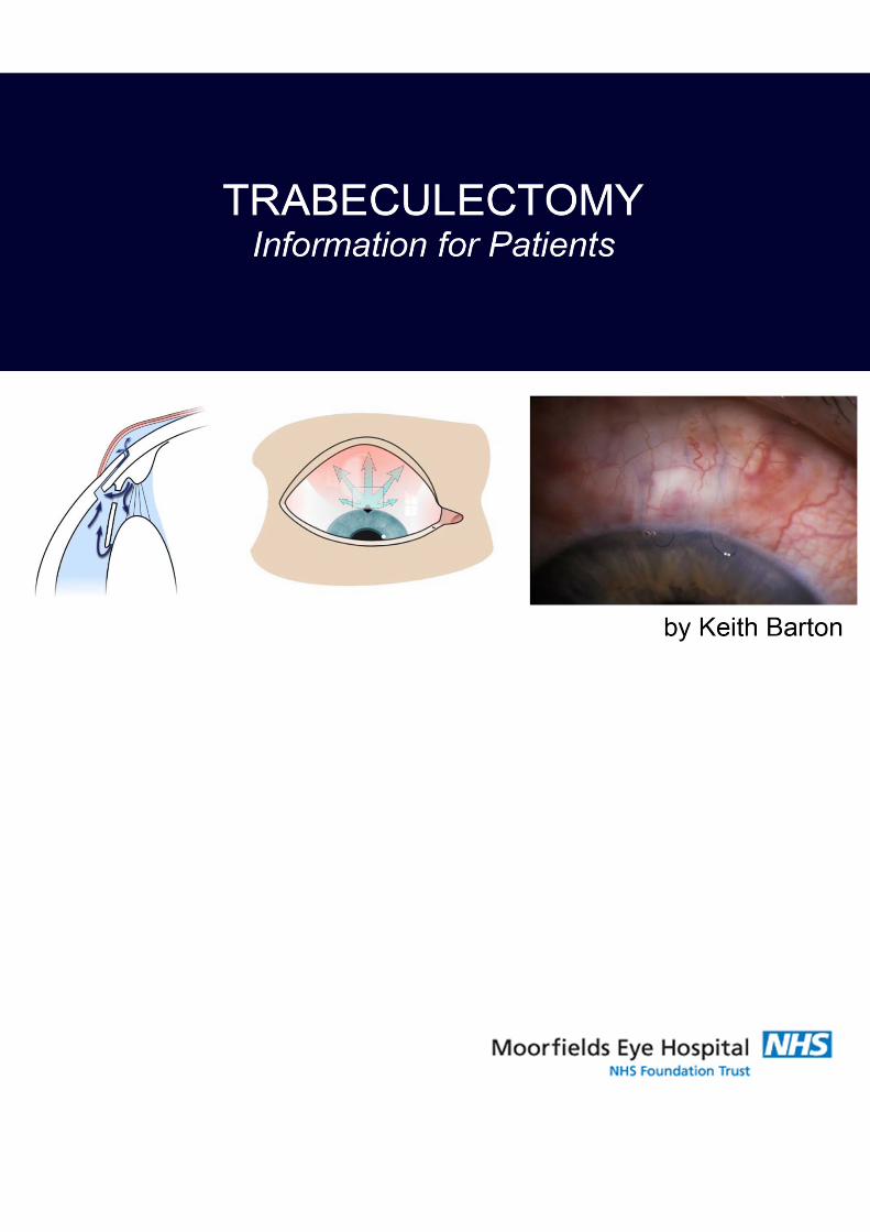
Trabecule
1. In
Trabecu
(intraoc
hole in t
inside th
reservoi
trap‐doo
too quic
By drain
the opti
glaucom
Please n
vision a
The aqu
Waterin
caused
reduces
The aqu
reservoi
Diagramthrough
ctomy, K Ba
ntroduct
ulectomy i
cular press
the eye w
he eye kno
ir or bleb j
or is sutur
ckly.
ning aqueo
ic nerve an
ma.
note that c
lready los
ueous hum
ng of the e
by high pr
s the eye p
ueous hum
ir betwee
m showingh a norma
rton, Moorfi
tion – Wh
s a surgica
sure) in pa
all (sclera
own as aq
just under
red (stitch
ous humo
nd preven
control of
t from gla
mor is a flu
eye is caus
ressure in
pressure b
mor that d
n the scle
g flow of Al trabecul
elds Eye Hos
hat is a T
al operatio
atients wit
), covered
queous hu
r the eye s
hed) in a w
r the trab
nts further
f the eye p
aucoma.
uid inside t
sed by tea
the aqueo
by draining
rains thro
ra and the
Aqueous lectomy
spital NHS Fo
Trabecule
on which
th glaucom
d by a thin
mor, drai
surface, h
way that p
beculectom
r damage
pressure w
the eye an
ars, not aq
ous humo
g aqueous
ough the t
e surface
Drainaafter a
oundation Tr
ectomy?
lowers th
ma. This is
n trap‐doo
ns throug
idden by t
revents aq
my operat
and furth
with a trab
nd is not r
queous hu
or inside th
s humor fr
rabeculec
layer of tis
age bleb va trabecul
rust, June 20
?
e pressure
s achieved
r in the sc
h the trap
the eyelid
queous hu
ion reduc
er loss of
beculectom
related to
mor. Glau
he eye. Tr
rom the ey
tomy accu
ssue that
visible on lectomy
013
e inside th
d by makin
clera. The
p‐door to a
d (see belo
umor from
es the pre
vision in
my will no
the tears
ucoma is o
rabeculect
ye.
umulates
covers the
lifting the
3
he eye
ng a small
fluid
a small
ow). The
m draining
essure on
ot restore
.
often
tomy
in a
e eyeball
e eyelid
g
Trabeculectomy, K Barton, Moorfields Eye Hospital NHS Foundation Trust, June 2013 4
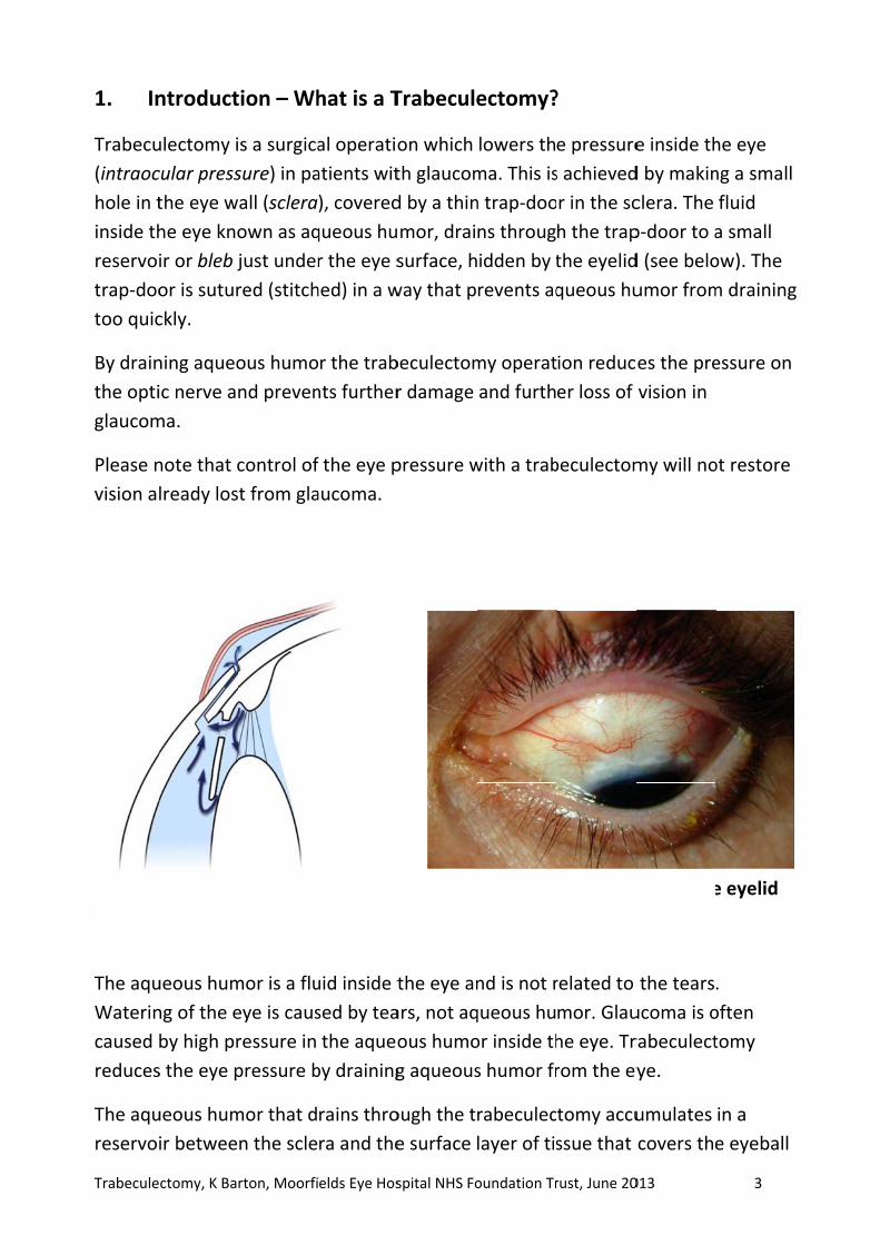
(the conjunctiva) to form a small drainage bleb that is usually hidden under the
upper eyelid (see illustrations on page 3).
2. What is the appearance of the eye after a trabeculectomy?
Initially the eye will be red and swollen to a variable degree after surgery. The
eyelid may also droop partially. This resolves over a period of weeks to months.
The drainage bleb is not usually visible to the naked eye after the trabeculectomy
operation. The bleb may, however, be seen if the patient looks in the mirror and
raises the upper eyelid.
After surgery, most patients feel no sensation from the presence of the drainage
bleb. Rarely, patients are aware of the drainage bleb. Should this occur, steps can
be taken to make the bleb more comfortable; this is discussed further under
complications (see below).
3. Medication Prior to Surgery
Prior to undergoing surgery, patients are asked to continue all drops and tablets in
accordance with their normal treatment regimen until the morning of the
operation. Blood thinning medications such as Aspirin, Warfarin and Clopidogrel
should also be continued. Patients who are taking Warfarin are advised to have
their level (eg. INR) checked at least 2 weeks prior to surgery to ensure it is within
the correct therapeutic range.
If patients opt to have the surgery performed under general anaesthesia, a
preoperative assessment of their general health will be carried out just before
surgery. Underlying medical conditions including cardiac disease, uncontrolled
high blood pressure or diabetes will need to be addressed prior to scheduling of
surgery.
Trabeculectomy, K Barton, Moorfields Eye Hospital NHS Foundation Trust, June 2013 5
4. The surgery itself
Trabeculectomy surgery typically lasts up to 45 minutes.
Anaesthesia
Trabeculectomy is often performed under local anaesthesia, though it may also be
performed under general anaesthesia.
Patients who have their surgery under local anaesthesia will be awake during the
operation but will have the option of requesting light sedation. The eye will be
anaesthetised first with eye drops and then an injection of anaesthetic will be
administered around the eye. The anaesthetic injection itself may cause some mild
discomfort; a slight sensation of pressure as the anaesthetic is delivered. The
injection anaesthetises the eye, preventing not only pain but also excessive eye
movement during surgery. During surgery patients are covered by a sterile sheet,
or drape, which keeps the operation site sterile and also prevents patients from
seeing any of the surgery. Patients will be aware of the surgeon working around
the eye, but should not feel pain. In the event of any pain or discomfort, the
patient may calmly raise a hand and the surgeon will stop the surgery and top‐up
the anaesthetic if needed. Patients may also hear the surgeon speaking to the
scrub nurse and other members of the surgical team.
Mitomycin C
During the surgery, Mitomycin C may be applied to the surface of the eye for a
brief period of time (usually 2‐3 minutes). Mitomycin C is a drug that was originally
used to treat cancer, but it is also used in glaucoma surgery to reduce scarring.
Scarring prevents the trabeculectomy from functioning in the long term, as it
prevents the aqueous humor from being absorbed back into the circulation. The
Mitomycin C is then washed away from the eye with sterile water so that no
residual drug remains.
Trabeculectomy, K Barton, Moorfields Eye Hospital NHS Foundation Trust, June 2013 6
5. After surgery – Postoperative care
The day of surgery and the next day
Patients are usually discharged home from hospital either the same day as the
surgery or the day after.
Please note; all patients need to be examined one day after surgery so a further
visit to the hospital the following day is required for those having day case surgery.
Patients travelling from afar will have the option to request overnight
accommodation at the time of booking the surgery.
The eye is normally padded after surgery and the eye pad is removed the following
day. If the unoperated eye does not see well, then the operated eye will not be
padded. Instead, a clear shield will be placed on the operated eye so that it is still
possible to see after surgery.
Patients are advised to ask a friend or relative to accompany them home after
surgery, especially patients who have poor sight in the unoperated eye or those
who have had general anaesthesia.
What should I expect to feel during the postoperative period?
It is normal for the vision to be blurred and the eye to be uncomfortable after
surgery. The period of blurring is variable. The vision may be particularly blurred
for 1‐2 weeks following surgery, and then start to improve. It can take 2‐3 months
for the eye to feel completely normal and the vision to stabilise completely.
The patient will also be asked to wear a shield at night for the first 2 weeks or so;
this is to prevent any accidental harm to the operative site whilst sleeping.
Soreness in the eye after surgery is partly due to the surgery itself, and partly due
to the stitches (or sutures). The sutures do not dissolve and are usually removed in
the clinic 2 to 3 weeks after surgery (this takes 2 – 3 minutes in clinic with the eye
anaesthetised using eye drops). The eye usually starts to feel more comfortable
after the sutures have been removed.
Trabeculectomy, K Barton, Moorfields Eye Hospital NHS Foundation Trust, June 2013 7
Eye drops after surgery
Eye drops will be prescribed to use regularly after surgery. These start the day after
surgery, after the post‐operative examination. It is not usually necessary to use eye
drops the first night after the surgery. Acetazolamide (Diamox) tablets or any
glaucoma medication to the operated eye should also be stopped the night after
surgery unless advised otherwise.
It is important that any eye drops for the unoperated eye are continued unless
advised otherwise.
The postoperative eye drops will usually consist of an antibiotic
(eg.chloramphenicol) and anti‐inflammatory steroid (eg. dexamethasone). The
steroid eye drop will initially be used intensively (every 2 hours or about 8 times
daily) and the antibiotic four times daily. During the period of intensive usage
preservative‐free drops are normally used. When drops are prescribed to take
intensively after surgery, it is usually intended that they are taken during the day
only. If overnight intensive use is intended, then the patient will be advised of this
separately.
Patients are given a supply of postoperative eye drops on leaving the hospital;
these should last one month. The postoperative eye drops will normally need to be
taken for 2 to 3 months. Patients are advised at each post‐operative visit whether a
change in the dosage of drops is required. The drops should not be stopped or the
dosage changed without consulting the doctor.
Postoperative clinic visits
Patients are usually seen once weekly for the first 4 weeks, and may be seen more
frequently if the eye pressure is either too high or too low.
During this time sutures may be removed to adjust the pressure and additional
injections of steroids or 5‐Fluororacil (a drug that reduces healing), may be given
around the eye to counteract the body’s natural healing process. The injections
are performed after the administration of anaesthetic eyedrops, during the clinic
appointment itself.
Trabeculectomy, K Barton, Moorfields Eye Hospital NHS Foundation Trust, June 2013 8
Patients who live a long distance from the hospital will likely be able to alternate
postoperative appointments between their surgeon and a local ophthalmologist.
Activity after Surgery
It is important to avoid strenuous activity during the early post‐operative period
including swimming, tennis, jogging and contact sports.
It is permissible to watch television and read, as these will not harm the eye. For
patients who wish to pray, it is better to kneel but not to bow the head down to
the floor in the first 2 – 3 weeks. Bending over can cause significant pain when the
eye is still inflamed after surgery. Similarly, activities such as yoga that require
head‐down posturing should be avoided.
As patients will be monitored closely following surgery, it is recommended that
they consult their surgeon before commencing strenuous activity. If the eye
pressure is very low after surgery the surgeon may suggest refraining from all
exertion and remaining sedentary until the pressure is restored.
When can I go back to work?
The duration of time off work will depend on a number of factors such as the
nature of the patient’s employment, the state of the vision in the other eye and
the pressure in the operated eye.
Typically someone working in an office environment would require 2 weeks off, if
the postoperative course is smooth. Someone whose occupation involves heavy
manual work or work in a dusty environment may require a month or more (e.g.
construction workers, farmers).
Contact lens wear after trabeculectomy surgery
It is usually possible to restart contact lens wear around 4 weeks and sometimes
sooner after trabeculectomy surgery. Not everyone can continue to wear contact
lenses after trabeculectomy surgery, so this is something to consider before having
a trabeculectomy operation. If contact lens wear is essential, then other
alternatives to trabeculectomy should be considered.
Trabeculectomy, K Barton, Moorfields Eye Hospital NHS Foundation Trust, June 2013 9
Whether or not contact lenses can be worn after surgery depends on the
appearance and shape of the drainage bleb. The surgeon will usually be able to
advise on this by 6 ‐8 weeks after surgery.
Flying after surgery
Although it is safe to fly after surgery, patients should bear in mind that their
surgeon will wish to see them for a number of post‐operative visits to ensure that
the eye pressure is at the correct level.
When is the eye back to normal?
In most cases, it takes 2 to 3 months for the eye to feel completely normal and
sometimes longer in more complicated cases. At this point a refraction (spectacle
test) is usually required as the spectacle prescription may have changed slightly
from the pre‐surgery prescription.
5. Success rates and complications
Success rates
Long‐term studies suggest that most people will achieve a low eye pressure
without the need for additional glaucoma medication after trabeculectomy
surgery. In clinical trials, trabeculectomy has proven consistently more successful
at lowering intraocular pressure than either medication or laser.1;2 The success rate
of trabeculectomy at controlling the pressure varies according to a number of risk
factors including the type of glaucoma, previous surgery, race, age and other
conditions.
In one study of trabeculectomy success, after 20 years almost 90% were still
successful.3 Just under two thirds of theses required no glaucoma medication to
control the pressure, wheras one third still required medication. In the author’s
practice, roughly 10‐12% will require further surgery for uncontrolled pressure.
Uncommonly, a patient will develop a pressure that is too low, requiring further
surgery to elevate the pressure.
Trabeculectomy, K Barton, Moorfields Eye Hospital NHS Foundation Trust, June 2013 10
Complications
Severe complications are rare and may happen either if the eye pressure drops
very low or very quickly during the early postoperative period, or if the eye
becomes infected.
Very low eye pressure is the biggest risk in the early postoperative period.
Although it is often painless, it may be associated with a dull aching feeling or a
throbbing sensation within the operated eye. Patients who notice severe blurring
of vision, distortion or a fluctuating curtain in their visual field should attend the
eye casualty department as soon as possible for further assessment.
Very low pressure or a precipitous drop in pressure can result in bleeding at the
back of the eye (choroidal haemorrhage). This is a very severe complication but
rare. In order to ensure that this does not happen the surgeon will often suggest
further intervention if the pressure becomes very low. Such intervention may
consist of a return to the operating theatre to have the trap‐door sutures
tightened. Sometimes the surgeon will inject a viscoelastic gel into the eye and
wait to observe the result before deciding on further adjustment of the trap‐door
sutures, as the eye pressure will often stabilise by itself. Sometimes a simple
adjustment of medication is sufficient, in which case, neither of the above will be
required.
About 5% of trabeculectomy patients at Moorfields require a return to the
operating theatre in the first month after surgery for adjustment, either because
the pressure is too high or too low.
The risk of serious infection or serious bleeding in the eye from trabeculectomy at
Moorfields is rare (approximately 1 in 250).4
Longer‐Term Risks
The longer‐term risks of trabeculectomy are infection, discomfort, cataract and
change in glasses prescription. Low pressure occasionally develops in the longer
term, but generally the risk of low pressure is highest in the early postoperative
period rather than later.
Trabeculectomy, K Barton, Moorfields Eye Hospital NHS Foundation Trust, June 2013 11
Infection
While the risk of infection after surgery is rare, there is a very small on‐going life‐
time risk that the drainage bleb might become infected.
If a patient who has had a trabeculectomy subsequently develops a red, sticky or
painful eye, it is important they have their eye examined immediately by an
ophthalmologist, as this may be a sign of an infection. While infection is rare, it
may be very serious and can result in visual loss. The earlier any infection is
treated, the better the outcome for the eye.
Discomfort
The drainage bleb may become large. Occasionally this may extend below the
eyelid or cause the eyelid to be raised or droopy.
A large drainage bleb may cause interference with the tear film on the eye surface,
and can create a feeling of discomfort or drying of the eye. This occurs in about
10% of patients and is usually treatable with artifical tear drops. Occasionally, the
discomfort is more severe and requires surgery to make the drainage bleb smaller.
Cataract
In patients who have not had cataract surgery, there is a risk that trabeculectomy
may worsen an existing cataract3.
Raised eye pressure and glaucoma medications have been shown to cause cataract
in population studies. In a study of 607 patients, the likelihood of needing cataract
surgery within 7.7 years of a trabeculectomy operation was 20%, compared to 12%
in those treated with eyedrops only.2
Astigmatism and other changes in glasses prescription
Most patients require a small change in their glasses prescription after
trabeculectomy. Patients should refrain from changing their glasses until at least 3
months after the surgery and only once the eye pressure has stabilised. It is
advisable to check with the doctor before changing glasses. Rarely, a patient who
does not require glasses before surgery develops a need for glasses after surgery.
Trabeculectomy, K Barton, Moorfields Eye Hospital NHS Foundation Trust, June 2013 12
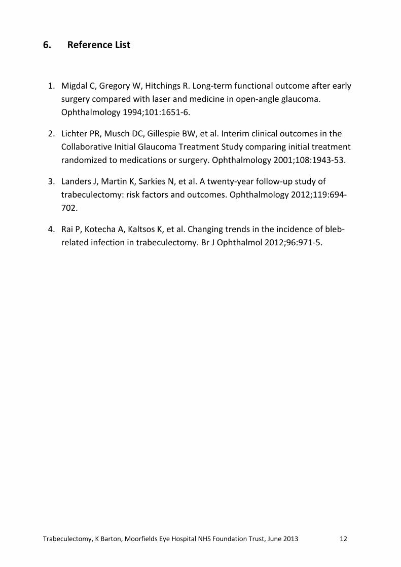
6. Reference List
1. Migdal C, Gregory W, Hitchings R. Long‐term functional outcome after early
surgery compared with laser and medicine in open‐angle glaucoma.
Ophthalmology 1994;101:1651‐6.
2. Lichter PR, Musch DC, Gillespie BW, et al. Interim clinical outcomes in the
Collaborative Initial Glaucoma Treatment Study comparing initial treatment
randomized to medications or surgery. Ophthalmology 2001;108:1943‐53.
3. Landers J, Martin K, Sarkies N, et al. A twenty‐year follow‐up study of
trabeculectomy: risk factors and outcomes. Ophthalmology 2012;119:694‐
702.
4. Rai P, Kotecha A, Kaltsos K, et al. Changing trends in the incidence of bleb‐
related infection in trabeculectomy. Br J Ophthalmol 2012;96:971‐5.
Trabeculectomy, K Barton, Moorfields Eye Hospital NHS Foundation Trust, June 2013 13
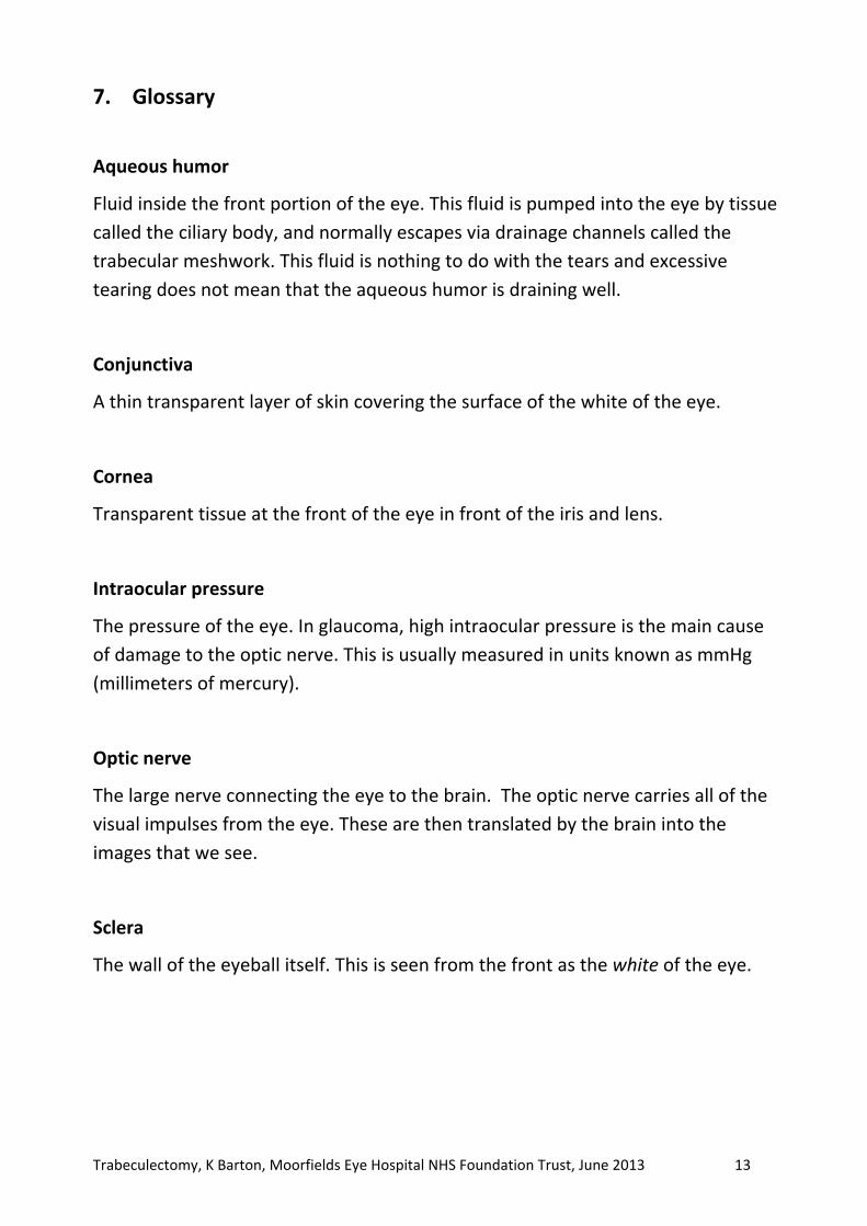
7. Glossary
Aqueous humor
Fluid inside the front portion of the eye. This fluid is pumped into the eye by tissue
called the ciliary body, and normally escapes via drainage channels called the
trabecular meshwork. This fluid is nothing to do with the tears and excessive
tearing does not mean that the aqueous humor is draining well.
Conjunctiva
A thin transparent layer of skin covering the surface of the white of the eye.
Cornea
Transparent tissue at the front of the eye in front of the iris and lens.
Intraocular pressure
The pressure of the eye. In glaucoma, high intraocular pressure is the main cause
of damage to the optic nerve. This is usually measured in units known as mmHg
(millimeters of mercury).
Optic nerve
The large nerve connecting the eye to the brain. The optic nerve carries all of the
visual impulses from the eye. These are then translated by the brain into the
images that we see.
Sclera
The wall of the eyeball itself. This is seen from the front as the white of the eye.
Trabeculectomy, K Barton, Moorfields Eye Hospital NHS Foundation Trust, June 2013 14
8. Acknowledgements
The author would like to thank Emma Jones, Abigail Mackrill, Rashmi Mathew,
Kirithika Muthusamy, Chris Smith and Eleanor Wilkinson as well as a number of
patients and their relatives for their help in the preparation of this document.
9. Disclaimer
Accuracy
While every step has been taken to compile accurate information and to keep it up
to date, we cannot guarantee its correctness and completeness. The information
provided in this information sheet is designed as an adjunct to, and not a
substitute for professional healthcare advice, by a qualified doctor or other
healthcare professional, which will be tailored to a patient's individual
circumstances. Keith Barton and Moorfields Eye Hospital NHS Foundation Trust
cannot take responsibility if you rely solely on the information in this information
sheet.
Document Last Modified 3rd June 2013