Embed Size (px)
Citation preview
European Respiratory Society Annual Congress 2013, Barcelona, Spain
Tracheal stenosis.
Prevention and
treatment
I. Motus, N.Giss Urals Research Institute for Phthisiopulmonology, Lung Surgery Clinic of Regional Pulmonologic
Centre , Hospital # 23, Ekaterinburg, Russia
Objectives. Tracheal stenosis occurred in 2-10% of patients who were
subjected to prolonged ventilation via tracheostomy or endotracheal tube. This rate does not reduce during last years so that tracheal stenosis still remains a problem in Russia. Clinical situations and anatomic variants in tracheal stenosis (TS) are quite various so different approaches for prevention and treatment are required.
The severity of tracheal stenosis strongly depends on technique of the tracheostomy and further care. The patients involved often have to be canulated for a long time or forever. Endotracheal stenting in the treatment of TS usually is not of substantial help (granulation, dislocation). If possible circular resection is to be undertaken. The main task is to prevent formation of TS and provide proper treatment when is has developed.
The aims of this study is to find a way for prevention and
treatment of tracheal stenosis. Namely:
- to elucidate the rate of tracheostomies and variants of this operation in intensive care units in Ekaterinburg;
- to ascertain the subsequent clinical course of these patients after discharging from the hospital;
2
- to investigate the rate of tracheal stenosis after different variants of tracheostomy;
- to assess the degrees of tracheal impairment; - to improve the early detection and treatment; - to find a way of prophylaxis severe tracheal impairment and ventilatory
disorders; - to find optimal conditions for surgical manipulation as well as adequate
gas exchange which are of a great importance for successful surgical treatment of tracheal stenosis.
Materials and methods. The data of 848 patients subjected to tracheostomy in three Ekaterinburg hospitals from 2006 to 2011 years were under the study. Fifty-four of them (6,4%) were found to have cicatrical tracheal stenosis. (I group). In order to reveal and treat early posttracheostomy alterations threatening with stenosis development we examined 149 patients also subjected to tracheostomy and prolonged artificial lung ventilation (II group). The complex of diagnostic facilities (CT-scan or MR imaging, laboratory investigation) was also employed. Where some risk symptoms (see below) were revealed subsequent endoscopic treatment was performed. The endoscopic treatment consisted of cryoapplication, granulematous tissue debridement and bougienage of the stenotic area. The alterations concerned were found in 28 cases. The patients who had the severe consequences of the illnesses or traumatic damage and could not be subjected to examination were excluded from the study.
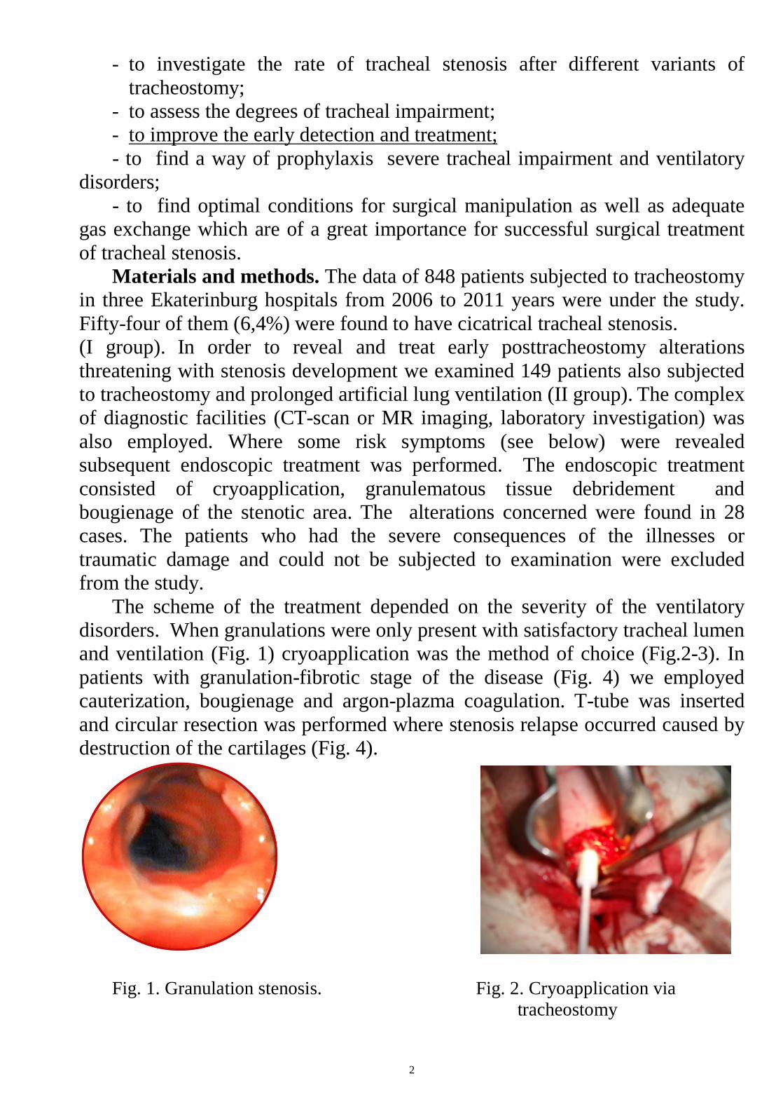
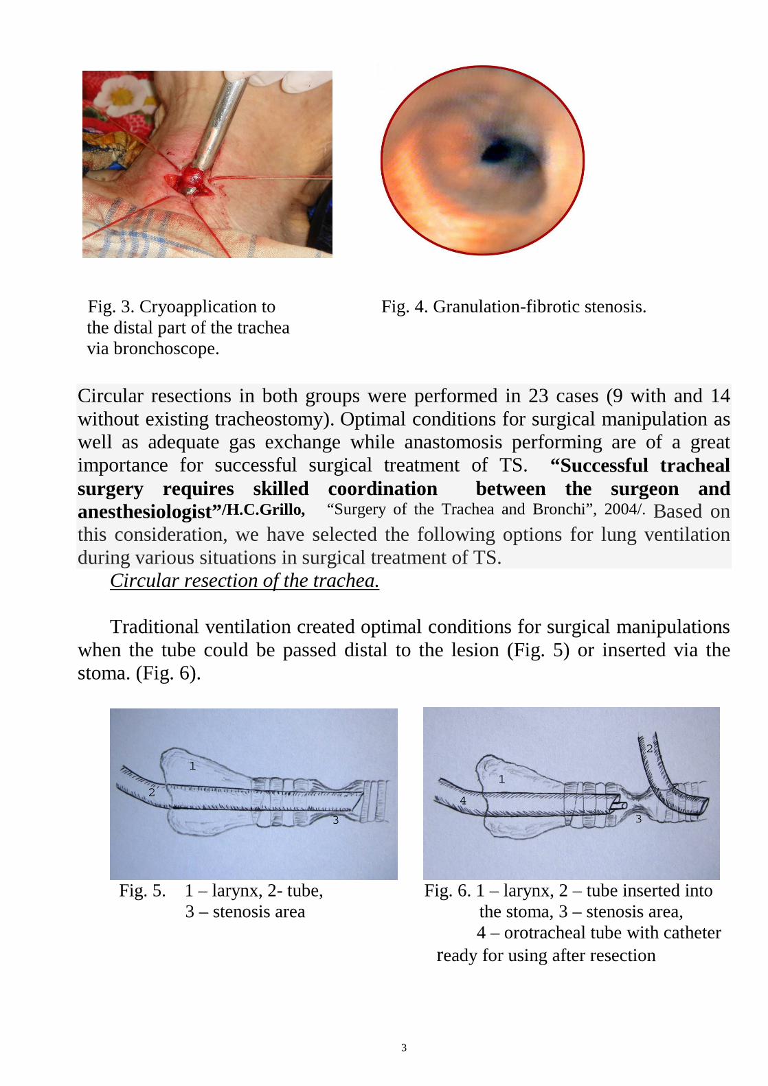
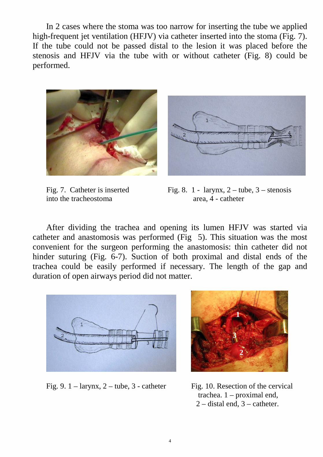
The scheme of the treatment depended on the severity of the ventilatory disorders. When granulations were only present with satisfactory tracheal lumen and ventilation (Fig. 1) cryoapplication was the method of choice (Fig.2-3). In patients with granulation-fibrotic stage of the disease (Fig. 4) we employed cauterization, bougienage and argon-plazma coagulation. T-tube was inserted and circular resection was performed where stenosis relapse occurred caused by destruction of the cartilages (Fig. 4).
Fig. 1. Granulation stenosis. Fig. 2. Cryoapplication via tracheostomy
3
Fig. 3. Cryoapplication to Fig. 4. Granulation-fibrotic stenosis. the distal part of the trachea via bronchoscope.
Circular resections in both groups were performed in 23 cases (9 with and 14 without existing tracheostomy). Optimal conditions for surgical manipulation as well as adequate gas exchange while anastomosis performing are of a great importance for successful surgical treatment of TS. “Successful tracheal surgery requires skilled coordination between the surgeon and anesthesiologist”/H.C.Grillo, “Surgery of the Trachea and Bronchi”, 2004/. Based on this consideration, we have selected the following options for lung ventilation during various situations in surgical treatment of TS.
Circular resection of the trachea. Traditional ventilation created optimal conditions for surgical manipulations
when the tube could be passed distal to the lesion (Fig. 5) or inserted via the stoma. (Fig. 6).
Fig. 5. 1 – larynx, 2- tube, Fig. 6. 1 – larynx, 2 – tube inserted into 3 – stenosis area the stoma, 3 – stenosis area, 4 – orotracheal tube with catheter ready for using after resection
4
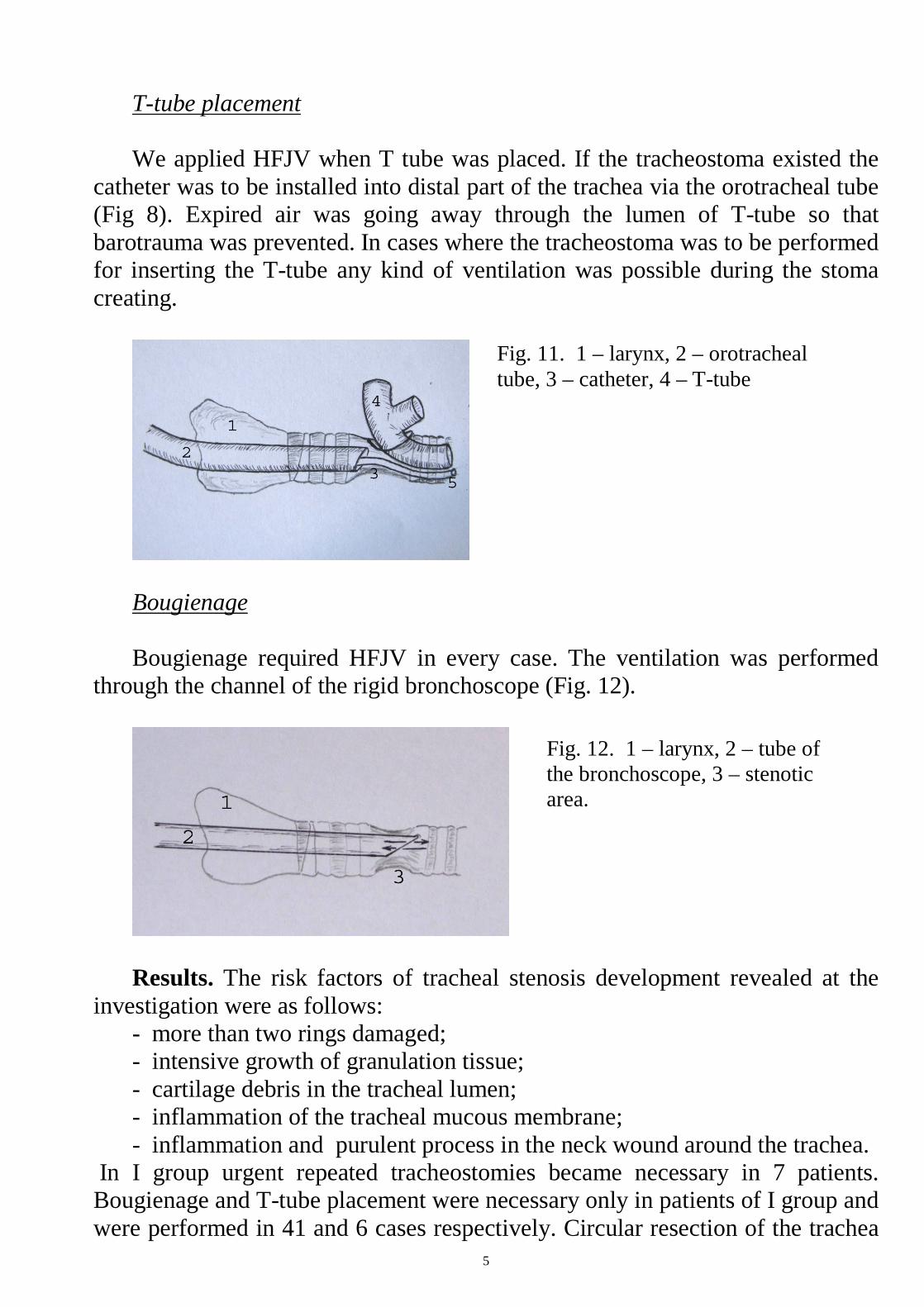
In 2 cases where the stoma was too narrow for inserting the tube we applied high-frequent jet ventilation (HFJV) via catheter inserted into the stoma (Fig. 7). If the tube could not be passed distal to the lesion it was placed before the stenosis and HFJV via the tube with or without catheter (Fig. 8) could be performed.
Fig. 7. Catheter is inserted Fig. 8. 1 - larynx, 2 – tube, 3 – stenosis into the tracheostoma area, 4 - catheter After dividing the trachea and opening its lumen HFJV was started via
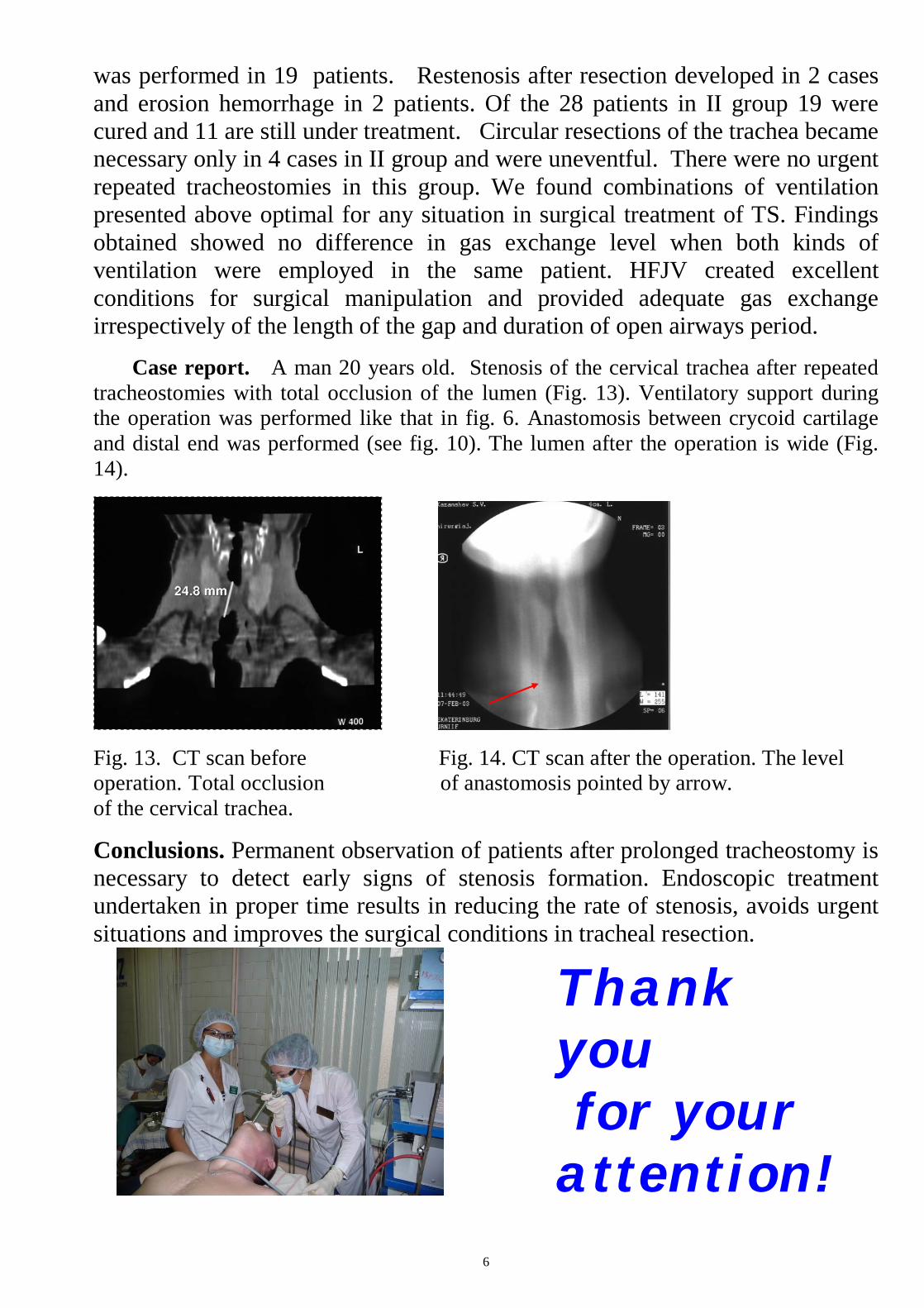
catheter and anastomosis was performed (Fig 5). This situation was the most convenient for the surgeon performing the anastomosis: thin catheter did not hinder suturing (Fig. 6-7). Suction of both proximal and distal ends of the trachea could be easily performed if necessary. The length of the gap and duration of open airways period did not matter.
Fig. 9. 1 – larynx, 2 – tube, 3 - catheter Fig. 10. Resection of the cervical trachea. 1 – proximal end, 2 – distal end, 3 – catheter.
1
2
3
5
T-tube placement We applied HFJV when T tube was placed. If the tracheostoma existed the
catheter was to be installed into distal part of the trachea via the orotracheal tube (Fig 8). Expired air was going away through the lumen of T-tube so that barotrauma was prevented. In cases where the tracheostoma was to be performed for inserting the T-tube any kind of ventilation was possible during the stoma creating.
Bougienage Bougienage required HFJV in every case. The ventilation was performed
through the channel of the rigid bronchoscope (Fig. 12).
Results. The risk factors of tracheal stenosis development revealed at the
investigation were as follows: - more than two rings damaged; - intensive growth of granulation tissue; - cartilage debris in the tracheal lumen; - inflammation of the tracheal mucous membrane; - inflammation and purulent process in the neck wound around the trachea.
In I group urgent repeated tracheostomies became necessary in 7 patients. Bougienage and T-tube placement were necessary only in patients of I group and were performed in 41 and 6 cases respectively. Circular resection of the trachea
Fig. 11. 1 – larynx, 2 – orotracheal tube, 3 – catheter, 4 – T-tube
Fig. 12. 1 – larynx, 2 – tube of the bronchoscope, 3 – stenotic area.
6
was performed in 19 patients. Restenosis after resection developed in 2 cases and erosion hemorrhage in 2 patients. Of the 28 patients in II group 19 were cured and 11 are still under treatment. Circular resections of the trachea became necessary only in 4 cases in II group and were uneventful. There were no urgent repeated tracheostomies in this group. We found combinations of ventilation presented above optimal for any situation in surgical treatment of TS. Findings obtained showed no difference in gas exchange level when both kinds of ventilation were employed in the same patient. HFJV created excellent conditions for surgical manipulation and provided adequate gas exchange irrespectively of the length of the gap and duration of open airways period.
Case report. A man 20 years old. Stenosis of the cervical trachea after repeated tracheostomies with total occlusion of the lumen (Fig. 13). Ventilatory support during the operation was performed like that in fig. 6. Anastomosis between crycoid cartilage and distal end was performed (see fig. 10). The lumen after the operation is wide (Fig. 14).
Fig. 13. CT scan before Fig. 14. CT scan after the operation. The level operation. Total occlusion of anastomosis pointed by arrow. of the cervical trachea.
Conclusions. Permanent observation of patients after prolonged tracheostomy is necessary to detect early signs of stenosis formation. Endoscopic treatment undertaken in proper time results in reducing the rate of stenosis, avoids urgent situations and improves the surgical conditions in tracheal resection.
Thank you for your attention!