Embed Size (px)
Citation preview

453
List of Abbreviations ATP: Adenosine triphosphate, BDZ: Benzodiazipine, Capnography: Monitoring of the concentration or partial pressure of CO2 in the respiratory gases, CNS: Central Nervous System, DPG: 2,3-Diphosphoglycerate organo phosphate, EtCO2: End-tidal CO2 tension, GABA: Gamma-aminobutyric acid neuro transmitter, GABAA: Gamma-aminobutyric acid neuro transmitter receptor, Hypercapnia: A state of increased CO2, Hypoxia: A state of reduced oxygen tension, ICU: Intensive care unit, NADH: Nicotinamide Adenine Dinucleotide, PaCO2: Partial Pressure of CO2, PO2: Partial Pressure of oxygen, pH: Measure of acidity, SpO2: Oxygen saturation
IntroductionDental anxiety is very common, and prevents many from accessing dental care. In a Gallup Poll, 22% of people surveyed suffered anxiety levels that were so high that they had delayed seeking dental treatment [1] Approximately 5% of people who attend the dentist would, at some time, require sedation [2]. Properly provided Conscious Sedation is safe, valuable and effective for dental patients. (Dental Sedation Teachers Group) The current standards of care involve appropriate monitoring and the observation of respiratory excursion [3]. Sedation may cause profound respiratory depression and hypoventilation. Therefore accurate monitoring of ventilatory status of sedated patients is desirable. One study has reported the difficulty in detecting hypoventilation in patients undergoing sedation [4].
Conscious Sedation is defined as: A technique in which the use of a drug or drugs produces
a state of depression of the central nervous system enabling treatment to be carried out, but during which verbal contact with the patient is maintained throughout the period of sedation. The drugs and techniques used to provide conscious sedation for dental treatment should carry a margin of safety wide enough to render loss of consciousness unlikely.
The level of sedation must be such that the patient remains conscious, retains protective reflexes, and is able to respond to verbal commands [5].
Effect of BDZs on the CNS and Respiratory Drive
Breathing is the exchange of air between the atmosphere and the lungs. The aim of gas exchange is to collect oxygen from the environment, so as to provide the final electron acceptor in the electron transport chain during cellular respiration. Respiration consists of the oxidation of mitochondrial NADH which is coupled by the electron transport chain to the pumping of protons out across the mitochondrial inner membrane [6]. This produces the desired product; energy in the form of ATP to expel CO2 which is a by-product of cellular respiration from the body tissues to the environment. Cells undergo a variety of biological responses when placed in hypoxic conditions, including activation of signaling pathways that regulate proliferation, angiogenesis and death. An increase in CO2 can lead to hypercapnia, Hypercapnic respiratory failure (Type II) is characterised by a PaCO2 higher than 50 mm Hg [7]. Hypoxemic respiratory failure (Type I) is characterised by an arterial oxygen tension, which is the amount of oxygen dissolved in the blood, lower than 60 mm Hg. In contrast CO2 tension remains normal. During normal breathing, ventilation is constantly monitored and adjusted to maintain an appropriate arterial pH and Pao2. This homeostatic control system requires a series of sensors, a central controlling mechanism, and an effector system to carry out its commands. The afferent input into the central system is provided primarily by neural receptors, peripheral arterial chemoreceptors, chemoreceptors and muscle mechanoreceptors. The greatest increase in ventilation is seen in response to hypoxia, especially when PaO2 falls to 70 mm Hg [8].
Oxygen is carried in the blood by serum (plasma) but mainly by haemoglobin. CO2 dissolved in plasma is of the form bicarbonate and is attached to proteins as carbamino compounds. Bicarbonate is formed from the hydration of CO2, which in turn leads to the production of carbonic acid, which becomes ionized. The resulting hydrogen ions decrease the pH of the blood leading to acidosis.
This is represented by the equation CO2 + H2O → H2CO3 → H+ + HCO−3
There is a sigmoid relationship between the saturation of
Transcutaneous Carbon Dioxide Monitoring: Literature ReviewDavid Drysdale Special Care Dentistry Department, Dorset County Hospital, NHS Foundation Trust, Williams Avenue, Dorchester South, England, DT1 2JY, United Kingdom.
AbstractDental anxiety is very common, affecting 22% of the population. One way to increase access to dental care is by the use of BDZs in conscious sedation. These drugs are known to be safe when titrated. BDZs work by stimulating GABAA receptors within inhibitory pathways of the CNS. A consequence of this is a reduction in respiratory drive. Thus assessing saturation of peripheral oxygen (SpO2) is mandatory. A pulse oximeter is normally used to do this. However a pulse oximeter cannot detect changes in CO2 which could result from a reduction in ventilatory drive. Many practitioners prescribe supplemental oxygen to compensate for hypoventilation, which can inhibit the ability of pulse oximetry to detect hypoventilation. Transcutaneous CO2 monitoring is currently used in ICUs and neonatal units. It may, however, have a place in conscious sedation dentistry. It can be used to detect changes in CO2, and is not affected by supplemental oxygen.
Corresponding author: David Drysdale, Special Care Dentistry Department, Dorset County Hospital, NHS Foundation Trust, Williams Avenue, Dorchester South, England, DT1 2JY, United Kingdom; Tel: +4479009992104; e-mail: [email protected]
454
OHDM - Vol. 13 - No. 2 - June, 2014
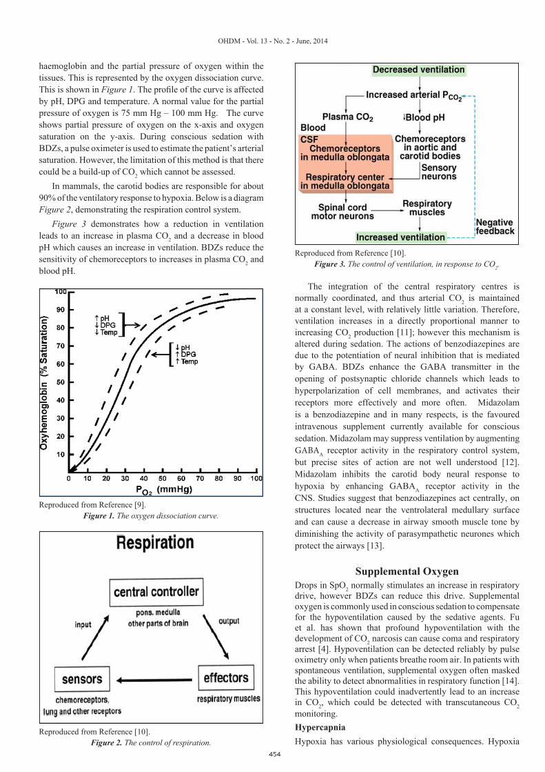
haemoglobin and the partial pressure of oxygen within the tissues. This is represented by the oxygen dissociation curve. This is shown in Figure 1. The profile of the curve is affected by pH, DPG and temperature. A normal value for the partial pressure of oxygen is 75 mm Hg – 100 mm Hg. The curve shows partial pressure of oxygen on the x-axis and oxygen saturation on the y-axis. During conscious sedation with BDZs, a pulse oximeter is used to estimate the patient’s arterial saturation. However, the limitation of this method is that there could be a build-up of CO2 which cannot be assessed.
In mammals, the carotid bodies are responsible for about 90% of the ventilatory response to hypoxia. Below is a diagram Figure 2, demonstrating the respiration control system.
Figure 3 demonstrates how a reduction in ventilation leads to an increase in plasma CO2 and a decrease in blood pH which causes an increase in ventilation. BDZs reduce the sensitivity of chemoreceptors to increases in plasma CO2 and blood pH.
The integration of the central respiratory centres is normally coordinated, and thus arterial CO2 is maintained at a constant level, with relatively little variation. Therefore, ventilation increases in a directly proportional manner to increasing CO2 production [11]; however this mechanism is altered during sedation. The actions of benzodiazepines are due to the potentiation of neural inhibition that is mediated by GABA. BDZs enhance the GABA transmitter in the opening of postsynaptic chloride channels which leads to hyperpolarization of cell membranes, and activates their receptors more effectively and more often. Midazolam is a benzodiazepine and in many respects, is the favoured intravenous supplement currently available for conscious sedation. Midazolam may suppress ventilation by augmenting GABAA receptor activity in the respiratory control system, but precise sites of action are not well understood [12]. Midazolam inhibits the carotid body neural response to hypoxia by enhancing GABAA receptor activity in the CNS. Studies suggest that benzodiazepines act centrally, on structures located near the ventrolateral medullary surface and can cause a decrease in airway smooth muscle tone by diminishing the activity of parasympathetic neurones which protect the airways [13].
Supplemental OxygenDrops in SpO2 normally stimulates an increase in respiratory drive, however BDZs can reduce this drive. Supplemental oxygen is commonly used in conscious sedation to compensate for the hypoventilation caused by the sedative agents. Fu et al. has shown that profound hypoventilation with the development of CO2 narcosis can cause coma and respiratory arrest [4]. Hypoventilation can be detected reliably by pulse oximetry only when patients breathe room air. In patients with spontaneous ventilation, supplemental oxygen often masked the ability to detect abnormalities in respiratory function [14]. This hypoventilation could inadvertently lead to an increase in CO2, which could be detected with transcutaneous CO2 monitoring. HypercapniaHypoxia has various physiological consequences. Hypoxia
Figure 1. The oxygen dissociation curve. Reproduced from Reference [9].
Reproduced from Reference [10].Figure 2. The control of respiration.
Reproduced from Reference [10].Figure 3. The control of ventilation, in response to CO2.
455
OHDM - Vol. 13 - No. 2 - June, 2014
Current Methods for Monitoring during Con-scious Sedation
Pulse oximetryPulse oximetry is the standard method of assessing ventilation in conscious sedation. It was introduced into medical care in the late 1980s to detect changes in oxygen saturation during anaesthesia, and at the time was regarded as a major innovation in patient safety [23]. The accuracy in being able to monitor true arterial oxygen saturation has always been a major problem. Potentially important abnormalities in respiratory activity are undetected with pulse oximetry [24].
Movement artifact, poor tissue perfusion, temperature abnormal haemoglobin, tissue pigmentation, probe site, and artificial light are all known to affect the accuracy of pulse oximeters, most commonly by altering the signal received by the probe [24]. Pulse oximetry effectively detects oxygen desaturation and hypoxia. It works by recording the absorbance of light of two different wavelengths, which are affected by the oxygen saturation of arterial blood. However, it would be inaccurate and potentially risky to assume that it is an appropriate parameter to detect alveolar ventilation, as this can occur in the presence of normal SaO2 measured by pulse oximetry [25].
Different Methods of Measuring CO2Arterial blood gasThe characteristics of the four standard methods of measuring carbon dixode are summarized in Table 1. ABG analysis is the gold standard for measuring the arterial PaCO2 [26]. It is the most acurate method we have available because it samples a harvested blood sample. Pa CO2 is detected using a modified pH electrode which is covered in Teflon. CO2 diffuses through the Teflon membrane, combines with the electrolyte, and alters the pH [27]. The change in pH is displayed as the partial pressure of CO2. ABG analysis can be time consuming, painful for the patient and bears the risk of catheter-related infection, haemorrhage, ischemia, arterio-venous fistula and pseudoaneurysm formation [27]. ABG only provides a one-off reading for PaCO2 and not a continuous reading, which would be required for conscious sedation. End-Tidal CO2 Tension (EtCO2)In patients with endotracheal intubation, EtCO2 is routinely performed and this has been shown to correlate with PaCO2 [28]. However EtCO2 may inaccurately estimate PaCO2 in the case of ventilation/perfusion mismatch found in patients with obstructive lung disease and acute respiratory distress syndrome [29]. Side stream capnographyMeasurement of EtCO2 using side stream capnography in non-intubated sedated patients has the risk of oral leakage
causes peripheral vascular beds to dilate which induces a compensationary tachycardia and a subsequent increase in cardiac output to improve oxygen delivery. Hypoxia can lead to cellular injury through the production of reactive oxygen species; this can result in tissue inflammation and cell death. The early detection of ventilation reduction leads to the prevention of hypoxia, which is predicted by pulse oximetry with a certain delay [15]. This is mandatory for patients undergoing diagnostic or therapeutic interventions, because their respiratory function is influenced by anaesthetic and sedative agents [16]. A consequence of suppressed ventilation is a decrease in PaO2 and an increase in PaCO2, which could lead to hypercapnia. Hypercapnia is defined as a PCO2 above 45 mmHg and normally triggers a reflex which increases breathing and access to oxygen, however BDZs inhibit CB neural response to hypoxia by enhancing GABAA receptor activity [17], causing CO2 to increase. Hypercapnia will rapidly cause an intracellular acidosis in all cells of the body. The cerebral effects of hypercapnia result in dyspnoea, disorientation, acute confusion, headache, mental obtundation or even focal neurologic signs. The cardiovascular signs a clinician should be aware of are warming, the patient appearing flushed or sweaty. The patient may become tachycardic and have a bouncing pulse.
Hypercapnia at risk goups: Hypercapnia can have a detrimental effect on patients with underlying medical conditions. These include:
ObesitySickle cell anaemia COPDObese patients with sleep hypoventilation have an
increased risk of acute hypercapnic respiratory failure. Early diagnosis and implementation of non-invasive positive pressure ventilation is recommended for these patients [18]. This could be a hazard if undergoing conscious sedation, as respiratory drive will be diminished. Wijesinghe et al. showed in a study of 24 severely obese patients with obesity related hypoventilation that supplemental oxygen caused a large increase in CO2 [19]. In patients with sickle cell anaemia, sickling may precipitate with hypoxia, hypercapnia and sickle cell anaemia is sometimes associated with respiratory dysfunction [20]. Boyle et al suggests patients may also benefit from such pre-oxygenation prior to IV sedation, to minimise the risk of respiratory dysfunction. Respiratory muscle weakness in patients with COPD can lead to hypoventilation with subsequent hypercapnia and hypoxia [21]. Chronic hypercapnia in patients with COPD has been associated with a poor prognosis [22].
Dental Setting Invasive Accuracy Calibration
Cutaneous CO2 Tension Yes No Yes Slow
Arterial Blood Gas Analyzer No Yes Yes N/A
End Tidal Volume CO2 tension No No Yes N/A
End Tidal Volume side Stream Capnography No No No N/A
Table 1. A comparison of the four different clinical methods of measuring CO2.
456
OHDM - Vol. 13 - No. 2 - June, 2014
when patients alternate between oral and nasal breathing and can yield incorrect measurements [30].
Overview of Transcutaneous Carbon Dioxide Monitoring
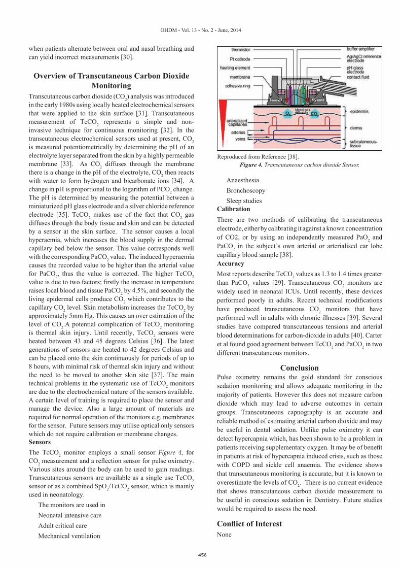
Transcutaneous carbon dioxide (CO2) analysis was introduced in the early 1980s using locally heated electrochemical sensors that were applied to the skin surface [31]. Transcutaneous measurement of TcCO2 represents a simple and non-invasive technique for continuous monitoring [32]. In the transcutaneous electrochemical sensors used at present, CO2 is measured potentiometrically by determining the pH of an electrolyte layer separated from the skin by a highly permeable membrane [33]. As CO2 diffuses through the membrane there is a change in the pH of the electrolyte, CO2 then reacts with water to form hydrogen and bicarbonate ions [34]. A change in pH is proportional to the logarithm of PCO2 change. The pH is determined by measuring the potential between a miniaturized pH glass electrode and a silver chloride reference electrode [35]. TcCO2 makes use of the fact that CO2 gas diffuses through the body tissue and skin and can be detected by a sensor at the skin surface. The sensor causes a local hyperaemia, which increases the blood supply in the dermal capillary bed below the sensor. This value corresponds well with the corresponding PaCO2 value. The induced hyperaemia causes the recorded value to be higher than the arterial value for PaCO2, thus the value is corrected. The higher TcCO2 value is due to two factors; firstly the increase in temperature raises local blood and tissue PaCO2 by 4.5%, and secondly the living epidermal cells produce CO2 which contributes to the capillary CO2 level. Skin metabolism increases the TcCO2 by approximately 5mm Hg. This causes an over estimation of the level of CO2.A potential complication of TcCO2 monitoring is thermal skin injury. Until recently, TcCO2 sensors were heated between 43 and 45 degrees Celsius [36]. The latest generations of sensors are heated to 42 degrees Celsius and can be placed onto the skin continuously for periods of up to 8 hours, with minimal risk of thermal skin injury and without the need to be moved to another skin site [37]. The main technical problems in the systematic use of TcCO2 monitors are due to the electrochemical nature of the sensors available. A certain level of training is required to place the sensor and manage the device. Also a large amount of materials are required for normal operation of the monitors e.g. membranes for the sensor. Future sensors may utilise optical only sensors which do not require calibration or membrane changes.SensorsThe TcCO2 monitor employs a small sensor Figure 4, for CO2 measurement and a reflection sensor for pulse oximetry. Various sites around the body can be used to gain readings. Transcutaneous sensors are available as a single use TcCO2 sensor or as a combined SpO2/TcCO2 sensor, which is mainly used in neonatology.
The monitors are used inNeonatal intensive care Adult critical care Mechanical ventilation
Anaesthesia Bronchoscopy Sleep studies
CalibrationThere are two methods of calibrating the transcutaneous electrode, either by calibrating it against a known concentration of CO2, or by using an independently measured PaO2 and PaCO2 in the subject’s own arterial or arterialised ear lobe capillary blood sample [38]. AccuracyMost reports describe TcCO2 values as 1.3 to 1.4 times greater than PaCO2 values [29]. Transcutaneous CO2 monitors are widely used in neonatal ICUs. Until recently, these devices performed poorly in adults. Recent technical modifications have produced transcutaneous CO2 monitors that have performed well in adults with chronic illnesses [39]. Several studies have compared transcutaneous tensions and arterial blood determinations for carbon-dioxide in adults [40]. Carter et al found good agreement between TcCO2 and PaCO2 in two different transcutaneous monitors.
ConclusionPulse oximetry remains the gold standard for conscious sedation monitoring and allows adequate monitoring in the majority of patients. However this does not measure carbon dioxide which may lead to adverse outcomes in certain groups. Transcutaneous capnography is an accurate and reliable method of estimating arterial carbon dioxide and may be useful in dental sedation. Unlike pulse oximetry it can detect hypercapnia which, has been shown to be a problem in patients receiving supplementary oxygen. It may be of benefit in patients at risk of hypercapnia induced crisis, such as those with COPD and sickle cell anaemia. The evidence shows that transcutaneous monitoring is accurate, but it is known to overestimate the levels of CO2. There is no current evidence that shows transcutaneous carbon dioxide measurement to be useful in conscious sedation in Dentistry. Future studies would be required to assess the need.
Conflict of InterestNone
Figure 4. Transcutaneous carbon dioxide Sensor.Reproduced from Reference [38].
457
OHDM - Vol. 13 - No. 2 - June, 2014
References1. Delfino J. Public attitudes toward oral surgery: results of a
Gallup poll. Journal of Oral and Maxillofacial Surgery. 1997; 55: 564–567.
2. Goodwin M, Pretty IA. Estimating the need for dental sedation. 3. Analysis of factors contributing to non-attendance for dental treatment in the general population, across 12 English primary care trusts. British Dental Journal. 2011; 211: 599-603.
3. Koniaris LG, Wilson S, Drugas G, Simmons W. Capnographic monitoring of ventilator status during moderate (conscious) sedation. Surgical Endoscopy. 2003; 17: 1261-1265.
4. Fu ES, Downs JB, Schweiger JW, Miguel RV, Smith RA. Miguel Robert A. Smith Supplemental oxygen impairs detection of Hypoventilation by Pulse Oximetry Chest. 2004; 126: 1552-1558.
5. General Dental Council. Maintaining Standards. 1999; Revised.
6. http://hyperphysics.phy-astr.gsu.edu/hbase/biology/etrans.html7. Harris AL. Hypoxia--a key regulatory factor in tumour growth.
Nature Reviews Cancer. 2002; 2: 38-47.8. Caruana-Montaldo B, Gleeson K, Zwillich CW. The Control
of Breathing in Clinical Practice. Chest. 2000; 117: 205-225.9. http://www.bio.davidson.edu/Courses/anphys/1999/Dickens/
Oxygendissociation.htm 10. Caruana-Montaldo B1, Gleeson K, Zwillich CW. The
Control of Breathing in Clinical Practice. Chest. 2000; 117: 205-225.11. http://d3jonline.tripod.com/29-Clinical_Neuroscience/
Brainstem_Control_of_Autonomic_Function.htm12. Mackenzie N. The Role of Sedation During Regional
Anaesthesia: Original Papers Sedation during regional anaesthesia: indications, advantages and methods. European Journal of Anaesthesiology. 1996; 13: 2-7.
13. Haxhiu MA, van Lunteren E, Cherniack NS, Deal EC. Benzodiazepines acting on ventral surface of medulla cause airway dilation. American Journal of Physiology. 1989; 257: 810-815.
14. Fu ES, Downs JB, Schweiger JW, Miguel RV, Smith RA. Miguel Robert A. Smith Supplemental oxygen impairs detection of Hypoventilation by Pulse Oximetry. Chest. 2004; 126: 1552-1558.
15. Kim V, Benditt JO, Wise RA, Sharafkhaneh A. Oxygen Therapy in Chronic Obstructive Pulmonary Disease. Proceedings of the American Thoracic Society. 2008; 5: 513-518.
16. American Society of Anesthesiologists. Practice Guidelines for Sedation and Analgesia by Non-Anesthesiologists. Anesthesiology. 2002; 96: 1004-1017.
17. Igarashi A, Zadzilka N, Shirahata M. Benzodiazepines and GABA-GABAA receptor system in the cat carotid body. Advances in Experimental Medicine and Biology. 2009; 648: 169-175.
18. Esquinas AM. Obesity hypoventilation and acute hypoventilation. Minerva Anestesiologica. 2011; 77: 4-5.
19. Wijesinghe M, Williams M, Perrin K, Weatherall M, Beasley R. The Effect of Supplemental Oxygen on Hypercapnia in Subjects With Obesity-Associated Hypoventilation: A Randomized, Crossover, Clinical Study. Chest. 2011; 139: 1018-1024.
20. Bryant C, Boyle C. Sickle cell disease, dentistry and conscious sedation. Dental Update. 2011; 38: 491-492.
21. Nizet TA, van den Elshout FJ, Heijdra YF, van de Ven MJ, Mulder PG, Folgering HT. Survival of Chronic Hypercapnic COPD Patients Is Predicted by Smoking Habits, Comorbidity, and Hypoxia. Chest. 2005; 127: 1904-1910.
22. Nizet TA, van den Elshout FJ, Heijdra YF, van de Ven MJ,
Mulder PG, Folgering HT. Survival of Chronic Hypercapnic COPD Patients Is Predicted by Smoking Habits, Comorbidity, and Hypoxia. Chest. 2005; 127: 1904-1910.
23. Sivarajan, VB. Bohn D. Monitoring of standard heamodynamic paremeters: Heart rate, systemic blood pressure, aterial pressure, pulse oximetry, and end-tidal Co2. Hemodynamic Monitoring. 2011; 12: S2-S11.
24. Chhajed PN, Miedinger D, Baty F, Bernasconi M, Heuss LT, Leuppi JD, Tamm M. Comparison of combined oximetry and cutaneous capnography using a digital sensor with arterial blood gas analysis. Scandinavian Journal of Clinical & Laboratory Investigation. 2010; 70: 60-64.
25. Clark VL, Kruse JA. Aterial catheterization. Critical Care Clinics. 1992; 8: 687-697.
26. Campbell FA, McLeod ME, Bissonnette B, Swatz JS. End-tidal CO2 measurement in infants and children and after general anaesthesia. Canadian Journal of Anesthesia. 1994; 41: 107-110.
27. Liu SY, Lee TS, Bongard F. Accuracy of capnography in nonintubated surgical patients. Chest; 1992; 102: 1512-1515.
28. Heuss LT, Chhajed PN, Scnieper P, Hirt T, Beglinger C. Combined pulse oximtry/cutaneous CO2 tension monitoring during colobosscopies: pilot study with a smart ear clip. Digestion. 2005; 70: 152-158.
29. Bucher HU, Fanconi S, Fallenstein F, Duc G. Transcutaneous CO2 tension in newborn infants: reliability and safety of continuous 24-hour measurement at 42 degrees C. Pediatrics. 1986; 78: 631-635.
30. Rennie JM. Transcutaneous CO2 monitoring. Archives of Disease in Childhood. 1990; 6: 345-346.
31. Eberhard P, Schäfer R. A sensor for noninvasive monitoring of carbon dioxide. Journal of Clinical Engineering. 1980; 5: 224-226.
32. Chhajed PN, Miedinger D, Baty F, Bernasconi M, Heuss LT, Leuppi JD, Tamm M. Comparison of combined oximetry and cutaneous capnography using a digital sensor with arterial blood gas analysis. Scandinavian Journal of Clinical & Laboratory Investigation. 2010; 70: 60-64.
33. Chhajed PN, Heuss LT, Tamm M. Cutaneous CO2 monitoring in adults. Current Opinion in Anaesthesiology. 2004; 17: 521-525.
34. International Organization for Standardization. Skin temperature at the pulse oximeter probe. 2006.
35. Carter B, Hochmann M, Osborne A, Nisbet A, Campbell N. A comparison of two transcutaneous monitors for the measurement of arterial PO2 and PCO2 in neonates. Anaesthesia and Intensive Care. 1995; 23: 708-714.
36. Chow JL, Kost GJ, Kenny MA. Monitoring of transcutaneous carbon dioxide tension. American Journal of Clinical Pathology.1983; 80: 832-838.
37. Clark JS, Votteri B, Ariagno RL, Cheung P, Eichhorn JH, Fallat RJ, Lee SE, Newth CJ, Rotman H, Sue DY. Noninvasive assessment of blood gases. American Review of Respiratory Disease. 1992; 145: 220-232.
38. http://dev.ersnet.org/uploads/Document/2c/WEB_CHE-MIN_2560_1194522858.pdf
39. Carroll P. Procedural sedation. Capnography's heightened role. Modern Medicine. 2002; 65: 54-58.
40. Ray M, Saha E. Complications following general anaesthesia in paediatric patients. Indian Journal of Anaesthesia. 2004; 48: 400-405.