Embed Size (px)
Citation preview
European Journal of Dentistry486
Cherubism is a familial disorder of the jaws, which was first identified by Jones in 1933.1 The term “cherubism” has arisen from the char-acteristic cherubic appearance of the patients. Cherubism is an autosomal dominant disease, and mutation of the exon 9 of the SH3BP2 gene has been identified in cherubism patients. Although penetrance of cherubism is known to be 100% for males and 50% to 70% for females, some clini-cal studies have shown that mutation in this gene does not have 100% penetrance in males.2-4 Com-mon clinical and radiographic signs of patients
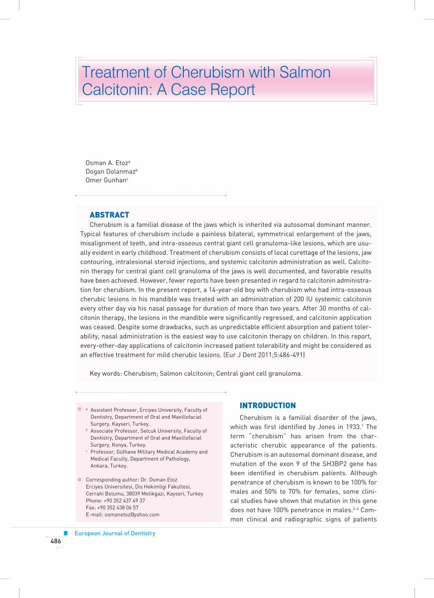
AbstrActCherubism is a familial disease of the jaws which is inherited via autosomal dominant manner.
Typical features of cherubism include a painless bilateral, symmetrical enlargement of the jaws, misalignment of teeth, and intra-osseous central giant cell granuloma-like lesions, which are usu-ally evident in early childhood. Treatment of cherubism consists of local curettage of the lesions, jaw contouring, intralesional steroid injections, and systemic calcitonin administration as well. Calcito-nin therapy for central giant cell granuloma of the jaws is well documented, and favorable results have been achieved. However, fewer reports have been presented in regard to calcitonin administra-tion for cherubism. In the present report, a 14-year-old boy with cherubism who had intra-osseous cherubic lesions in his mandible was treated with an administration of 200 IU systemic calcitonin every other day via his nasal passage for duration of more than two years. After 30 months of cal-citonin therapy, the lesions in the mandible were significantly regressed, and calcitonin application was ceased. Despite some drawbacks, such as unpredictable efficient absorption and patient toler-ability, nasal administration is the easiest way to use calcitonin therapy on children. In this report, every-other-day applications of calcitonin increased patient tolerability and might be considered as an effective treatment for mild cherubic lesions. (Eur J Dent 2011;5:486-491)
Key words: Cherubism; Salmon calcitonin; Central giant cell granuloma.
Osman A. Etoza
Dogan Dolanmazb
Omer Gunhanc
Treatment of Cherubism with Salmon Calcitonin: A Case Report
a Assistant Professor, Erciyes University, Faculty of Dentistry, Department of Oral and Maxillofacial Surgery, Kayseri, Turkey.b Associate Professor, Selcuk University, Faculty of Dentistry, Department of Oral and Maxillofacial Surgery, Konya, Turkey.c Professor, Gülhane Military Medical Academy and Medical Faculty, Department of Pathology, Ankara, Turkey.
Corresponding author: Dr. Osman Etoz Erciyes Universitesi, Dis Hekimligi Fakultesi, Cerrahi Bolumu, 38039 Melikgazi, Kayseri, Turkey Phone: +90 352 437 49 37Fax: +90 352 438 06 57E-mail: [email protected]
IntroductIon
October 2011 - Vol.5487
European Journal of Dentistry
with cherubism are painless bilateral expansion of the jaws, displaced and/or missing teeth or tooth germs, and multilocular well-defined radiolucent lesions. Histopathologic features of the afore-mentioned lesions are similar to central giant cell granuloma (CGCG). Despite their stromal struc-ture, cherubism lesions are spongier than CGCG lesions, and eosinophilic fibrolitic thickening has been evident.5 However, cherubism and CGCG le-sions have similar findings and similar treatment options. Conventional treatment of CGCG com-prises local curettage and long-term follow-up. Despite the high success rate of surgical interven-tions in the management of CGCG, conservative approaches like intralesional steroid injections,6 subcutaneous administration of α-interferon fol-lowing surgery,7 and systemic calcitonin therapy8 have been advocated, and favorable results have been achieved.
It has been reported that calcitonin therapy is a feasible treatment option in the management of CGCG, especially for larger aggressive or multiple lesions.9 However, few reports have been pub-lished with regard to successfully treated CGCG-like lesions of cherubism by calcitonin administra-tion. In this case report, treatment of cherubism with 200 IU salmon calcitonin via nasal adminis-tration is presented.
cAsE rEPortA 14-year-old boy was referred to our clinic
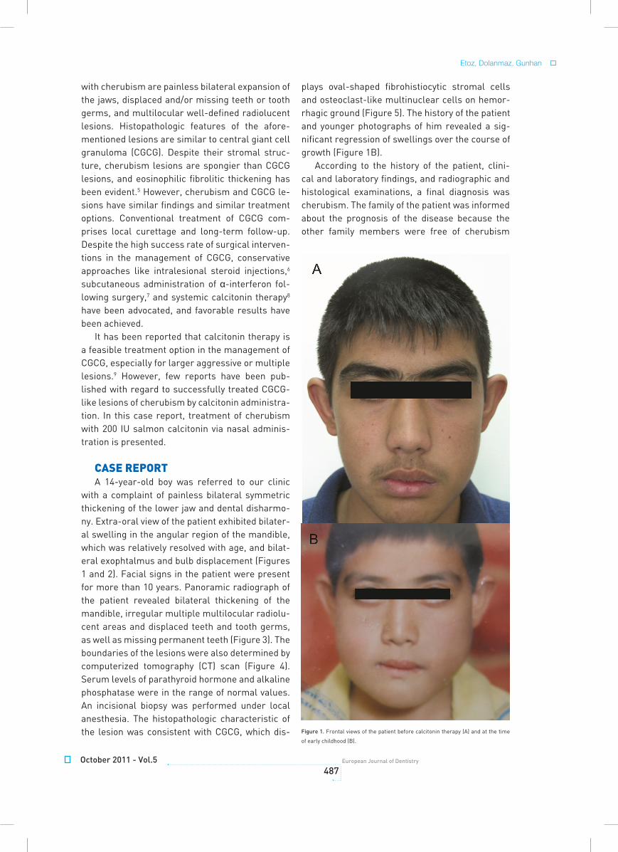
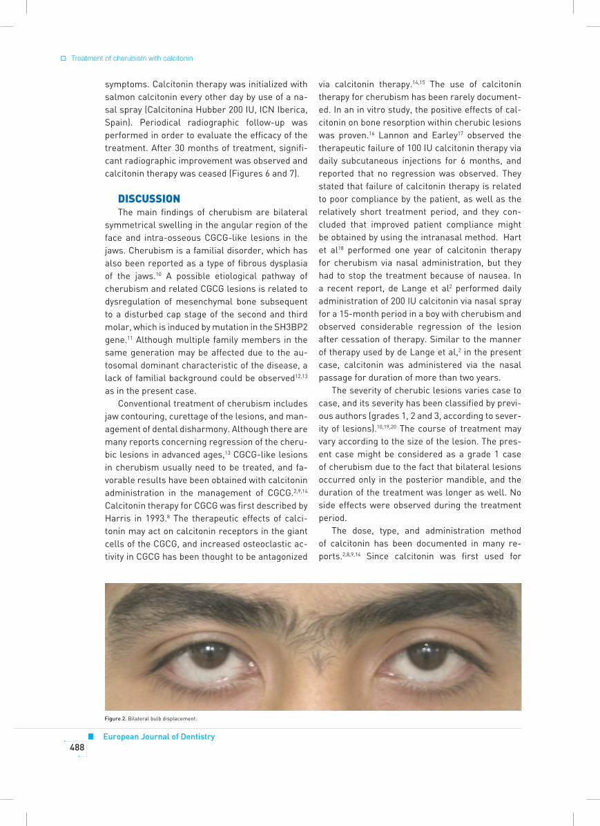
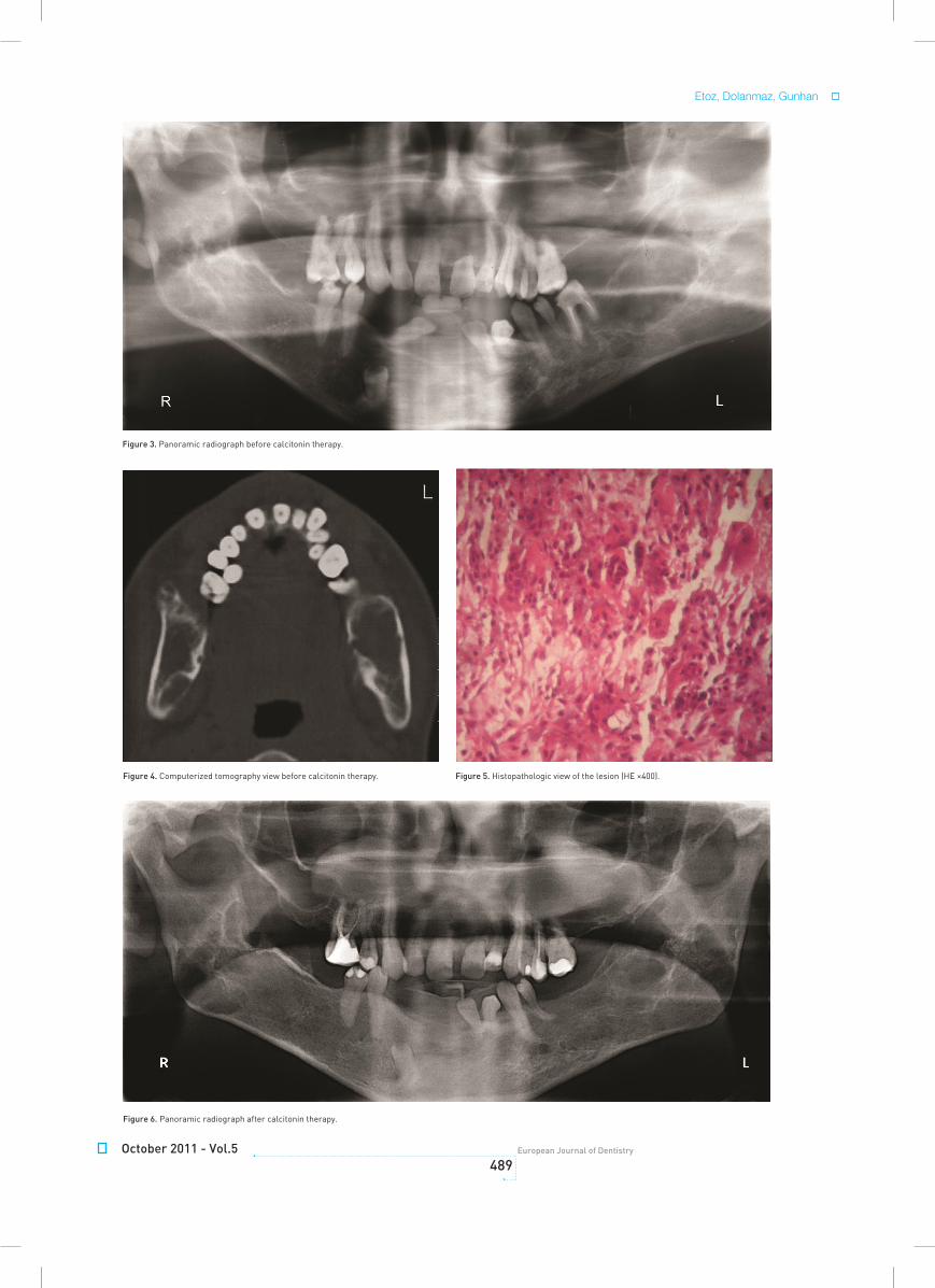
with a complaint of painless bilateral symmetric thickening of the lower jaw and dental disharmo-ny. Extra-oral view of the patient exhibited bilater-al swelling in the angular region of the mandible, which was relatively resolved with age, and bilat-eral exophtalmus and bulb displacement (Figures 1 and 2). Facial signs in the patient were present for more than 10 years. Panoramic radiograph of the patient revealed bilateral thickening of the mandible, irregular multiple multilocular radiolu-cent areas and displaced teeth and tooth germs, as well as missing permanent teeth (Figure 3). The boundaries of the lesions were also determined by computerized tomography (CT) scan (Figure 4). Serum levels of parathyroid hormone and alkaline phosphatase were in the range of normal values. An incisional biopsy was performed under local anesthesia. The histopathologic characteristic of the lesion was consistent with CGCG, which dis-
plays oval-shaped fibrohistiocytic stromal cells and osteoclast-like multinuclear cells on hemor-rhagic ground (Figure 5). The history of the patient and younger photographs of him revealed a sig-nificant regression of swellings over the course of growth (Figure 1B).
According to the history of the patient, clini-cal and laboratory findings, and radiographic and histological examinations, a final diagnosis was cherubism. The family of the patient was informed about the prognosis of the disease because the other family members were free of cherubism
Figure 1. Frontal views of the patient before calcitonin therapy (A) and at the time
of early childhood (B).
Etoz, Dolanmaz, Gunhan
European Journal of Dentistry488
symptoms. Calcitonin therapy was initialized with salmon calcitonin every other day by use of a na-sal spray (Calcitonina Hubber 200 IU, ICN Iberica, Spain). Periodical radiographic follow-up was performed in order to evaluate the efficacy of the treatment. After 30 months of treatment, signifi-cant radiographic improvement was observed and calcitonin therapy was ceased (Figures 6 and 7).
dIscussIonThe main findings of cherubism are bilateral
symmetrical swelling in the angular region of the face and intra-osseous CGCG-like lesions in the jaws. Cherubism is a familial disorder, which has also been reported as a type of fibrous dysplasia of the jaws.10 A possible etiological pathway of cherubism and related CGCG lesions is related to dysregulation of mesenchymal bone subsequent to a disturbed cap stage of the second and third molar, which is induced by mutation in the SH3BP2 gene.11 Although multiple family members in the same generation may be affected due to the au-tosomal dominant characteristic of the disease, a lack of familial background could be observed12,13 as in the present case.
Conventional treatment of cherubism includes jaw contouring, curettage of the lesions, and man-agement of dental disharmony. Although there are many reports concerning regression of the cheru-bic lesions in advanced ages,13 CGCG-like lesions in cherubism usually need to be treated, and fa-vorable results have been obtained with calcitonin administration in the management of CGCG.2,9,14 Calcitonin therapy for CGCG was first described by Harris in 1993.8 The therapeutic effects of calci-tonin may act on calcitonin receptors in the giant cells of the CGCG, and increased osteoclastic ac-tivity in CGCG has been thought to be antagonized
via calcitonin therapy.14,15 The use of calcitonin therapy for cherubism has been rarely document-ed. In an in vitro study, the positive effects of cal-citonin on bone resorption within cherubic lesions was proven.16 Lannon and Earley17 observed the therapeutic failure of 100 IU calcitonin therapy via daily subcutaneous injections for 6 months, and reported that no regression was observed. They stated that failure of calcitonin therapy is related to poor compliance by the patient, as well as the relatively short treatment period, and they con-cluded that improved patient compliance might be obtained by using the intranasal method. Hart et al18 performed one year of calcitonin therapy for cherubism via nasal administration, but they had to stop the treatment because of nausea. In a recent report, de Lange et al2 performed daily administration of 200 IU calcitonin via nasal spray for a 15-month period in a boy with cherubism and observed considerable regression of the lesion after cessation of therapy. Similar to the manner of therapy used by de Lange et al,2 in the present case, calcitonin was administered via the nasal passage for duration of more than two years.
The severity of cherubic lesions varies case to case, and its severity has been classified by previ-ous authors (grades 1, 2 and 3, according to sever-ity of lesions).10,19,20 The course of treatment may vary according to the size of the lesion. The pres-ent case might be considered as a grade 1 case of cherubism due to the fact that bilateral lesions occurred only in the posterior mandible, and the duration of the treatment was longer as well. No side effects were observed during the treatment period.
The dose, type, and administration method of calcitonin has been documented in many re-ports.2,8,9,14 Since calcitonin was first used for
Figure 2. Bilateral bulb displacement.
Treatment of cherubism with calcitonin
October 2011 - Vol.5489
European Journal of Dentistry
Etoz, Dolanmaz, Gunhan
Figure 3. Panoramic radiograph before calcitonin therapy.
Figure 4. Computerized tomography view before calcitonin therapy. Figure 5. Histopathologic view of the lesion (HE ×400).
Figure 6. Panoramic radiograph after calcitonin therapy.
European Journal of Dentistry490
CGCG, daily subcutaneous administration of hu-man calcitonin has become the most common method of therapy. However, the use of salmon calcitonin has recently become more popular than using human calcitonin because of its increased potency and availability.21 The correct dose of cal-citonin has been determined at 100 IU per day, which is based on the regimens previously used for Paget’s disease.22 On the other hand, the use of daily 200 IU salmon calcitonin via nasal spray for cherubism has been recently reported.2 In the present case, 200 IU calcitonin was adminis-tered via nasal spray every-other-day to make an equivalent daily dose of 100 IU, because a 100 IU calcitonin nasal spray dosage was not available in the country. However, it is well known that daily subcutaneous administration of 100 IU calcitonin is considerably more effective than an every-oth-er-day nasal dose of 200 IU. As a result, the two treatment regimens are not comparable due to the lower bio-availability of nasal spray. In gen-eral, subcutaneous application was preferred in order to achieve efficient absorption. Neverthe-less, the absorbed amount of calcitonin might be unpredictable because of the condition of the na-sal mucosa. However, systemic calcitonin dosage via nasal spray is considered the easiest method of management, especially in children.
To our knowledge, the present report is the second in which cherubic lesions were treated with systemic calcitonin administration via nasal spray for duration of more than one year. After approximately three years of treatment, calcito-nin therapy was discontinued since radiographic regression of the lesions and growth of the pa-tient had ended, and the patient was scheduled
for follow-up. However, regression of the lesions through the natural course of the disease, due to the relatively older age of the patient, may have contributed to the successful outcome of the treatment. The dose and application method of calcitonin therapy would be dependent on the case and patient tolerability. A dosage of 200 IU calci-tonin via nasal spray may be administered every other day in order to improve patient tolerability and continuity of the treatment, especially in mild cases of cherubism. Further studies will be nec-essary to determine the ideal regimen of calcito-nin therapy for cherubic individuals.
rEFErEncEs1. Jones WA. Familial multilocular cystic disease of the jaws.
Am J Cancer 1933;17:946.
2. De Lange J, van den Akker H, Scholtemeijer M. Cherubism
treated with calcitonin: report of a case. J Oral Maxillofac
Surg 2007;65:1665-1667.
3. Anderson DE, McClendon JL. Cherubism-hereditary fi-
brous dysplasia of the jaws; I. genetic considerations. Oral
Surg Oral Med Oral Pathol 1962;15(suppl.2):5-16.
4. De Lange J, van Maarle M, van den Akker HP, Redeker
EJW. A new mutation in the SH3BP2 gene showing reduced
penetrance in a family affected with cherubism. Oral Surg
Oral Med Oral Pathol 2007;103:378-381.
5. Gunhan O. Oral ve Maksillofasiyal Patoloji (1st edn). Ankara:
Atlas Kitapcilik; 2001.
6. Carlos R, Sedano HO. Intralesional corticosteriods as an
alternative treatment for central giant cell granuloma.
Oral Surg Oral Med Oral Pathol 2002;93:161-166.
7. Kaban LB, Mulliken JB, Ezekowitz RA et al. Antiangiogenic
therapy of a recurrent giant cell tumor of the mandible with
interferon alfa-2a. Pediatrics 1999;103:1145-1149.
8. Harris M. Central giant cell granulomas of the jaws re-
gress with calcitonin therapy. Br J Oral Maxillofac Surg
1993;31:89-94.
9. Pogrel MA. Calcitonin therapy for central giant cell granu-
loma. J Oral Maxillofac Surg 2003;61:649-653.
10. Von Wowern N. Cherubism: a 36-year long term follow-up
of 2 generations in different families and review of the lit-
erature. Oral Surg Oral Med Oral Pathol 2000;90:765-772.
11. Hyckel P, Berndt A, Schleier P, Clement JH, Beensen V,
Peters H, et al. Cherubism – new hypotheses on pathogen-
esis and therapeutic consequences. J Craniomaxillofac Surg
2005;33:61-68.
12. Grünebaum M, Tiqva P. Non-familial cherubism: report of
a case. J Oral Surg 1973;31:632-635.
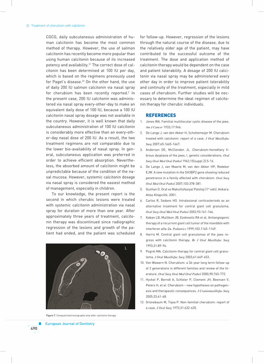
Figure 7. Computerized tomography view after calcitonin therapy
Treatment of cherubism with calcitonin
October 2011 - Vol.5491
European Journal of Dentistry
Etoz, Dolanmaz, Gunhan
13. Kozakiewicz M, Perezynska-Partyka W, Kobos J.
Cherubism – clinical picture and treatment. Oral Diseases
2001;7:123-130.
14. Pogrel MA, Regezi JA, Harris ST, Goldring SR. Calcitonin
treatment for central giant cell granulomas of the mandi-
ble: report of two cases. J Oral Maxillofac Surg 1999;57:848-
853.
15. Flanagan AM, Nui B, Tinkler SMB, Horton MA, Williams DM,
Chambers TJ. The multinucleate cell in giant cell granulo-
mas of the jaw are osteoclast. Cancer 1988;62:1139-1145.
16. Southgate J, Sarma U, Townend JV, Barron J, Flanagan
AM. Study of the cell biology and chemistry of cherubism. J
Clin Pathol 1998;51:831-837.
17. Lannon DA, Earley MJ. Cherubism and its charlatans. Br J
Plast Surg 2001;54:708-731.
18. Hart W, Schweitzer DH, Slootweg PJ, et al. Man with
cherubism. Ned Tijdschr Geneeskd 2000;144:34-38.
19. Motamedi MHK. Treatment of cherubism with locally ag-
gressive behavior presenting in adulthood: report of four
cases and a proposed new grading system. J Oral Maxil-
lofac Surg 1998;56:1336-1342.
20. Seward GR, Hankey GT. Cherubism. Oral Surg Oral Med
Oral Pathol 1957;10:952-974.
21. Azria M. The calcitonines, physiology and pharmacology.
London: Karger;1989.
22. De Lange J, Rosenberg AJWP, van den Akker HP, Koole R,
Wirds JJ, van den Berg. Treatment of giant cell granuloma
with calcitonin. Int J Oral Maxillofac Surg 1999;28:372-376.