Embed Size (px)
Citation preview
British Journal of Ophthalmology, 1987, 71, 290-294
Treatment of ptosis in chronic progressive externalophthalmoplegiaCAROL M LANE AND J R 0 COLLIN
From Moorfields Eye Hospital, High Holborn and City Road, London
SUMMARY Seventeen patients with ptosis as a feature of chronic progressive external ophthalmo-plegia were managed in accordance with a new protocol. An anterior approach levatoradvancement was performed on seven patients (13 lids) with more than 4 mm of levator functionand a brow suspension procedure on eight patients (14 lids) with minimal levator function, in whomthe frontalis muscle was relatively spared. Ptosis props were prescribed for two patients with a veryweak orbicularis and poor lid closure. Six patients required long term lubricants, and onedeveloped a postoperative corneal abscess associated with a poor Bell's phenomenon. Satisfactoryelevation of the lids was achieved in 16 patients (25 lids).
The surgeon's choice of operation for ptosis correc-tion is normally based on the degree of ptosis and theamount of levator function.' However, in chronicprogressive external ophthalmopiegia (CPEO)increasing weakness of the levator palpebraesuperioris is accompanied by deterioration in func-tion of the frontalis, orbicularis oculi, and externalocular muscles. There is a significant risk of post-operative corneal exposure due to lagophthalmosand a poor Bell's phenomenon. Surgery is indicatedonly if ptosis interferes with vision or causes consider-able social embarrassment. In these circumstances asmall degree of ptosis correction often provides amarked improvement in the patient's quality of life.This paper presents a protocol for the management ofptosis in patients with CPEO and the results of its usein 17 patients.The treatment protocol was based on the relative
power of the affected muscles. Patients with goodlevator function were treated with an anteriorapproach levator advancement. The anteriorapproach was used to help the identification ofstructures, since the levator muscle and aponeurosisoften show fatty infiltration. It also avoids interfer-ence with the conjunctiva and the tear film, which isoften compromised in patients with CPEO.2 Thosewith poor levator function but preserved frontalisaction were treated by a brow suspension procedure.Lid props were prescribed when severe orbicularisweakness was demonstrable.Correspondence to C M Lane, FRCS, Moorfields Eye Hospital,High Holborn, London WCIV 7AN.
CPEO may be considered as a gradually progres-sive external ophthalmoplegia which occurs inseveral disease states with numerous associated find-ings, hence Drachman's term, 'ophthalmoplegiaplus'.3 Some of the more common associations andother disorders which occasionally cause CPEO arelisted in Table 1. Patients with myotonic dystrophymay give a history of autosomal dominant inherit-ance and have a classical long thin facies, frontalbalding, and cataract.4 Ptosis is marked, and theremay be slowing or limitation of external ocularmovements.' Kearns and Sayre6 described a syn-drome in which CPEO and heart block occurredbefore the age of 20 years, with diverse associatedfeatures and the later development of pigmentaryretinopathy. Similar hereditary diseases include
Table 1 Conditions associated with orcausing CPEO
Conditions Associated with CPEOMyotonic dystrophyKearns Sayre syndromeStephens syndromeOculopharyngeal dystrophyDisorders rarely causing external ophthalmoplegiaCongenital disorderse.g. Abetalipoproteinaemia
Refsum's diseaseExtraocular fibrosis syndromeMoebius syndrome
Progressive supranuclear palsyEndocrine exophthalmosMyasthenia gravisMultiple sclerosis
290
on 12 April 2019 by guest. P
rotected by copyright.http://bjo.bm
j.com/
Br J O
phthalmol: first published as 10.1136/bjo.71.4.290 on 1 A
pril 1987. Dow
nloaded from
Treatment ofptosis in chronic progressive external ophthalmoplegia
Stephens syndrome7 of CPEO, cerebellar ataxia andperipheral neuropathy, and oculopharyngealdystrophy in which ptosis, mild restriction of eyemovements, and dysphagia with autosomal dominantinheritance occur after 40 years of age.8 Abetalipo-proteinaemia9 and Refsum's disease'0 are among rarecongenital disorders which sometimes featureexternal ophthalmoplegia. Patients with ocular fibro-sis syndrome have static restriction of ocular move-ment, with reduced movement of the globe on forcedduction.These syndromes must be differentiated from
acquired brainstem disease, such as progressivesupranuclear palsy, and from multiple sclerosis.Dysthyroid eye disease may rarely present in clinic-ally euthyroid" ptotic patients, but the symptom ofdiplopia, uncommon in other forms of CPEO,together with particular restriction of elevationand abduction of the globe, may assist diagnosis.Myasthenia gravis is recognisable by the presence ofmuscle fatigue with a reduced number of acetylcho-line receptors and a positive response to edropho-nium, an anticholinesterase. 12 Weakness of theexternal ocular muscles is often asymmetrical andmomentary retraction of the upper eyelid may beobserved during refixations from down to straightahead.'3 Surgical correction of myasthenic ptosis isinfluenced by the medical status of the patient andhas an unpredictable outcome; patients with myast-henia gravis were therefore excluded from the study.
Subjects and methods
Seventeen patients with severe ptosis and CPEOpresented to the Lid Clinic at Moorfields EyeHospital over a period of six years (Table 2). Patientswith mild ptosis were not included in the study.Eleven patients had features of systemic disease: fourhad the Kearns-Sayre syndrome, two had oculo-pharyngeal dystrophy, four had myotonic dystrophy,and one had progressive multiple sclerosis. In allcases the diagnosis was confirmed by a neurologist.Specimens from triceps muscle biopsy in fivepatients, four of whom had Kearns-Sayre syndrome,had shown ragged red fibres, a marker for mito-chondrial abnormalities. 14 At presentation a fullclinical history was taken from each patient. Specificquestions referred to the duration and variability ofthe ptosis, the use of props, diplopia, and the degreeof disability suffered.The palpebral aperture was measured with the
patient looking straight ahead and levator functionwas measured from full downgaze to full upgaze withthe frontalis muscle held fixed against the brow. Thedistance from the lid margin to the upper lid skincrease was noted when the latter was present. Bell's
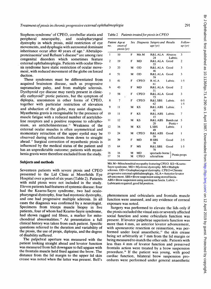
Table 2 Patients treatedforptosis in CPEO
Patient Age at Sex Diagnosis Surgery and Results Follow-no. onset of age (yr) up (yr)
ptosis (yr)
1 10 F Mit.M R&L ALA Abscess 324 Lubric.
2 19 F MD R&LALA Good 255
3 33 M OD R&LALA Good 346
4 51 M OD R&LALA Good 359
5 41 F CPEO RALA Lubric. 1-561
6 43 F MD R&LALA Good 251
7 58 F CPEO R&LALA Good 373
8 7 F CPEO R&L SBS Lubric. 0-562 1-5
9 11 M KS R&LABS Lubric. 141
10 11 F KS R&LABS Lubric. 715
11 12 M KS R&LABS Bandscut 520 Ptotic+
12 16 M KS L SBS Lubric. 149
13 24 M CPEO R&L ABS Good 159
14 39 F MD R SBS Good 2-539
15 64 F MS R&L SBS Good 1-578
16 16 M MD spectacle-borne Ptosis props17 51 M CPEO scleral lens
Mit.M= Mitochondrial myopathy featuring CPEO. KS= Kearns-Sayre syndrome. MD=Myotonic dystrophy. MS=Multiplesclerosis. OD= Oculopharyngeal dystrophy. CPEO=Chronicprogressive external ophthalmoplegia. ALA=Anterior elevatoradvancement. SBS=Brow suspension using stored fascia.ABS=Brow suspension using autologous fascia. Lubric. -Lubricants required; good lid position.
phenomenon and orbicularis and frontalis musclefunction were assessed, and any evidence of cornealexposure was noted.
Surgery was performed to elevate the lids only ifthe ptosis occluded the visual axis or severely affectedsocial function and some orbicularis function waspresent. If levator palpebrae superioris function wasmore than 4 mm, an anterior levator advancement,with aponeurotic resection or reinsertion, was per-formed under local anaesthetic,'9 the skin creasebeing set arbitrarily at 7 mm from the lid margin orbeing measured to match the other side. Patients withless than 4 mm of levator function and preservedfrontalis action were treated by a brow suspensionprocedure.'6 If the patient was young, with goodcardiac function, bilateral brow suspension pro-cedures were performed under general anaesthetic
291
on 12 April 2019 by guest. P
rotected by copyright.http://bjo.bm
j.com/
Br J O
phthalmol: first published as 10.1136/bjo.71.4.290 on 1 A
pril 1987. Dow
nloaded from
CarolMLane andJ R 0 Collin
by the Crawford technique'7 and autologous fascialata. The anaesthetist was consulted at an early stage,and halothane, barbiturates, and suxamethoniumwere avoided.'" In older patients, and where generalanaesthesia was contraindicated, homologous fascialata, stored in 70% alcohol and then soaked in salinefor 12 hours and rinsed in soframycin 2% for 1 hourpreoperatively, was inserted under local anaesthesiaby the technique described by Fox.20 Strips of storedfascia- were cut 3 mm wide to ensure adequatestrength, and the patients receiving them were givena five-day course of systemic antibiotic.With both an anterior approach advancement and
a brow suspension procedure the eyelid level wasadjusted at surgery under local anaesthesia so that itwas just clear of the pupillary axis with the patientvoluntarily opening the eye. With a brow suspensionunder general anaesthesia the upper eyelid waspositioned so that the lid was closed but just begin-ning to open at the end of the procedure. A 4/0 silksuture, inserted into the lower eyelid over a bolsterand drawn up to the brow,2' protected the corneaunder the dressing. When the dressing was taken off48 hours after surgery, the suture was taped to thecheek for 24 hours and removed at the end of thisperiod if there was no evidence of corneal exposure;traction to the lower lid could be reapplied if thisoccurred. All patients were prescribed local anti-biotic and lubricant drops on a 1/2 to 2 hourly basis.
All patients were reviewed at three or fourmonthly intervals in the clinic, with particular atten-tion being paid to corneal exposure, and they wereinstructed to report immediately should the eyebecome painful, red, or start discharging pus.
Results
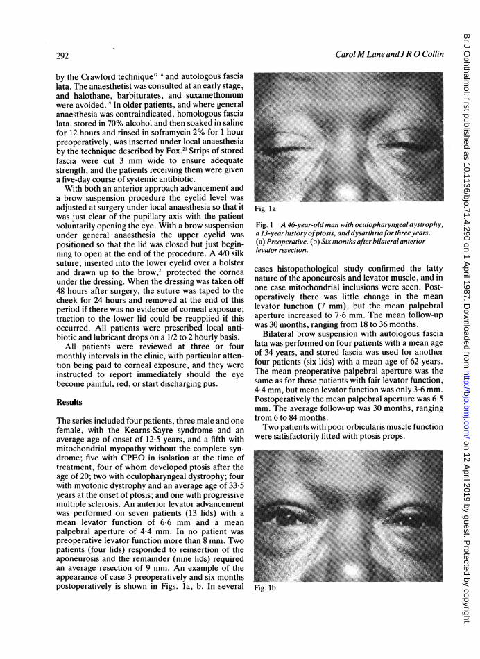
The series included four patients, three male and onefemale, with the Kearns-Sayre syndrome and anaverage age of onset of 12-5 years, and a fifth withmitochondrial myopathy without the complete syn-drome; five with CPEO in isolation at the time oftreatment, four of whom developed ptosis after theage of 20; two with oculopharyngeal dystrophy; fourwith myotonic dystrophy and an average age of 33.5years at the onset of ptosis; and one with progressivemultiple sclerosis. An anterior levator advancementwas performed on seven patients (13 lids) with amean levator function of 6-6 mm and a meanpalpebral aperture of 4-4 mm. In no patient waspreoperative levator function more than 8 mm. Twopatients (four lids) responded to reinsertion of theaponeurosis and the remainder (nine lids) requiredan average resection of 9 mm. An example of theappearance of case 3 preoperatively and six monthspostoperatively is shown in Figs. la, b. In several
Fig. la
Fig. 1 A 46-year-old man with oculopharyngeal dystrophy,a 13-year history ofptosis, and dysarthriafor three years.(a) Preoperative. (b) Six months after bilateral anteriorlevator resection.
cases histopathological study confirmed the fattynature of the aponeurosis and levator muscle, and inone case mitochondrial inclusions were seen. Post-operatively there was little change in the meanlevator function (7 mm), but the mean palpebralaperture increased to 7-6 mm. The mean follow-upwas 30 months, ranging from 18 to 36 months.
Bilateral brow suspension with autologous fascialata was performed on four patients with a mean ageof 34 years, and stored fascia was used for anotherfour patients (six lids) with a mean age of 62 years.The mean preoperative palpebral aperture was thesame as for those patients with fair levator function,4-4 mm, but mean levator function was only 3.6 mm.Postoperatively the mean palpebral aperture was 6-5mm. The average follow-up was 30 months, rangingfrom 6 to 84 months.Two patients with poor orbicularis muscle function
were satisfactorily fitted with ptosis props.
Fig. lb
292
on 12 April 2019 by guest. P
rotected by copyright.http://bjo.bm
j.com/
Br J O
phthalmol: first published as 10.1136/bjo.71.4.290 on 1 A
pril 1987. Dow
nloaded from
Treatment ofptosis in chronicprogressive external ophthalmoplegia
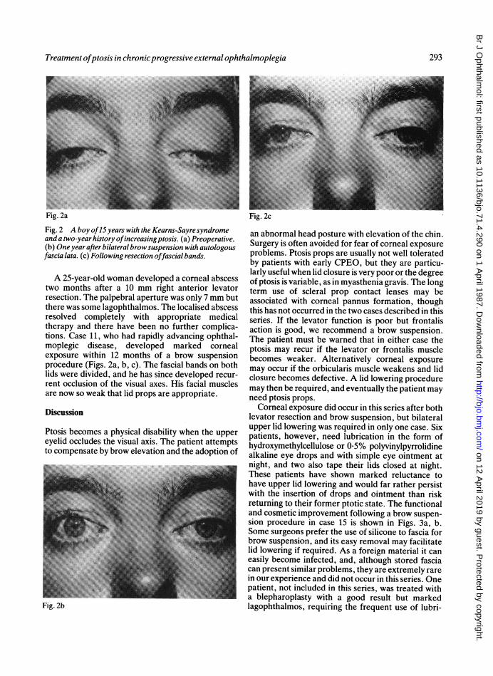
Fig. 2a Fig. 2cFig. 2 A boy of15 years with the Kearns-Sayresyndromeand a two-year history ofincreasingptosis. (a) Preoperative.(b) One yearafter bilateral browsuspension with autologousfascia lata. (c) Following resection offascial bands.
A 25-year-old woman developed a corneal abscesstwo months after a 10 mm right anterior levatorresection. The palpebral aperture was only 7 mm butthere was some lagophthalmos. The localised abscessresolved completely with appropriate medicaltherapy and there have been no further complica-tions. Case 11, who had rapidly advancing ophthal-moplegic disease, developed marked cornealexposure within 12 months of a brow suspensionprocedure (Figs. 2a, b, c). The fascial bands on bothlids were divided, and he has since developed recur-rent occlusion of the visual axes. His facial musclesare now so weak that lid props are appropriate.
Discussion
Ptosis becomes a physical disability when the uppereyelid occludes the visual axis. The patient attemptsto compensate by brow elevation and the adoption of
Fig. 2b
an abnormal head posture with elevation of the chin.Surgery is often avoided for fear of corneal exposureproblems. Ptosis props are usually not well toleratedby patients with early CPEO, but they are particu-larly useful when lid closure is very poor or the degreeof ptosis is variable, as in myasthenia gravis. The longterm use of scleral prop contact lenses may beassociated with corneal pannus formation, thoughthis has not occurred in the two cases described in thisseries. If the levator function is poor but frontalisaction is good, we recommend a brow suspension.The patient must be warned that in either case theptosis may recur if the levator or frontalis musclebecomes weaker. Alternatively corneal exposuremay occur if the orbicularis muscle weakens and lidclosure becomes defective. A lid lowering proceduremay then be required, and eventually the patient mayneed ptosis props.
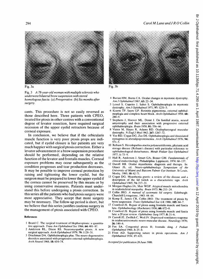
Corneal exposure did occur in this series after bothlevator resection and brow suspension, but bilateralupper lid lowering was required in only one case. Sixpatients, however, need lubrication in the form ofhydroxymethylcellulose or 0 5% polyvinylpyrrolidinealkaline eye drops and with simple eye ointment atnight, and two also tape their lids closed at night.These patients have shown marked reluctance tohave upper lid lowering and would far rather persistwith the insertion of drops and ointment than riskreturning to their former ptotic state. The functionaland cosmetic improvement following a brow suspen-sion procedure in case 15 is shown in Figs. 3a, b.Some surgeons prefer the use of silicone to fascia forbrow suspension, and its easy removal may facilitatelid lowering if required. As a foreign material it caneasily become infected, and, although stored fasciacan present similar problems, they are extremely rarein our experience and did not occur in this series. Onepatient, not included in this series, was treated witha blepharoplasty with a good result but markedlagophthalmos, requiring the frequent use of lubri-
293
on 12 April 2019 by guest. P
rotected by copyright.http://bjo.bm
j.com/
Br J O
phthalmol: first published as 10.1136/bjo.71.4.290 on 1 A
pril 1987. Dow
nloaded from
CarolM Lane andJ R 0 Collin
Fig. 3a Fig. 3b
Fig. 3 A 78-year-old woman with multiple sclerosis whounderwent bilateral brow suspension with storedhomologous fascia. (a) Preoperative. (b) Six months aftersurgery.
cants. This procedure is not so easily reversed asthose described here. Three patients with CPEO,treated for ptosis in other centres with a conventionaldegree of levator resection, have required surgicalrecession of the upper eyelid retractors because ofcorneal exposure.
In conclusion, we believe that if the orbicularismuscle function is very poor ptosis props are indi-cated, but if eyelid closure is fair patients are verymuch happier with surgical ptosis correction. Either alevator advancement or a brow suspension procedureshould be performed, depending on the relativefunction of the levator and frontalis muscles. Cornealexposure problems may occur subsequently as thecondition progresses and tear production decreases.It may be possible to improve corneal protection byraising and tightening the lower eyelid, but thesurgeon must be prepared to lower the upper eyelid ifthe cornea cannot be preserved by this means or byusing conservative measures. Patients must under-stand this before undergoing a ptosis correction. Inthis series all the patients who had ptosis surgery weremost appreciative. They accept that more surgerymay be necessary. The follow-up period is short, butwe believe that this series justifies cautious surgery inthe management of ptosis associated with CPEO.
References
1 Beard C. The surgical treatment of blepharoptosis: a quantita-tive approach. Trans Am Ophthalmol Soc 1966; 64: 401-87.
2 Anderson RL, Dixon RS. Neuromyopathic ptosis. A newsurgical approach. Arch Ophthalmol 1979; 79:1129-31.
3 Drachman DA. Ophthalmoplegia plus. The neuro-degenerativedisorders associated with progressive external ophthalmoplegia.Arch Neurol 1968; 18: 654-74.
4 Burian HM, Burns CA. Ocular changes in myotonic dystrophy.Am J Ophthalmol 1967; 63: 22-34.
5 Lessel S, Copetto J, Sahet S. Ophthalmoplegia in myotonicdystrophy. Am J Ophthalmol 1971; 97: 1231-5.
6 Kearns TP, Sayre GP. Retinitis pigmentosa, external ophthal-moplegia and complete heart block. Arch Ophthalmol 1958; 60:280-9.
7 Stephens J. Hoover ML, Denst J. On familial ataxia, neuralamyotrophy and their association with progressive externalophthalmoplegia. Brain 1958; 81: 556-66.
8 Victor M, Hayes R, Adams RD. Oculopharyngeal musculardystrophy. N Engl J Med 1962; 267: 1267-72.
9 Yee RD, Cogan DG, Zee DS. Ophthalmoplegia and dissociatednystagmus in abetalipoproteinemia. Arch Ophthalmol 1976; 94:571-5.
10 Refsum S. Heredopathia atactia polyneuritiformis, phytanic acidstorage disease (Refsum's disease) with particular reference toophthalmological disturbances. Metab Pediatr Syst Ophthalmol1977; 1: 73-9.
11 Hall R, Anderson J. Smart GA, Besser GM. Fundamentals ofclinical endocrinology. Philadelphia: Lippincott, 1974: 68-137.
12 Daroff RB. Ocular myasthenia: diagnosis and therapy. In:Glaser JS, ed. Neuro-ophthalmology Symposium of theUniversity of Miami and Bascom Palmer Eye Institute. St Louis,Mosby, 1980; 10: 62-71.
13 Cogan DG. Myasthenia gravis: a review of the disease and adescription of the lid twitch as a characteristic sign. ArchOphthalmol 1965; 74: 217-21.
14 Morgan-Hughes JA, Mair WGP. Atypical muscle mitochondriain oculoskeletal myopathy. Brain 1973; 96: 215-24.
15 Collin JRO. A manual of systematic lid surgery. Edinburgh:Churchill Livingstone, 1983: 44-64.
16 Kemp E, James CR, Collin JRO. The treatment of ptosis bybrow suspension. Trans Ophthalmol Soc UK 1986; 105: 84-7.
17 Crawford JS. Repair of ptosis using frontalis muscle and fascialata. Ophthalmology (Rochester) 1956; 60: 672-8.
18 Crawford JS. Repair of ptosis using frontalis muscle and fascialata: a 20 year review. Ophthalmic Surg 1977; 8: 31-6.
19 Carroll JE, Zwillich C, Weil JV. Depressed ventilatory responsein oculocraniosomatic neuro-muscular disease. Neurology 1976;26: 140-6.
20 Fox SA. Congenital ptosis II; frontalis sling. J PediatrOphthalmol 1966; 3: 25-8.
21 Frost AD. Supporting suture in ptosis operations. Am JOphthalmol 1934; 17: 633.
Acceptedfor publication 26 June 1986.
294
on 12 April 2019 by guest. P
rotected by copyright.http://bjo.bm
j.com/
Br J O
phthalmol: first published as 10.1136/bjo.71.4.290 on 1 A
pril 1987. Dow
nloaded from


![ptosis [emedicine]](https://img.pdfslide.net/doc/110x75/577cdd4a1a28ab9e78acb3ee/ptosis-emedicine.jpg)