Embed Size (px)
Citation preview
Chapter 3
Treatments in Infectious and Allergic Conjunctivitis:Is Immunomodulation the Future?
Concepcion Santacruz, Sonia Mayra Perez-Tapia,Angel Nava-Castañeda, Sergio Estrada-Parra andMaria C. Jimenez-Martinez
Additional information is available at the end of the chapter
http://dx.doi.org/10.5772/52263
1. Introduction
The ocular surface is a functional unit mainly formed by the conjunctival and corneal epi‐thelium (structural component), and tear film (soluble component). Microorganisms andenvironmental allergens can interact with the tear film, reach the structural componentand generate an immune response against them. Understanding the cellular and solublemediators that are involved in these inflammatory responses not only helps in under‐standing the mechanisms of current treatments, but also is needed to identification anddevelopment of new therapeutics targets. The aim of this review was to investigate thenovel and developing therapies, with special emphasis in immunomodulatory drugs/molecules that could have some clinical indication in the treatment of infectious and aller‐gic conjunctivitis in few years.
2. Novel therapies in infectious keratoconjunctivitis
2.1. Interferons (IFN) and adenoviral conjunctivitis
Interferons were first described as the major effector cytokines of the host immune responseagainst viral infections. IFN are well recognized by their potent antiviral properties, howev‐er IFN production is also induced in response to bacterial ligands of innate immune recep‐tors and/or bacterial infections, indicating a broader physiological role for these cytokines inhost defence and homeostasis than was originally described.
© 2013 Santacruz et al.; licensee InTech. This is an open access article distributed under the terms of theCreative Commons Attribution License (http://creativecommons.org/licenses/by/3.0), which permitsunrestricted use, distribution, and reproduction in any medium, provided the original work is properly cited.
Three main types of cytokines compose the IFN family: type I, type II and type III IFN.Type I IFN family is composed of 16 members, namely 12 IFNα subtypes, IFNβ, IFNε,IFNκ and IFNω. By contrast, the type II IFN family includes only one cytokine: IFNγ,which also exhibits antiviral activities. The third type of IFN is the IFNλ family, which in‐cludes IFNλ1 (also known as IL-29), IFNλ2 (also known as IL-28A) and IFNλ3 (also knownas IL-28B). On the basis of protein sequence and structure, type III IFN are markedly differ‐ent from type I and type II IFN and are more similar to members of the interleukin-10(IL-10) family; however, they provoke antiviral responses and induce the activation of IFN-stimulated genes. [1]
Epidemic keratoconjunctivitis (EKC) is a severe ocular infection, caused by highly conta‐gious adenoviruses Ad8, Ad19, and Ad37. Adenoviral infection of the eye induces keratitisand conjunctivitis, accompanied by pain, lacrimation, red and swollen eye, as well as de‐creased vision that may last for months or even years. No specific antiviral drugs are cur‐rently available for the treatment of EKC or any other infection caused by adenoviruses.Interestingly, it has been suggested that five strains of different serotypes of adenovirus,types 3 (AdV3; species B), 4 (species E), 8, 19a and 37 (species D) involved in acute kerato‐conjunctivitis are highly inhibited by IFN-b and IFN-g in the A549 cell line, [2] However,IFN therapy in adenoviral keratoconjunctivitis has not been evaluated in clinical trials yet.
2.2. Glycan interactions and EKC
The initial event leading to EKC is binding of the viruses to glycans that contain sialic acidmoieties on epithelial cells in the cornea or conjunctiva through trimeric fiber structures ex‐tending from the viral particles. The receptor-binding domain is located at the C terminus ofeach fiber and contains three separate pockets that each can accommodate one sialic acidresidue. Ad37 was recently shown to bind to cell-surface glycoproteins carrying a glycanstructure named GD1a due to similitude to GD1a ganglioside. The GD1a glycan is abranched hexasaccharide with a terminal sialic acid residue on each of its two arms. Struc‐tural studies showed that the two sialic acid moieties dock into two of three sialic acid bind‐ing sites in the trimeric knob of the Ad37 fiber protein. Most likely, multiple fiber proteinssimultaneously engage several host-cell epitopes containing terminal sialic acids; internali‐zation and subsequent infection follow. In this context, the molecules named ME0322,ME0323, and ME0324 were synthetized as a tri- and tetravalent sialic acid compounds, andinterestingly all of theses molecules inhibited the attachment of Ad37 virions to HCE cells ina dose-dependent manner and were at least two orders of magnitude more effective thansialic acid, suggesting a promissory inhibitor of Ad37 infection on corneal cells, composedby a multivalent sialic acid conjugate. If these compounds could be useful as a topical treat‐ment is not known and needs further investigation. [3]
2.3. Vaccines and Herpetic Stromal Keratitis (HSK)
The disease course in herpetic stromal keratitis (HSK) begins with a primary infection byherpes simplex virus (HSV) followed by a period during which the virus enters latency insensory and autonomic ganglia, after that a reactivation from the trigeminal ganglia follow‐
Common Eye Infections46
ing primary infection induce virus transportation to the ocular mucosa via antero-grademovement from the ganglia, ultimately causing herpetic keratitis, conjunctivitis and otherocular sequelae [4]
Many studies have shown that clinical disease is the result of a recruitment of inflammatorycells, mainly polymorphonuclear cells (PMN), macrophages, and T cells to the corneas of pa‐tients with HSK. [5] Due to HSK could lead to a potentially blinding disease; several thera‐peutical strategies are in development to control ocular damage at initial steps ofinflammatory process, i.e. vaccination with different HSV epitopes.
Since the early nineties many attempts have been made to develop a vaccine that would beeffective in preventing HSK. Most of these vaccines were useful to prevent primary HSKwhen given prior to HSV infection however failed to prevent recurrent HSK lesions. [6, 7, 8]Recently, a novel construct with a DNA vaccine expressing herpes simplex virus type 1gDand IL-21, appears to be effective in protect from primary lesions, and also ameliorates her‐pes keratitis severity and time course after corneal infection with HSV-1 in the animal model[9] Nevertheles, future studies are needed in humans HSK to study efficacy of this vaccine.
2.4. Lipids mediators and HSK
Resolvins are lipid mediators that are derived from the v-3 polyunsaturated fatty acids eico‐sapentaenoic acid and do- cosahexaenoic acid [10] The name of these lipid mediators is re‐lated to their main function, control of inflammation. Resolvins are involved in preventionof diapedesis, regulation of dendritic cell costimulatory factors, [11], increased macrophagephagocytosis of apoptotic neutrophils, inhibition of host tissue inflammatory responses,with the release of chemokines and cytokines, [12] promotion of tissue repair, and preven‐tion of host tissue cell death during stress. [13] Interestingly, topical therapy with resolvinsin corneas infected with HSV showed a diminished lesion severity and corneal neovasculari‐zation when compared with non-treated eyes. Therapy with resolvins, induced a decreasedinflux of effector CD4+ T cells and neutrophils to corneal tissue; a diminished production ofproinflammatory cytokines and molecules involved in ocular neovascularization were alsoobserved during this treatment in the animal model, suggesting resolvins as promissorymolecules in the treatment of HSK.
2.5. Dialyzable Leuckocyte Extracts (DLE) and HSK
DLE were described by Lawrence in 1955, who proved that the extract obtained from a dia‐lyzed of viable leukocytes from a health donor presenting a positive percutaneous tubercu‐lin test was able to transfer to a healthy receptor the ability to respond to this test [14] DLEare constituted by a group of numerous molecules all of them with a molecular weight be‐tween 1-12 KDa. DLE have been widely used as adjuvant for treating patients with infec‐tious diseases, and deficient cell-mediated immune response. [15]
The most consistent effects of DLE on the immune system are expression of delayed-typehypersensitivity (DTH) and production of cytokines. [16] Despite DLE have been extensive‐ly studied in worldwide, in our country, only Transferon® has been approved for human
Treatments in Infectious and Allergic Conjunctivitis: Is Immunomodulation the Future?http://dx.doi.org/10.5772/52263
47
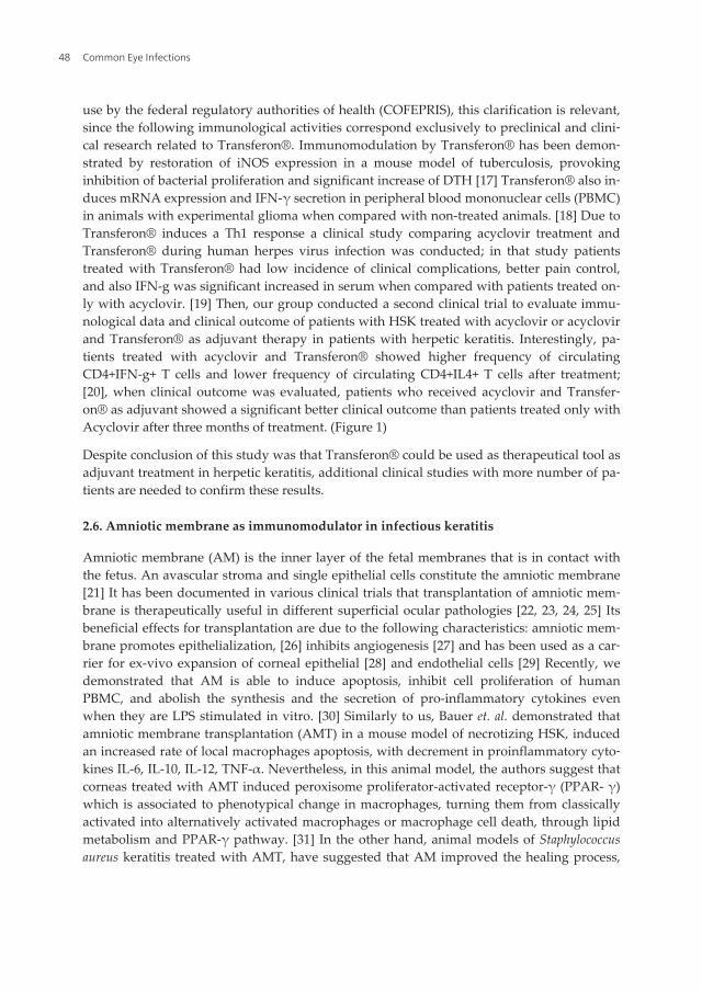
use by the federal regulatory authorities of health (COFEPRIS), this clarification is relevant,since the following immunological activities correspond exclusively to preclinical and clini‐cal research related to Transferon®. Immunomodulation by Transferon® has been demon‐strated by restoration of iNOS expression in a mouse model of tuberculosis, provokinginhibition of bacterial proliferation and significant increase of DTH [17] Transferon® also in‐duces mRNA expression and IFN-γ secretion in peripheral blood mononuclear cells (PBMC)in animals with experimental glioma when compared with non-treated animals. [18] Due toTransferon® induces a Th1 response a clinical study comparing acyclovir treatment andTransferon® during human herpes virus infection was conducted; in that study patientstreated with Transferon® had low incidence of clinical complications, better pain control,and also IFN-g was significant increased in serum when compared with patients treated on‐ly with acyclovir. [19] Then, our group conducted a second clinical trial to evaluate immu‐nological data and clinical outcome of patients with HSK treated with acyclovir or acyclovirand Transferon® as adjuvant therapy in patients with herpetic keratitis. Interestingly, pa‐tients treated with acyclovir and Transferon® showed higher frequency of circulatingCD4+IFN-g+ T cells and lower frequency of circulating CD4+IL4+ T cells after treatment;[20], when clinical outcome was evaluated, patients who received acyclovir and Transfer‐on® as adjuvant showed a significant better clinical outcome than patients treated only withAcyclovir after three months of treatment. (Figure 1)
Despite conclusion of this study was that Transferon® could be used as therapeutical tool asadjuvant treatment in herpetic keratitis, additional clinical studies with more number of pa‐tients are needed to confirm these results.
2.6. Amniotic membrane as immunomodulator in infectious keratitis
Amniotic membrane (AM) is the inner layer of the fetal membranes that is in contact withthe fetus. An avascular stroma and single epithelial cells constitute the amniotic membrane[21] It has been documented in various clinical trials that transplantation of amniotic mem‐brane is therapeutically useful in different superficial ocular pathologies [22, 23, 24, 25] Itsbeneficial effects for transplantation are due to the following characteristics: amniotic mem‐brane promotes epithelialization, [26] inhibits angiogenesis [27] and has been used as a car‐rier for ex-vivo expansion of corneal epithelial [28] and endothelial cells [29] Recently, wedemonstrated that AM is able to induce apoptosis, inhibit cell proliferation of humanPBMC, and abolish the synthesis and the secretion of pro-inflammatory cytokines evenwhen they are LPS stimulated in vitro. [30] Similarly to us, Bauer et. al. demonstrated thatamniotic membrane transplantation (AMT) in a mouse model of necrotizing HSK, inducedan increased rate of local macrophages apoptosis, with decrement in proinflammatory cyto‐kines IL-6, IL-10, IL-12, TNF-α. Nevertheless, in this animal model, the authors suggest thatcorneas treated with AMT induced peroxisome proliferator-activated receptor-γ (PPAR- γ)which is associated to phenotypical change in macrophages, turning them from classicallyactivated into alternatively activated macrophages or macrophage cell death, through lipidmetabolism and PPAR-γ pathway. [31] In the other hand, animal models of Staphylococcusaureus keratitis treated with AMT, have suggested that AM improved the healing process,
Common Eye Infections48
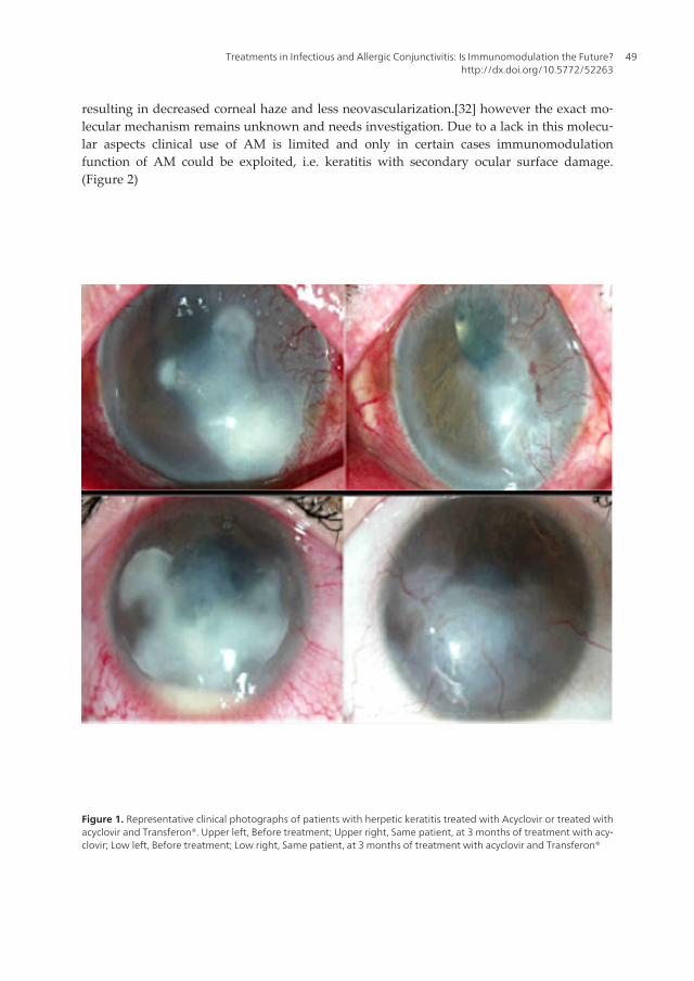
resulting in decreased corneal haze and less neovascularization.[32] however the exact mo‐lecular mechanism remains unknown and needs investigation. Due to a lack in this molecu‐lar aspects clinical use of AM is limited and only in certain cases immunomodulationfunction of AM could be exploited, i.e. keratitis with secondary ocular surface damage.(Figure 2)
Figure 1. Representative clinical photographs of patients with herpetic keratitis treated with Acyclovir or treated withacyclovir and Transferon®. Upper left, Before treatment; Upper right, Same patient, at 3 months of treatment with acy‐clovir; Low left, Before treatment; Low right, Same patient, at 3 months of treatment with acyclovir and Transferon®
Treatments in Infectious and Allergic Conjunctivitis: Is Immunomodulation the Future?http://dx.doi.org/10.5772/52263
49
Figure 2. Clinical photographs of AMT in 67 year old female patient with a history of peripheral infectious keratitissecondary to trichiasis. Left, AMT covering the lower peripheral corneal defect. Amniotic membrane was folded sever‐al times over the cornea to increase their anti-inflammatory properties. Right, Same patient, 15 days after AMT, clinicalphotograph showing apparent control of hyperaemia and inflammation
2.7. MIF-CD74 blockade in Pseudomona aeurginosa keratitis
Macrophage migration inhibitory factor (MIF) is an integral component of inflammatory re‐sponses. MIF induces and sustains expression of several pro-inflammatory cytokines.[33]trough interaction with a receptor complex composed by CD74/CD44 [34] CD74 was firstdescribed as class II invariant chain, while CD44 is an adhesion molecule that binds hyalur‐onic acid and other matrix metalloproteinases. Interaction of MIF with CD74/CD44 results inactivation of Mitogen-Activated Protein Kinase (MAPK), production of PGE214 and furtherinduction of inflammatory mediators [35]
Corneal infections by Pseudomonas aeruginosa are more difficult to treat and result in worsevisual outcome than other bacterial corneal ulcers. Unfortunately the existing therapies failto control the inflammation secondary to P. aeruginosa keratitis and novel interventions areneeded to alleviate tissue damage resulting from local inflammation, recently two studiessuggest that blockade of MIF-CD74 ligation ameliorate the disease-associated pathology bydecreased proinflammatory mediators and reduced bacterial presence in the cornea [36, 37]
3. Novel therapies in allergic conjunctivitis
Treatment of allergic conjunctivitis can be a challenge by the diverse immunological mecha‐nisms of damage involved in ocular allergic diseases, reviewed in [38]. To date, a wide rangeof antiallergic drops treatments are available and can be confusing due to lack of improve‐ment at the ocular surface in terms of avoiding anatomical changes in severe cases and con‐trol of symptoms in the long time period, reviewed in [39, 40, 41]
Hence our primary goal for treating allergic patients should be preferently to recognize al‐lergy background and ocular inflammation status at the time visit to better establish the
Common Eye Infections50
type and source of antigenic stimuli. In this way, primary action such as avoidance andclearance of antigen with lubrication is recommended preferently in acute but also in thelate stage of the chronic forms when dry eye could be implicated. Secondary treatment algo‐rithm includes topical antiallergic agents, which are used towards the reaction characterizedby mast cell activation, release of preformed and newly formed mediators such as hista‐mine, prostaglandins, leukotrienes, production of chemokines and expression of adhesionmolecules. The aim of treatment in seasonal allergic conjunctivitis and perennial allergicconjunctivitis is directed to symptom relief and control, whereas the objective in the chronicforms of vernal keratoconjunctivitis and atopic keratoconjuctivitis will be also to preventvisual complications or try to identify in early stages possible implication of cornea injury.Therefore the efficacy of therapeutic agents varies from patient to patient in terms of gradeof severity at the ocular surface, reviewed in [38] and actual local and systemic status activi‐ty of the immune system making the choice of treatment depending on multiple variables,each case must be individualized. In general ocular allergic diseases involve mast cell degra‐nulation that will initiate through inflammatory mediators activation of enzymatic cascades,giving rise to pro-inflammatory mediators and in consequence antihistamines, mast cell sta‐bilizers, non-steroidal anti-inflammatory agents, corticosteroids are agents of common usefor acute and chronic conjunctivitis.
Nonetheless this wide range of drugs, management of allergic conjunctivits is still a chal‐lenge and immune modulation could be the missing link in the therapeutical approach ofocular allergic diseases.
3.1. Calcineurin inhibitors and atopic keratoconjunctivitis
Calcineurin inhibitors are capable of inducing local immunosuppression more than immu‐nomodulation. Topical [42] and systemic cyclosporine a (CsA) [43] have been suggested inthe treatment of severe atopic keratoconjunctivitis. Cyclosporine is effective in controllingocular allergic inflammation by blocking Th2 lymphocyte proliferation and IL-2 production.It also reduces eosinophils production via inhibition of IL-5 production. Use of CsA appearsto be safe and the clinical goal for its use is to eliminate the need/dependence of steroids andfavourably alter the long-term prognosis of patients with AKC.
Others calcineurins inhibitors that appears to be well tolerated by patients with severe atop‐ic blepharoconjunctivitis [44] and severe atopic keratoconjunctivitis [45] and acceptable clin‐ical outcome are tacrolimus and pimecrolimus, both of them were used first in atopicdermatitis treatment [46]. To date the real impact of anti-allergic treatment with calcineurininhibitors is unknown.
3.2. Mapracorat and eosinophils in ocular allergy
Mapracorat is a novel selective glucocorticoid receptor agonist that maintains a beneficialanti-inflammatory activity but seems to be less effective in transactivation, resulting in alower potential for side effect; it has been proposed for the topical treatment of inflammato‐ry skin disorders. In vitro, Mapracorat inhibited eosinophil migration and IL-8 release from
Treatments in Infectious and Allergic Conjunctivitis: Is Immunomodulation the Future?http://dx.doi.org/10.5772/52263
51
eosinophils or the release of IL-6, IL-8, CCL5/RANTES, and TNF-α from a human mast cellline with equal potency as dexamethasone, whereas it was clearly less potent than this glu‐cocorticoid in inducing annexin I and CXCR4 expression on the human eosinophil surface;in other hand, animal model of allergic conjunctivitis showed that mapracorat was similar todexamethasone eye drops in analogous reduction in clinical symptoms of allergic conjuncti‐vitis and conjunctival eosinophil accumulation. [47] The authors suggest this novel gluco‐corticoid receptor agonist as a candidate to be used in clinical trials of ocular allergy.
3.3. Omalizumab and allergic diseases
Omalizumab is a biological engineered molecule, targeting the Cε3 domain of the IgE mole‐cule. It binds with free IgE and prevents free IgE from attaching to high-affinity IgE receptor(FcεRI) on effector cells such as mast cells, basophils and also on dendritic cells. An IgE-anti-IgE complex is formed, and as a result, free IgE is decreased. [48] Omalizumab has been wellstudied and used in treatment of asthma [49, 50, 51] and other allergic diseases such as uriti‐caria and and stational rhinitis [52] Like other immunomodulators mentioned above, clinicaltrials with allergic conjunctivitis patients are needed to asses the real impact in ocular aller‐gic diseases.
4. Ocular complications with topical or systemic treatments
Allergic reactions to medication could generate ocular manifestations ranging from mild tosevere and it would not be considered infrequent. Demonstration of allergy to topical medi‐cations could not be easily evaluated by allergen test, but give some information. Directprovocation in conjunctiva with suspicious drug has been reported, [53] the authors of thisreview do not recommend this method as a diagnostic protocol, however this test could beused as a research tool to investigate immune responses during allergy to topical medica‐tion. To evaluate ocular allergy to drug medications, epicutaneous allergen test and immedi‐ate-reading intradermal tests are carried out to diagnose immediate hypersensitivityreactions, while atopy patch tests are usually performed to evaluate delayed reactions, re‐viewed in [38, 54] with this diagnostic methodology, Wijnmaalen et al reported that themost frequent medication-associated allergies were directed against tobramycin, neomycinsulphate and thimerosal. [55]
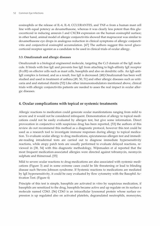
Mild to severe ocular reactions to drug-medications are also associated with systemic medi‐cations (Figure 3) and in some extreme cases could be life threatening or lead to blindingdisease such Stevens Johnson syndrome. If Systemic reactions to medications are mediatedby IgE hypersensitivity, it could be easy evaluated by flow cytometry with the Basophil Ac‐tivation Test. (Figure 4)
Principle of this test is simple, basophils are activated in vitro by suspicious medication, ifbasophils are sensitized to the drug, basophils became active and up regulate on its surface amolecule named CD63. [56] CD63 is an intracellular lysosomal protein whose surface ex‐pression is up regulated also on activated platelets, degranulated neutrophils, monocytes,
Common Eye Infections52
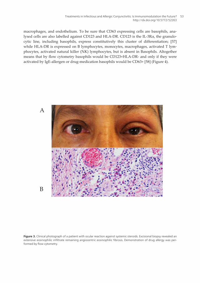
macrophages, and endothelium. To be sure that CD63 expressing cells are basophils, ana‐lysed cells are also labelled against CD123 and HLA-DR. CD123 is the IL-3Rα, the granulo‐cytic line, including basophils, express constitutively this cluster of differentiation; [57]while HLA-DR is expressed on B lymphocytes, monocytes, macrophages, activated T lym‐phocytes, activated natural killer (NK) lymphocytes, but is absent in Basophils. Altogethermeans that by flow cytometry basophils would be CD123+HLA-DR- and only if they wereactivated by IgE-allergen or drug-medication basophils would be CD63+ [58] (Figure 4).
A
B
Figure 3. Clinical photograph of a patient with ocular reaction against systemic steroids. Excisional biopsy revealed anextensive eosinophilic infiltrate remaining angiocentric eosinophilic fibrosis. Demonstration of drug allergy was per‐formed by flow cytometry.
Treatments in Infectious and Allergic Conjunctivitis: Is Immunomodulation the Future?http://dx.doi.org/10.5772/52263
53
Figure 4. Representative cytometer data of Basophil activation test. Analysis gates are shown at upper dot plots. Up‐per left, a gate was drawn on CD123 positive cells according to SSC characteristics; these cells correspond mainly tobasophils. Upper right, A second gate is performed on HLA-DR negative cells; Dot plots of gated CD123+HLA-DR- cells(basophils) are displayed. Low left, negative test; Low right, Positive test, markedly up regulated expression of CD63 isobserved.
5. Conclusions
As the prevalence of allergic disease increases around the world, resistance to antibiotics/antivirals/or antimicotic drugs grows, and virulence of microorganisms improves its capaci‐ty of infection, it is clear that more effective therapies and disease-modifying agents areneeded. Only treatment evolution will be obtained understanding immune pathophysiologi‐cal mechanism underlying infectious and allergic diseases. The authors of this review are
Common Eye Infections54
convinced that immunomodulation is part of our future as health professionals and areworking today to make it posible as soon as posible.
Acknowledgments
C Santacruz and SM Perez-Tapia should be considerer as a first authors indistinctly. Thiswork was supported in part by “Fundación Conde de Valenciana” and Transfer FactorProject.
Author details
Concepcion Santacruz1, Sonia Mayra Perez-Tapia2, Angel Nava-Castañeda1,Sergio Estrada-Parra2 and Maria C. Jimenez-Martinez3*
1 Research Unit, Institute of Ophthalmology “Conde de Valenciana”, Mexico City, Mexico
2 National School of Biological Sciences, IPN, Mexico City, Mexico
3 Faculty of Medicine, UNAM and Research Unit, Institute of Ophthalmology “Conde deValenciana”, Mexico City, Mexico
References
[1] González-Navajas JM, Lee J, David M, Raz E. Immunomodulatory functions of type Iinterferons. Nat Rev Immunol. 2012 Jan 6;12(2):125-35. doi: 10.1038/nri3133.
[2] Uchio E, Inoue H, Fuchigami A, Kadonosono K. Anti-adenoviral effect of interferon-β and interferon-γ in serotypes that cause acute keratoconjunctivitis. Clin Experi‐ment Ophthalmol 2011 May-Jun;39(4):358-63. doi: 10.1111/j.1442-9071.2010.02457.x.Epub 2011 Jan 14.
[3] Sara Spjut, Weixing Qian, Johannes Bauer, Rickard Storm, Lars Frängsmyr, ThiloStehle, Niklas Arnberg, and Mikael Elofsson A Potent Trivalent Sialic Acid Inhibitorof Adenovirus Type 37 Infection of Human Corneal Cells. Angew Chem Int Ed Engl.2011 July 11; 50(29): 6519–6521.
[4] Labetoulle M, Kucera P, Ugolini G, et al. Neuronal propagation of HSV1 from the or‐al mucosa to the eye. Invest. Ophthalmol. Vis. Sci 2000;41(9):2600–2606.
[5] Divito SJ, Hendricks RL. Activated inflammatory infiltrate in HSV-1-infected corneaswithout herpes stromal keratitis. Investigative Ophthalmology and Visual Science.2008;49(4):1488–1495
Treatments in Infectious and Allergic Conjunctivitis: Is Immunomodulation the Future?http://dx.doi.org/10.5772/52263
55
[6] Inoue Y, Ohashi Y, Shimomura Y, et al. Herpes simplex virus glycoprotein D. Protec‐tive immunity against murine herpetic keratitis. Investigative Ophthalmology andVisual Science. 1990;31(3):411–418
[7] Heiligenhaus, Wells PA, Foster CS. Immunisation against HSV-1 keratitis with a syn‐thetic gD peptide. Eye. 1995;9(1):89–95.
[8] Walker J, Leib DA. Protection from primary infection and establishment of latency byvaccination with a herpes simplex virus type 1 recombinant deficient in the virionhost shutoff (vhs) function. Vaccine. 1998;16(1):1–5.
[9] Hu K, He X, Yu F, Yuan X, Hu W, Liu C, Zhao F, Dou J. Immunization with DNAvaccine expressing herpes simplex virus type 1 gD and IL-21 protects against mouseherpes keratitis. Immunol Invest. 2011;40(3):265-78. Epub 2011 Jan 4.
[10] Serhan, C. N., N. Chiang, and T. E. Van Dyke. 2008. Resolving inflammation: dual an‐ti-inflammatory and pro-resolution lipid mediators. Nat. Rev. Immunol. 8: 349–361.
[11] Vassiliou, E. K., O. M. Kesler, J. H. Tadros, and D. Ganea. 2008. Bone marrow- de‐rived dendritic cells generated in the presence of resolvin E1 induce apoptosis of acti‐vated CD4+ T cells. J. Immunol. 181: 4534–4544
[12] Bannenberg, G., and C. N. Serhan. 2010. Specialized pro-resolving lipid mediators inthe inflammatory response: an update. Biochim. Biophys. Acta 1801: 1260–1273.
[13] Zhang, F., H. Yang, Z. Pan, Z. Wang, J. M. Wolosin, P. Gjorstrup, and P. S. Reinach.2010. Dependence of resolvin-induced increases in corneal epithelial cell migrationon EGF receptor transactivation. Invest. Ophthalmol. Vis. Sci. 51: 5601–5609.
[14] Lawrence, HS. (1955). The transfer in humans of delayed skin sensitivity to strepto‐coccal M substance and to tuberculin with disrupted leucocytes. J Clin Invest. pp.219- 230.
[15] Wilson, GB., Fudenberg, HH. & Keller, RH. (1984). Guidelines for immunotherapy ofantigen-specific defects with transfer factor. J Clin Lab Immunol. pp. 51-58. Berron-Perez, R., Chavez-Sanchez, R., Estrada-Garcia, I., Espinosa-Padilla, S., Cortez-Gomez, R. et al. (2007). Indications, usage, and dosage of the transfer factor. RevAlerg Mex. pp. 134-139.
[16] Kirkpatrick, CH. (1993) Structural nature and functions of transfer factors. Ann N YAcad Sci. pp. 362-368.
[17] Fabre, RA., Pérez, TM., Aguilar, LD., Rangel, MJ., Estrada-Garcìa, I., et al.(2004)Transfer factors as immunotherapy and supplement of chemotherapy in experimen‐tal pulmonary tuberculosis. Clin Exp Immunol. pp. 215-23.
[18] Pineda B, Estrada-Parra S, Pedraza-Medina B, Rodriguez-Ropon A, Pérez R, ArrietaO. Interstitial transfer factor as adjuvant immunotherapy for experimental glioma. JExp Clin Cancer Res. 2005 Dec;24(4):575-83.
Common Eye Infections56
[19] Estrada-Parra S, Nagaya A, Serrano E, Rodriguez O, Santamaria V, Ondarza R, Cha‐vez R, Correa B, Monges A, Cabezas R, Calva C, Estrada-Garcia I. Comparativestudy of transfer factor and acyclovir in the treatment of herpes zoster. Int J Immuno‐pharmacol. 1998 Oct;20(10):521-35.
[20] Luna-Baca GA, Linares M, Santacruz-Valdes C, Aguilar-Velazquez G, Chavez R, Per‐ez-Tapia M, Estrada-Garcia I, Estrada-Parra S, Jimenez-Martinez MC. Immunologicalstudy of patients with herpetic stromal keratitis treated with Dialyzable LeukocyteExtracts. In 13th International Congress of Immunology – ICI. Proceedings Immunol‐ogy 2007, CD: ISBN:978-88-7587-380-6, Book: ISBN:978-88-7587-379-0
[21] Ellies, P., Anderson, D., Dighiero, P., Legeais, J. M., Renard, G., Tseng, S. G. (2001).Mise au point sur la membrane amniotique humaine dans la prise en charge despathologies de la surface oculaire. J. Fr. Opthalmol. 24(5):546–556.
[22] Burman, S., Tejwani, S., Vemuganti, G.K., Gopinathan, U., Sangwan, V.S. (2004).Ophthalmic application of preserved human amniotic membrane: A review of cur‐rent indications. Cell Tissue Bank 5(3):161–175
[23] Dua, H. S., Gomes, J. A. P., King, A. J., Maharajan, V. S. (2004). The amniotic mem‐brane in ophthalmology. Surv. Ophthalmol. 49(1):510–577.
[24] Dua, H. S., Gomes, J. A. P., King, A. J., Maharajan, V. S. (2004). The amniotic mem‐brane in ophthalmology. Surv. Ophthalmol. 49(1):510–577.
[25] Giasson, C. J., Bouchard, C., Boisjoly, H., Germain, L. (2006). Amnios et problèmes desurface oculaire. Med. Sci. 22(6–7):639–644.
[26] Grueterich, M., Espana, E. M., Tseng, S. C. G. (2003). Ex vivo expansion of limbal epi-thelial stem cells: Amniotic membrane serving as a stem cell niche. Surv. Ophthal‐mic- mol. 48(6):631–646.
[27] Ma, D. H. K., Yao, J. Y., Yeh, L. K., et al. (2004). In vitro antiangiogenic activity in exvivo expanded human limbocorneal epithelial cells cultivated on human amnioticmembrane. Invest. Ophthalmol. Vis. Sci. 45(8):2586–2595.,
[28] Koizumi, N., Fullwood, N. J., Bairaktaris, G., Inatomi, T., Kinoshita, S., Quantock, A.J. (2000). Cultivation of corneal epithelial cells on intact and denuded human amni-otic membrane. Invest. Ophthalmol. Vis. Sci. 41(9):2506–2513.
[29] Ishino, Y., Sano, Y., Nakamura, T., Connon, C. H., Rigby, H., Fullwood, N. J., Kinosh‐ita, S. (2004). Amniotic membrane as a carrier for cultivated human corneal endothe‐lial cell transplantation. Invest. Ophthalmol. Vis. Sci. 45(3):800–806.
[30] Garfias Y, Zaga-Clavellina V, Vadillo-Ortega F, Osorio M, Jiménez-Martinez MC.Amniotic Membrane is an immunosuppressor of peripheral blood mononuclearcells. Immunological Investigations 2011, 40(2): 1-14.
[31] Bauer D, Hennig M, Wasmuth S, Baehler H, Busch M, Steuhl KP, Thanos S, Heiligen‐haus A. Amniotic membrane induces peroxisome proliferator-activated receptor-γ
Treatments in Infectious and Allergic Conjunctivitis: Is Immunomodulation the Future?http://dx.doi.org/10.5772/52263
57
positive alternatively activated macrophages. Invest Ophthalmol Vis Sci. 2012 Feb21;53(2):799-810.
[32] Barequet IS, Habot-Wilner Z, Keller N, Smollan G, Ziv H, Belkin M, Rosner M. Effectof amniotic membrane transplantation on the healing of bacterial keratitis. InvestOphthalmol Vis Sci. 2008 Jan;49(1):163-7.
[33] Bernhagen, J., Calandra, T., & Bucala, R. The emerging role of MIF in septic shockand infection. Biotherapy 8, 123–127 (1994).
[34] Leng, L. et al. MIF signal transduction initiated by binding to CD74. The Journal ofexperimental medicine 197, 1467–1476 (2003).
[35] Shi, X. et al. CD44 is the signalling component of the macrophage migration inhibito‐ry factor-CD74 receptor complex. Immunity 25, 595–606 (2006).
[36] Gadjeva M, Nagashima J, Zaidi T, Mitchell RA, Pier GB. Inhibition of macrophagemigration inhibitory factor ameliorates ocular Pseudomonas aeruginosa-inducedkeratitis. PLoS Pathog. 2010 Mar 26;6(3):e1000826.
[37] Zaidi T, Reidy T, D'Ortona S, Fichorova R, Pier G, Gadjeva M. CD74 deficiency amel‐iorates Pseudomonas aeruginosa-induced ocular infection. Sci Rep. 2011;1:58.
[38] Robles-Contreras A, Santacruz C, Ayala J, Bracamontes E, Godinez V, Estrada-GarcíaI, Estrada-Parra S, Chávez R, Perez-Tapia M, Bautista-De Lucio V, Jimenez-MartínezMC. Allergic conjunctivitis: an immunological point of view. Book: Conjunctivitis: AComplex and Multifaceted Disorder. 2011, InTech. ISBN 978-953-307-750-5
[39] Bielory Leonard, Lien WK,Bigelsen S. Allergic conjunctivitis Drugs 2005:65 (2):215-228.
[40] Manzouri B, H Flynn T, Larkin Frank, J Ono S, Wyse R. Pharmacotherapy of allergiceye disease. Expert Opin Pharmacother. (2006) 7 (9): 1191-1200
[41] Abelson MB, McLaughlin JT, Gomes PJ. Antihistamines in ocular allergy: are they allcreated equal? Curr Allergy Asthma Rep. 2011 Jun;11(3):205-11.
[42] Tzu JH, Utine CA, Stern ME, Akpek EK. Topical calcineurin inhibitors in the treat‐ment of steroid-dependent atopic keratoconjunctivitis Cornea. 2012 Jun;31(6):649-54.
[43] Cornish KS, Gregory ME, Ramaesh K. Systemic cyclosporin A in severe atopic kera‐toconjunctivitis. Eur J Ophthalmol. 2010 Sep-Oct;20(5):844-51.
[44] Virtanen HM, Reitamo S, Kari M, Kari O. Effect of 0.03% tacrolimus ointment on con‐junctival cytology in patients with severe atopic blepharoconjunctivitis: a retrospec‐tive study. Acta Ophtalmol Scand. 2006;84(5):693–695
[45] García DP, Alperte JI, Cristóbal JA, Mateo Orobia AJ, Muro EM, Valyi Z, Del Rio BJ,Arnao MR. Topical tacrolimus ointment for treatment of intractable atopic keratocon‐junctivitis: a case report and review of the literature. Cornea. 2011 Apr;30(4):462-5.
Common Eye Infections58
[46] Reynolds NJ, Al-Daraji WI. Calcineurin inhibitors and sirolimus: mechanism of ac‐tion and applications in dermatology. Clin Exp Dermatol. 2002;27(7):555–561 Reita‐mo S. Tacrolimus: a new topical immunomodulatory therapy for atopic dermatitis. JAllergy Clin Immunol. 2001;107(3):445–448.
[47] Baiula M, Spartà A, Bedini A, Carbonari G, Bucolo C, Ward KW, Zhang JZ, GovoniP, Spampinato S. Eosinophil as a cellular target of the ocular anti-allergic action ofmapracorat, a novel selective glucocorticoid receptor agonist. Mol Vis.2011;17:3208-23. Epub 2011 Dec 14.
[48] Vichyanond P. Omalizumab in allergic diseases, a recent review. Asian Pac J AllergyImmunol. 2011 Sep;29(3):209-19.
[49] Fahy JV, Fleming HE, Wong HH, Liu JT, Su JQ, Reimann J, et al. The effect of an anti-IgE monoclonal antibody on the early- and late-phase responses to allergen inhala‐tion in asthmatic subjects. Am J Respir Crit Care Med. 1997;155:1828-34.
[50] Holgate S, Buhl R, Bousquet J, Smith N, Panahloo Z, Jimenez P. The use of omalizu‐mab in the treatment of severe allergic asthma: A clinical experience update. RespirMed. 2009;103:1098-113.
[51] Humbert M, Beasley R, Ayres J, Slavin R, Hebert J, Bousquet J, et al. Benefits of oma‐lizumab as add-on therapy in patients with severe persistent asthma who are inade‐quately controlled despite best available therapy (GINA 2002 step 4 treatment):INNOVATE. Allergy. 2005;60:309-16.
[52] Casale TB, Busse WW, Kline JN, Ballas ZK, Moss MH, Townley RG, et al. Omalizumab pretreatment decreases acute reactions after rush immuno‐therapy for ragweed-induced seasonal allergic rhinitis. J Allergy Clin Immunol.2006;117:134-40.
[53] Petersen PE, Evans RB, Johnstone MA, Henderson WR Jr. Evaluation of ocular hy‐persensitivity to dipivalyl epinephrine by component eye-drop testing. J Allergy ClinImmunol. 1990 May;85(5):954-8
[54] Ventura MT, Viola M, Gaeta F, Di Leo E, Buquicchio R, Romano A. Hypersensitivityreactions to ophthalmic products. Curr Pharm Des. 2006;12(26):3401-10.
[55] Wijnmaalen AL, van Zuuren EJ, de Keizer RJ, Jager MJ. Ophthalmic Res. 2009;41(4):225-9. Epub 2009 May 15. Cutaneous allergy testing in patients suspected of an aller‐gic reaction to eye medication.
[56] Stain C, Stockinger H, Scharf M, et al. Human blood basophils display a unique phe‐notype including activation linked membrane structures. Blood. 1987;70:1872-1879
[57] Smith WB, Guida L, Sun Q, et al. Neutrophils activated by granulocyte-macrophagecolony-stimulating factor express receptors for Interleukin-3 which mediate class IIexpression. Blood. 1995;86:3938-3944.
Treatments in Infectious and Allergic Conjunctivitis: Is Immunomodulation the Future?http://dx.doi.org/10.5772/52263
59

[58] Sainte-Laudy J, Vallon C, Guerin JC. Diagnosis of latex allergy: comparison of hista‐mine release and flow cytometric analysis of basophil activation. Inflamm Res. 1996;45:S35-S36.
Common Eye Infections60